Role of classical cadherins in epidermal junction and barrier formation
Inaugural-Dissertation
zur
Erlangung des Doktorgrades
der Mathematisch-Naturwissenschaftlichen Fakultät der Universität zu Köln
vorgelegt von Christian Michels
aus Trier
Köln 2010
Berichterstatter: Prof. Dr. Mats Paulsson Prof. Dr. Manolis Pasparakis
Tag der mündlichen Prüfung: 02.06.2010
Classical cadherins mediate Ca
2+-dependent intercellular adhesion and are essential for tissue morphogenesis and maintenance. They are key components of adherens junctions (AJs). In vitro studies in simple epithelial cells indicated an essential role for E-cadherin not only in the formation of AJs but also other intercellular contacts, such as desmosomes and tight junctions. In contrast, in vivo tissue specific knockout studies did not reveal a necessity of E-cadherin in the formation of intercellular junctions, raising the question if classical cadherins are necessary or if other classical cadherins can compensate for the loss of E- cadherin. Therefore, the aim of this thesis was to ask how E-cadherin regulates tight junctions and if E-cadherin has a specific function in the formation of tight junctions. In addition, the question was asked if classical cadherin function is necessary for the formation of other intercellular contacts, such as desmosomes.
Using primary keratinocytes as a model for de novo junction formation, it was found that loss of E-cadherin prevents the formation of a functional tight junctional barrier. Surprisingly, the basic assembly of tight junctions was not affected, suggesting that E-cadherin regulates a late step in the formation of a functional barrier. One pathway through which E-cadherin may regulate the functional barrier is by controlling expression levels of the barrier promoting claudin-14. However, knockdown of claudin-14 is insufficient to reduce barrier function, suggestion that other mechanisms contribute to E-cadherin controlled barrier function.
Loss of E-cadherin in combination with knock down of the only other epidermal classical cadherin, P-cadherin, resulted in an almost complete loss of intercellular contacts showing that classical cadherins are crucial for desmosome formation.
Re-expression of either E- or P-cadherin can rescue not only desmosome formation but also tight junction function, showing that levels but not specific classical cadherin expression is crucial for the functional formation of intercellular junctions.
E-cadherin is a tumor suppressor and found to be down regulated in many tumors.
In this thesis it was found that loss of E-cadherin in primary keratinocytes is
insufficient to enhance migration and proliferation, suggesting that in cancer cells
E-cadherin interacts with other, E-cadherin independent pathways in the regulation
of growth and migration.
Klassische Cadherine sind Calcium abhängige Zelladhäsionsmoleküle und ein wichtiger Bestandteil der zonula adherens. Funktionale Studien zeigten eine wichtige Rolle für E-Cadherin bei der Initiierung und Erhaltung von Zell-Zell Adhäsion in einfachen Epithelien. Dabei hing nicht nur die Bildung von zonula adherens, sondern auch die von Desmosomen und zonula occludens von der funktionalen Aktivität von E-Cadherin ab. Im Gegensatz dazu war die Ausbildung von Desmosomen in der Epidermis nicht von E-Cadherin abhängig. Nur die Barrierefunktion der zonula occludens im stratum granulosum der Epidermis war nach genetischer Inaktivierung von E-Cadherin beeinträchtigt. Dies führte zur Frage, ob E-Cadherin für die Bildung epidermaler Desmosomen entbehrlich ist und wie die spezifische Regulation von epidermalen zonula occludens stattfindet.
Mithilfe primärer Maus Keratinozyten konnte in dieser Arbeit gezeigt werden dass E-Cadherin für die Bildung epidermaler Desmosomen entbehrlich ist, was auf eine funktionale Kompensation durch P-Cadherin zurück zuführen ist. Entweder E- oder P-Cadherin Expression wurde benötigt, um die Bildung von Desmosomen in primären Keratinozyten auszulösen.
Obwohl zonula occludens in Abwesenheit von E-cadherin strukturell vorhanden sind, konnte gezeigt werden dass diese Strukturen keine funktionelle Barriere bilden. Expression von entweder E- oder P-cadherin war erforderlich um eine funktionale Barriere in primären Maus Keratinozyten zu bilden. Die klassischen Cadherine E- und P-Cadherin zeigen funktionelle Redundanz bei sowohl Desmosomenbildung als auch bei der Bildung einer funktionalen zonula occludens Barriere.
Neben der Regulation von Zelladhäsion spielt E-cadherin eine wichtige Rolle bei
der Regulation anderer zellulärer Prozesse wie Proliferation und Migration. E-
cadherin gilt als Tumorsupressor und seine Expression ist in einer Reihe von
Tumoren vermindert. Diese Arbeit zeigt das E-cadherin kein direkter Regulator der
Keratinozyten Proliferation und Migration ist. Daher erscheint es wahrscheinlich,
dass E-cadherin diese zellulären Prozesse im Zusammenspiel mit anderen, E-
cadherin unabhängigen, Mechanismen reguliert.
TABLE OF CONTENTS
1. INTRODUCTION 1
1.1 Adherens Junctions 2
1.1.1 Classical cadherins 2
1.1.2 Catenins 3
1.1.2.1 p120ctn 3
1.1.2.2 β-catenin 4
1.1.2.3 α-catenin 4
1.1.3 The nectin/afadin system 5
1.1.4 Regulation adherens junctions 6
1.1.4.1 Regulation of gene expression 7
1.1.4.2 Regulation of interactions in the core AJ complexes by phosphorylation 7 1.1.4.3 Regulation through alterations in cytoskeletal dynamics. 8 1.1.4.4 The Regulation of cell surface transport and endocytosis of adherens junction
components 9
1.1.4.5 Regulation by cleavage of adherens junction components 10
1.2 Desmosomes 11 1.3 Tight Junctions 12
1.3.1 Transmembrane components of the tight junction 13 1.3.2 Cytoplasmic scaffold proteins at the tight junction 14 1.3.3 Signaling and polarity complexes at the tight junction 15
1.4 Regulation of Rho family GTPases by intercellular junctions. 16 1.5 Intercellular junctions in the epidermis 17
1.5.1 Adherens junctions in the epidermis 18 1.5.2 Desmosomes in the epidermis 19 1.5.3 Tight junctions in the epidermis 19
1.6 Cadherin mediated regulation of intercellular junctions and barrier
formation 20
1.7 Regulation of growth and migration by classical cadherins 22
1.8 Aims of this thesis 22
2. RESULTS 24
2.1 Classical cadherins are required for intercellular junction formation in primary mouse keratinocytes 24
2.1.1 Isolation of E-cadherin negative primary mouse keratinocytes 24 2.1.2 Adherens junctions form in the absence of E-cadherin 25 2.1.3 Delay in desmosome formation in the absence of E-cadherin 27 2.1.4 Impaired adherens junction and desmosome formation in the absence of both E- and P-
cadherin 28
2.1.5 Desmosome formation in primary mouse keratinocytes depends on classical cadherin
levels 30
2.2 In vitro barrier formation is impaired in E-cadherin deficient primary
mouse keratinocytes 33
2.2.1 Recruitment of TJ proteins is not affected in E-cadherin deficient keratinocytes 33 2.2.2 Ultrastructural Tight Junctions form in the absence of E-cadherin. 34 2.2.3 No in vitro barrier in E-cadherin deficient keratinocytes 36 2.2.4 Normal barrier formation upon knock down of P-cadherin in primary keratinocytes 37 2.2.5 Re-expression of either E-cadherin or P-cadherin rescues barrier formation 38 2.2.6 Expression of the E-cadherin cytoplasmic tail interferes with adhesion 41 2.2.7 No Rescue of barrier formation by overexpression of aPKC 42 2.2.8 Decreased Rac activity in Ecad-/- keratinocytes 43 2.2.9 No obvious alterations in actin cytoskeletal architecture in the absence of E-cadherin 45 2.2.10 Inhibition of Myosin-ATPase, but not PI3-kinase affects barrier formation in primary
keratinocytes 46
2.3 E-cadherin regulates claudin-14 expression in vivo and in vitro 49
2.3.1 Claudin-14 is down regulated in the absence of E-cadherin 49 2.3.2 Partial knock down of claudin-14 does not affect barrier formation in mouse keratinocytes51 2.3.3 No restoration of barrier function upon expression of human claudin-14 in Ecad-/-
keratinocytes 52
2.4 E-cadherin is dispensable for mouse keratinocyte migration and
proliferation 54
2.4.1 Proliferation and proliferative potential not affected in the absence of E-cadherin 54 2.4.2 Keratinocyte migration is not affected upon loss of E-cadherin 55
3. DISCUSSION 58
3.1 Desmsome formation in primary keratinocytes depends on classical
cadherin expression levels 59
3.2 The Role of classical cadherins in epidermal barrier formation 61
3.2.1 E-cadherin is not required for structural tight junction formation in primary keratinocytes 61 3.2.2 E-cadherin is required for epidermal barrier formation 62 3.2.3 Keratinocyte barrier formation depends on classical cadherin levels 65 3.2.4 E-cadherin regulates claudin-14 expression 66
3.3 E-cadherin does not regulate keratinocyte proliferation and migration 68
4. MATERIAL AND METHODS 69 4.1 Cell Culture and lentiviral transduction 69
4.1.1 Isolation and culture of primary keratinocytes 69
4.1.2 Splitting 69
4.1.3 Differentiation and induction of intercellular contact formation by Ca2+ switch 70 4.1.4 Production of lentivirus and lentiviral transduction 70 4.1.5 Lentiviral gene silencing by shRNA transduction 70
4.2 Protein Analysis 71
4.2.1 Immunoblot analysis of primary keratinocytes 71 4.2.2 Immunofluorescence analysis of keratinocytes 72 4.2.3 Rac GTPase activity assay 72 4.2.4 Antibodies and antisera 73 4.2.4.1 Primary antibodies 73 4.2.4.2 Secondary antibodies 74
4.3 Barrier assays 75
4.3.1 Trans epithelial resistance measurement (TER) 75 4.3.2 Paracellular diffusion of nonionic tracers 75
4.4 Electron microscopy 75
4.4.1 Thin section transmission electron microscopy 75 4.4.2 Freeze fracture electron microscopy 76
4.5 Molecular cloning 76
4.5.1 Bacterial Transformation 76 4.5.2 Recombinant DNA techniques 76 4.5.3 Polymerase Chain Reaction (PCR) 77 4.5.4 Constructs and cloning strategies 77
4.6. Reverse transcription and Real time PCR 79
4.6.1 RNA isolation from tissues and cells 79
4.6.2 cDNA synthesis 79 4.6.3 Semi-quantitative RT-PCR 79 4.6.4 Quantitative real time PCR 79 4.6.5 RT-PCR primer list 79
4.7 Cell migration analysis 80
4.7.1 Single cell random migration 80
4.7.2 Scratch assay 81
4.8 Analysis of growth and proliferative potential 81
4.8.1 Cell viability assay 81 4.8.2 BrdU incorporation assay 81 4.8.3 Colony forming assay 81
5. ABRREVIATIONS 82
6. REFERENCES 85
7. ACKNOWLEDGEMENTS 91
8. ERKLÄRUNG 92
9. CURRICULUM VITAE 93
1. Introduction
Physical cohesion between cells is an essential requirement for the formation and maintenance of complex tissues and organs and is provided by adhesive protein complexes that form intercellular junctions. These intercellular junctions must be tightly regulated not only during morphogenesis but, depending on the physiological requirements and cellular functions of a tissue, also during tissue homeostasis. Different types of intercellular junctions serve distinct functions.
Adherens junctions and desmosomes are primarily involved in physical cohesion whereas tight junctions serve as paracellular and membrane diffusion barriers. Gap junctions form channels between cells and thereby allowing intercellular communication of small solutes (fig. 1).
This study mainly addresses the role of adherens junctions, and more specifically its cell adhesion receptors, the classical cadherins in the regulation of not only adherens junctions but also desmosomes and tight junctions as well as their requirement in epidermal barrier formation by using loss of function approaches in primary mouse keratinocytes.
Figure 1: Intercellular junctions in simple epithelia.
Left: Schematic representation of intercellular junction complexes in polarized simple epithelia.
Right: Electron micrograph showing intercellular ultrastructures. Mv: Microvilli; TJ:tight junction; AJ:
adherens junction; DS: desmosome. Taken from (Tsukita, Furuse et al. 2001)
1.1 Adherens Junctions
As the name implies, adherens junctions are intercellular adhesive structures that mediate physical cohesion between cells. First identified in 1963, the adherens junctions were characterized by electron microscopy as part of a tripartite complex in simple epithelia (Farquhar and Palade 1963) (fig. 1). On the ultrastructural level, adherens junctions appear as parallel membranes separated by an intercellular space of ~ 20 nm. Together with the apical localized tight junctions and the more basally found desmosomes, this tripartite structure was called the apical junctional complex. As predicted at the time based on their ultrastructural appearance, it is now widely accepted that adherens junctions are crucial for providing adhesion between cells and thereby establish physical integrity of tissues. They are dynamically regulated structures that serve as coordinating signaling platforms that regulate the actin cytoskeleton, signaling complexes and polarity cues, thereby regulating directly and indirectly cell shape and a variety of cellular processes.
Adherens junctions consist of transmembrane adhesion receptors of the classical cadherin and nectin family which bind and recruit cytoplasmic scaffold and signaling molecules via their cytoplasmic domain. Among these recruited factors, actin binding proteins provide either direct or indirect connections to the actin cytoskeleton. In the following paragraphs, I will describe the molecular composition of the adherens junction as well as several mechanisms of their regulation.
1.1.1 Classical cadherins
Classical cadherins are type I transmembrane proteins that belong to the cadherin super family of Ca
2+dependent adhesion molecules, which are characterized by a Ca
2+binding extracellular motive, the so called cadherin repeat (EC). The number of extracellular repeats varies between family members, ranging form 4 to 34.
Classical cadherins have 5 extracellular repeats which mediate homophilic
interaction between cadherins of adjacent cells in a Ca
2+dependent manner. The
EC1 domain plays a crucial role in adhesive bond formation as well as in mediating
binding specificity. In addition to trans interaction between cells, the cadherin
extracellular domain is capable to interact in cis laterally in the membrane resulting
in cadherin dimers that are thought to represent the basic adhesive units (Brieher,
Yap et al. 1996).
The most prominent members of the classical cadherin sub family are E(pithelial)- cadherin, N(euronal)-cadherin and P(lacental)-cadherin. Although initially named after the tissue where they were discovered, they show a broader expression pattern. Homophilic binding of differentially expressed cadherins was thought to be the basic driving force behind cellular sorting and segregation processes, for example in mesoderm induction, where a switch from E- to N-cadherin induces segregation from the ectoderm. Similarly, E- to N-cadherin switch is observed in many carcinomas and is this is likely to be a key step in tumor progression leading to invasiveness. However, other studies report that classical cadherins are able to interact also in a heterophilic fashion, suggesting that the molecular mechanisms that drive sorting processes are not that simple (Niessen and Gumbiner 2002).
1.1.2 Catenins
The cadherin cytoplasmic domain forms a basic complex with catenins, and this interaction is crucial for full adhesive capacity. The Ch1 domain in the juxtamembrane position interacts with p120ctn. The more distal, c-terminal Ch2 domain interacts with the armadillo repeat protein β-catenin, which in turn recruits the actin binding protein α-catenin. Thereby, dynamic, indirect association with the actin cytoskeleton is achieved. In the following, the different catenins and their role in the adherens junctions are discussed.
1.1.2.1 p120ctn
p120ctn binding to the cadherin has emerged as a critical regulation of the
cadherin cell surface stability (Xiao, Oas et al. 2007). Loss of p120ctn results in
increased cadherin turnover by regulating access to the endocytotic machinery
(Reynolds 2007). p120 comprises 10 armadillo repeats that are crucial for the
interaction with the cadherin (fig. 3). Regulation of cadherin dynamics at the cell
surface occurs via intensive tyrosine and serine/threonin phosphorylation
(Fukumoto, Shintani et al. 2008). In addition to its regulatory function in the
cadherin core complex, p120ctn can translocate to the nucleus and regulates gene
expression through binding to the transcription factor kaiso. Furthermore p120ctn
turned out to mediate the regulation of Rho family GTPases upon cadherin
engagement. In particular, RhoA becomes inhibited by binding to p120ctn, whereas Rac is activated by a less understood mechanism (Anastasiadis 2007).
1.1.2.2 β -catenin
β-catenin binds to the distal region of the cadherin cytoplasmic domain and this interaction serves several functions. It protects the cadherin cytodomain from degradation, promotes cadherin trafficking from the ER to the plasma membrane, and recruits α-catenin to the core complex. In addition to these functions that all affect adhesive capacity of the adherens junction, β-catenin mediates gene regulation as a signal transducer in the wnt signaling pathway which plays crucial roles in several developmental processes. In the absence of wnts, cytosolic β- catenin that is not bound to the cadherin gets phosphorylated by the APC complex, thereby targeting it to ubiquitinylation and proteasomal degradation. Extracellular wnt signals inhibit the APC complex, allowing β-catenin to shuttle to the nucleus where it acts as a transcriptional cofactor by binding to TCF/Lef transcription factors, resulting in activation of wnt responsive genes (Nelson and Nusse 2004).
1.1.2.3 α -catenin
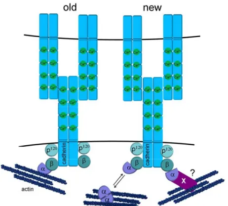
The actin binding protein α-catenin is indirectly associated with the cadherin via binding to β-catenin. Loss of α-catenin results in reduced adhesion, although the cadherin β-catenin complex is still present at the cell surface. Since pull down experiments showed simultaneous precipitation of β-catenin and actin together with α-catenin, it was believed for a long time and led to a textbook model that the cadherin core complex is directly connected to the actin cytoskeleton (fig 2, left).
However, studies showed that α-catenin exists in a monomeric state that interacts
with β-catenin, and a dimeric state that interacts with actin, and these distinct
interactions were shown to be mutually exclusive. Thus, no stable connection
between the actin cytoskeleton and the cadherin core complex could be
demonstrated in vitro, suggesting that cadherin mediated actin regulation is much
more dynamic than previously believed (Drees, Pokutta et al. 2005; Yamada,
Pokutta et al. 2005) (fig. 2, right).
Figure 2: Schematic presentation of the cadherin core complex.
Textbook model versus newer model for how the cadherin complex interacts with actin. In the text book model (old) the cadherin complex was directly bound to actin via α-catenin. Recent data indicate that binding of α-catenin to either β-catenin or actin is mutually exclusive, resulting in a much more dynamic view on actin regulation by classical cadherins (new). In this model, actin binding either takes place in the vicinity of adherens junctions through dynamic exchange of α- catenin and binding is through other actin-binding proteins that link α-catenin to actin.
1.1.3 The nectin/afadin system
The nectin/afadin system represents another basic adhesive unit of the adherens junction. The nectin family belongs to the IgG-like super family of intercellular adhesion receptors and consists of 4 nectin variants (nectin 1-4) together with the closely related nectin like molecules (NECL1-5). Initially identified as receptors for α-herpes and polio viruses, they were later implicated in the regulation of intercellular adhesion. Nectins form lateral homo- or heterodimer and are capable to engage in either hetero or homophilic adhesion with other nectins, which is, in contrast to the cadherins, independent of Ca
2+(Takai, Miyoshi et al. 2008).
They consist of an extracellular domain comprising three IgG-like loops, and a
transmembrane domain and a cytoplasmic domain with a c-terminal PDZ binding
motif. The actin binding protein afadin interacts with its PDZ domain and this links
the nectin directly to the actin cytoskeleton. Blocking of nectin functions interferes
with cadherin mediated adhesion in vitro, suggesting that nectins might represent a first scaffold in the formation of adherens junctions. On the other hand, adherens junction formation is Ca
2+dependent, showing the requirement for cadherin engagement and suggesting a more cooperative mechanism of both complexes in the formation of adherens junctions. This could be mediated by direct interaction of the complexes since afadin binds directly to α-catenin and p120 catenin. However, the exact mechanism of such a regulation is not known.
Figure 3: Overview of the domain structures and protein interaction binding sites of adherens junction components.
Domain structure of the adherens junction core components. Bars indicate sites of protein interaction. IG, immunoglobulin-like domain; Dil, dilute domain; EC, cadherin extracellular repeat;
PDZ, PSD95/Dlg/ZO-1 domain; RA, Ras association domain; FHA, Forkhead associated domain;
VH, vinculin homology domain and PR, proline-rich domain.
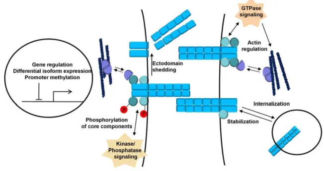
1.1.4 Regulation adherens junctions
Adherens junctions are tightly regulated during morphogenetic processes in
development and tissue growth and maintenance. Especially classical cadherins
play a key role in the dynamic regulation of intercellular contacts during processes that involve cell rearrangements, like cell sorting, epithelial to mesenchymal transition (EMT) and migration (Gumbiner 2005).
Regulation of the adherens junctions occur on multiple levels ranging from gene expression via alterations of core complex components by post translational modification to regulation of cell surface expression by either trafficking or cleavage (fig. 4). A short overview of the various mechanisms is given in the following chapters.
1.1.4.1 Regulation of gene expression
Epithelial to mesenchymal transition (EMT) is a reversible developmental process in which cells lose their epithelial characteristics and gain mesenchymal properties.
Hallmarks of EMT are loss of polarity and of adherens junctions concomitant with cytoskeletal rearrangements and increased cell motility. A key feature of EMT is the loss of E-cadherin. This process is also thought to reversibly occur during cancer progression when cells gain migratory and invasive properties. Signals that induce EMT upregulate transcriptional repressors of different families, for example Snail, which bind to the promoter of E-cadherin and several other key epithelial markers, thereby resulting in reversible silencing of these promoters (Peinado, Olmeda et al. 2007). Importantly, downregulation of E-cadherin is crucial to gain migratory/invasive properties. Epigenetic changes, such as promoter methylation, result in long-term silencing of gene expression. The best known adherens junction component for which this occurs is E-cadherin. The promoter of E-cadherin is silenced by CpG methylation in a range of cancers (Berx, Nollet et al. 1998).
1.1.4.2 Regulation of interactions in the core AJ complexes by phosphorylation
Adherens junction assembly/disassembly and maintenance is directly influenced
by phosphorylation-dependent alterations in the interactions between the core
complex components of the cadherin and nectin adhesion complexes. This may
occur directly at adherens junctions since both kinases and phosphatases are
associated with core components. In general, serine/threonine phosphorylation is
considered to strengthen adherens junctions whereas tyrosine kinase activity is
often associated with disassembly of junctions. However, several examples exist
where increased tyrosine kinase activity promotes adherens junctions (McLachlan and Yap 2007). Phosphorylation of cadherin core complex components alters the affinity of the different core complex components for each other. The cadherin binds β-catenin while passing through the endoplasmic reticulum. Serine/threonine phosphorylation of the cadherin cytoplasmic domain or the C-terminus of β-catenin further increases the affinity of this already strong interaction, thus strengthening adhesion and AJ formation/maintenance (Daugherty and Gottardi 2007). Tyrosine phosphorylation of β-catenin, however, lowers the affinity for either the cadherin or α-catenin, thus promoting disassembly of adherens junctions.
1.1.4.3 Regulation through alterations in cytoskeletal dynamics.
As the ultra structure reveals adherens junctions are closely connected to the actin cytoskeleton and regulate actin dynamics at sites of cell–cell contacts. Vice versa, actin rearrangements are crucial for the formation of adherens junctions. The Rho and Rap subfamilies of the Ras super family of small GTPases, key coordinators of cytoskeletal activity, have emerged as important regulators of dynamic cell–cell adhesion and adherens junction formation and maintenance (Braga and Yap 2005). Vice versa, cadherin or nectin engagement can control the activity of members of these small GTPase subfamilies, suggesting a close reciprocal relationship. Early AJ formation is driven by lamellipodia that are formed by localized Rac activity at sites of initial cell–cell contacts. This controls actin dynamics through actin filament nucleators such as the Arp2/3 complex. Cadherin engagement then directly activates Rac1 at sites of forming cell–cell contacts thereby establishing a positive reinforcing loop that enlarges the contact size between cells. Expansion of contacts requires Rho-regulated actomyosin contractions resulting in compaction of the intercellular contacts (Nelson 2008).
Cadherin binding itself recruits different myosins and thereby further promote
adherens junction stabilization. However, actomyosin contractions also drive local
dissociation of adherens junctions.
1.1.4.4 The Regulation of cell surface transport and endocytosis of adherens junction components
Transport of components to and removal from the cell surface through the vesicle transport machinery are dynamic processes that cells use to regulate adherens junctions. After protein synthesis, cadherin/β-catenin complexes are delivered to different destinations at the cell surface. For example, in simple epithelia E- cadherin is sorted to the basolateral membrane, which is essential for the establishment and maintenance of basolateral polarity. Control of cadherin exocytosis occurs on three levels (Delva and Kowalczyk 2009). First, the cadherin cytodomain encodes sorting signals to direct the cadherin to the correct membrane compartment. The most consistent one is a dileucine motif in E-cadherin that targets E-cadherin to basolateral membranes. Second, the cadherin–catenin complex travels along microtubules that may target specific cell surface localizations. Third, specific protein assemblies clustered on the cell surface may specify vesicle fusion. The best example is the so-called exocyst complex. This complex regulates cell surface expression of Drosophila cadherin and is localized to the apical junctional complex.
Endocytosis of cadherins has also emerged as a crucial regulatory step in the maintenance and stability of adherens junctions. Cadherins have two main destinations upon endocytosis: the lysosomal degradation pathway and recycling to the cell surface. Although lysosomal degradation likely contributes to disassembly of adherens junctions, several lines of evidence indicate that dynamic cadherin recycling is crucial not only for dynamic cell rearrangements but also essential for maintaining adherens junction stability (Wirtz-Peitz and Zallen 2009).
How the decision is made between sending cadherin to the lysosomal
compartment versus recycling is at present not clear. Nevertheless, since p120ctn
is crucial for cell surface stability of cadherins, mechanisms that lower the
interaction of p120 with the cadherin are likely to promote endocytosis. Although it
is at present unclear if endocytosis of nectins regulates adherens junctions, nectin
activity appears important in the regulation of cadherin endocytosis.
1.1.4.5 Regulation by cleavage of adherens junction components
Both nectins and cadherins are subject to cleavage at defined sites by different proteases resulting in a permanent loss of full-length adhesive molecules from the cell surface. In a first step, the extracellular domains of cadherins and nectins are cleaved close to the membrane resulting in release of the extracellular domain, a process called ectodomain shedding (Reiss, Ludwig et al. 2006). Subsequently, the cytoplasmic domains can be cleaved by other proteases, such as caspases and presenilin. This cleavage also generates several novel cadherin and nectin fragments that may have biological functions by themselves. For example, the cadherin extracellular domain itself promotes cell motility in development and in cancer cells. Although at present unclear if the released cadherin cytoplasmic domain has a physiological relevant function, several reports have shown that it can inhibit the activity of certain transcription factors by retaining them in the cytoplasm. Other reports have shown that the cytoplasmic domain translocates to the nucleus where it binds deoxyribonucleic acid (DNA).
Figure 4: Cadherin regulation.
Different mechanisms regulate adherens junction formation and maintenance. Adherens junctions are regulated on multiple levels ranging from regulation of gene expression, trafficking to posttranslational modification and proteolytic cleavage.
1.2 Desmosomes
Desmosomes also form intercellular adhesive structures but unlike adherens junctions that link to the actin cytoskeleton, these junctions anchor the intermediate filament cytoskeleton to the plasmamembrane. In polarized simple epithelial cells desmosomes localize to the lateral membrane and appear ultrastructurally as electron dense plaque. On the cytoplasmic site an outer dens plaque (ODP) and an inner dense plaque (IDP) can be distinguished. Between the two plasma membranes of adjacent cells, the trans-interacting cadherin domains appear as an electron dense midline.
Like adherens junctions, they are formed by adhesion receptors belonging to the cadherin super family. Two subfamilies have been identified: desmogleins and desmocollins. Similar to classical cadherins, they have five extracellular cadherin repeats that mediate Ca
2+dependent adhesion between cells.
Desmogleins and Desmocollins cooperate to form an adhesive interface. The cytodomains of both family members bind to the armadillo family member plakoglobin and to plakophillins. Plakoglobin, also referred to as γ-catenin, preferentially associates with desmosomal cadherins but can also interact with classical cadherins, where it substitutes for β-catenin by occupying its binding site (Nathke, Hinck et al. 1994).
Desmoplakin, a member of the plakin protein family, binds to plakoglobin and
bridges the desmosomal complex to the intermediate filament system. Plakophillins
offer a more complex repertoire of interactions. Like plakoglobin, they bind to
desmoplakin but can also interact with intermediate filaments directly. Lateral
interactions among the armadillo proteins in the desmosome increase the stability
and adhesive strength of the structure. Overall, desmosomes are tightly connected
to the intermediate filaments, providing firm adhesion and physical integrity to
tissues. This is highlighted by the findings that mutations in desmosomal
components often result in tissue fragility, like blistering diseases in the skin (Green
and Simpson 2007).
Figure 5: Schematic drawing of the molecular composition of desmosomes superimposed on a desmsosomal ultrastructure.
Dsg: Desmoglein, Dsc: Desmocollin, Pg:Plakoglobin, PKP; Plakophilin, DP: Desmoplakin, IF:
intermediate filaments. Taken from (Green and Simpson 2007)
1.3 Tight Junctions
The third and most apically located intercellular junctional structure in the tripartite complex described by Farquhar and Palade are tight junctions. In thin section electron microscopy tight junctions appear as close intercellular contacts where the two opposing membranes come in such close proximity that the extracellular space almost seems obliterated. These contacts resemble the so called kissing points that have been described on the ultrastructural level for tight junctions in simple epithelia (Farquhar and Palade 1963). The formation of a network of ultrastructurally discernible interconnected strands which can be detected by freeze fracture replica electron microscopy are considered to form the basis of the seal of tight junctions (Staehelin, Mukherjee et al. 1969).
Relative to adherens junctions and desmosomes, tight junctions are most apical
localized in simple epithelia (Farquhar and Palade 1963). Tight junctions form an
ion and size-selective diffusion barrier and are the main regulators of paracellular
permeability. In simple, polarized epithelia they are found at the border of the
apical and baso-lateral membrane domains, which differ in their protein and lipid
composition. In addition to restricting paracellular diffusion by having a “gate
function”, tight junctions are also thought to restrict intermixing of constituents of
the two different membrane domains, which is referred to as its “fence function”
(Anderson, Van Itallie et al. 2004). Like adherens junctions and desmosomes, tight junctions consist of protein complexes comprising transmembrane adhesion receptors and cytoplasmic scaffold that are linked to the cytoskeleton.
1.3.1 Transmembrane components of the tight junction
Three types of structural transmembrane components are enriched at the tight junctions and have the potential to mediate cell adhesion: the IgG like family of junctional adhesion molecules and the claudin and occludin families. Although not homologous in sequence, occludin and claudins share a topology with four transmembrane domains and two extracellular repeats (Schneeberger and Lynch 2004).
Claudins are critical for not only tight junction strand formation, but also for providing ion and size- selective permeability properties of the tight (Van Itallie, Rahner et al. 2001). The claudin family comprises at least 24 members in the human genome, where as 21 have been identified in mice. They all show tissue specific expression patterns. Claudins engage in either homophilic or heterophilic interaction across opposing membranes as well as laterally in the same cell. Thus, depending on the amount of claudin isoforms that are expressed and their relative expression levels, the claudin composition determines barrier properties of the tight junction and enables this protein family to account for the different barrier requirements of different tissues.
The function of occludin at the tight junction is less clear. Occludin was the transmembrane protein that was identified to specifically localize to the tight junction (Furuse, Hirase et al. 1993). Overexpression or mutations in occludin affected TER in vitro (Balda, Whitney et al. 1996). However, it appeared dispensable for the ultrastructural strand formation and for the establishment of a paracellular diffusion barrier in vivo. Nevertheless, occludin deficient mice revealed several phenotypes such as growth retardation, mineral deposits in the brain, male sterility and gastritis, which may implicates barrier regulation (Saitou, Furuse et al.
2000).
Tricellulin, an occludin related molecule, was shown to enrich at tricellular junctions
in simple epithelia and to contribute to barrier function at these specialized sites
(Ikenouchi, Furuse et al. 2005). Both molecules share a MARVEL transmembrane domain which is thought to mediate their preferential accumulation in cholesterol rich membrane micro domains. Based on this structural feature, very recently another protein belonging to this family, MarvelD3, was discovered by bioinformatic approaches and was shown to specifically localize to the tight junctions and to have partially overlapping functions with occludin and tricellulin, thus defining the occludin family as tight junction associated MARVEL proteins (TAMP) (Raleigh, Marchiando et al.).
Junctional adhesion molecules (JAM) represent another type of adhesion receptors at the tight junction. They belong to the family of IgG like adhesion molecules and engage both in homophilic and heterophilic adhesion. Unlike claudins, they do not induce ultrastructural strand formation when expressed in fibroblasts and there expression is not confined to cell types that form tight junctions. They have been implicated in the regulation of migration and polarity (Ebnet, Suzuki et al. 2004).
1.3.2 Cytoplasmic scaffold proteins at the tight junction
An important group of tight junction scaffold molecules are the zonula occludens (ZO) proteins. These proteins belong to the membrane associated guanylate kinase-like homologs and are characterized by three N-terminal PDZ domains, an SH3 domain and a guanylate kinase domain (GUK). The first PDZ domains interact with claudins and the GUK domain with occludin. The C-terminus interacts with actin, thereby linking the tight junction transmembrane components to the actin cytoskeleton. The second PDZ domain was shown to mediate homo- and hetero dimerization of ZO proteins and the third PDZ domains interacts with Jam-1 (Ebnet, Schulz et al. 2000). ZO-1 and ZO-2, but not ZO-3, were shown to be crucial for the formation of claudin based strands and for the establishment of a tight junctional barrier, and this was dependent on the first PDZ domain. (Umeda, Ikenouchi et al. 2006). ZO-1 represents one direct link between tight junctions and adherens junctions since it can also interact with α-catenin (Rajasekaran, Hojo et al. 1996).
Several other PDZ domain containing proteins, such as MUPP1 and MAGI proteins
are associated with the tight junction cytosolic plaque and can directly interact with
one or more of the tight junctional transmembrane components (Schneeberger and
Lynch 2004). It is at present unclear if these molecules are directly involved in the formation of the tight junctions or serve a more regulatory function.
Cingulin, a non-PDZ tight junctional plaque protein, interacts with ZOs, JAMs and actin as well as myosin. As such, this protein may be a regulator of tight junctional dynamics during actomyosin contraction (Clayburgh, Barrett et al. 2005).
1.3.3 Signaling and polarity complexes at the tight junction
At the interface of two distinct membrane domains in simple epithelia, tight junctions serve as a spatial landmark which recruits protein complexes that are essential for the formation of apico-basal polarity. Ternary polarity complexes like the Par3/Par6/aPKC and the Cumbs/Pals/Patj complexes localize to the tight junction and are required for epithelial polarity. Many studies highlight the close interrelationship between intercellular junction formation and the formation of apico-basal polarity in simple epithelia. Adherens junctions and tight junctions might cooperate in the process of epithelial polarization, since JAM-1 and nectins have been reported to recruit Par3 to the apical junction complex which might represent an initial step in the setup of epithelial polarity (Ebnet, Suzuki et al. 2001;
Takekuni, Ikeda et al. 2003). On the other hand, functional interference with the
polarity complexes affects paracellular permeability, indicating a role of these
complexes in the structural formation of the tight junction and pointing out a
concerted action of junction formation and polarization in simple epithelia
(Anderson, Van Itallie et al. 2004).
Figure 6: Schematic representation of the basic structural components of the tight junctions.
ZO-1 or ZO-2 is important for clustering of claudins and occludin. The role of other scaffolding proteins (ZO-3/MAGI/MUPP1) is less clear. The ZOs and cingulin can provide a direct link to the actin cytoskeleton. Taken from (Niessen 2007).
1.4 Regulation of Rho family GTPases by intercellular junctions.
The Rho family belongs to the Ras superfamily of small GTPases. These GTPases switch between a GDP bound inactive state and a GTP bound active state. This transition is regulated by proteins that interact with the small GTPase which do either enhance its GTPase activity (GTPase activating protein, Gap), or do facilitate the exchange of GDP for GTP (Guanidine nucleotide exchange factor, Gef). In the GTP bound form the small GTPases specifically interacts with downstream effector molecules that then mediate various cellular responses by having multiple signaling outputs. In addition, small GTPases can be activated by several upstream signaling events. Thus, small GTPases serve as signal integrators that are controlled by tight spatio-temporal regulation.
RhoA family GTPases emerged as critical regulators of the actin cytoskeleton,
thereby directly affecting cell shape and migration. Their most intensively studied
members are RhoA, Rac and cdc42, which have been shown to organize distinct
actin structures in vitro. Rac1 is an activator of Arp2/3 mediated actin assembly
and induces lamellipodia formation, whereas RhoA is thought to act in actin
nucleation responsible for the formation of stress fibers. Cdc42 stimulates filopodia assembly (Etienne-Manneville and Hall 2002). Intense crosstalk between Rho family GTPases and intercellular junction formation has been reported in simple epithelia.
For instance, both cadherin and nectin engagement activate Rac1 and Cdc42, which in turn is required for proper organization of the cortical actin cytoskeleton.
RhoA can either be activated or inhibited upon cadherin engagement depending on the cell type that was used in the study. In addition, Rac inhibits E-cadherin endocytosis, thereby affecting junction stability and remodeling. Cadherin mediated regulation of Rho GTPases was shown to be mainly mediated by p120ctn, although alternative pathways implicating PI3K have been discussed (Gavard, Lambert et al. 2004; Anastasiadis 2007).
Similarly, the formation of macromolecular complexes at the tight junction by ZO-1 and Jam localization affect Rho and cdc42 activation (Miyoshi and Takai 2008). In addition, properly compartmentalized regulation of Rho family GTPases is important for tight junction homeostasis since overexpression of either dominant active or negative variants of each Rho family GTPase disrupts tight junctions in vitro (Jou, Schneeberger et al. 1998). Both Rac and Cdc42 directly interact with the Par complex and thereby regulate epithelial polarity and tight junction function (Suzuki and Ohno 2006).
Conflicting results regarding activation or inhibition of specific GTPases come from studies using different cell types, thus, GTPase regulation might be highly cell context dependent (Braga and Yap 2005).
1.5 Intercellular junctions in the epidermis
The epidermis of the skin is a multilayered epithelium that serves as the outermost
barrier to the environment. Its function is to establish protective barriers against
invading pathogens and chemical or physical assaults as well as to prevent
dehydration by trans epidermal water loss. The epidermis is a continuously self
renewing tissue that is composed of mitotically active keratinocytes in the
innermost basal layer that enter a program of terminal differentiation in order to
form more differentiated suprabasal layers. Upon detachment from the basement
membrane the cells withdraw from the cell cycle and enter the spinous layer,
thereby assembling a durable cytoskeletal framework that provides mechanical integrity. By entering the granular layer, the cells flatten and are characterized by accumulation of keratin macrofibrils and lipid containing lamellar bodies. Finally, in the outermost stratum corneum, structural proteins of the so called corneocytes are irreversibly crosslinked by transglutaminases. Lipids that extrude from the lamellar bodies seal the extracellular space around the corneocytes, thereby creating an insoluble meshwork that results in the formation of an epidermal barrier (Segre 2003).
Throughout the process of epidermal differentiation, intercellular junctions are maintained and actively remodelled to provide physical integrity to the tissue. In the following, the different types of intercellular adhesive structures in the epidermis and the localization of their structural components and functions in the epidermis will be discussed.
1.5.1 Adherens junctions in the epidermis
Two classical cadherins, E- and P-cadherin are expressed in the epidermis. While E-cadherin is expressed in all layers of the epidermis, P-cadherin expression is confined to the basal layer. P120ctn, β-catenin and α-catenin are associated with E-cadherin in all viable layers of the epidermis. Thus, functional adherens junctions are maintained during epidermal homeostasis in all viable layers. Conditional ablation of adherens junction components did not only affect mechanical stability of the epidermis and their appendages, but also signaling mechanisms that affect differentiation, proliferation and inflammation.
Loss of E-cadherin results in hair loss due to impaired adhesion, but also altered epidermal differentiation (Young, Boussadia et al. 2003; Tinkle, Lechler et al. 2004;
Anastasiadis 2007). Loss of P-cadherin did not cause any obvious skin phenotype (Radice, Ferreira-Cornwell et al. 1997). However, mutations in human P-cadherin are associated with hair disorder and with ectodermal displasia (Sprecher, Bergman et al. 2001; Kjaer, Hansen et al. 2005).
Epidermal deletion of the cadherin associated catenins revealed overlapping and
specific functions in mice. Knock out of α-catenin resulted in loss of adherens
junctions, reduced desmosome formation, hyperproliferation and altered growth
factor signaling (Vasioukhin, Bauer et al. 2001). Loss of p120ctn reduced adherens
junction but also activated an inflammatory responses, which was independent of its cadherin complex function but linked to increased NF-kB signalling in keratinocytes (Perez-Moreno, Davis et al. 2006). (Perez-Moreno, Davis et al.
2006). Ablation of β-catenin in the epidermis highlighted its importance in the Wnt signalling pathway during hair morphogenesis and stem cell regulation. No differences in intercellular adhesion were observed which is likely explained by the replacement of β-catenin by plakoglobin in the cadherin complex (Huelsken, Vogel et al. 2001). Taken together, the results indicate that adherens junction components may couple structural integrity to intracellular signalling events.
1.5.2 Desmosomes in the epidermis
Structural desmosomes can be found in all layers of the epidermis, thereby providing firm adhesion to the tissue. However, appearance and size of the desmosomes vary between the different layers. Smaller, less organized desmosomes in cells of the basal layer are replaced by larger, more electron dense desmosomes in the suprabasal layers. Also the composition of desmosomal constituents varies between the layers. Desmoglein 1 and Desmocollin 1 expression increases in the more differentiated suprabasal layers and show only little expression in the basal layer. Desmocollin 2, 3 and Desmoglein 3 show opposite expression patterns with high expression in the basal layer and decreasing expression in the suprabasal layers (Green and Simpson 2007).
Desmoglein 4 is concentrated in the granular and cornified layer whereas desmoglein 2 is only observed in the basal layer. The importance of epidermal desmosomes in the maintenance of epidermal tissue stability is highlighted by findings that ablation or mutation in desmosomal components is often associated with blistering diseases (Green and Simpson 2007).
1.5.3 Tight junctions in the epidermis
Ultrastructurally discernable tight junctions can only be found in the granular layer
of the epidermis and tracer penetration assays confirmed this layer as a site of a
functional permeability barrier (Proksch, Brandner et al. 2008). Tight junction
molecular key components like claudin-1 and ZO-1 are expressed in all viable
layers of the epidermis, whereas claudin-4 and Occludin show specific localization to the membrane of the granular layer (Brandner 2009).
For a long time it was assumed that the lipid envelop of the stratum corneum was sufficient to provide a structural barrier to the epidermis. However, conditional deletion of claudin-1 in the epidermis of mice resulted in death due to trans epidermal water loss. This revealed for the first time a requirement for functional tight junctions in the epidermis to maintain inside-out barrier function (Furuse, Hata et al. 2002).
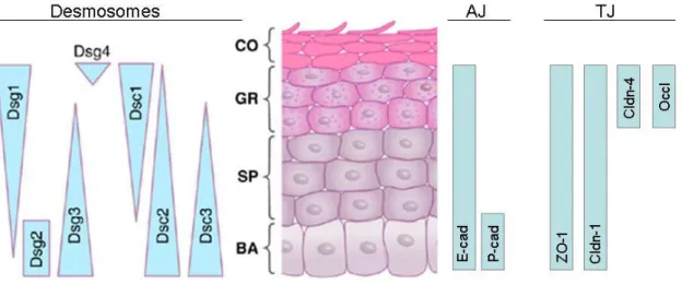
Figure 7: Expression of structural components of desmosomes, adherens junctions (AJ) and tight junctions (TJ) in the different layers of the epidermis,
Ultrastructural adherens junctions and desmosomes are found in all viable layers of the epidermis, whereas ultrastructural tight junctions are only found in the stratum granulosum.
BA: stratum basale; SP: stratum spinosum; GR: stratum granulosum; CO: stratum corneum
Dsg: Desmosglein; Dsc: Desmocollin; cad: cadherin; ZO-1: zonula occludens 1; Cldn (claudin), Occl: occludin. Taken from (Green and Simpson 2007)), modified.
1.6 Cadherin mediated regulation of intercellular junctions and barrier formation
Classical cadherins have been implicated not only in the formation and regulation
of adherens junctions, but also in the formation of other types of intercellular
junctions, such as desmosomes and tight junctions. E-cadherin was thought to be
specifically required for the formation of intercellular junctions in simple epithelia
(Gumbiner, Stevenson et al. 1988), whereas cooperative roles for E- and P-
cadherin were suggested in stratifying epithelia (Lewis, Jensen et al. 1994). In vivo
epidermal deletion of α-catenin confirmed the requirement for adherens junctions
in epidermal desmosome formation, since these structures were largely reduced in numbers (Vasioukhin, Bauer et al. 2001). Whereas P-cadherin appeared to be dispensable for epidermal junction formation in vivo (Radice, Ferreira-Cornwell et al. 1997), only tight junctions were affected upon epidermal deletion of E-cadherin (Vasioukhin, Bauer et al. 2001; Tunggal, Helfrich et al. 2005).
Epidermal deletion of E-cadherin in the epidermis resulted in perinatal death due to trans epidermal waterloss, which closely resembled the phenotype of the claudin-1 deficient epidermis (Tunggal, Helfrich et al. 2005). Interestingly, the structural formation of tight junction was not completely disturbed. Occludin, claudin-4 and ZO-1 still localized to the cell surface as judged by immunofluorescence, albeit with changed staining patterns for the latter two molecules. More importantly, tight junction like structures could still be detected in electron microscopy, suggesting that E-cadherin is not required for their initial structural formation in the first place, but may regulates maturation at later stages. Intercellular cohesion was unaffected in E-cadherin deficient epidermis, since no blistering or fragility could be detected.
This was explained by upregulation of P-cadherin in the basal layer and the
formation of ultrastructurally normal desmosomes which might have been
reinforced by upregulation of desmosomal components. Taken together, E-
cadherin appeared dispensable for epidermal cohesion and junction formation and
turned out to be a critical regulator of epidermal tight junctions by regulating the
specific incorporation of claudins in an adhesion independent manner. Indeed, the
mislocalization of the tight junction regulators Rac and phosphorylated aPKC in E-
cadherin deficient epidermis suggested that E-cadherin regulates epidermal tight
junctions by proper recruitment or activation of tight junction regulatory molecules
that then might mediate claudin incorporation and thereby barrier formation
(Tunggal, Helfrich et al. 2005). The results raise the question whether classical
cadherins are indeed required for epidermal desmosome and tight junction
formation, and whether there are specific or overlapping functions of E- and P-
cadherin. Furthermore, since conditional ablation techniques were used, it remains
unclear whether classical cadherins regulate de novo junction formation in
keratinocytes, a question which is difficult to address in vivo.
1.7 Regulation of growth and migration by classical cadherins
Especially E-cadherin was implicated in the regulation of various cellular processes like proliferation and migration. In simple epithelia, cadherin engagement and subsequent formation of intercellular contacts restricts proliferation, a phenomenon called contact inhibition of proliferation (Perrais, Chen et al. 2007) (Liu, Jia et al.; St Croix, Sheehan et al. 1998).
Control of cell motility is crucial for tissue integrity and morphogenesis. Epithelial cells undergoing epithelial to mesenchymal transition during organogenesis disassemble intercellular contacts and are characterized by a marked down regulation of E-cadherin, mediated by the repression of gene expression via the transcription factor snail (Cano, Perez-Moreno et al. 2000; Peinado, Olmeda et al.
2007). Similarly, E-cadherin is mutated in a variety of human cancers, and down regulation occurs during invasive tumor progression, again accompanied by acquisition of a mesenchymal phenotype and increased motility (Batlle, Sancho et al. 2000). Thus, E-cadherin is considered to act as a tumor suppressor and as a determinant of epithelial cell shape. Combined epithelial deletion of E-cadherin and p53 resulted in accelerated development of invasive mammary tumors (Derksen, Liu et al. 2006). However it remains unclear whether loss of E-cadherin is primarily causing enhanced migration or whether this occurs in cooperation with other oncogenic mutations.
1.8 Aims of this thesis
In vitro studies in simple epithelial cells indicated an essential role for E-cadherin
not only in the formation of AJs but also other intercellular contacts, such as
desmosomes and tight junctions. In contrast, in vivo tissue specific knockout
studies did not reveal a necessity of E-cadherin in the formation of intercellular
junctions, raising the question if classical cadherins are necessary or if other
classical cadherins can compensate for the loss of E-cadherin in the formation of
other junctions. Previous work in the laboratory had revealed that in vivo loss of E-
cadherin in a stratifying epithelium, the epidermis, resulted in loss of tight junctional
function but not desmosomes. Since in vivo P-cadherin expression but not
localization was upregulated, this may potentially compensate for E-cadherin in
desmosome assembly. Alternatively, these results may suggest that cadherins are
dispensable for desmosome formation. In addition, in cancer cells loss of E- cadherin alters the growth and invasive properties of cells but in vivo studies suggested that loss of E-cadherin in non-transformed tissues do not affect these functions.
The overall aim of this thesis was to address the roles of classical cadherins in intercellular junction and barrier formation, and migration. Isolated keratinocytes from control and epidermal specific E-cadherin knockout mice provided a unique model system to study the consequences of cadherin loss in primary epithelial cells. Switching keratinocytes from low Ca
2+conditions to high Ca
2+conditions activates cadherin-dependent intercellular adhesion and enables de novo intercellular junction formation. This allows one to follow the kinetics of junction formation and function. In addition, one can compare growth and migration properties under cadherin adhesion non-permissive or permissive conditions.
Specifically, the following questions were asked:
1. Is there a requirement of classical cadherins in keratinocyte de novo desmosome formation and are there specific or overlapping functions for E- or P-cadherin?
2. Is the regulation of tight junctions a specific function of E-cadherin?
3. How does E-cadherin regulate tight junctions and thus epidermal barrier function?
4. How does loss of E-cadherin affect growth and migration properties of
primary epithelial cells?
2. Results
In order to address these questions, primary mouse keratinocytes are utilized as a model system since de novo junction formation is difficult to assess in vivo but can be controlled in vitro. The so called Ca
2+switch protocol allows for precise induction of intercellular contact formation and enables to follow their kinetics.
A combined approach of conditional gene deletion and shRNA mediated silencing is used to address the specific roles of E- and P-cadherin, respectively.
Furthermore, gene delivery by lentiviral transduction of keratinocytes allows testing for the significance of potential candidate molecules or protein domains that are involved in the regulation of epidermal desmosome and barrier formation.
2.1 Classical cadherins are required for intercellular junction formation in primary mouse keratinocytes
2.1.1 Isolation of E-cadherin negative primary mouse keratinocytes
Primary mouse keratinocytes were isolated as described in materials and methods.
Keratinocytes derived from mice of the genotype K14Cre-Ecad
fl/+, K14Cre-Ecad
-/+or Ecad
fl/fland Ecad
fl/-were used as control (Ctr), whereas keratinocytes isolated
from K14Cre-E-cad
fl/fland K14Cre-Ecad
fl/-were termed E-cadherin negative
(Ecad
-/-). Western blot analysis confirmed that E-cadherin protein expression is
indeed absent in Ecad
-/-keratinocytes (fig. 8). In phase contrast microscopy, no
morphological difference was observed between Ecad
-/-and control keratinocytes
when cells were cultured in low Ca
2+concentration (50 μM) (fig. 2-1). This is not
unexpected since under these conditions cadherins cannot engage in intercellular
adhesion.
Figure 8: Keratinocytes isolated from K14Cre-Ecad fl/fl mice are deficient for E-cadherin.
(A) Phase contrast images of Control and Ecad-/- keratinocytes cultured under low Ca2+ condition (50 µM). Bar = 25 µm. (B) Western blot analysis of total cell lysates using anti-E-cadherin antibody.
Actin was used as loading control.
2.1.2 Adherens junctions form in the absence of E-cadherin
To address the role of E-cadherin in intercellular epidermal junction formation, kinetics of adherens junction formation was analyzed in primary Ecad
-/-keratinocytes. Control and Ecad
-/-keratinocytes were subjected to a Ca
2+-switch by replacing the medium containing 50 μM (low Ca
2+) with medium containing 1.8 mM Ca
2+(high Ca
2+medium). The Ca
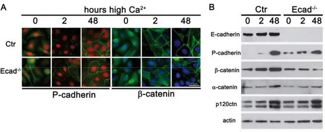
2+-switch not only induces differentiation of keratinocytes but also allows the formation of intercellular junctions. Using imunofluoresence microscopy this allows examining the subcellular localization of adherens junction and desmosomal components and examining their recruitment to sites of cell-cell contact.
In both control and Ecad
-/-keratinocytes, membrane staining was observed for β- catenin 2 hours after Ca
2+-switch and membrane staining was even more intense after 48 hours in high Ca
2+, suggesting that initiation of adherens junction formation takes place with similar kinetics in the absence of E-cadherin (fig. 9A). Since all classical cadherins can interact with the catenins it was examined if P-cadherin was responsible for the observed recruitment of β-catenin. Even though recruitment of P-cadherin to sites of intercellular contacts was observed 2 hours after Ca
2+-switch in both control and Ecad
-/-keratinocytes, staining was more intense in the absence of E-cadherin. Both control and Ecad
-/-keratinocytes displayed similar recruitment of P-cadherin 48h after Ca
2+-switch.
Epidermal deletion of E-cadherin resulted in upregulation of P-cadherin in the basal layer of the epidermis. To test whether P-cadherin expression was altered in Ecad
-/-
keratinocytes, protein levels of P-cadherin as well as its associated catenins were
assessed by western blot analysis. Indeed, similar to in vivo situation, P-cadherin was up regulated in undifferentiated keratinocytes and 2h after Ca
2+switch in Ecad
-/-
when compared to controls, may reflecting an attempt to compensate for the loss of E-cadherin (fig.9B). Similar expression levels for control and Ecad
-/-keratinocytes were observed 48h after Ca
2+switch, showing that P-cadherin is expressed in differentiated keratinocytes in vitro. In both control and Ecad
-/-keratinocytes P-cadherin expression increased with longer Ca
2+induced differentiation periods, in contrast to the in vivo situation where expression of P- cadherin is confined to basal, undifferentiated keratinocytes (Tunggal, Helfrich et al. 2005).
α-catenin and β-catenin are known to be stabilized when associated with classical cadherins in the adherens junction complex, thus reflecting indirectly classical cadherin protein levels. Western blot analysis of control and Ecad
-/-keratinocytes revealed a down regulation of these two proteins at all observed time points of differentiation, suggesting that despite the upregulation of P-cadherin overall classical cadherin levels are reduced in the absence of E-cadherin. Expression of p120ctn, which expression levels have been shown to be independent of classical cadherin levels, was unchanged (fig. 9B).
These results show that adherens junctions are assembled in the absence of E- cadherin, most likely because P-cadherin was upregulated.
Figure 9: Adherens junction formation upon loss of E-cadherin.
(A) Immunofluorescence analysis of adherens junction components P-cadherin and β-catenin Bar=15 µm. (B) Western blot analysis of total cell lysates for adherens junction proteins. Cells were differentiated in high Ca2+ for the indicated time points.
2.1.3 Delay in desmosome formation in the absence of E-cadherin
In vivo loss of epidermal cadherins did not show an obvious change in number and appearance of desmosomes in newborn skin. However, it is not possible to follow the kinetics of desmosome formation in vivo. To examine if membrane recruitment of desmosomal components, used to assess desmosome formation, is altered by the loss of E-cadherin the localization and expression of desmoglein 3 and plakoglobin were analyzed (fig. 10A). Interestingly, whereas control keratinocytes were able to recruit these desmosomal components to the cell surface after 2 hours of Ca
2+stimulation, no membrane localization was observed in E-cadherin deficient keratinocytes at this time point, suggesting a delay in desmosome formation. However, membrane localization of these components was indistinguishable between E-cad
-/-and control keratinocytes after 48 hours, indicating that desmosomes can form in E-cadherin deficient keratinocytes, albeit with a delay in kinetics. Indeed, thin section electron microscopy analysis revealed the presence of desmosomes in the absence of E-cadherin, which were ultrastructurally indistinguishable from control cells when differentiated for 48 hours, showing that E-cadherin is dispensable for desmosome formation in vivo (Tunggal et al., 2005) and in vitro in primary keratinocytes (Michels, Buchta et al.
2009)(fig. 11C).
To asses whether the observed delay in desmosomal protein recruitment was
caused by alterations in desmosomal component expression, western blot analysis
was performed (fig. 10B). No obvious difference was found in plakoglobin
expression 2 hours after Ca
2+switch, suggesting that its absence form intercellular
junctions at this timepoint was not caused by differences in the expression of this
protein. In both control and Ecad
-/-keratinocytes, expression of desmoglein 1 and 2
was only detectable 48 hours after Ca
2+switch, indicating that other desmosomal
cadherins mediate desmosome formation at initial phases of junction formation.
Figure 10: Desmsome formation upon loss of E-cadherin.
(A) Immuno fluorescence analysis of the desmosomal components desmoglein 3 and plakoglobin.
Bar=15 µm. (B) Western blot analysis of total cell lysates for desmosomal proteins desmosglein 1/2 and plakoglobin. Cells were differentiated in high Ca2+ for the indicated time points.