Characterization of Synaptopodin 2 Subcellular Trafficking during Phenotype Modulation of
Vascular Smooth Muscle Cells and Beyond
Inaugural-Dissertation zur
Erlangung des Doktorgrades (Dr. rer. nat.) der Mathematisch-Naturwissenschaftlichen Fakultät
der Universität zu Köln
Vorgelegt von
Mostafa Abootorabi Ardestani
Cologne, Germany, 2015
Thesis Supervisor: Prof. Dr. Gabriele Pfitzer Thesis Committee Chair: Prof. Dr. Thorsten Hoppe Thesis Reader: Prof. Dr. Günter Schwarz
Thesis Reader: Prof. Dr. Angelika A. Noegel
Date of Oral Exam: 2015-04-14
Anleitung von Frau Prof. Dr. Gabriele Pfitzer und Frau Dr. Mechthild M. Schroeter am Institute für vegetative Physiologie der medizinischen Fakultät der Universität Köln,
angefertigt.]
[This study was undertaken under the guidance of Prof. Dr. Gabriele Pfitzer and Dr.
Mechthild M. Schroeter in the period between November 2011 and January 2015 at the Institute of Vegetative Physiology of the Medical faculty of the University of Cologne.]
To My Beloved
Family
“Imagination is more important than knowledge. For knowledge is limited to all we now know and understand, while imagination embraces the entire
world, and all there ever will be to know and understand.”
Albert Einstein
Table of Contents_Toc410319457
1 Introduction 18
1.1 Muscle 18
1.1.1 Vascular smooth muscle 18
1.1.2 Molecular basis of contraction and relaxation 20
1.1.3 Smooth muscle cells phenotype plasticity and disease 22
1.1.4 Actin cytoskeleton 23
1.1.4.1 Actin and actin filaments 23
1.2 Synaptopodin family 25
1.2.1 Synaptopodin 26
1.2.2 Synaptopodin 2 26
1.2.3 Synaptopodin 2-like 28
1.3 Protein-Protein interaction 29
1.3.1 Förster resonance energy transfer 30
1.3.2 Bimolecular fluorescence complementation (BIFC) 31
1.4 Aim of the project 33
2 Materials and Methods 34
2.1 General materials 34
2.2 Methods 49
2.2.1 Molecular biology methods 49
2.2.1.1 Total RNA isolation and cDNA synthesis 49
2.2.1.1.1 Total RNA isolation by MAXWELL machine 49
2.2.1.1.2 cDNA synthesis 50
2.2.1.2 Preparative PCR 50
2.2.1.2.1 Purification of PCR products 51
2.2.1.2.2 Sub-Cloning 52
2.2.1.2.3 Transformation 53
2.2.1.2.4 Colony PCR 53
2.2.1.2.5 Preparation of plasmid DNA 54
2.2.1.2.6 Test digest screening 55
2.2.1.2.7 DNA sequencing 55
2.2.1.3 Quantitative polymerase chain reaction (qRT-PCR) 55
2.2.1.3.1 Analysis of qRT-PCR data 56
2.2.2 Cell biology methods 57
2.2.2.1 Isolation of vascular smooth muscle cells from rat aorta 57
2.2.2.2 Cell culture and treatments 59
2.2.2.3 Immunofluorescence staining 61
2.2.2.4 Confocal microscopy 62
2.2.3 Biochemical and analytical methods 63
2.2.3.1 Characterization of isolated VSMCs 63
2.2.3.2 Fluorescence activated cell sorting assay (FACS) 63
2.2.3.3 Subcellular fractionation 63
2.2.3.4 SDS-PAGE and Western blot analysis 65
2.2.3.5 Estimation of F/G-Actin ratio 66
2.2.4 Preparation of collagen I and laminin coated polyacrylamide matrices 67 2.2.5 Stiffness measurement of polyacrylamide by atomic force microscopy 67
2.2.6 Bimolecular fluorescence complementation assay (BIFC) 68
2.2.7 FACS-BIFC based assay 69
2.2.8 Statistics 70
3 Results 71
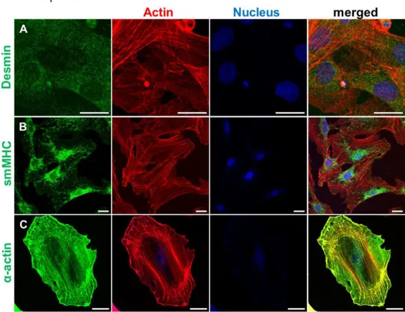
3.1 Over 95% of the isolated cells were vascular smooth muscle cells 71 3.2 Isolated primary VSMCs expressed the smooth muscle marker proteins 72 3.3 Immunofluorescence studies demonstrated co-localization of SYNPO2, smMHC, and α-SMA 73
3.4 Alteration of marker proteins during phenotype switch 73 3.5 Phenotype switch of VSMCs alters subcellular distribution of SYNPO2 75
3.6 Controlling of phenotype switch using ECM proteins 77
3.7 Cultivation the cells on ECM proteins pre-coated matrices alter subcellular
localization of SYNPO2 80
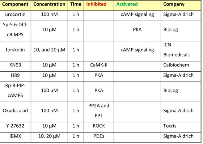
3.8 Actin cytoskeleton turnover affects subcellular distribution of SYNPO2 81 3.9 Heat Shock Stress did not affect the Subcellular Localization of SYNPO2 in VSMCs 83 3.10 Subcellular distribution of SYNPO2 is controlled by cAMP 84 3.11 Inhibition of PKA and CaMK-II reduced the accumulation of SYNPO2 in the nucleus of VSMCs 86
3.12 ROCK signaling does not affect subcellular localization of SYNPO2 88 3.13 Treatment with okadaic acid resulted in a reduction of nuclear SYNPO2- displaying cells 89 3.14 Characterization of expression level of 14-3-3 isoforms in VSMCs 90 3.15 Analysis of subcellular localization of SYNPO2 and its interaction with 14-3-3β/γ, and importinα using fluorescent fused proteins and BIFC assay 91 3.16 Subcellular localization of YFP-fused SYNPO2 & CFP-fused 14-3-3γ/β, importinα 92 3.17 Analysis of SYNPO2, 14-3-3 γ/β and importinα interaction by using BIFC 95 3.17.1 SYNPO2 interacts with 14-3-3γ in the cytoplasm and not in the nucleus 95 3.17.2 SYNPO 2 and 14-3-3β accumulate and form loop-like complexes in the nucleus 96 3.17.3 Preferred interaction of SYNPO2 and importinα in the nucleus 97 3.18 Visualization of SYNPO2 homo-dimer using BIFC and FACS-BIFC based assay 99
4 Discussion 100
4.1 Current state of knowledge as relevant to this work 100 4.2 Phenotype switch of VSMCs affects the subcellular distribution of SYNPO2 102 4.3 Actin dynamics and subcellular distribution of SYNPO2 in VSMCs 105 4.4 The cAMP mediated signaling is involved in the subcellular shuttling of SYNPO2
between the nucleus and the cytoplasm 108
4.5 Imaging of SYNPO2, 14-3-3γ/β, and Importinα interaction in living cells 111
4.6 Visualization of SYNPO2 homo-dimerizartion using BIFC assay in living cells 116
5 Conclusion and Outlook 118
6 Bibliography 121
7 Supplementary information 141
7.1 Synaptopodin 2 141
7.2 14-3-3γ 142
7.3 14-3-3β 142
7.4 Importinα 143
7.5 Plasmids 144
7.5.1 pEYFP-N1-SYNPO2 and pEYFP-C1-SYNPO2 144
7.5.2 pECFP-N1-14-3-3γ and pECFP-C1-14-3-3γ 145
7.5.3 pECFP-N1-14-3-3β and pECFP-C1-14-3-3β 146
7.5.4 pECFP-N1-importinα and pECFP-C1-importinα 147
7.5.5 ΔC- and ΔN-YFP-SYNPO2 148
7.5.6 ΔC- and ΔN-YFP-14-3-3γ 149
7.5.7 ΔC- and ΔN-YFP-14-3-3β 150
7.5.8 ΔC- and ΔN-YFP- Importinα 151
7.6 BIFC control experiments 152
8 Acknowledgment 154
9 Erklärung 156
10 Curriculum Vitae 157
I. List of Figures
Fig. 1 Schematic illustration of (A) skeletal and (B) smooth muscles (according to Lodish
4th Ed). ______________________________________________________________________ 18 Fig. 2 Schematic illustration of the signals that regulate and control the smooth muscle
cell contraction and relaxation (according to Puetz et al 2009)16. ________________________ 21 Fig. 3 Schematic illustration of actin polymerization/depolymerization. _________________ 25 Fig. 4 Schematic illustration of synaptopodin 2 isoforms in human. ______________________ 27 Fig. 5 Principle of FRET assay used in vivo. __________________________________________ 31 Fig. 6 Schematic illustration of the BIFC assay used in this work according to Hu, CD. et al.
2003 130. _____________________________________________________________________ 32 Fig. 7 Schematic illustration: Isolation of vascular smooth muscle cell from rat aorta using enzymatic method. ____________________________________________________________ 58 Fig. 8 Fluorescence-activated cell sorting (FACS) analysis of isolated rat aorta vascular
smooth muscle cells versus fibroblasts. ____________________________________________ 71 Fig. 9 Indirect immunofluorescence analysis of marker proteins of isolated VSMCs. ________ 72 Fig. 10 Visualization of SYNPO2 and the contractile marker proteins, smMHC and α-SMA. ___ 73 Fig. 11 Expression analysis of marker proteins in primary rat VSMCs. ____________________ 75 Fig. 12 Subcellular localization of synaptopodin 2 during isolation and cultivation of
primary VSMCs. _______________________________________________________________ 77 Fig. 13 Characterization of VSMCs cultivated on polyacrylamide matrices coated with
extracellular matrix proteins. ____________________________________________________ 79 Fig. 14 On laminin coated polyacrylamide matrices cultivated VSMCs develop a contractile phenotype. ___________________________________________________________________ 80 Fig. 15 Subcellular distribution of SYNPO2 during actin cytoskeleton turnover. ____________ 82 Fig. 16 Distribution of smooth muscle cells SYNPO2 is independent of heat stress. _________ 83 Fig. 17 cAMP-induced shuttling of SYNPO2 in VSMCs. ________________________________ 85 Fig. 18 Inhibition of both PKA and CaMKII reduced nuclear accumulation of SYNPO2 in the isolated VSMCs. _______________________________________________________________ 87 Fig. 19 Inhibition of ROCK had no consequences on the subcellular localization of SYNPO2. __ 88
Fig. 20 Okadaic acid induced accumulation of SYNPO2 in the cytoplasm. _________________ 89 Fig. 21 Expression analysis of 14-3-3 isoforms in primary rat VSMCs._____________________ 90 Fig. 22 Schematic illustration of constructs used for SYNPO2, 14-3-3γ/β & importinα
interaction and localization analysis by BIFC and a FACS-BIFC based assays. _______________ 91 Fig. 23 Expression analysis of fluorescent fused proteins. ______________________________ 92 Fig. 24 Subcellular distribution of fluorescent fusion proteins in living cells detected by
confocal laser scanning microscopy. _______________________________________________ 94 Fig. 25 Interaction between SYNPO2 and 14-3-3γ takes place in the cytoplasm of HEK293
cells. ________________________________________________________________________ 96 Fig. 26 SYNPO2 and 14-3-3β localized and formed a loop structure in the nucleus. _________ 97 Fig. 27 Imaging of SYNPO2, importinα interaction using BIFC assay. _____________________ 98 Fig. 28 Imaging of self-association of SYNPO2 to form homo-dimers in living cells. _________ 99 Fig. 29 Schematic illustration of actin cytoskeleton dynamics depending on Rho-Rock
signaling pathway (modified from Alan Hall, science, 1998). __________________________ 104 Fig. 30 Schematic illustration of PKA (R= regulatory domain, C= catalytic domain) and
CaMKII-depending phosphorylation of SYNPO2 used in this work (modified from Faul et
al. 2007). ____________________________________________________________________ 109 Fig. 31 Schematic illustration of nuclear-cytoplasmic trafficking of SYNPO2 (According to
Faul et al. 2005). ______________________________________________________________ 114
II. List of Tables
Table 1 All chemicals used in this work. ... 34
Table 2 List of Kits and Materials ... 37
Table 3 All buffers and solutions used in this PhD work ... 38
Table 4 Bacterial and mammalian media ... 40
Table 5 List of used instruments during this work. ... 40
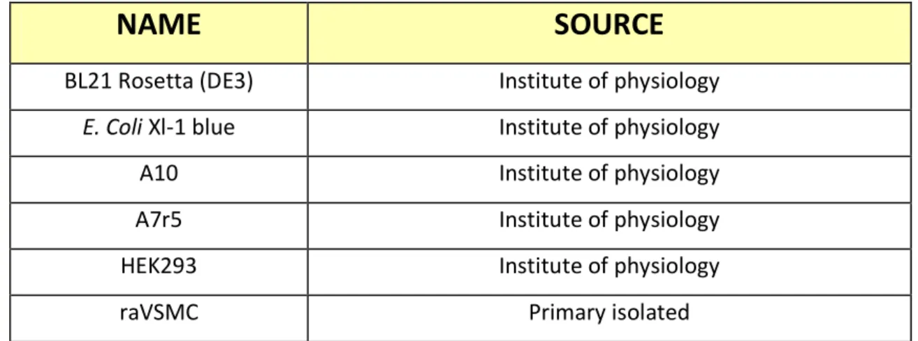
Table 6 List of used bacterial for cloning and sub-cloning process. ... 42
Table 7 List of vectors and plasmids used in this work ... 42
Table 8 List of primers used in this work... 43
Table 9 List of used antibodies in this work. ... 47
Table 10 General PCR Mixture used in this work ... 50
Table 11 PCR Programs used in this work ... 51
Table 12 Concentration of TAE agarose for DNA purification ... 51
Table 13 Digestion mixture and using times ... 52
Table 14 Used ligation reaction mixture in this work ... 53
Table 15 Colony PCR reaction mixture ... 54
Table 16 General Real-Time PCR Mixture used in This Work ... 56
Table 17 Real-Time PCR Program used in This Work ... 56
Table 18 Incubation times and treatment conditions used for this study ... 59
Table 19 Used combinations of BIFC-constructs in this work. ... 68
Table 20 Alteration of the marker proteins expression level in the smooth muscle cells during phenotype switch and/or diseases ... 100
III. Abbreviations
αSMA Smooth muscle α-actin
ΔC-YFP Yellow Fluorescent Protein aa 1-158 ΔN-YFP Yellow Fluorescent Protein aa 159-238
µl Microliter
ABPs Actin binding proteins
ADP Adenosine diphosphate
APS Ammonium persulfate
Arp2/3 Complex Actin-Related Proteins ARP2 and ARP3
ATP Adenosine triphosphate
BIFC Bimolecular fluorescence complementation BLAST Basic Local Alignment Search Tool
BSA Bovine serum albumin
CalP Calponin
CaMKII Ca2+-Calmodulin dependent kinase II
Cc Critical concentration
CCD Cytochalasin D
CD90 Cluster differentiation 90, Thy-1
cDNA Complementary DNA
CFP Cyan fluorescent protein
CHAP Cytoskeletal heart-enriched actin-associated protein dNTP Deoxyribonucleotide triphosphate
DTT Dithiothreitol
EDTA Ethylenediaminetetraacetic acid
EtBr Ethidium bromide
FACS Fluorescence Activated Cell Sorting Assay
FBS Fetal bovine serum
FRET Förster resonance energy transfer GFP Green fluorescent protein
HBSS HEPES-buffered saline solution
hCaD High Caldesmon
HEK Human embryonic kidney
IF Immunofluorescence
IPTG Isopropyl-thio-β-d-galactoside
JASP jasplakinolide
kb Kilo base
kDa kilodalton
LB Lysogeny broth
lCaD Low Caldesmon
MLC20 Myosin light chain 20 MLCK Myosin light chain kinase MLCP Myosin light chain phosphatase
ml Milliliter
mRNA Messenger RNA
NEB New England Biolabs
PAGE Polyacrylamide gel electrophoresi PBS Phosphate-buffered saline
PCR Polymerase chain reaction
PFA Paraformaldehyde
PIC Protease inhibitor cocktail PMSF Phenylmethylsulfonyl fluoride
PP1 Protein phosphatase 1
PP2A Protein phosphatase 2A
RNA Ribonucleic acid
ROCK Rho-associated protein kinase
RT Room temperature
SDS Sodium dodecyl sulfate SEM Standard error of the mean
SMC Smooth muscle Cell
smMHC Smooth muscle myosin heavy chain
Smtn Smoothelin
SYNPO Synaptopodin
SYNPO2 Synaptopodin 2
SYNPO2L Synaptopodin 2-like
TAE Tris-acetate-EDTA
TBS Tris-buffered saline
TEMED N,N,N´,N´-tetramethylethylenediamine
Tm Melting temperature
Tris Tris (hydroxymethyl) aminomethane
UV Ultraviolet
V Volts
VSMCs Vascular Smooth Muscle Cells
Vim Vimentin
WB Western blot
WT Wild-type
IV. Zusammenfassung
In eukaryotischen Zellen stellt Aktin das Protein mit der höchsten Abundanz dar, besitzt diverse Funktionen und ist Hauptbestandteil des Zytoskeletts. Daher spielt Aktin eine wichtige Rolle für die Entstehung und Erhaltung der Zell Form, der Zellmotilität, der Zelladhäsion, der Muskelkontraktion und der Zytokinese. Die Struktur des Aktinnetzwerks und seine zellulären Funktionen werden von Proteinen reguliert, welche allgemein als Aktin bindende Proteine (ABP) bekannt sind.
Gegenstand dieser Arbeit war die Analyse eines neuen ABPs Synaptopodein 2 (SYNPO2) und seine Relevanz für die Phänotyp Modulation in vaskulären Glattmuskelzellen (vascular smooth muscle cell; VSMC). SYNPO2 ist maßgeblich an der Regulation von Aktin Nukleation und dem Umbau des Aktin Zytoskeletts beteiligt. SYNPO2 ist ein Dual- Kompartiment Protein, welches seine subzelluläre Lokalisation zwischen dem Nukleus und dem Zytoplasma in Herz- und Skelettmuskelzellen ändert. Dieser Prozess geschieht in Abhängigkeit einer cyclischen Adenosinmonophosphats (cAMP)-abhängigen Phosphorylierung des SYNPO2 durch Protein kinase A (PKA) und Calcium/calmodulin- dependent protein kinase II (CaMKII). Die unterschiedliche SYNPO2 Lokalisation spielt in vielen Prozessen, wie der Zelldifferenzierung, der Antwort auf Hitzeschock und unter pathophysiologischen Bedingungen, wie z. B. Blasen- und Prostatakrebs eine bedeutende Rolle. SYNPO2 wird abundant in VSMCs exprimiert, wobei seine Rolle in diesen Zellen jedoch bis jetzt unbekannt ist.
In der vorliegenden Arbeit zeigen wir, dass der cAMP-abhängige PKA Signalweg am Pendeln von endogenem SYNPO2 zwischen Zellkern und Zytoplasma in beiden Richtungen beteiligt ist. Wie bereits beschrieben, konnten in Folge von erhöhtem cAMP eine Akkumulation von SYNPO2 im Zellkern detektieren werden. Des Weiteren legen wir dar, dass die Inhibierung der Protein Phosphatase 1 (PP1), der Protein Phosphatase 2A (PP2A) und/oder der Inhibierung von Phosphodiesterasen (PDEs) einen Einfluss auf die Anreicherung von SYNPO2 im Zellkern hat. Im Gegensatz zu Weins et al. 2001, die eine nukleäre Lokalisation von SYNPO2 in Folge eines Hitzeschocks zeigten, konnten wir nach einem Hitzeschock keine Veränderung in der Lokalisation des endogenen SYNPO2
feststellen. Zudem verursacht die Phänotypänderung der VSMCs nicht nur eine subzelluläre Lokalisationsänderung des endogenen SYNPO2, sondern beeinflusst auch dessen Expressionsrate. Kontraktile VSMCs (Tag 1) weisen eine starke Akkumulation von SYNPO2 im Zytoplasma auf, während nicht kontraktile (proliferative) VSMCs aus Passage 3 eine starke Reduktion von zytoplasmatischem SYNPO2 und einen Anstieg von nukleärem SYNPO2 aufweisen. Wir konnten feststellen, dass die Aussaht von VSMCs (Passage 3) auf einer lamininbeschichteten Matrix zu einer Umkehrung des Phänotyps von nicht-kontraktil zu kontraktil und somit zu einer Akkumulation von SYNPO2 im Zytoplasma führt. Im Vergleich dazu, verbleiben VSMCs nach Aussaht auf Collagen I Beschichtung, im nicht-kontraktilen Zustand und weisen eine Akkumulation von SYNPO2 im Nukleus auf. Interessanterweise wurde darüber hinaus eine Hochregulation von SYNPO2 im kontraktilen Phänotyp bei frisch isolierten VSMCs (Tag 1), als auch bei VSMCs, die auf Laminin kultiviert wurden, nachgewiesen. Um die subzelluläre Lokalisation von SYNPO2 und dessen Interaktion mit potentiellen Bindungspartnern zu analysieren wurde ein bimolekularer fluoreszenzkomplementations (bimolecular fluorescence complementation; BIFC) Ansatz etabliert. Es konnte eine Interaktion zwischen SYNPO2, 14-3-3β und Importinα im Nukleus beobachtet werden, während SYNPO2 mit 14-3-3β ausschließlich im Zytoplasma interagiert. Diese Interaktion von SYNPO2 mit 14-3-3β wurde in Form von ‚nuclear bodies‘-ähnlichen Strukturen, welche nicht für den SYNPO2- Importinα Komplex festgestellt werden konnte. Interessanterweise, formen beide Komplexe SYNPO2-14-3-3β und SYNPO2-Importinα ringförmige Strukturen. Zuletzt konnten wir zeigen, dass SYNPO2 Homodimere bildet, die hauptsächlich im Zytoplasma vorkommen.
Zusammenfassend ist zu sagen, dass das neue Aktin bindende Protein SYNPO2 eine potentiell wichtige Rolle in mindestens drei Prozessen spielt, die mit einer Phänotypänderung einhergehen. Zum Ersten, während der Zelldifferenzierung und der vaskulären Entwicklung, des Weiteren für die Reparatur nach Gefäßverletzung und zuletzt unter pathologischen Umständen wie Arteriosklerose oder Krebs. Abschließend können wir sagen, dass SYNPO2, durch Aktin selbst und durch das Aktinnetzwerk vermittelte
Transkription, einen wesentlichen Teil zu der frühen und späten Differenzierung von VSMCs beiträgt.
Abstract
In eukaryotic cells actin is the most abundant protein, forming different cytoskeletal structures and having different functions. Thus, actin plays a crucial role in processes such as the establishment of cellular shape, cell motility, cell adhesion, muscle contraction and cytokinesis. The structures of actin networks and their cellular functions are regulated by other proteins, generally known as actin binding proteins (ABPs).
Subject of this work was the investigation of the novel ABP synaptopodin 2 (SYNPO2) and its relevance in vascular smooth muscle cell phenotype modulation. SYNPO2 is involved in the regulation of actin nucleation and remodeling of actin cytoskeleton. SYNPO2 is a dual compartment protein that alters its subcellular localization or shuttles between the cell nucleus and cytoplasm in cardiac and skeletal muscle cells depending on cAMP dependent kinases like protein kinase A (PKA), and calcium/Calmodulin- dependent kinase II (CaMKII). SYNPO2 shuttling occurs in several processes like differentiation, heat shock response, as well as under pathophysiological conditions, such as bladder and prostate cancer. SYNPO2 is abundantly expressed in VSMCs, whereas its functional role in these cells remains elusive.
Our results demonstrate that the cAMP-mediated PKA pathway is involved in the shuttling of endogenous SYNPO2 between the cell nucleus and the cytoplasm in both directions. As described before, we detected that increased cAMP causes nuclear accumulation of SYNPO2 in VSMCs. Additionally, we show that inhibition of phosphatases like protein phosphatases 1 and 2A (PP1 and PP2A) and/or inhibition of phosphodiesterases (PDEs) affected the accumulation of SYNPO2 in the cytoplasm.
Contrary to Weins et al. 2001, reporting a nuclear localization of GFP-myopodin upon heat schock, we did not detect any changes in the localization of endogenous SYNPO2 due to heat shock. Furthermore, the phenotype switch of VSMCs not only causes an alteration of subcellular localization of endogenous SYNPO2 but it also affects its expression level. Contractile VSMCs (day one) showed a strong accumulation of SYNPO2 in the cytoplasm, while non-contractile (proliferative) VSMCs from passage 3 displayed a strong reduction of cytoplasmic SYNPO2 and an increase in the number of cells with nuclear-localized SYNPO2. We observed that plating of VSMCs (passage 3) on laminin-
coated matrices led to a back-switch from a synthetic phenotype to a contractile phenotype, which subsequently induces an accumulation of SYNPO2 in the cytoplasm. In contrast VSMCs plated on collagen I remain non-contractile, displaying nuclear accumulation of SYNPO2. Interestingly, we found an upregulation of SYNPO2 in the contractile phenotype of fresh isolated VSMCs (day one), as well as VSMCs (passage 3) grown on laminin. A new bimolecular fluorescence complementation (BIFC) based assay was established in order to analyze the subcellular localization and the interaction of SYNPO2 with its binding partners. An interaction between the SYNPO2, 14-3-3β, and importinα was detected in the nucleus, while SYNPO2 interacts with 14-3-3γ exclusively in the cytoplasm. The nuclear interaction of SYNPO2 with 14-3-3β was detected in form of nuclear bodies-like structures, which were not observed for the SYNPO2-importinα complexes. Interestingly, both SYNPO2-14-3-3β and SYNPO2-importinα complexes formed a loop-like structure. Finally, we show a self-association of SYNPO2 in the form homo-dimers mainly in the cytoplasm.
Taken together, our findings indicate that the novel actin binding protein SYNPO2 plays a potential role in at least three different processes in which phenotype switch is essential, like during cell differentiation and vascular development, after vascular injury (vascular repair) and under pathological conditions like atherosclerosis, cancer. In summary, we may conclude that SYNPO2 is essential for early and late differentiation of VSMCs via mechanisms involving actin and actin-mediated transcription networks.
1 Introduction
1.1 Muscle
There are three types of muscle tissues in human and other vertebrates: skeletal, cardiac and smooth muscle. Skeletal muscles are the most abundant muscles in the body, which control and regulate positioning, and moving the skeleton, while cardiac muscle is only present in the heart and is responsible for blood streaming in the vascular system.
Smooth muscles are found in various internal organs like stomach, urethra, and blood vessels.
1.1.1 Vascular smooth muscle
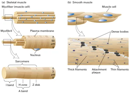
By using light microscopy, we are able to see differences between smooth muscle cells (SMCs) and skeletal muscles. In skeletal muscle, the arrangement of actin and myosin filaments cause a myofibrilar striated structure while in smooth muscle cells appear homogeneous. This difference can be explained by morphological differences of the individual muscle fibers between the skeletal muscle and SMCs. In the muscle fibers of skeletal muscle, the contractile elements like myofibrils are arranged in parallel, which showing a longitudinal view of bundles. However, in smooth muscle, the myofilaments form a cross-linked structure within the cells. Furthermore, SMCs contain actin as much as skeletal muscle cells per unit of cell volume, but five times less myosin1-3. The SMCs are spindle-shaped with a factor of 20 smaller than skeletal muscle cells (Fig. 1).
Fig. 1 Schematic illustration of (A) skeletal and (B) smooth muscles (according to Lodish 4th Ed).
The single nucleus of SMCs is centrally located in the cell, while in the multinucleated skeletal muscle cells, the nucleus are located directly at the periphery of the cell (Fig. 1).
In smooth muscle, the actin filaments are anchored to the cell membrane by the dense plaques and intracellular to the dense bodies via an actin binding protein known as α- actinin. Dense plaques and dense bodies contain α-actinin and vinculin, and they are therefore considered to be equivalent to the Z-discs of skeletal muscle. Based on the size of the protein structures, three types of filaments can be distinguished in the smooth muscles as follows: (A) The thin filaments having a diameter of Ø 5-8 nm consist mainly of filamentous actin, tropomyosin and actin-binding proteins, for example, caldesmon.
These thin filaments form together with (B) the thick myosin filaments (Ø 16 nm) the contractile apparatus of SMCs. Furthermore, actin filaments are connected with (C) intermediate filaments (Ø 10 nm) by adapter proteins. Intermediate filaments are a part of the cytoskeleton, which are not involved in the contraction of muscles, but have structural functions. Desmin and vimentin are two abundant intermediate filaments proteins in smooth muscle4.
SMCs can be functionally divided into two types, which in turn can perform different functions.
The "multi-unit" type, which is usually stimulated vegetatively, and this type is without spontaneous activity. They mainly occur in blood vessels with a larger diameter and the bronchi.
The "single-unit" type is of the hollow organs of the gastrointestinal tract (GI track), in which the SMCs are connected via gap junctions. They can almost simultaneously contract together and demonstrate a rhythmic activity of spontaneously active pacemaker cells.
Smooth muscles can be further distinguished in phasic and tonic contractile smooth muscle. Phasic contraction underlies the rhythmic contractions of for instance, the intestine, the portal vein and the ureter. Tonic contractions occur in most blood vessels, the trachea, and in the sphingters of the gastrointestinal tract5. The vascular smooth muscle cells are a main component of the vascular system like blood vessels, which regulate vessel tone, blood pressure and distribution of blood flow in the vascular system
like arteries, veins and capillaries6, 7. The vascular smooth muscle cells control and/or regulate blood stream through the vessels by contraction and relaxation, and therefore changes in the vessel volume8, 9. In the mature vessels, smooth muscle cells are mainly in the contractile state (also known as differentiated) and have an extremely low non- contractile phenotype8, 10. However, unlike skeletal muscle and cardiac cells, SMCs retain remarkable plasticity and can therefore change their phenotype during different pathophysiological conditions6, 10-13.
1.1.2 Molecular basis of contraction and relaxation
To analyze and to comprehend the function of smooth muscle cells in normal physiological conditions and in diseases, it is not easy without a complex study about cellular events, extracellular matrix components and their interactions together and with membrane components. The general principle of contractility of SMCs is the same as that described for skeletal muscle depends on ATP/ADP exchanges. Each myosin protein head possesses ATPase activity and controls its binding to actin in a cyclical manner that couples ATP binding and its hydrolysis into ADP and inorganic phosphate (Pi) to a conformational change in the myosin head14, 15.
In absence of ATP, the head of the heavy chains of myosin binds tightly to actin. Briefly, after binding of ATP, the myosin head can detach from the actin whereas the ATP is hydrolytically cleaved into ADP and Pi. The cleavage products initially remain on myosin, while myosin is weakly bound to actin. Only the sequential release of Pi and ADP leads to the formation of strongly bound myosin to actin, force-generating cross-bridges, conformational changes in the area of myosin heads and therefore the so-called "Power stroke", which finally causes the shortening of sarcomeres and thus the whole muscle14,
15.
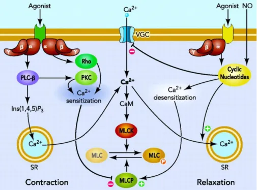
Fig. 2 illustrates a general signaling that regulates the smooth muscle contractile machinery depending on both Ca2+ and GTPase protein signaling16. Regulation of smooth muscle cell tone is mainly depends on calcium concentration and Ca2+ sensitization and desensitization, which in turn are regulated by various agonists bind to the specific receptors and in the subsequent activation of the SMCs contraction mechanism17, 18. An
increase in the intracellular Ca2+ concentration, in response to a wide spectrum of extracellular signals, activates Ca2+-Calmodulin depended myosin light chain kinase (MLCK), and subsequent phosphorylation of the regulatory light chain of myosin (MLC20) at serine 19, which is the main regulatory mechanism in activating smooth muscle cell contraction18-20. In contrast to contraction, relaxation is initiated by dephosphorylation of MLC20 by myosin light chain phosphates (MLCP). MLCP belongs to the family of hydrolases and is a heterotrimer of three subunits: a PP1c catalytic subunit, a 20-kDa subunit of unknown function, and a regulatory subunit, myosin phosphatase type 1 (MYPT1), which targets MLCP to myosin18 (Fig. 2).
In addition, G proteins increase Ca2+ sensitivity and therefore contractility in the smooth muscle cells due to the inhibition of MLCP21-23. Further studies demonstrated that this inhibition is controlled by the phosphorylation state of MYPT124, 25. Moreover, several groups reported that GTPase-induced Ca2+-sensitivity and therefore contractility of SMCs is based on the activation of Rho and Rho-associated protein kinase (ROCK) signaling pathways, in which ROCK inhibits MLCP by phosphorylation26-31.
Fig. 2 Schematic illustration of the signals that regulate and control the smooth muscle cell contraction and relaxation (according to Puetz et al 2009)16.
1.1.3 Smooth muscle cells phenotype plasticity and disease
SMCs in the vascular system and other systems in adult animals are differentiated cells, whose principal function are the contraction and regulation of blood vessel tone, blood pressure, and blood flow7, 8, 10, 32. Notably, unlike other cells that are terminally differentiated, smooth muscle cells retain their ability to change their phenotype from a contractile to a non-contractile phenotype (synthetic phenotype) and vice versa according to the physiological and pathophysiological conditions10, 11, 13, 33. The phenotype plasticity of SMCs (also known as phenotype modulation) plays a crucial role during vascular development, regulation of the vascular system (vascular remodeling) during blood flow, blood pressure, and also during diseases such as injuries, atherosclerosis, cancer, and diabetes10-12, 34-39. SMCs in a differentiated state express a variety of contractile proteins and contractile-associated marker proteins that regulate and control the correct function of these cells. In other words, in mature vessels contractile or differentiated SMCs are characterized by the expression of marker proteins such as smooth muscle α-actin (αSMA), smooth muscle myosin heavy chain (smMHC), calponin, h-caldesmon, SM2240, 41, and smoothelin42-44. Consequently, many studies have also shown that up and/or down regulation of these proteins are correlated by phenotype modulation of VSMCs and diseases45-47. For instance, upon injury, VSMCs dedifferentiate into non-contractile smooth muscle cells, with an increased rate of proliferation, migration and also synthesis of extra cellular matrix components13, 48, while at the same time VSMCs showed a reduction in expression levels of marker proteins12, 41.
Many groups reported specific factors and regulatory pathways, which directly or indirectly promote and control SMCs phenotype modulation during cellular processes and diseases8, 10, 11, 32. Tacking together, these results suggested that the phenotype modulation plays a crucial role in vascular development10, 49 and repair of vascular diseases like injury36, 37. Furthermore, the model that has evolved in the last years for the regulation and controlling of VSMCs phenotype modulation is extremely complex. It depends on environmental cues, signaling pathways that control expression of genes characteristic for VSMCs. Therefore, we are just beginning to understand some of the
molecular mechanisms that control phenotype modulation of VSMCs during pathophysiological conditions.
1.1.4 Actin cytoskeleton
1.1.4.1 Actin and actin filaments
Actin is the most abundant protein in eukaryotic cells (up to 20% of the total protein)50, 51. It is a highly conserved protein family with six different actin proteins that is divided into three isoforms: 3 α-, 1 β-, 2 γ, which are tissue specific52. Alpha actins are found in muscle (α-skeletal, α-aortic smooth, α-cardiac), and γ2-enteric smooth, whereas β and γ isoforms are dominant in non-muscle cells. Smooth muscle α-actin is mainly expressed in vascular smooth muscle53, whereas in visceral smooth muscle, a high proportion of γ-actin is found. The β-isoform of actin is expressed in all smooth muscle tissues and appears to be a component of the cytoskeleton54. Actin, as a major component in cytoskeleton, is the best investigated protein in motility systems, and regulation of mechanical and biomechanical signals through the cells. However, its exact functions in different pathophysiological conditions, development, aging and diseases are not completely understood. Actin exists in two forms, monomeric, G-actin with a molecular mass of 42 kDa and filamentous, F-actin.
Actin, as an involved protein in the cell movements, controls contraction/relaxation mechanism by two actions: A) actin polymerization and depolymerization and B) interaction with motor proteins like myosin family. The average length of actin filaments in the contractile state of VSMCs is about 4.5 µm, whereas myosin filament length is only 1.6 µm 1, 54, 55. Actin filaments, Actin binding proteins (ABPs), myosin and other proteins that interact with the actin cytoskeleton, make stress fibers in the cells50, 56, 57, which are dominant in cell culture conditions57. The actin cytoskeleton contains two types of filaments, cross-linked filaments and cortical actin filaments50. Cross-linked filaments control cytoskeletal deformation, transmit forces and stabilize subcellular localization of cellular organelles, while cortical filaments stabilize the form of cells during different physiological conditions, cellular development and inhibit lateral movement of organelles in the cells50, 56.
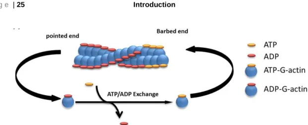
Actin filaments extend when ATP-G-actin monomers bind to the barbed end, while actin filaments disassemble when ADP-G-actin monomers lose from the pointed end58-60 (Fig.
3). Actin filaments are highly dynamic and their elongation involves both polymerization and depolymerization61-63. The polymerization of actin filaments occurs in three sequential steps: (A) nucleation step, (B) elongation step, and (C) steady state step61, 64. The first step (A) initiates with the formation of a trimer of G-actin monomers that is also known as a polymerization nucleus, which in the second step (B) will be used to elongate a filament by the addition of actin monomers to both of its ends. Finally, as actin filaments grow, (C) the concentration of G-actin monomers decreases until it reaches a steady state with the F-actin filaments (critical concentration) that are important to control assembly and disassembly of actin monomers56, 64, 65. With this notion that both the formation of a stable actin trimer as the initial point for polymerization and elongation are dependent on the critical concentration of monomers, dynamics of this concentration play a major role in assembly and disassembly processes.
Actin nucleation and Cc therefore require additional proteins known as 'actin binding proteins' that control and/or regulate the formation of a stable actin nucleus and Cc in the cells. Beside actin, actin binding proteins are the other abundant protein super family, which account for 10% of total cellular protein56. For instance, the Arp2/3 complex66, formins67, 68, and synaptopodin 2 (SYNPO2) as a novel actin protein regulate nucleation of actin and ADF/cofilin69, 70 disassembles actin filaments. While the function of actin nucleators such as the Arp2/3 complex65, 71, and formins has been extensively studied in cells, the role of SYNPO2 is not completely clear. The detailed biochemistry and function of the synaptopodin family on the actin dynamics is under investigation. Further detail about the synaptopodin family is discussed in the next section.
Fig. 3 Schematic illustration of actin polymerization/depolymerization. ADP binds with ~10-fold lower affinity to the nucleotide binding site of Mg-G-actin and ~200-fold weaker to the Ca-G-actin monomers as compared to the ATP and ADP-Pi, which is crucial for actin polymerization and depolymerization60.
1.2 Synaptopodin family
As described above, the functions of actin and the actin cytoskeleton are regulated or/and controlled during a variety of cellular processes which are involved in different pathophysiological conditions by ABPs50, 56, 72. The synaptopodin family comprises a conserved family of proteins that fall into three different members; Synaptopodin, Synaptopodin 2 (SYNPO2), and Synaptopodin 2 like proteins (SYNPO2l), which are also divided by different splicing in various isoforms73. The main feature of the synaptopodin family seems to be the natively unfolded form of these proteins, which is supposed to be necessary for their potential functions in the cells. Accordingly, the synaptopodin family may have different binding partners73. Although different genes, located on different chromosomes, express synaptopodin proteins they show a high similarity73. The intrinsically unfolded structure of the synaptopodin family members depends on position and/or the high proline content.
Since their discovery, the synaptopodin family members have been studied in different cell types. The synaptopodin family members play various roles in spine motility74, actin organization75, 76, cell differentiation77, cancer78-80, and heart development and somitogenesis81, 82. The detailed biochemistry and function of synaptopodin family is discussed in the following sections.
1.2.1 Synaptopodin
Synaptopodin (SYNPO) as the first member of the synaptopodin family was reported by Mundel et al. in 1997 as a novel actin binding protein which expresses in both differentiated kidney podocytes, associated with the actin cytoskeleton of postsynaptic densities, and in telencephalic synapses of the brain, associated with dendritic spines74, 83. Synaptopodin is expressed by the gene SYNPO, located on chromosome 5 (human).
Synaptopodin binds either directly to F-actin and α-actinin on several independent binding sites84 or indirectly to F-actin via binding to α-actinin85. At this time, the published human protein sequence included a 685-aa polypeptide with a calculated molecular mass of 73.7 kDa and an isoelectric point of 9.38. The appropriate murine homolog showed a 690-aa protein with a calculated molecular mass of 74.0 kDa and an isoelectric point of 9.27. In SDS-PAGEs SYNPO2 migrated with an app. molecular mass of 100 kDa. A sequence analysis showed a 84% sequence similarity between the human and murine synaptopodin. Furthermore, 2005 the Mundel group reported85, that murine synaptopodin includes three splice variants that are tissue-specific, short-variant (685 amino acids) neuronal, long-variant (903 amino acids) and variant T (181 amino acids) renal. Functional analysis demonstrated that synaptopodin caused a connection between the actin cytoskeleton and spine apparatuses of telencephalic neurons86, which may suggest a crucial role for synaptopodin in case of regulation of long-term potentiation (LTP). Further anaylsis showed that the lack of synpatopodin in synpo-/- mice caused a complete lack of spine apparatuses and in turn a loss of LTP and learning impairments87.
1.2.2 Synaptopodin 2
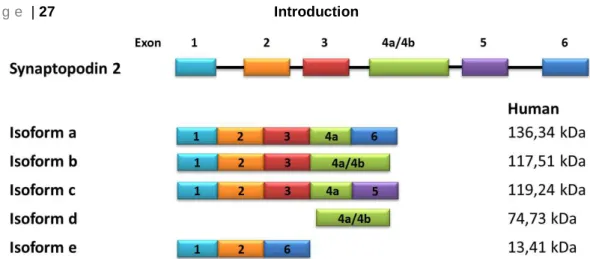
The novel actin binding protein synaptopodin 273, 75 (known as myopodin77 or fesselin88) regulates actin nucleation and polymerization in a Ca2+-Calmodulin manner89-91. SYNPO2 is expressed by the gene SYNPO2, located on chromosome 4 (human). Sequence analysis of SYNPO2 demonstrated a 47% amino acid similarity to synaptopodin. De Gank reported in 2008 three different isoforms which seem to be a result of different splicing events79, however Chalovich and Schroeter reported in 2010, 5 isoforms of SYNPO2 (Fig. 4 ).
SYNPO2 binds directly to F-actin and α-actinin at several binding sites92, 93.
Fig. 4 Schematic illustration of synaptopodin 2 isoforms in human.
SYNPO2 is a tumor suppressor protein80, 94, 95
and shuttles between the nucleus and the cytoplasm during differentiation and under stress such as heat shock77, 96. The suppressor activity of synaptopodin 2 on cell growth and cell motility seems to be dependent on phosphorylation and interaction of myopodin by/with integrin-linked kinases95. It has been shown that the differentiation of myoblasts to myotubes affects translocation of SYNPO2 from the nucleus to the cytoplasm77. In smooth muscle cells SYNPO2 is expressed in the cytoplasm where it is localized in dense bodies97 (and Z-disc in striated muscle77, 93).
Additionally it has been observed that SYNPO2 accumulated in the cell cytoplasm during tumor development and metastases79, 80, 94. Notably, loss of nuclear myopodin is connected to the clinical outcome94. However, its exact function is still unclear and under intensive investigation.
Detailed analyses of the biochemical and biophysical properties of SYNPO2 from turkey gizzards showed that it can control critical functions of actin, such as nucleation and elongation of monomeric G-actin as well as bundling and crosslinking of F-actin filaments73, 75, 90, 98
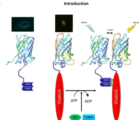
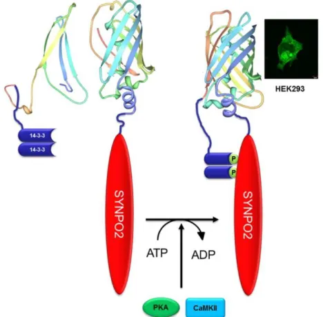
. A further link to the actin cytoskeleton is established by the ability of SYNPO2 to bind to myosin76, α-actinin93, 99, and filamin93. The binding to α-actinin is important for the regulation of the transport of SYNPO2 to the nucleus, which was observed in C2C12 cells and cardiac myocytes93, 100. This shuttle has been observed after the application of heat shock77, 96, upon which murine SYNPO2 is phosphorylated by protein kinase A (PKA) and Ca2+-calmodulin dependent kinase II (CaMK II)100. This posttranslational phosphorylation releases SYNPO2 from α-actinin and induces the
binding of SYNPO2 to the 14-3-3 protein100-102. Binding of SYNPO2 to 14-3-3 requires the presence of two 14-3-3 binding motifs73, 100, 101. Afterwards the 14-3-3-SYNPO2-complex binds to the protein importin-α, and shuttles to the nucleus96, 100-102.
Interestingly, the human SYNPO2 provides only one 14-3-3 binding motif, the required second motif is missing. Therefore, the mechanism by which SYNPO2 is translocated to the nucleus of human cells is still unclear.
1.2.3 Synaptopodin 2-like
The synaptopodin 2-like protein also known as cytoskeletal heart-enriched actin- associated protein (CHAP) or tritopodin is the third member of the synaptopodin family81,
103. A gene (SYNPO2L) located on chromosome 10 in human expresses CHAP protein.
CHAP is a new Z-disc protein that is expressed in differentiated heart, skeletal muscle during embryonic development81 and in adult aorta, carotid and coronary arteries in vascular smooth muscle cells, but not during embryonic development82. This protein exhibits approximately 31% similarity to SYNPO2 and 30% to synaptopodin. This sequence similarity may suggest similar features for CHAPs81, 82. Beqqali reported two isoforms of murine CHAP (mCHAP) that are produced by alternative splicing. CHAPa resulted from exons 1, 2, 3, and 5 and has a molecular weight about 140 kDa, while CHAPb started with a start-codon in exon 4 and produced a protein with 110 kDa. A sequence analysis of both isoforms showed a classical NLS sequence (KKRR) for both CHAPa and CHAPb and a PDZ domain for CHAPa. However, this NLS was not crucial for nuclear import of mCHAP. A protein expression analysis of different isoforms showed that CHAPb was only expressed during early skeletal muscle and heart development, while the long isoform CHAPa was expressed around day E 17.5 and a high level of CHAPa protein was detected in adult heart and skeletal muscle, whereas CHAPb protein was undetectable.
Immunofluorescence studies demonstrated a low nuclear localization and a strong colocalization of mCHAP proteins and α-actinin within the Z-discs in the sarcomers of embryonic cardio myocytes. Although, CHAP contains a C-terminal actin-binding domain, it interacts with actin via binding to α-actinin. Zebra fish studies using antisense morpholino oligonucleotides demonstrated a crucial role of CHAP during heart
development and somitogenesis. Downregulation of CHAP in zebra fish caused dysfunction of cardiac contractility, myofibrillar disarray, disruption of somite boundaries, sarcomeric integrity and notably muscle defects in case of the contraction.
1.3 Protein-Protein interaction
All cellular processes, including development, pathophysiological conditions and diseases are controlled by signaling pathways, which in turn are based on protein-protein interactions. Correct localization of proteins, protein-protein interactions and their dynamic at the plasma membrane and along the signaling cascade play important roles in cell signal transductions49, 104, 105
. Although, many biochemical techniques like immunoprecipitation, pull-down assay, yeast two-hybrid and other in vitro assays elucidated many details about protein-protein interactions, the existence of protein complexes and their physiological roles in living cells is of greater interest, especially in cases of diseases and development studies106, 107. Despite the evidences and results from biochemical methods, there are many details about the exact roles and functions of proteins that remain unclear.
To investigate the existence of protein complexes and in turn also their physiological roles in cells, the methods often rely on Fluorescent tagged proteins and therefore conventional fluorescence imaging methods to detect the distribution and/or protein- protein interaction in a cellular context108-110. Due to their sensitivity and selectivity, fluorescence imaging techniques like fluorescence and confocal microscopy stand out among the other optical techniques used in characterization of structures, detection and quantification of processes like protein-protein interaction109-112. Notably, these techniques provide useful information on diversity of process and involved proteins that can be related to their physiological functions. Regarding to these evidences, förster resonance energy transfer (FRET) and more recently bimolecular fluorescence complementation assay, BIFC, are two powerful assays that facilitate and realize live imaging of protein interactions in the cellular context. It has been recently reported that 14-3-3 proteins as chaperone-like adaptor proteins are able to bind a specific sequence of SYNPO2101, which in turn is crucial for the interaction of importinα with NLSs of SYNPO2
and its shuttling from the cell cytoplasm to the nucleus96, 101. This synergy strongly depends on the phosphorylation form of SYNPO2 as shown by in vitro experiments100. Nevertheless, this evidence waited for more physiological information about these proteins in the cellular context.
We will use specific techniques that should allow us to analyze the interaction between SYNPO2, 14-3-3 and importinα in a cellular context. In this way we will be able to increase our understanding of both the basic mechanisms of interaction between SYNPO2, 14-3-3 and importinα in cells and also how this interaction controls subcellular distribution of SYNPO2.
1.3.1 Förster resonance energy transfer
FRET is a commonly used method to investigate molecular dynamics in biochemistry113, protein-DNA interactions114, protein-protein interactions, and protein conformation changes115 in both in vitro and in vivo116-118. It is describing an energy transfer between two fluorescent (fluorophores) that are tagged to proteins of interest. Notably, the introduction of fluorescent proteins like GFP and GFP variants revolutionized one more time the use of this method for protein-protein interaction studies and the study of protein conformational changes in cellular context116, 119. In principle, FRET occurs when emission and absorption spectra of two different chromophores, usually two green fluorescent protein variants, overlapped. However, this is only achieved if two chromophores fused to two different proteins or two different sites of one protein, which it becomes a distance range between 10-80 Å according to the conformational changes of protein or the interaction of two proteins (Fig. 5). Combination of fluorescent conjugated proteins and FRET assay offer a unique chance to visualize and to monitor a real-time protein-protein interaction, conformational changes, and localization studies of proteins in living cells117, 118, 120, 121.
Fig. 5 Principle of FRET assay used in vivo. Two fluorescent fusion proteins were used to perform FRET measurements with CFP as a donor and YFP as an acceptor. FRET occurs as a non-radioactive energy transfer between two fluorophores, in which the excitation of the donor induces an energy transfer from the donor to the acceptor resulting in the reduction of the donor and an increase of acceptor fluorescence.
The critical point is 10 nm distance.
1.3.2 Bimolecular fluorescence complementation (BIFC)
Although, FRET is a powerful method for colocalization and protein-protein interaction studies of the fluorescent conjugated proteins, its limitations include the crucial distance
~ 10 nm, and choice of the best sites for ligation of fluorescent proteins (chromophores) that may lead to false-positive or -negative, remains a significant challenge117, 122. For instance, false-negative can occur when the distance between two Fluorescent labeled components in large protein complexes is too far. Therefore optimizing and characterizing the best FRET constructs can be time-consuming. The second technique, known as bimolecular fluorescence complementation (BIFC), is an alternative method that eliminates these limitations in case of distance and false negative123, 124. Notably, this method offers a simultaneous visualization of multiple protein interaction in living cells125,
126. It is based on fluorescence development, according to complementation of two non- fluorescent fragments when they associate together by the interaction between interested proteins fused to each fragment123, 127. Therefore, the reconstitution of the
fluorescent protein is activated by the direct interaction between the two target proteins.
Since BIFC introduction, different BIFC-based assays have been developed and used in a cellular context. For instance, BIFC was used to visualize the molecular mechanism of protein-protein interaction in living cells128, protein dimerization/oligomerization, protein ternary complex formation129.
Fig. 6 Schematic illustration of the BIFC assay used in this work according to Hu, CD. et al. 2003 130. BIFC fluorescence development is based on the interaction between protein A (14-3-3) and protein B (SYNPO2) in a cellular context.
1.4 Aim of the project
Vascular smooth muscle cell (VSMC) modulation is essential during vascular development, contributes to the vascular lesion formation and plays an essential role in diseases such as atherosclerosis, post-angioplasty restenosis and also during vascular repair. The mechanisms regulating the modulation of this cell type are therefore of great interest.
Several studies have shown that alterations in the function and/or expression level of contractile and cytoskeletal proteins are associated with VSMCs modulation. Moreover, several studies have revealed that actin is associated with phenotype modulation of VSMCs. The structures of actin networks and their cellular functions are regulated by other proteins, generally known as actin binding proteins (ABPs). One novel class of such proteins is synaptopodin 2 (SYNPO2), of which there are at least five isoforms.
The functional features and significant roles of SYNPO2 in VSMCs, characterized by an abundant expression of SYNPO2, are so far unknown.
The purpose of this work is to find out (A) the consequences of the VSMCs phenotype switch on the subcellular distribution of SYNPO2, (B) whether SYNPO2 plays an active role in this process, (C) whether cAMP dependent signaling pathways like PKA, are required for SYNPO2 trafficking in VSMCs, and finally (D) to investigate the molecular mechanism of SYNPO2’s shuttling by using biophysical techniques such BIFC and FACS-BIFC assay in a cellular context.
2 Materials and Methods
2.1 General materials
In this work, the materials and reagents were purchased from Qiagen, New England Bio- Lab, Sigma Aldrich, Merck, Invitrogen, and Roth. Sequencing of DNA fragments were performed by MWG. Primers were designed with snap-gene viewer software and ordered form Invitrogen and MWG.
CHEMICALs
Table 1 All chemicals used in this work.
NAME PROVIDER
3-Isobutyl-1-methylxanthin (IBMX) Sigma-Aldrich
Acrylamide/Bisacrylamide AppliChem
Agarose Sigma-Aldrich
Ammonium peroxodisulfate (APS) Serva
Ammonium sulfate AppliChem
Ampicillin AppliChem
β-mercaptoethanole Serva
Bacto-agar Roth
Bacto-tryptone Sigma-Aldrich
BCA reagents Pierce
Bradford reagent BioRad
Chloramphenicol Roth
Collagenase I, II Worthington
Collagen I Upstate
Complete protease inhibitor cocktail Roche
Coomassie Brilliant Blue BioMol Feinchem. GmbH Cover Slip Mounting Medium Poly Science
Dimethyl sulfoxide (DMSO) Sigma-Aldrich
Dried milk powder (fat free) AppliChem
Dithiothreitol (DTT) AppliChem
ECL Cell Attachment Matrix Millipore Ethylenediaminetetraacetic acid disodium
salt Dehydrate (EDTA)
Sigma-Aldrich
Ethanol (absolute p.a.) J.T. Baker
Fetal Bovin Serum Sigma-Aldrich
Formaldehyde Sigma-Aldrich
Forskolin Tocris
Fungicide Gibco
Glutathione Roth
Glycerol Sigma-Aldrich
H89 Tocris
Hank's Balanced Salt Solution (HBSS) Gibco
HCl J.T. Baker
HEPES Gerbu Biotechnik GmbH
Hoechst 33342 Sigma
Isopropyl-thio-β-d-galactoside (IPTG) Roth
Isopropanol Roth
Kanamycin Gerbu
Leptomycin B (LMB) Santa Cruz
LB-Agar Roth
LB-Medium Roth
L-Glutamine 200mM Gibco
Kanamycin AppliChem
KCl Sigma-Aldrich
KH2PO4 Sigma-Aldrich
K2HPO4 Sigma-Aldrich
Laminin I Sigma-Aldrich, L2020
Methanol (absolute p.a.) J.T. Baker
MgCl2 Sigma-Aldrich
N2 (liquid) Air Liquid
NaCl J.T. Baker
NaHCO3 Sigma-Aldrich
NaH2PO4 Sigma-Aldrich
Na2HPO4 Sigma-Aldrich
NaN3 Sigma-Aldrich
NaOH J.T. Baker
NP-40 AppliChem
Paraformaldehyde (PFA) Sigma
Penicillin Streptomycin (Pen Strep) Invitrogen
Phalloidin Invitrogen
Phosphate buffered saline (PBS) Invitrogen Phenylmethylsulfonyl fluoride (PMSF) Sigma-Aldrich
Polyacrylamide Roth
Ponceau S Sigma
Sodium dodecyl sulfate (SDS) AppliChem Tetramethylethylen di amine (TEMED) Serva Trypsin-EDTA Solution (1x) 0.05% Gibco
Tryptone MP Biomedicals
Tris Roth
Triton-X100 Sigma-Aldrich
Tween-20 Calbiochem
Trypan blue Sigma-Aldrich
KITs AND MATERIALs
Table 2 List of Kits and Materials
KIT PROVIDER
10 cm dishes for Agar Plates BD Biosciences
BCA Protein Assay Kit Pierce/Thermo Scientific
Cell culture dishes (10 cm) BD Biosciences
Cell culture dishes (6 cm) BD Biosciences
Cell culture dishes (24-well) BD Biosciences Cell culture dishes (12-well) BD Biosciences Cell culture dishes (6-well) BD Biosciences
Cover glass 10 mm VWR
Cover glass 12 mm VWR
G-Actin/F-actin In Vivo Assay Biochem Kit Cytoskeleton
Gel Purification Kit Qiagen
GoTaq® qPCR MasterMix Promega
Maxwell Simply RNA Promega
Micro-Amp Fast 96-Well Reaction Plate (0.1 ml) Invitrogen
Mini-Prep PeqLab,Qiagen
Micro tubes (1.5 ml) Eppendorf
Micro tubes (2 ml) Eppendorf
PCR Purification Kit Qiagen
PCR Soft-tubes 0.2 ml clear Biozym
Stripettes 5, 10, 25 (ml) Falcon
Tube 15, 50 (ml) Falcon
BUFFERs AND SOLUTIONs
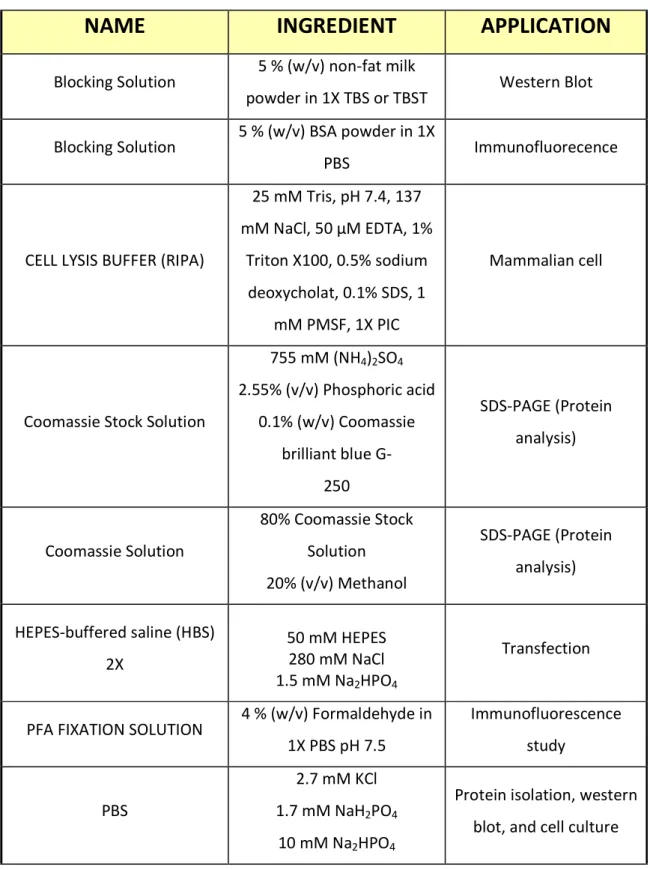
Table 3 All buffers and solutions used in this PhD work
NAME INGREDIENT APPLICATION
Blocking Solution 5 % (w/v) non-fat milk
powder in 1X TBS or TBST Western Blot Blocking Solution 5 % (w/v) BSA powder in 1X
PBS Immunofluorecence
CELL LYSIS BUFFER (RIPA)
25 mM Tris, pH 7.4, 137 mM NaCl, 50 µM EDTA, 1%
Triton X100, 0.5% sodium deoxycholat, 0.1% SDS, 1
mM PMSF, 1X PIC
Mammalian cell
Coomassie Stock Solution
755 mM (NH4)2SO4
2.55% (v/v) Phosphoric acid 0.1% (w/v) Coomassie
brilliant blue G- 250
SDS-PAGE (Protein analysis)
Coomassie Solution
80% Coomassie Stock Solution 20% (v/v) Methanol
SDS-PAGE (Protein analysis)
HEPES-buffered saline (HBS) 2X
50 mM HEPES 280 mM NaCl 1.5 mM Na2HPO4
Transfection
PFA FIXATION SOLUTION 4 % (w/v) Formaldehyde in 1X PBS pH 7.5
Immunofluorescence study
PBS
2.7 mM KCl 1.7 mM NaH2PO4
10 mM Na2HPO4
Protein isolation, western blot, and cell culture
137 mM NaCl, pH 7.5
SDS-loading buffer (2X)
8 M Urea, 100 mM Tris-HCl, pH 6.8, 2% SDS (w/v), 0.2%
bromophenol blue, 20 mM DTT, 1 mM PMSF, 1X PIC
Visualization of proteins by SDS-PAGE
SDS-Separation buffer (4X) 1.5 M Tris, pH 8.8
0.4% (w/v) SDS SDS-PAGE
SDS-Running buffer (10X) 250 mM Tris, 1.92 M
glycine, 1% SDS SDS-PAGE
TAE buffer (1X)
40 mM Tris acetate, 2 mM EDTA,
pH 8
TBS 150 mM NaCl
10 mM Tris, pH 7.5 Western blot
TBST
150 mM NaCl 10 mM Tris, pH 7.5
0.01% Tween 20
Western blot
Western blot running buffer (1X)
25 mM Tris-HCl 192 mM Glycine 20 % (v/v) MeOH
0.01% SDS
Western blot
MEDIA
All media used in this work are shown in the table. All media were autoclaved or sterile filtered before use.
Table 4 Bacterial and mammalian media
NAME INGREDIENTS
Cell Culture Medium (A10, A7r5)
DMEM (Gibco)10 % FBS (Sigma-Aldrich) 1% Penicillin (100 U) Streptomycin (100 µg)
(Pen/Strep, Invitrogen)
Cell Culture Medium (HEK293)
DMEM/F12 1:1 (Gibco) 10 % FBS (Sigma-Aldrich)
1% Penicillin (100 U) Streptomycin (100 µg) (Pen/Strep, Invitrogen)
Cell Culture Medium (raVSMCs)
DMEM/F12 1:1 (Gibco) 10 % FBS (Sigma-Aldrich)
1% Penicillin (100 U) Streptomycin (100 µg) (Pen/Strep, Invitrogen)
LB media for 1 L 25 g LB-dry powder, ddH20 to 1 L, pH 7.0
LB agar plates 15 g/l bacto agar
50 mg/l ampicillin or 35 mg/l Kanamaycin
INSTRUMENTs
Table 5 list of used instruments during this work.
INSTRUMENT PROVIDER
10, 20, 100, 200, 1000 µl, pipette Eppendorf
Autoclave Varioklav
Avanti Centrifuge G-52 I Beckman Coulter