antagonists
Marie Gienger
1, Harald Hübner
1, Stefan Löber
1, Burkhard König
2& peter Gmeiner
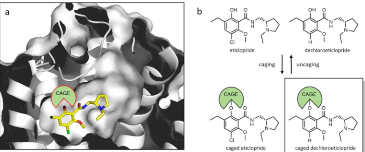
1*Dopamine is a neurotransmitter of great physiological relevance. Disorders in dopaminergic signal transduction are associated with psychiatric and neurological pathologies such as parkinson’s disease, schizophrenia and substance abuse. therefore, a detailed understanding of dopaminergic neurotransmission may provide access to novel therapeutic strategies for the treatment of these diseases. caged compounds with photoremovable groups represent molecular tools to investigate a biological target with high spatiotemporal resolution. Based on the crystal structure of the D
3receptor in complex with eticlopride, we have developed caged D
2/D
3receptor ligands by rational design. We initially found that eticlopride, a widely used D
2/D
3receptor antagonist, was photolabile and therefore is not suitable for caging. Subtle structural modification of the pharmacophore led us to the photostable antagonist dechloroeticlopride, which was chemically transformed into caged ligands. Among those, the 2-nitrobenzyl derivative 4 (MG307) showed excellent photochemical stability, pharmacological behavior and decaging properties when interacting with dopamine receptor-expressing cells.
Photopharmacology substantially contributes to our understanding of receptor function, potentially paving the way for new therapeutics
1. Hence, photoswitchable small molecules and neuropeptides have facilitated optical control of GPCR function
2. Moreover, photoactivable agonists and antagonists (caged ligands) have been devel- oped
3. The rapid spatiotemporal control of such ligands upon photo-uncaging provides valuable insights into kinetics of association, dissociation as well as receptor-induced signalling. In vivo photopharmacology has been a significant challenge, because delivery of UV light to deep tissue infusion is technically challenging. However, new wireless devices being able to co-deliver light and drug or prodrug simultaneously may be a major break- through
4. Caged compounds consist of a biologically active molecule masked by a photolabile protective group, to prevent target binding and thus attenuate biological activity. Upon suitable illumination, photolytic cleavage of the cage leads to rapid release of the active molecule towards cellular targets via concentration jumps, ideally within the time span of a light pulse
5,6. Most prominent photosensitive masking groups are nitrobenzyl deriva- tives. These well-established cages have previously been introduced to a wide range of functionalities including ions
7,8, phosphates
9, phenols
10–13, amines
13and carboxylic acids
14. Photolytic cleavage of nitrobenzyl-type cages proceeds via a radical mechanism and is triggered by UV illumination with excitatory wavelengths ranging from 300 to 400 nm
15. Simple structural modifications involving formal introduction of two methoxy substituents allowed a cleavage with light of longer wavelengths
13,15,16.
The neurotransmitter dopamine is critically involved in the regulation of movement, fine-motor control, emotions and behavior. Its physiological effects are mediated via five G protein-coupled receptors (GPCRs), the dopamine receptors D
1– D
5. Irregularities in the dopaminergic system are related to psychiatric and neurolog- ical pathologies including Parkinson’s disease, schizophrenia and substance abuse
17,18. Whereas dopaminergic agonists are successfully used for the treatment Parkinson’s disease, D
2/D
3receptor antagonists reduce positive symptoms of schizophrenia and are of interest to treat addiction
19,20. Hence, the discovery of selective ligands for D
2/D
3receptors is still an active field of drug research
21–29.
Caged dopamine derivatives have been employed for kinetic experiments on neurotransmitter release and clearance
30–34, for electrophysiological experiments
34and for the mapping of dopamine receptors in brain slice preparations
31. However, the repertoire is limited to caged dopamine. In order to expand the range of such studies
1