New Phthalimide-based Sensors for Chiral and Achiral Anions and Peroxides
Inaugural-Dissertation
Zur Erlangung des Doktorgrades
der Mathematisch-Naturwissenschaftlichen Fakultät der Universität zu Köln
vorgelegt von
Yrene Hortencia Díaz Pérez aus Caracas
(Venezuela)
Köln 2009
Berichterstatter: Prof. Dr. A. G. Griesbeck Prof. Dr. B. Goldfuß
Tag der mündlichen Prüfung: 3.02.2010
Gedruckt mit Unterstützung des Deutschen
Akademischen Austauschdienstes
For my Parents and Claus Miara
Acknowledgements
First of all, I would like to thank the Deutscher Akademischer Austausch Dienst (DAAD) for giving me the possibility to conduct my Ph.D in Germany in the research group of Prof. Dr.
Axel Griesbeck and especially my referee Veronica Metje.
Next, I would like to express my gratitude to Prof. Dr. Axel Griesbeck for giving me the opportunity to perform this work in his group and for his qualified and valuable help and the excellent working conditions in his group.
I owe special thanks to Prof. Dr. Bernd Goldfuß for the fruitful cooperation and for accepting to act as referee of my thesis as well as Prof. Dr. Klaus Meerholz and Dr. Dirk Blunk for being part of the evaluation committee of my thesis.
Another important person that I would like to thank is Dr. Franklin Vargas in Venezuela for his support and for his right orientation to my professional career.
It is also important to me to thank my colleagues Dr. Angela Raabe, Elmar Zimmerman, Johannes Uhlig, Dr. Miyeon Cho, Dr. Raúl Pérez, Dr. Alberto Soldevilla, Dr. Oliver Höinck, Marco Franke, Olga Hinze, Alan de Kiff, Viktor Schlundt, Sarah Strohmeier and Nestor Nazarov for the very nice time together in the laboratory and the good atmosphere. Special thanks go to Dr. Angela Raabe, Elmar Zimmerman, Dr. Raúl Pérez, Dr. Alberto Soldevilla and Sebastian Hanft for the help and friendly cooperation on my work.
I would like to give my thanks to the NMR department consisting of Dr. Nils Schlörer, Kathrin König and Gunter Arnold-Hässlich for the help by the NMR experiments, as well as Christoph Schmitz for his help with the elemental analysis and Andreas Adler for the micropipette. Dr. Jörg Neudörfl for the X-Ray measurements and Maria Schumacher for the theoretical calculations.
In the Physical Chemistry department, I would like to thank Dr. Dirk Hertel for his help and dedication to the measurements of lifetimes and Georgios Liaptsis for conduction of the mass spectrometry. In the Biochemistry department, I would like to thank Dr. Kay Marin for his help and his availability in the chemoluminescence measurements.
The luminol project was a joint work, which is why I would like to thank Robert Fichtler for the nice time that we worked together, for his help and collaboration. I would like to thank Dr.
Axel Jacobi von Wangelin, who was a part of the Luminol project, for his help and friendship.
I would like to thank Tobias Robert, Stefanie Ritter, Jutta Schütte, Dorina Köbele-Milas and
Tobias Hermann for helping me correct my work and for the very, very nice time we have
shared together.
For their great support, I would like to say my Venezuelan friends thousand thanks.
I would like to thank Inger Miara on becoming a great guide for me, now that my parents have become so far.
For the support, understanding, help, dedication and thousand reasons more since I came to Germany and especially in the last months I would like to thank my husband Claus Miara.
A last thank goes to my parents (Nery de Díaz and Aquiles Díaz) as well as my brother Pablo
Díaz and all my familiy members for the absolute support and help during my study in
Venezuela and during my Ph.D., I am very grateful for all that.
Explanation
This work was performed from October 2006 to December 2009 under the supervision of Prof. Dr. Axel G. Griesbeck at the Department of Chemistry, Institute of Organic Chemistry, University of Cologne.
In the experimental part names in the format pydr[number] refer to the enumeration in the lab-journal.
Abbreviations
1
H NMR Proton Nuclear Magnetic Resonance Spectroscopy
13
C NMR Carbon Nuclear Magnetic Resonance Spectroscopy
Abs. Absorption
Ar. Aromatic
ACN/CH
3CN Acetonitrile b.p. Boiling point (°C)
cat.
Catalyst
Cbz Carbonylbenzyloxy
CL Chemoluminescence
CT Charge transfer
n-Bu n-Butyl
t-Bu t-Butyl
d Doublet
dd Doublet of Doublet
DA Diels-Alder
DABCO 1,4-Diazabicyclo[2,2,2]octane DBU 1,8-Diazabicyclo[5.4.0]undec-7-ene DCC Dicyclohexylcarbodiimide
DCM Dichlormethan
DMAP
N,N-DimethylaminopyridineDMBA
N,N-Dimethyl(phenyl)methanamine1,2-DMB 1,2-Dimethoxybenzene
1,3-DMB 1,3-Dimethoxybenzene 1,4-DMB 1,4-Dimethoxybenzene
DMBA
N,N-dimethyl(phenyl)methanamineDMPAA 2-(3,4-dimethoxyphenyl)acetic acid DMSO Dimethylsulfoxide
Em. Emission
equiv. Equivalent
eq. Equation
E
sSinglet energy
EtOAc Ethylacetate
EtOH Ethanol
Et
3N Trimethylamine
Exc. Excitation
F Fluorescence intensity
FRET Fluorescence Resonance Energy Transfer GC/MS Coupled gas chromatography-mass spectrometry
GP General procedure
h Hour
HMQC Heteronuclear Multiple-Quantum Coherence Experiment HOMO Highest Occupied Molecular Orbital
HRMs High Resolution Mass Spectrometry IC Internal conversion
ICT Internal Charge Transfer
IR Infrared spectrum
ISC Intersystem Crossing
JCoupling constant (Hz)
KDStern-Volmer constant
kq
Bimolecular quenching constant K
CTConstant of CT complex
k
FFluorescence rate constant
LUMO Lowest Unoccupied Molecular Orbital MeOH Methanol
M Molar concentration
m Multiplet
min. Minute
mmol Milli mole
M.p. Melting Point
MPAA 2-(4-methoxyphenyl)acetic acid
MS Mass Spectrometry
NMP
N-methyl-2-pyrrolidinoneNMR Nuclear Magnetic Resonance ns Nano second (10
-9s)
PET Photoinduced Electron Transfer
Q Quencher
q Quartet
RET Resonance Electron Trensfer R
fRate of flow (retention factor)
r.t. Room Temperature
s Second or singlet (in NMR) S
0Singlet ground state
S
1First excited singlet state
T Temperature
T
1First excited triplet state
TBA Tetrabutylammonium
t Triplet
THF Tetrahydrofuran
TLC Thin-layer Chromatography TSA
p-Toluenesulfonic acidUV Ultraviolet
UV-vis Ultraviolet visible
λ
Wavelength
ε
Molar extinction coefficient
µ Micro (10
-6)
τ
Lifetime
* Excited state
Φf
Fluorescence Quantum Yield
Abstract
The first part of this work describes the synthesis of fluorescent and non-fluorescent phthalimide derivatives via straightforward synthetic routes, including multicomponent reactions (MCRs) (scheme 1-a), and aromatic substitutions and reductions (scheme 1-b).
a.-
R1 NH2 O
+ H O R2
CO2Me
CO2Me
+ O
O
O O NH O R1 R2
R2 TSA, Ac2O,NMP 120 °C, 24 h
O O
O O NH O R1 R2
R2 MnO2120 °C, toluene
+
NH O R1 R2
R2 N O
O R3 H2N-R3
b.-
O O
O
NH2 N
O
O NO2 NO2
+ + H2 Pd/C N
O
O NH2 NEt3
toluene EtOH
R R R
OH O NH O
O
NH N R O
O H O
N O O
Ac2O N R
O
O NH O
Scheme 1
In the second part the synthesis of new photocages based on aminophthalimide-serine was carried out and the fluorescence quenching behaviour of these photocages was investigated (scheme 2)
N O
COO- OAc O
R1 R2
N O
O R1
R2
hν + CO2 + AcO-
Scheme 2
In order to obtain new chiral sensors for achiral and chiral anion recognition the fluorescent
sensors 107, 109-112 were synthesized in the third part of this work. The syntheses are based on
urea-activated phthalimides with stereogenic centers that were synthesized using an efficient
procedure involving a Curtius rearrangement (scheme 3).
N HO2C
O
O
N HN
O
O HN
O R 2. , 25 °C 1. PhOCOCl, NaN3 t-BuONa/DME, 75°C
R-NH2
Scheme 3
The non-fluorescent sensor 123 based on a thiourea-activated phthalimide with a stereogenic center was synthesized following a synthetic route involving five steps each of which could be performed with good yields (scheme 4).
NH O
O
H2SO4 / HNO3
NH O
O O2N
N O
O O2N
Br K2CO3, KI
N O
O H2N
H2,Pd/C
N O
O SCN
Cl Cl S
N O
O HN
HN S
NH2 Dioxan, Ar
Scheme 4
This work demonstrates the capability of a new series of fluorescent and non-fluorescent chiral sensors obtained through the previously described synthetic routes to recognize achiral and chiral anions and peroxides.
Photophysical properties of the sensors such as absorption (abs), excitation (exc), emission (em) wavelengths (
λ), Stokes shifts, singlet energies (E
s), fluorescence lifetimes (
τF), quantum fluorescence yields (Φ
F) and fluorescence rate constants (k
F) were determined in several solvents in order to compare the solvent effects on the different photophysical properties of the sensor.
The recognition of the achiral and chiral anions was performed through absorption, fluorescence and
1H NMR experiments. To consolidate the experimental results, theoretical calculations based on DFT methods at B31YP/6-31G
*level were carried out.
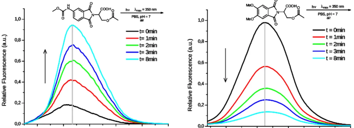
Recognition of peroxides was conducted by fluorescence experiments before and after
irradiation of the sensor−peroxide solutions at 350 nm.
NH NH NH2 O
O
NH NH NH2 O
O
NH i-Pr NH
NH2
i-Pr O
O
NH Bn NH
NH2
Bn O
O NH NH NH2 O
O NH
NH NH2 O
O
S NH2
NH NH
O O
Scheme 5
In the last part of this thesis the photophysical properties of luminol derivatives were
compared with the parent luminol. Furthermore, comparative studies of the chemoluminescence
efficiency of these luminol derivatives were carried out (scheme 5).
Kurzzusammenfassung
Im ersten Teil dieser Arbeit wurden verschiedene fluoreszierende und nicht-fluoreszierende Phthalimidderivate über vergleichsweise einfache synthetische Routen hergestellt. Eine der verwendeten Routen verlief über eine Multikomponeten-Reaktion (MCR, Abbildung 1-a).
Davon abgesehen, wurden überwiegend aromatische Substitution und Reduktionen eingesetzt (Abbildung 1-b).
a.-
R1 NH2 O
+ H O R2
CO2Me
CO2Me
+ O
O
O O NH O R1 R2
R2 TSA, Ac2O,NMP 120 °C, 24 h
O O
O O NH O R1 R2
R2 MnO2120 °C, toluene
+
NH O R1 R2
R2 N O
O R3 H2N-R3
b.-
O O
O
NH2 N
O
O NO2 NO2
+ + H2 Pd/C N
O
O NH2 NEt3
toluene EtOH
R R R
OH O NH O
O
NH N R O
O H O
N O O
Ac2O N R
O
O NH O
Abbildung 1
Im zweiten Teil der Arbeit wurde eine Synthese für neue Aminophthalimid-Serin Systeme, die als sogenannte photocages eingesetzt werden konnten, entwickelt und durchgeführt. Für diese photocages wurden eine Reihe von Fluoreszenzlöschungs-Experimenten durchgeführt (Abbildung 2).
N O
COO- OAc O
R1 R2
N O
O R1
R2 hν
+ CO2 + AcO-
Abbildung 2
Neue chirale Sensoren 107, 109-112 für die Erkennung von achiralen und chiralen Anionen
wurden im dritten Teil dieser Arbeit synthetisiert und untersucht. Die verwendete Synthese
zielte auf Harnstoff-aktivierte Phthalimide mit einem stereogenenen Zentrum, die unter Anwendung einer effizienten Methode (Curtius-Umlagerung) hergestellt wurden (Abbildung 3).
N HO2C
O
O
N HN
O
O HN
O R 2. , 25 °C
1. PhOCOCl, NaN3 t-BuONa/DME, 75°C
R-NH2
Abbildung 3
Der nicht fluoreszierende Sensor 123 basiert auf einem über Thioharnstoff aktivierten Phtalimid mit einem stereogenen Zentrum und wurde durch Fünfstufensynthese in guten Ausbeuten hergestellt (Abbildung 4).
NH O
O
H2SO4 / HNO3
NH O
O O2N
N O
O O2N
Br K2CO3, KI
N O
O H2N
H2,Pd/C
N O
O SCN
Cl Cl S
N O
O HN
HN S
NH2 Dioxan, Ar
Abbildung 4
In der vorliegenden Arbeit wurde das Potential dieser neuen fluoreszierenden und nicht fluoreszierenden chiralen Sensoren für chirale und achirale Anionen und Peroxide untersucht.
Die photophysikalischen Eigenschaften der Sensoren wie Absorption (abs), Anregung (exc), Wellenlänge (
λ), Stokes-Verschiebung, Singulett Energie (E
s), Fluoreszenz-Lebensdauer (
τF), Fluoreszenz-Quantenausbeute (Φ
F) and Fluoreszenz-Geschwindigkeits-konstante (k
F) wurden in verschiedenen Lösungsmitteln gemessen, um Lösungsmitteleffekte auf die verschiedenen photophysikalischen Eigenschaften der Sensoren zu vergleichen.
Die Erkennung von achiralen und chiralen Anionen wurde über Absorption, Fluoreszenz und
1
H-NMR Experimente bestimmt. Um die experimentellen Ergebnisse zu stützen, wurden DFT- theoretische Berechnungen auf B31YP/6-31G* Niveau durchgeführt.
Die Erkennung der Peroxide wurde durch Fluoreszenzexperimente vor und nach der
Belichtung von Sensor-Peroxid Proben bei 350 nm durchgeführt.
NH NH NH2 O
O
NH NH NH2 O
O
NH i-Pr NH
NH2
i-Pr O
O
NH Bn NH
NH2
Bn O
O NH NH NH2 O
O NH
NH NH2 O
O
S NH2
NH NH
O O
Abbildung 5
Der letzte Teil der Arbeit behandelt die Untersuchung der photophysikalischen Eigenschaften
von Luminol-Derivaten und vergleicht diese mit denen des Grundkörpers Luminol. Dazu wurde
unter anderem eine vergleichenden Studie der Chemolumineszenzeffizienz dieser Luminol-
Derivate durchgeführt (Abbildung 5).
CONTENT
1 INTRODUCTION 1
1.1 Electronic States 1
1.2 Energy level diagrams for photoluminescent molecules (Jablonski Diagram) 2 1.3 The Franck-Condon principle, absorption and emission spectra 3
1.4 Fluorescence 4
1.4.1 Characteristic of fluorescence emission 6
1.4.2 Fluorescence quenching 8
1.4.3 Resonance energy transfer (RET) 11
1.4.4 Mechanisms of quenching 12
1.5 Fluorescence lifetimes and quantum yields 14
1.6 Fluorescence sensing 15
1.6.1 Mechanisms of sensing 15
1.6.2 Cations sensing 16
1.6.3 Anions sensing 23
1.6.4 Chiral recognition 36
1.6.5 Hydrogen peroxide recognition 46
1.7 Phthalimides as chromophore 49
1.8 Multicomponent reactions 50
1.8.1 One-pot reactions with dienophilic acetylendicarboxylates 51 1.8.2 One-pot reactions with dienophilic maleimide and methyl maleimide 52
1.9 Chemoluminescence 53
2 AIM OF THE WORK 57
3 RESULTS AND DISCUSSION 59
3.1 Synthesis of nitro- and amino-substituted phthalimide derivatives from benzylamine 59 3.2 Synthesis of phathalimide derivativess from 4-amino-2-benzylisoindoline-1,3-dione 59
3.3 Synthesis of nitro-, amino-substituted phthalimide deriva-tives from 2,2-
Diphenylhydrazine 61
3.4 Quenching study of amino- and acetamido-substituted phthalimide derivatives 61 3.4.1 Quenching study of 4-amino-2-benzylisoindoline-1,3-dione 62 3.4.2 Quenching study of N-(2-Benzyl-1,3-dioxoisoindolin-4-yl)acetamide 64 3.4.3 Preliminary study: fluorescence activation of 2-(diphenyl-amino)-4-aminoisoindoline-1,3-
dione through cation coordination 67
3.5 From 1-(2-Aminonaphthalen-1-yl) naphthalene-2-amine to chiral phthalimides 71
3.6 Multicomponent Reaction 73
3.6.1 One-pot reaction with dienophilic dimethyl acetylenedi-carboxylate 73
3.7 Synthesis of Phthalimide-Serine Couples 78
3.7.1 Irradiation of caged acetates 3-acetoxy-2-(5,6-dimethoxy-1,3-dioxoisoindolin-2- yl)propionic acid and 3-acetoxy-2-(5-methoxycarbonylamino-1,3-dioxoisoindolin-2-yl)
propanoic acid 80
3.8 Synthesis of Chiral Phthalimide-Urea-Conjugates 85
3.9 Photophysical properties, anion sensing and chiral recognition by chiral phthalimide-
urea-conjugate 87
3.9.1 Photophysical properties 87
3.9.2 Anion Sensing 89
3.9.3 Chiral recognition 108
3.10 Fluorescence study of the sensors 107 and 109-112 with different peroxides 116
3.11 Synthesis of Chiral Phthalimide-Thiourea-Conjugate 123
3.12 Photophysical Properties, Anion Sensing and Chiral Recognition by Chiral Phthalimide-
Thiourea-Conjugates 123
3.13 Synthesis and Photophysical Properties of Luminol Derivates 132
3.13.1 Photophysical data and spectroscopic properties 134
3.13.2 pH-Dependence on absorption and steady-state fluorescence of 129-135 136
3.13.3 Chemoluminescence (CL) 138
4 CONCLUSION 145
5 EXPERIMENTAL PART 153
5.1 General Remarks 153
5.2 General Procedures 155
5.3 Synthesis of nitro; amino- and acetamide-substituted phthalimide derivatives 158 5.4 Preliminary quenching study of 4-amino-2-benzylisoindo-line-1,3-dione, N-(2-benzyl-
1,3-dioxoisoindolin-4-yl)acetamide. 164
5.5 Preliminary study for fluorescence activation of 2-(diphenylamino)-4-
aminoisoindoline-1,3-dione through cation coordination 164 5.6 Reactions with 1-(2-Aminonaphthalen-1-yl) naphthalene-2-amine to get chiral
phthalimides 165
5.7 Multicomponent coupling with dienophilic dimethyl acetylenedicarboxylate 168
5.8 Anilines via MnO2-mediated oxidation 170
5.9 Synthesis of Phthalimides from aniline derivatives 172
5.10 Reactions of Dimethyl 3-acetamido-4,6-diethylbenzene-1,2-dioate with acid and basic 174
5.11 Synthesis of Phthalimide-Serine Couples 175
5.12 Synthesis of chiral phthalimide-Urea-Conjugates 184
5.13 Photophysical properties, anion sensing and chiral recognition by chiral phthalimide-
urea-conjugate 191
5.14 Fluorescence study of chiral phthalimide-urea-conjugate with different peroxides 192 5.15 Fluorescence study of 107 and 109-112 with hydrogen peroxides 193
5.16 Synthesis of Chiral Phthalimide-Thiourea-Conjugate 193
5.17 Photophysical Properties, anion sensing and chiral recognition by Chiral Phthalimide-
Thiourea-Conjugates 197
5.18 Synthesis and Photophysical Properties of Luminol Derivates 197
6 APPENDIX 203
7 LITERATUR 207
1 Introduction
1.1 Electronic States
A full understanding of photochemical reactions requires an appreciation of the nature and properties of electronically excited states. Quantum mechanical concepts are invaluable in the analysis of the behavior of electronically excited molecules and can be used to rationalize experimental observations despite the approximations involved.
Each electron in a molecule carries a spin angular momentum with a spin quantum number s = 1/2. A point charge moving in a Coulomb field gives rise to a magnetic moment which, in the presence of a magnetic field, may take up one of two orientations. The magnetic moment may be aligned in the direction of the lines of force of the applied magnetic field or opposed to it, giving rise to two different energy states of the electron. A transition between the two energy levels corresponding to these states involves a change of alignment of the electron magnetic moment and is the basis of electron spin resonance.
The term electron spin refers to the alignment of the electron magnetic moment with respect to an imaginary magnetic field. If no field is present, there can be no splitting in the electron spin energy levels but the individual moments will still be present and will still dictate how the electrons interact with each other and with the nucleus.
The total spin angular momentum possessed by a many-electron atom or molecule is represented by the total spin quantum number S, which may be calculated as the vector sum of all the individual contributions from each electron. Two electrons, each possessing s = 1/2, may be present with their spins parallel or opposed. If the spins are opposed the total quantum number S is zero. If the electron spins are parallel the total quantum number S is 1/2 + 1/2 = 1.
The spin multiplicity gives the number of states expected in the presence of an applied magnetic field and is given by 2S+1. Thus, a molecule with all electrons spin-paired (which will be the case for the ground electronic state of most organic molecules) possesses S = 0 and a spin multiplicity of 1. Such an electronic state is referred to as a singlet state. The combination of ground state and singlet state is abbreviated by the symbol S
0.
[1]The Pauli Exclusion Principle states that two electrons in an atom can not have the same set
of four quantum numbers. This restriction requires that no more than two electrons can fit in one
orbital; furthermore, the two must have opposed spin states.
[2]S
2S
1S
0T
2T
1It is common to present these different spin states by a simplified molecular energy level diagram to which the appropriate labels are attached. An example of such a state diagram is illustrated in figure 1 for a generalized unsaturated hydrocarbon.
Figure 1: General state diagram of the (relative) energy of the lowest vibrational level
The excited T
1state is indicated to have an energy lower than that of the excited S
1state.
This lowering of the T
1state energy is due to spin correlation. It is a consequence of the operation of the Pauli principle, and is summarized by Hund´s rule
[1]of maximum multiplicity.
Even in the present situation where the two unpaired electrons occupy different orbitals, there is a minimum energy of electron-electron repulsion when their spins are parallel. This repulsion energy will determine the energy difference between the excited and singlet state and will depend on the extent of space between the orbitals involved.
1.2 Energy level diagrams for photoluminescent molecules (Jablonski Diagram)
Excitation of molecules are initiated by absorption of two modes of radiation, one centered around the wavelength
λ1(S
0→S
1), and the second around a shorter wavelength
λ2(S
0→S
2).
What happens to an electronically excited molecule that does not undergo some kind of chemical reaction? The molecule cannot persist in an excited state indefinitely, since it represents a situation unstable with respect to the ground state. Electron de-excitation must occur somehow, the excess energy being released as thermal or radiative energy. Transitions involving the de-excitation of electronically excited states that do not involve the emission of radiation are called nonradiative transitions.
The emitted radiation is called fluorescence if it originates in the de-excitation of an excited
state that has the same spin multiplicity as the ground state, and the emission is called
phosphorescence if it originates from the de-excitation of an excited state of spin multiplicity
different from that of the ground state (for example, T
1….S
0).
We can use the state diagram in figure 1 and indicate all the possible transitions that may occur between different energy levels. The result (figure 2) is called a Jablonski diagram.
Figure 2: Jablonski Diagram [3]
Radiative transitions are “vertical” transitions and involve a change in the total energy of the molecule due to the absorption and emission of a photon. Nonradiative transitions are
“horizontal” transitions and involve conversion from one state to another at a constant energy. A conversion between states of the same spin multiplicity is called internal conversion (IC) and for states of different spin multiplicity the term intersystem crossing (ISC) is used. In the solution phase the excess vibrational energy is rapidly removed by collisions with solvent molecules, a process sometimes referred to as vibrational relaxation (VR).
1.3 The Franck-Condon principle, absorption and emission spectra
The Franck-Condon principle states for the classical electronic transition of a vibrating molecule:
“Since electronic motions are much faster than nuclear motion, electronic transitions occur most favorably when the nuclear structure of the initial and final state are most similar”.
The conversion of electronic energy into vibrational energy may be the rate determining
step in an electronic transition between states of different nuclear geometries. A description of
the Franck-Condon principle would be that (a) for radiative transitions, nuclei geometries do not
change during the time it takes for a photon to “hit”, “be absorbed” and cause an electron to
jump; and (b) for nonradiative transitions, nuclear motions do not change during the time it
takes an electron to jump from one orbital to another. Figure 3 shows the potential energy curves of the possible Franck-Condon transition.
Absorption and emission spectra are not sharp lines with respect to the frequency
νof the absorbed or emitted light of the equation ∆E = h ν from the postulate that only one electron is excited or de-excited in an individual absorption or emission event.
In a set of molecules, an electronic transition is not as “pure” as it is in a single atom or molecule. This is due to the fact that, in order to describe the electronic states of a molecule, the motions of nuclei relative to one another (e.g. vibrations, rotations, collisions) must be considered. The sharp line or band which characterizes atomic transitions is replaced by a set of closely spaced lines in molecular absorption which may be only partially resolved or even completely unresolved. For organic molecules in solution - i.e. our case - this latter situation is common.
In analogy to absorption, the most probable emissions will be those which occur vertically.
In contrast to absorption, the equilibrium separation of the ground state potential-energy curve minimum is smaller than that of the excited state curve, so that the most probable vertical transitions produce an elongated ground state, while absorption produces a compressed excited state immediately after transition.
[4]Figure 3: Franck-Condon diagram [5]
1.4 Fluorescence
The electron in the excited orbital is paired (by opposite spin) to the second electron in the
ground state orbital. Consequently, return to the ground state is spin allowed and occurs rapidly
by emission of a photon. The emission rates of fluorescence are typically 10
8s
-1, so that a typical fluorescence lifetime is close to 10 ns.
[6]Some typical fluorescent substance (fluorophores) are quinine, fluorescein, rhodamine B, pyridine 1 etc, as shown in figure 4. The first observation of fluorescence from a quinine solution in sunlight was reported by Sir John Frederick William Herschel in 1845.
[6],[7]The experiment consisted of a observation of a glass of tonic water that was exposed to sunlight; a faint blue glow is frequently visible at the surface. The quinine in tonic water is excited by the ultraviolet light from the sun. Upon return to the ground state the quinine emits blue light with a wavelength near 450 nm. Due to this discovery, quinine was responsible for stimulating the development of the first spectrofluorometers that appeared in the 1950s.
N
HO N
MeO H
O
HO O
CO2H
O
(C2H5)2N N(C2H5)2
CO2H
N C2H5
(CH CH)2 N(CH3)2
ClO4-
Quinine Fluorescein Rhodamine B Pyridine 1
Figure 4: Typical fluorescent compounds
Other fluorophores are encountered in daily life like fluorescein and rhodamine.
Polynuclear aromatic hydrocarbons, such as anthracene and perylene, are also fluorescent, and the emission from such species is used for environmental monitoring of oil pollution. Pyridine 1 and rhodamine are frequently used in dye lasers.
The most intense and useful fluorescence is found in compounds containing aromatic functional groups with low energy
π→π*transition levels. Compounds containing aliphatic and alicyclic carbonyl structures or highly conjugated double bond structures may also exhibit fluorescence, but the number of these is small compared to the number of existing aromatic system. Another group which can fluorescence in solution are the unsubstituted aromatic hydrocarbon, the quantum efficiency usually increasing with the number of the rings and their degree of condensation.
The simple heterocycles, such as pyridine, furane, thiophene and pyrrole (figure 5) do not
exhibit fluorescence, on the other hand, fused ring structures ordinarily do. Fluorescence is
observed for compounds like quinoline, isoquinoline, and indole.
N
N
N
HN pyridine furan thiophene pyrrole
quinoline isoquinoline indole
non-fluorescent
fluorescent
O S H
N
Figure 5: Fluorescent and non-fluorescent molecules
The influence of halogen substitution is striking: the decrease in fluorescence with increasing atomic number of the halogen is thought to be in part due to the heavy atom effect, which increases the probability for intersystem crossing to the triplet state because of large spin- orbit coupling contribution.
[2]Substitution of a carboxylic acid or carbonyl group on an aromatic ring generally inhibits fluorescence. In these compounds, the energy of the n
, π∗transition is less than that of the
π, π*transitions.
[2]1.4.1 Characteristic of fluorescence emission
A fluorescence emission spectrum is a plot of the fluorescence intensity vs. wavelength (nm) or wavenumber (cm
-1). Emission spectra are dependent on the chemical structure of the fluorophore and the solvent in which it is dissolved. Another important feature of fluorescence is its highly sensitive detection. Fluorescence has some more general characteristics that will be describe in the following sections.
The Stokes Shift
The energy of emission is generally lower than that of absorption, and thus, fluorescence
occurs at lower energies or longer wavelengths.
[6]This shift in wavelength to a lower frequency
is called the Stokes shift (figure 6).
Figure 6: Absorption and emission spectra, Stokes shift [8]
Emission spectra are typically independent of the excitation wavelength
The same fluorescence emission spectrum is generally observed irrespectivly of the excitation wavelength (Kasha´s rule).
[9],[6]Upon excitation into higher electronic and vibrational levels, the excess energy is quickly dissipated, leaving the fluorophore in the lowest vibrational level of S
1. Because of this rapid relaxation (about 10
-12s), emission spectra are usually independent of the excitation wavelength.
There are exceptions, such as fluorophores that exist in two ionization states, each of which display distinct absorption and emission spectra. Also, some molecules are known to emit from the S
2level, but such emissions are rare and generally not observed in biological molecules.
[6]Effect of pH on Fluorescence
The wavelength and the emission intensity are likely to be different for the ionized and nonionized forms of the compounds, if they have acidic or basic substituents which are pH- dependent. For example, aniline has several resonance forms while the anilinium ion has only one. This resonance form leads to a more stable first excited state; fluorescence in the ultraviolet region is the consequence.
Experiments with pH-sensors have been used for the detection of end points in acid/base titrations. Changes in acid or base dissociation constants with excitation are common and occasionally as large as four to five orders of magnitude.
[2]Effect of solvent polarity
The effects of solvent polarity are one origin of the Stokes shift. Figure 7 shows a plot of
emission spectra of 4-dimethylamino-4´-nitrostilbene (DNS) in solvents of increasing polarity.
Figure 7: Emission spectra of DNS in H, hexane; CH, cyclohexane; T, toluene; EA, ethyl acetate; Bu, n-butanol [6]
Emission from fluorophores generally takes place at longer wavelengths than those at which absorption occurs. This loss of energy is due to a variety of dynamic processes that occur following light absorption. Figure 8 shows the Jablonski diagram for fluorescence with solvent relaxation.
Solvent effects shift the emission to even lower energies due to stabilization of the excited state by polar solvent molecules. In general, only fluorophores that are polar themselves display a large sensitivity to solvent polarity. Nonpolar molecules, such as unsubstituted aromatic hydrocarbons, are much less sensitive towards solvent polarity.
Figure 8: Jablonski diagram for fluorescence with solvent relaxation [6]
1.4.2 Fluorescence quenching
Fluorescence quenching refers to any process that decreases the fluorescence intensity of a
sample. Various processes of molecular interactions can result in quenching, for example:
•
Excited state reactions (chemical quenching)
•
Molecular rearrangements (chemical quenching)
•
Energy transfer (physical quenching)
•
Ground state complex formation (static quenching)
•
Collisional quenching (dynamic quenching)
In this opportunity two of the molecular interactions will be discussed, collisional or dynamic quenching and the ground state complex formation or static quenching.
Collisional quenching
Collisional quenching occurs when the excited state fluorophore is deactivated upon contact with some other molecule in solution, which is called quencher. The Jablonski diagram figure 9 illustrates this process and fluorescence resonance energy transfer (FRET).
Figure 9: Jablonski diagram with solvent relaxation and energy transfer (FRET) [6]
For collisional quenching the fluorophore is returned to the ground state during a diffusive encounter with the quencher. The molecule is not chemically altered in the process.
The decrease of intensity in this process is described by the Stern-Volmer equation (1):
F
0/ F = 1 + K [Q] = 1 + k
qττττ0[Q] (1) In this equation F
0is the emission intensity of the fluorophor without quencher, F is the emission intensity with quencher, K is the Stern-Volmer quenching constant, k
qis the bimolecular quenching constant,
τ0is the unquenched lifetime, and [Q] is the quenching concentration.
The Stern-Volmer quenching constant K indicates the sensitivity of the fluorophore to a
quencher. A linear Stern-Volmer plot is generally indicative of a single class of fluorophores, all
equally accessible to the quencher. It is important to recognize that observation of a linear Stern-Volmer plot does not prove that collisional quenching of fluorescence has occurred.
Static quenching
Static quenching can occur as a result of the formation of a nonfluorescent ground state complex between the fluorophore and the quencher. When this complex absorbs light it immediately returns to the ground state without emission of a photon.
Static quenching is described by equation 2:
F
0/ F = 1 +K
s[Q] (2)
Note that the dependency of F
0/ F on [Q] is linear, which is identical to the observed for dynamic quenching (eq. 1), except that the quenching constant is now the association constant.
The measurement of fluorescence lifetimes is the most definitive method to distinguish static and dynamic quenching. For static quenching τ
0/ τ = 1, in contrast to dynamic quenching F
0/ F = τ
0/ τ well as shown in figure 10.
Figure 10: Comparision of dynamic and static quenching [6]
Both quenching processes can also be distinguished by their differing dependence on temperature and viscosity. Higher temperatures result in faster diffusion and hence large amounts of collisional quenching. Higher temperatures will typically result in the dissociation of weakly bound complexes, and hence smaller amounts of static quenching.
The absorption spectrum of the fluorophor is, on careful examination, one additional
method to distinguish between static and dynamic quenching. Collisional quenching only
affects the excited states of the fluorophore, and thus no changes in the absorption spectra are
expected. In contrast, ground state complex formation will frequently result in perturbation of
the absorption spectrum of the fluorophore.
Combined dynamic and static quenching
It is possible that the fluorophore can be quenched both by collisions and by complex formation with the same quencher.
The Stern-Volmer plot is an upward curvature, concave towards the y-axis. The Stern- Volmer equation is modified to second order in [Q] (eq. 3), which accounts for the upward curvature observed when both static and dynamic quenching occur for the same fluorophore.
F
0/ F = 1 + (K
D +KS) [Q] + K
D KS[Q]
2(3) The dynamic component can generally be selected to compare the magnitude of the expected diffusion- controlled value of the solution, by the temperature or viscosity dependence of the values or from other available informations about the sample.
1.4.3 Resonance energy transfer (RET)
There is a process that causes a decrease in the fluorescence intensity of the donor and transfers the energy to an acceptor. This process is called resonance energy transfer (RET) and it can be considered a quenching process. The acceptor can be fluorescent or nonfluorescent, but in both cases the fluorescence intensity of the initially excited molecule is decreased.
Figure 11 shows that the fluorophore initially has two electrons in the highest-occupied (HO) molecular orbital. Absorption of light results in elevation of one electron to the lowest- unoccupied (LU) orbital.
Figure 11: Molecular orbital schematic for resonance energy transfer. The top row the size of fluorophores relative to the Föster distance R0 [6]
When RET occurs, the electron in the excited donor (D
R*) returns to the ground state.
Simultaneously an electron in the acceptor (A
R) goes into a higher excited-state orbital. If the
acceptor is fluorescent it may then emit. If the acceptor is nonfluorescent the energy is
dissipated as heat.
The important point is that quenching is due to short-range interactions between F and Q, shown in figure 12, and RET is due to long-range dipolar interactions between D
R*
and A
R. The rate of energy transfer is given by eq. 4
kT
(r) = 1 /
ττττD(R
0/ r)
6(4) where
τDis the donor lifetime in the absence of the acceptor, r is the center-to-center distance between D
Rand A
R, and R
0is the Förster distance.
The rate of quenching depends on the extent of interaction between the electron clouds in F and Q. The rate depends on the distance according to eq. 5
KE
(r) = A exp [-
ββββ(r - r
o)] (5) where r is the center-to-center distance between F and Q and r
c is the distance of the closestapproach at molecular contact. A is expected to have a value near 10
13s
-1and finally
βis typically near 1 Å
-1. This equation does not include the effect of diffusion on quenching and only describes the effect of distance on quenching but does not reveal the mechanisms of quenching.
Figure 12: Schematic of fluorescence quenching [6]
1.4.4 Mechanisms of quenching
There are at least three different mechanisms for singlet quenching:
1. Intersystem crossing (ISC) or the heavy atom effect 2. Electron exchange or Dexter interactions
3. Photoinduced electron transfer
The quenching process can occur by combination of these mechanisms.
[6]Intersystem crossing
ISC is a process in which the spin of an excited electron is reversed wich results in a change of the multiplicity of the molecules results. This process is most common in molecules that contain heavy atoms (the heavy atom effect). Apparently spin-orbital interactions become large in the presence of such atoms and a change in spin is thus more favorable. The presence of paramagnetic species such as molecular oxygen in solution also enhances intersystem crossing and consequently decreases fluorescence,
[2]as shown in figure 13.
Figure 13: Quenching by intersystem crossing [6]
Electron exchange
This interaction occurs between a donor D
Eand an acceptor A
E, where E indicates electron exchange. The excited donor has an electron in the LU orbital. This electron is transferred to the HO orbital of the acceptor, so the acceptor is left in an excited state.
Figure 14: Schematic for stepwise (top) or concerted (bottom) electron exchange [6]
At the same time or in a subsequent step an electron from the acceptor HO undergoes electron back transfer to the donor HO. Electron exchange is similar to RET because the energy is transferred to an acceptor. Figure 14 shows a schematic view for the electron exchange quenching.
Photoinduced electron transfer
In photoinduced electron transfer
(PET) a complex is formed between the electron donor D
Pand the electron acceptor A
p, yielding D
p +A
p-
. This charge transfer complex can return to the ground state without emission of a photon, but in some cases exciplex emission is observed.
Finally, the extra electron of the acceptor is returned to the electron donor. Figure 15 shows the molecular orbital diagram for photoinduced electron transfer.
Figure 15: Molecular orbital schematic for photoinduced electron transfer [6]
PET quenching can also occur by electron transfer from the excited fluorophore to the quencher. In PET the terms donor and acceptor do not classify which species is initially in the excited state. This is the difference from RET, where the fluorophore is always the donor.
1.5 Fluorescence lifetimes and quantum yields
The singlet lifetime determinates the time available for the fluorophore to interact with a substrate or diffuse in its environment, and hence the information available from its emission.
The lifetime of the excited state is defined by the average time wich the molecule spends in the excited state. Generally, fluorescenece lifetimes are between 100 ps and 10 ns.
[6]Quantum yield of fluorescence is the number of emitted photons relative to the number of
absorbed photons.
[6]For a highly fluorescent molecule such as fluorescein, the quantum yield
under several conditions approaches unity. Species that do not fluoresce have quantum yield
that approach zero.
[2]1.6 Fluorescence sensing
The design of fluorescent sensors has attracted considerable interest due to its importance to analytical chemistry, clinical biochemistry, medicine, industrial and environmental chemistry etc. Numerous chemical and biochemical analytes can be detected by fluorescence methods:
cations, anions, neutral molecules and gases.
[10]Fluorescence sensors are more sensitive than absorption sensors (colorimetric sensors) because the light absorbance is measured as the difference in intensity between light passing through the reference and the sample. In fluorescence the intensity is measured directly, without comparison with a reference beam.
[6]Fluorescence sensing requires a change in a spectral property in response to the analyte.
Changes can occur in the fluorescence intensity, excitation spectrum, emission spectrum, anisotropy, or lifetime of the sensing probe.
[6]In a fluorescence sensing approach, the fluorophore is the signaling species, i.e. it acts as a signal transducer that converts the information in the presence of an analyte (guest) into an optical signal expressed as the changes in the photophysical characteristics of the fluorophore.
[10]The receptor recognizes the guest and a behaviour change in the fluorescence signal is produced (figure 16).
Figure 16: Principle of fluorescence sensing
1.6.1 Mechanisms of sensing
There is a variety of signaling mechanisms the simplest mechanism is collisional quenching,
where the fluorophor is quenched by the analyte. Static quenching can also be used for sensing
but the lifetime does not change.
[6]Other mechanisms have been described such as ground state
charge transfer,
[11a,b]photoinduced electron transfer (PET),
[11c,g]excimer / exciplex formation,
[12]intramolecular charge transfer
[11g],[13]and excited-state proton transfer.
[14]Resonance energy transfer (RET) is perhaps the most general and valuable phenomenon for fluorescence sensors.
[6]Due to the mechanisms of sensing fluorescent sensors can be classified in three classes:
[10]•
Class 1: fluorophores that undergo quenching upon collision with an analyte (e.g.
O
2, Cl
-).
•
Class 2: fluorophores that can reversibly bind an analyte. If the analyte is a proton, the term fluorescent pH indicator is often used. If the analyte is an ion, the term
fluorescent chelating agent is appropriate. Fluorescence can either be quenchedupon binding (CEQ type: Chelation Enhancement of Quenching), or enhanced (CEF type: Chelation Enhancement of Fluorescence). In other cases, the compound is said to be fluorgenic (e.g. 8-hydroxyquinoline (oxime)).
•
Class 3: fluorophore linked, via a spacer or not, to a receptor. The design of such sensors, which are based on molecule or ion recognition by the receptor, requires special care in order to fulfill the criteria of affinity and selectivity. These aspects are revelant to the field of supramolecular chemistry. The changes in photophysical properties of the fluorophore upon interaction with the bound analyte are due to the perturbation by the latter of photoinduced processes such as electron transfer, charge transfer, energy transfer, excimer / exciplex formation or disappearance etc.
In this work it was tried to approach achiral and chiral anions, cations and peroxides- sensors.
1.6.2 Cations sensing
Detecting cations is of great interest for different areas such as chemistry, biology or medicine. Sodium, potassium, magnesium and calcium are involved in biological processes such as the transmission of nerve impulses, muscle contraction, regulation of cell activity, etc.
Zinc is an essential component of many enzymes; it plays a major role in enzyme regulation, gene expression, neurotransmission, etc.
On the other hand, there are some metal ions toxic to organisms (mercury, cadmium, etc.),
and early detection in the environment is desirable. Aluminum is also potentially toxic: it is
probably at the origin of some diseases such as osteomalacia, anemia, neurodegenerytive or
bone diseases. Control of aluminum content of is thus necessary in the production of
agricultural goods as well as in the pharmaceutical industry.
[10]Colorimetric determination of cations based on changes in color on complexation by dye reagents started to be popular a long time ago, especially in the case of alkaline earth metals ions, which are efficiently chelated by agents of the EDTA type. Sine fluorimetric techniques are more sensitive than photometric ones, numerous fluorogenic chelating reagents were studied and applied to practical cases.
Fluorescent molecular sensors of the EDTA type exhibit high selectivity for calcium with respect to other ions present in cells. In the late 1960s Crown ethers and cryptands were discovered
[10]and opened up new possibilities for cation recognition with improvement of selectivity, especially for alkali metal ions.
Fluorescent sensors emerged some years later with the design of a fluoroionophore. The ionophore moiety has been recognized by the fluorophore, this experienced changes in its fluorescence properties due to binding between ionophore and fluorophore.
The stability of a complex between ionophore and fluorophore depends on many factors:
nature of cation, nature of solvent, temperature, ionic strength and in some cases also pH.
The connection between the ionophore and the fluorophore is very important in aspects of sensing design, bearing in mind the search for the strongest perturbation of the photophysical properties of the fluorophore by the cation. The ionophore may be linked to the fluorophore via a spacer, but in many cases some atoms or groups participating in the complexation belong to the fluorophore. Therefore, the selectivity of binding often results from the whole structure involving both signaling and recognition moieties.
Fluorescence sensors of cations will be presented in the following part. The recognition of the cation can be explained by different mechanisms.
Stanculescu and coworkers
[15]used tetrandrine (6,6´,7,12-tetramethoxy-2,2´-dimethyl- berbaman; TET; figure 17) as fluorescent sensor. They characterized the binding properties of TET as host towards alkaline and alkaline earth metals, engaged in a molecular recognition process. The recognition has been studied by UV-vis and fluorescence spectroscopy.
After titration of TET with Na
+, K
+, Mg
2+and Ca
+, changes in the absorbance of TET were
observed in the region of 300-325 nm, where TET starts to absorb with the derived spectra
slightly shifted. These aspects lead to the conclusion that TET selectively complexes Ca
2+and
Mg
2+ions which can be classificated as hard acids.
N
O O O
O
N O
O
1
Figure 17: 6,6´,7,12-Tetramethoxy-2,2´-dimethyl-berbaman; TET (1)
The fluorescence emission showed a hypsochromic shift (~6 nm) upon addition of Ca
2+, accompanied by a 1.5- fold fluorescence enhancement. The blue shift and the increase in fluoresecence intensity are attributed to Ca
2+binding to TET which results in a more rigid structure of the ligand after complexation. The fluorescence enhancement may happen due to the suppression of the intramolecular phothoinduced electron transfer (PET) from the oxygen ion pairs. Another explanations is that the metal binding alters the rate of one or more relaxation processes from the excited state: radiative decay, internal conversion (IC) or intersystem crossing (ISC).
Zinc is one of the most important transition metal ions found in nature, where it has multiple roles in both extra- and intra-cellular functions. Gunnlaugsson and coworkers
[16]were active in the development of supramolecular luminescent chemosensors for zinc and other ions and molecules. In this case they chose to use 4-amino-1,8-naphthalimide (figure 18) as a photostable fluorophore reporter in designing 2 , as it absorbs in the visible region and emits in the green, with Stokes shift of ca.100 nm. Importantly, 2 does not respond to Ca
2+and Mg
2+, or many other transition metal ions.
N O O
HN
N
CO2- Na+ CO2- Na+
2
Figure 18: 4-amino-1,8-naphthalimide (2)
![Figure 7: Emission spectra of DNS in H, hexane; CH, cyclohexane; T, toluene; EA, ethyl acetate; Bu, n-butanol [6]](https://thumb-eu.123doks.com/thumbv2/1library_info/3669209.1504209/30.892.211.665.126.410/figure-emission-spectra-hexane-cyclohexane-toluene-acetate-butanol.webp)
![Figure 68: Normalized absorption bands of 107 ( ▲ ), excitation ( ● , λλλλ em = 520 nm) and emission ( ■ , λλλλ exc = 380 nm) spectra of the complex [107-F - ] in acetonitrile](https://thumb-eu.123doks.com/thumbv2/1library_info/3669209.1504209/115.892.291.643.264.525/figure-normalized-absorption-excitation-λλλλ-emission-λλλλ-acetonitrile.webp)
