AUS DEM LEHRSTUHL FÜR KIEFERORTHOPÄDIE PROF. DR. DR. PETER PROFF DER FAKULTÄT FÜR MEDIZIN DER UNIVERSITÄT REGENSBURG
Untersuchung zum relativen Einfluss mechanisch-kompressiver Deformation bzw. hypoxischer Zustände auf das Expressionsverhalten
parodontaler Ligamentfibroblasten im Rahmen der kieferorthopädischen Zahnbewegung
Inaugural-Dissertation zur Erlangung des Doktorgrades
der Zahnmedizin
der
Fakultät der Medizin der Universität Regensburg
vorgelegt von Niklas August Ullrich
2019
1
AUS DEM LEHRSTUHL FÜR KIEFERORTHOPÄDIE PROF. DR. DR. PETER PROFF DER FAKULTÄT FÜR MEDIZIN DER UNIVERSITÄT REGENSBURG
Untersuchung zum relativen Einfluss mechanisch-kompressiver Deformation bzw. hypoxischer Zustände auf das Expressionsverhalten
parodontaler Ligamentfibroblasten im Rahmen der kieferorthopädischen Zahnbewegung
Inaugural-Dissertation zur Erlangung des Doktorgrades
der Zahnmedizin
der
Fakultät der Medizin der Universität Regensburg
vorgelegt von Niklas August Ullrich
2019
2
Dekan: Prof. Dr. Dirk Hellwig
1. Berichterstatter: Priv.-Doz. Dr. Dr. Christian Kirschneck 2. Berichterstatter: Prof. Dr. Jonathan Jantsch
Tag der mündlichen Prüfung: 18. März 2020
3 Zusammenfassung
Hintergrund. Bei der kieferorthopädischen Zahnbewegung (KZB) wird eine mechanische Kraft auf die Zähne ausgeübt, die pseudoinflammatorische, osteoklastogene Prozesse und Knochenremodelling im parodontalen Ligament (PDL) induziert, welche von humanen Parodontalligamentfibroblasten (hPDLF) über die Expression verschiedener Signalmoleküle vermittelt werden. Bisher ist allerdings nicht bekannt, ob diese Prozesse maßgeblich durch mechanische Zellverformung (Mechanotransduktion) oder durch begleitende hypoxische Bedingungen durch eine Kompression der parodontalen Blutgefäße induziert werden.
Material und Methode. hPDLF wurden randomisiert auf konventionelle 6-Well-Zellkulturplatten mit O 2 -undurchlässigen Polystyrolmembranen und auf spezielle Platten mit gasdurchlässigen Membranen (Lumox ® , Sarstedt) ausgesät, was eine experimentelle Trennung von mechanotransduktiven und hypoxischen Effekten ermöglichte, die während der KZB gleichzeitig auftreten. Um physiologische kieferorthopädische Druckkräfte in Kompressionszonen des PDL während der KZB zu simulieren, wurden hPDLF nach 24h Vorinkubation für 48h bei 2g/cm 2 mechanisch stimuliert. Quantifiziert wurden die Zellviabilität mittels MTT-Assay sowie die Gen- und Proteinexpression für die KZB relevanter Markergene mittels RT-qPCR bzw. Western Blot/ELISA. Darüber hinaus wurde die hPDLF-vermittelte Osteoklastogenese (TRAP + -Zellen) in einer 72h-Kokultur mit RAW264.7-Osteoklastenvorläuferzellen bestimmt.
Ergebnisse. Die Expression von HIF-1α, COX-2, PGE2, VEGF, COL1A2, Kollagen, ALPL, das RANK-L/OPG-Verhältnis auf mRNA/Protein-Ebene sowie die hPDLF-vermittelte Osteoklastogenese wurden durch die mechanische Belastung sauerstoffunabhängig signifikant erhöht, während Hypoxie keinen signifikanten zusätzlichen Effekt zeigte.
Schlussfolgerungen. Die zellulär-molekulare Vermittlung der KZB durch hPDLF über die Expression
verschiedener Signalmoleküle scheint überwiegend durch die Krafteinwirkung selbst
(Mechanotransduktion) gesteuert zu werden, während hypoxische Effekte nur eine untergeordnete Rolle
zu spielen scheinen. Im Rahmen der KZB scheint der Hypoxiemarker HIF-1α nicht primär durch eine
reduzierte O 2 -Versorgung stabilisiert zu werden, sondern unterliegt vielmehr einer mechanisch
induzierten Stabilisierung.
4 Abstract
Background. For orthodontic tooth movement (OTM), mechanical forces are applied to teeth triggering pseudo-inflammatory, osteoclastogenic and remodelling processes in the periodontal ligament (PDL), mediated by PDL fibroblasts via the expression of various signalling molecules. So far it is unknown, whether these processes are mainly induced by mechanical cellular deformation (mechanotransduction) or by concomitant hypoxic conditions via a compression of periodontal blood vessels.
Material and Methods. Human primary PDL fibroblasts were randomly seeded onto conventional 6- well cell culture plates with O 2 -impermeable polystyrene membranes and on special plates with gas- permeable membranes (lumox ® , Sarstedt), enabling an experimental separation of mechanotransductive and hypoxic effects, which occur concomitantly during OTM. To simulate physiological orthodontic compressive forces, PDL fibroblasts were stimulated mechanically at 2g/cm 2 for 48h after 24h of pre- incubation. We quantified cell viability by MTT assay as well as gene and protein expression of OTM- relevant marker genes by RT-qPCR and Western-Blot/ELISA. In addition, PDL-fibroblast-mediated osteoclastogenesis (TRAP + cells) was determined in 72h coculture with RAW264.7 osteoclast precursor cells.
Results. Expression of HIF-1α, COX-2, PGE2, VEGF, COL1A2, Collagen, ALPL, the RANK-L/OPG ratio at the mRNA/protein level as well as PDL-fibroblast-mediated osteoclastogenesis were significantly elevated by mechanical loading irrespective of the oxygen supply present, whereas hypoxic conditions had no significant additional effect.
Conclusions. The cellular-molecular mediation of OTM by PDL-fibroblasts via the expression of
various signalling molecules is expected to be predominantly controlled by the force application itself
(mechanotransduction), whereas hypoxic effects seem to play only a minor role. In the context of OTM
the hypoxic marker HIF-1α does not appear to be primarily stabilised by reduced O 2 supply, but rather
mechanically.
5 Inhaltsverzeichnis
1 Einleitung ... 6
2 Material und Methoden ... 7
2.1 Gewinnung humaner parodontaler Ligamentfibroblasten (hPDLF)... 7
2.2 Versuchsaufbau ... 7
2.3 Bestimmung der Zellzahl und Zellviabilität mittels MTT-Assay ... 8
2.4 Genexpressionsanalyse mittels quantitativer Real-Time Polymerase Chain Reaction (RT-qPCR) ... 9
2.5 Enzyme-Linked Immunosorbent Assay (ELISA) ... 9
2.6 Quantifizierung des Gesamtkollagens im Zellkulturüberstand ... 10
2.7 Western-Blot ... 10
2.8 TRAP-Histochemie (hPDLF-vermittelte Osteoklastogenese) ... 11
2.9 Statistische Analyse ... 11
3 Ergebnisse ... 12
3.1 Effekt der Mechanotransduktion vs. Hypoxie auf die Zellzahl und Viabilität von hPDLF ... 12
3.2 Effekt der Mechanotransduktion vs. Hypoxie auf das hPDLF-Expressionsmuster und die Stabilisierung von HIF-1α ... 12
3.3 Effekt der Mechanotransduktion vs. Hypoxie auf die Expression von RANK-L/OPG durch hPDLF ... 15
3.4 Effekt der Mechanotransduktion vs. Hypoxie auf die Proteinexpression von membrangebundenen RANK-L und die hPDLF-vermittelte Osteoklastogenese ... 17
4 Diskussion ... 18 The role of mechanotransduction versus hypoxia during simulated orthodontic compressive strain - an in vitro study of human periodontal ligament fibroblasts………....
Danksagung ...
6 1 Einleitung
Bei der kieferorthopädischen Zahnbewegung wird eine mechanische Kraft auf einen Zahn ausgeübt, welche auf zellulär-molekularer Ebene in parodontalen Druckzonen eine pseudo- inflammatorische Reaktion, Osteoklastogenese und Remodellingprozesse hervorruft. 1
Humane parodontale Ligamentfibroblasten (hPDLF) sind die vorherrschenden Zellen des Parodontalligaments (PDL). 2 Sie sind für die Regulation der Gewebehomöostase verantwortlich, bilden kollagene Strukturproteine und spielen eine regulatorische Rolle bei der angeborenen Immunabwehr. 1,2 Diese Zellen haben auch in der Vermittlung der kieferorthopädischen Zahnbewegung 1,2 eine wichtige Rolle und wurden daher in der kieferorthopädischen Grundlagenforschung 3-7 intensiv untersucht, insbesondere im Hinblick auf ihre Reaktionen auf kieferorthopädische Druck- und Zugkräfte oder parodontale Krankheitserreger und ihre Toxine. 8-10 In Kompressionszonen des PDL werden hPDLF bei kieferorthopädischer Krafteinwirkung mechanisch verformt (Mechanotransduktion) und damit mechanosensitive Rezeptoren und Ionenkanäle in der Zellmembran stimuliert. 11 Eine hPDLF- vermittelte sterile pseudoinflammatorische Reaktion führt zu einer höheren Expression von IL- 6, IL-8 und COX-2, gefolgt von einem verstärkten extrazellulären Matrixumbau und Knochenresorption, ausgelöst durch erhöhte Expression von RANK-L (Receptor-Activator-of- NFκ-b-Ligand) und reduzierte Expression von OPG (Osteoprotegerin). 1,7 Gleichzeitig stört die damit einhergehende Kompression der Blutgefäße während der Zahnbewegung die Durchblutung an druckbeanspruchten Stellen des PDL, 12 wodurch die lokale Sauerstoffversorgung innerhalb des PDL reduziert wird (Hypoxie). 13 Es wird vermutet, dass diese lokale Reduktion der O 2 -Zufuhr auch eine bedeutende Rolle bei der zellulären Regulation der kieferorthopädischen Zahnbewegung spielt. 13
Obwohl die molekularen und zellulären Prozesse der kieferorthopädischen Zahnbewegung, welche durch hPDLF unter mechanischer Belastung vermittelt werden, bereits häufig untersucht wurden, 3,7,14 ist noch unklar, ob diese Prozesse hauptsächlich durch die mechanische Verformung der hPDLF (Mechanotransduktion) bei kieferorthopädischer Krafteinwirkung oder durch begleitende hypoxische Bedingungen über eine Kompression der parodontalen Blutgefäße und eine Störung der lokalen Blutzirkulation ausgelöst werden. Das Verständnis der relativen Bedeutung einer Mechanotransduktion bzw. von hypoxischen Zuständen für die molekularen Prozesse der Zahnbewegung könnte langfristig die therapeutischen und präventiven Möglichkeiten der kieferorthopädischen Behandlung erweitern.
In dieser Studie wurde daher das Expressionsverhalten von Genen und Proteinen, die bereits
7
zuvor mit der kieferorthopädischen Zahnbewegung assoziiert wurden, 1,15,16 nach Druckapplikation unter normaler oder reduzierter Sauerstoffzufuhr durch Verwendung konventioneller 6-Well-Zellkulturplatten oder spezieller Zellkulturplatten mit gaspermeablen Membranen untersucht. Ziel war es, ein besseres Verständnis der jeweiligen Rolle der Mechanotransduktion und Sauerstoffzufuhr für die molekularen und zellulären Prozesse in druckbelasteten Bereichen des PDL während der kieferorthopädischen Zahnbewegung zu erhalten.
2 Material und Methoden
2.1 Gewinnung humaner parodontaler Ligamentfibroblasten (hPDLF)
Primäre hPDLF wurden aus parodontalen Geweberesten extrahierter kariesfreier Zähne gewonnen. Die Gewebeproben wurden in 6-Well-Zellkulturplatten (37°C, 5% CO 2 , 100% H 2 O) in Vollmedium (DMEM high glucose, D5796, Sigma-Aldrich ® , St. Louis, MI, USA); 10% FCS (P30-3306, PAN-Biotech, Aidenbach), 1% L-Glutamin (SH30034.01, GE Healthcare Europe, München), 100 µM Ascorbinsäure (A8960, Sigma-Aldrich ® ) und 1%
Antibiotika/Antimykotika (A5955, Sigma-Aldrich ® ) bis zum proliferativen Auswachsen von adhärent wachsenden Fibroblasten kultiviert. Die Charakterisierung der Zellen erfolgte durch hPDLF-spezifische Markergene und ihre spindelförmige Morphologie. 15,39 Für die in-vitro- Experimente wurden hPDLF der dritten bis fünften Passage, die aus sechs Individuen (3 männlich, 3 weiblich, Alter: 17-27 Jahre) gepoolt waren, mit einer Dichte von 2.000 Zellen pro mm 2 entweder auf konventionellen 6-Well-Zellkulturplatten ohne Sauerstoffdurchlässigkeit der Polystyrol-Basis/Membran (353046, BD, Heidelberg) oder auf speziellen gaspermeablen Lumox ® -Platten (94.6077.331, Sarstedt, Nürnbrecht) mit einer gasdurchlässigen Basis/Membran ausgesät, welche eine kontinuierliche Sauerstoffversorgung für die Fibroblasten durch die Membran von der Unterseite der 6-Well-Zellkulturplatte sicherstellte.
2.2 Versuchsaufbau
Um eine kieferorthopädische Druckbelastung von hPDLF in Kompressionsbereichen zu
simulieren, wurde unter Zellkulturbedingungen (37°C, 5% CO 2 , 100% H 2 O, 2ml DMEM/Well)
ein physiologischer kieferorthopädischer Druck von 2g/cm 2 für 48h nach einer
Vorinkubationsphase von 24h mittels einer sterilisierten Glasscheibe nach einem etablierten
8
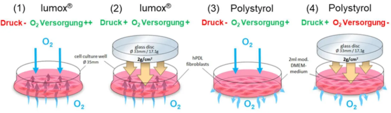
Modell aufgebracht (Abb. 1). 6,7,15,16,39,40. Vier Versuchsgruppen mit je 6-9 biologischen Replikaten (Proben) (n) in 2-3 aufeinanderfolgenden Experimenten (N) mit je 3 Replikaten wurden bei 70% Konfluenz für insgesamt 72h inkubiert: (1) keine mechanische kieferorthopädische Druckbelastung + Normoxie (Kontrolle, Lumox ® ); (2) mechanische Belastung + Normoxie (Lumox ® ); (3) keine mechanische Belastung + Normoxie (Kontrolle, Polystyrol); (4) mechanische Belastung + Hypoxie (Polystyrol) (Abb. 1). Bei herkömmlichen, nicht-gasdurchlässigen Polystyrol-Zellkulturplatten sowie in-vivo bewirken die aufgebrachten Druckkräfte nicht nur eine mechanische Verformung und Belastung der hPDLF, sondern unterbinden auch die Sauerstoffzufuhr (Gruppe 4), was bei Lumox ® -Platten mit intakter Sauerstoffversorgung über die gasdurchlässige Membran nicht der Fall ist (Gruppe 2) (Abb. 1).
Dieser Versuchsaufbau ermöglichte eine experimentelle Entkopplung von mechanotransduktiven und hypoxischen Effekten, welche während der Zahnbewegung gleichzeitig auftreten, und ermöglichte so eine Untersuchung ihrer jeweiligen Bedeutung.
Abb. 1. Versuchsaufbau der in-vitro-Experimente mit hPDLF zur Auswertung der vier Versuchsgruppen (1-4).
2.3 Bestimmung der Zellzahl und Zellviabilität mittels MTT-Assay
Die Zellzahl pro cm 2 wurde nach 72h Inkubation mit einem Beckman Coulter Counter Z2™
(Beckman Coulter GmbH, Krefeld) bestimmt. Die Zellviabilität der hPDLF wurde für alle experimentellen Gruppen mittels MTT-(3-(4,5-Dimethylthiazol-2-yl)-2,5- diphenyltetrazoliumbromid)-Assays bestimmt. Für die letzten fünf Stunden der 72h- Inkubationsphase wurden 400µl MTT-Lösung in PBS (5mg/ml, 4022.1, Carl Roth GmbH &
Co. KG, Karlsruhe) pro Well zugegeben. Nach Entfernung des Mediums wurde 1 ml DMSO
pro Well zugegeben. hPDLF wurden schließlich für weitere 5 Minuten bei 37°C inkubiert und
die Absorption bei 550nm mit einem ELISA-Reader (Multiscan GO Microplate
9
Spectrophotometer, Thermo Fisher Scientific, Schwerte) entsprechend der Zellviabilität quantifiziert.
2.4 Genexpressionsanalyse mittels quantitativer Real-Time Polymerase Chain Reaction (RT- qPCR)
Die Isolation und Aufreinigung der Gesamt-RNA erfolgte nach Ende der Inkubationszeit mit 1ml peqGOLD TriFast TM (PEQLAB Biotechnology) pro Well. Das gewonnene RNA-Pellet wurde in 20µl nukleasefreiem Wasser (T143, BioScience-Grade, Carl Roth GmbH & Co. KG, Karlsruhe, Germany) resuspendiert. Das zur Aufreinigung verwendete Protokoll führte zu einer hohen RNA-Integrität (RIN, 28S/18S-Verhältnis) ohne Verunreinigungen. 39 Nach cDNA- Synthese erfolgte die RT-qPCR mithilfe des Mastercycler ® -ep-realplex-S-Thermocyclers (Eppendorf) nach einem etablierten Protokoll in Doppelbestimmung. 44 Die bei diesem Vorgang erzeugte Fluoreszenz von SYBR ® Green wurde bei einer Wellenlänge von 521nm am Ende jedes Zyklus quantifiziert. Mithilfe der Software Realplex (Version 2.2, Eppendorf, CalqPlex Algorithmus) wurde ein C q Wert als Maximum der zweiten Ableitung der Fluoreszenzkurve ermittelt und aus den zwei ermittelten C q -Werten der Doppelbestimmung je Probe das arithmetische Mittel errechnet. Zur Standardisierung der Zielgene wurden die Referenzgene RPL22 und PPIB verwendet, für die bereits gezeigt werden konnte, dass sie von hPDLF unter diesen Versuchsbedingungen stabil exprimiert werden. 39 Die relative Genexpression wurde mit der Formel 2 -ΔCq mit ∆C q = C q (Zielgen) – C q (Mittelwert RPL22/PPIB) berechnet, und anschließend durch das arithmetische Mittel 2 -ΔCq der Kontrollgruppe dividiert, um eine relative Genexpression im Verhältnis zur Kontrollgruppe zu erhalten. 15,16 Sämtliche Primer wurden nach den MIQE-Qualitätsrichtlinien angefertigt. 41 Die nicht-modifizierten Primer wurden von der Firma Eurofins MWG Operon LLC synthetisiert und aufgereinigt. Für jedes Primerpaar und jede qPCR wurde eine No-Template-Control (NTC) ohne cDNA durchgeführt, um mögliche Fehler durch eine Kontamination auszuschließen. Die Primerspezifität wurde ebenfalls bereits durch Kirschneck et al. nachgewiesen. 39
2.5 Enzyme-Linked Immunosorbent Assay (ELISA)
Zur Quantifizierung von Osteoprotegerin (OPG), soluble RANK-L (sRANK-L), Alkalischer
Phosphatase (ALPL), Prostaglandin E2 (PGE2) und Vascular Endothelial Growth Factor
(VEGF) auf Translations-/Proteinebene im Zellkulturüberstand der hPDLF wurden
10
handelsübliche ELISA-Kits nach Herstellerangaben verwendet (OPG: EHTNFRSF11B, Thermo Fisher Scientific Inc.; sRANK-L: RD193004200R; Biovendor, Brno, Tschechische Republik, ALPL: OKEH00757; Aviva systems, San Diego, USA; PGE2: 514010; Cayman Chemical, Ann Arbor, USA VEGF-A: RAB0507; Sigma Aldrich). Es wurden je zwei unabhängige Experimente (N=2) mit jeweils 6 biologischen Replikaten verwendet (n=6). Die Proben zur Quantifizierung von OPG wurden zur photometrischen Vermessung 1:10 im dazugehörigen Puffer des ELISA-Kits verdünnt; die sRANK-L-Proben blieben dagegen unverdünnt. Die Menge an Protein wurde nach photometrischer Messung bei 450 und 550nm mithilfe einer Eichgeraden und unter Berücksichtigung der vorab ermittelten Zellzahl errechnet und schließlich als Induktion der Kontrollgruppe dargestellt.
2.6 Quantifizierung des Gesamtkollagens im Zellkulturüberstand
Für die Quantifizierung des Gesamtkollagens wurde ein handelsübliches Kit (K218-100, Biovision, Milpitas, USA) nach Angaben des Herstellers verwendet.
2.7 Western-Blot
Die Expression von membrangebundenem RANK-L und HIF-1α wurde mittels Immunoblot
mit einem RANK-L-, bzw. HIF-1α-spezifischen Antikörper untersucht. Das Gesamtprotein aus
den hPDLF wurde mit 100µl CelLytic™ M pro Well (C2978; Sigma-Aldrich ® ), ergänzt durch
Proteinasehemmer (Carl Roth GmbH & Co. KG), isoliert. Zur Reduktion der
Proteinaseaktivität wurden die Proteine auf Eis gehalten. Die Bestimmung der
Proteinkonzentration erfolgte mit RotiQuant (K015.3; Carl Roth GmbH & Co. KG) nach
Herstellerangaben. Für das Immunoblotting wurden unter reduzierenden Bedingungen gleiche
Mengen an Gesamtprotein auf einem 10%igen SDS-Polyacrylamid-Gel (RANK-L) und
8%igen SDS-Polyacrylamid-Gel (HIF-1α) getrennt und die Proteine mittels Elektroblotting auf
Polyvinylidendiflourid-(PVDF)-Membranen übertragen. Zur Reduktion unspezifischer
Bindungen wurden die Membranen mit 5%iger fettfreier Milch in trisgepufferter
Kochsalzlösung und 0,1% Tween 20 (pH 7,5; TBS-T) bei 4°C über Nacht geblockt. Danach
erfolgte die Inkubation der Membranen mit anti-RANK-L (1:2.000, ABIN500805, Antikörper-
online, Aachen), anti-HIF1α (1:2.000, Santa Cruz Biotech, Heidelberg), anti-HSP90 (Referenz,
1:500, Santa Cruz Biotech) und anti-ß-Actin (Referenz, 1:5.000, Sigma-Aldrich ® ) für jeweils
1h bei Raumtemperatur. Nach dreimaligem Waschen in TBS-T wurden die Blots für eine
weitere Stunde mit Meerrettich-Peroxidase-konjugiertem Anti-Kaninchen-IgG (Pierce,
11
Rockford, USA) verdünnt 1:5000 in 0,5%iger Milch in TBS-T bei Raumtemperatur inkubiert.
Antikörperbindungen wurden mit einem erweiterten Chemilumineszenzsystem visualisiert (Pierce, Rockford, USA).
2.8 TRAP-Histochemie (hPDLF-vermittelte Osteoklastogenese)
Am Ende der 72-stündigen Inkubationszeit und nach Ende der Kraftapplikation wurden die hPDLF jeder Versuchsgruppe gewaschen (PBS) und eine Osteoklastenvorläuferzelllinie (immortalisierte murine RAW264,7 Zellen, CLS Cell Lines Service, Eppelheim) in einer Konzentration von 70.000 Zellen pro Well hinzugefügt, wodurch eine mögliche nicht-RANK- L-, aber kraftinduzierte Induktion der RAW-Zelldifferenzierung vermieden wurde. 40 Die sich daraus ergebende Kokultur wurde dann für weitere 72h unter Zellkulturbedingungen inkubiert. 6,40 Die histochemische TRAP-Färbung (rot) wurde zum Nachweis differenzierter osteoklastenartiger Zellen verwendet. 42 TRAP-positive Zellen wurden bei einer Vergrößerung von ×100 mit einem Olympus-IX50-Mikroskop (Olympus) in zehn randomisierten Sichtfeldern pro Well (biologische Replikate) von einem verblindeten Beobachter quantifiziert und das arithmetische Mittel für weitere Analysen verwendet.
2.9 Statistische Analyse
Vor der statistischen Analyse wurden alle absoluten Datenwerte durch das jeweilige arithmetische Mittel der Lumox ® -Normoxie-Kontrollgruppe dividiert, um normierte Datenwerte in Bezug auf diese Kontrollen (=1) zu erhalten. Mit der Software SPSS ® Statistics 24 (IBM ® , Armonk, NY, USA) wurden alle Daten auf Normalverteilung (Shapiro-Wilk-Test, visuelle Beurteilung von Histogrammen) und Varianzhomogenität (Levene-Test) getestet.
Deskriptive Statistiken werden als Mittelwert (M) ± Standardabweichung (SD) angegeben. Die
Versuchsgruppen wurden mit einem Einweg-ANOVA verglichen, validiert durch den Welch-
Test. Games-Howell-Post-Hoc-Tests wurden für Paarvergleiche bei heterogener Varianz
verwendet. Die statistische Signifikanz wurde bei p ≤ 0,05 angenommen.
12 3 Ergebnisse
3.1 Effekt der Mechanotransduktion vs. Hypoxie auf die Zellzahl und Viabilität von hPDLF Die Mechanotransduktion (Druck) führte zu einer signifikanten Reduktion sowohl der Zellzahl pro cm 2 (p ≤ 0,007; Abb. 2a) als auch der Zellviabilität (p ≤ 0,001; Abb. 2b), die bei reduzierter O 2 -Zufuhr, insbesondere hinsichtlich der Zellviabilität, stärker ausgeprägt war.
Abb. 2. (a) Zellzahl pro cm 2 und (b) Zellviabilität von hPDLF nach 72h Inkubation (N=2, n=6).
Balken zeigen arithmetische Mittelwerte ± Standardabweichung. ** p ≤ 0,01, *** p ≤ 0,001.
AU = arbiträre Einheiten.
3.2 Effekt der Mechanotransduktion vs. Hypoxie auf das hPDLF-Expressionsmuster und die Stabilisierung von HIF-1α
Lumox ® - und Polystyrol-Kontrollgruppen (beide Normoxie, kein Druck) zeigten keine signifikanten Unterschiede in der Expression der untersuchten Gene/Proteine (p ≥ 0,086) oder in der Stabilisierung von HIF-1α (p = 0,081).
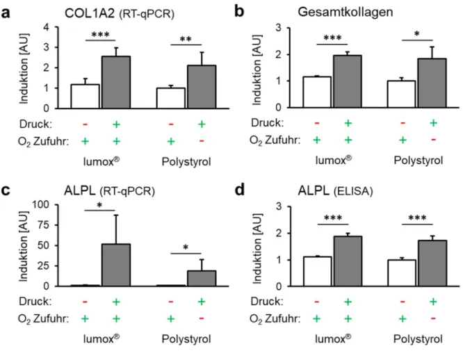
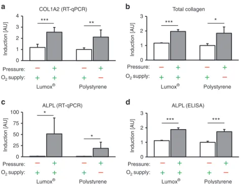
Die Expression von COL1A2 (Kollagen 1 Alpha 2) wurde durch Druckkraftapplikation
ebenfalls unabhängig von der O 2 -Zufuhr signifikant erhöht (p ≤ 0,003; Abb. 3a). Das Ausmaß
der O 2 -Versorgung während der Kompression hatte jedoch keinen signifikanten Einfluss auf
die COL1A2-Genexpression (p = 0,348). Die verstärkte COL1A2-Genexpression führte auch
zu einer höheren Menge an Gesamtkollagen mit (p = 0,019) oder ohne Einschränkung der O 2 -
Versorgung (p < 0,001; Abb. 3b). Die Änderung der O 2 -Zufuhr während der Druckapplikation
hatte keinen Einfluss auf diesen Effekt (p = 0,906).
13
Abb. 3. Effekt der Mechanotransduktion vs. Sauerstoffversorgung auf das Expressionsmuster der hPDLF. (a) COL1A2-mRNA, (b) Gesamtkollagen, (c) ALPL-mRNA und (d) ALPL- Proteinexpression mit Druckapplikation unter normoxischen (Lumox ® ) und hypoxischen (Polystyrol) Bedingungen (N=3, n=9). Balken zeigen arithmetische Mittelwerte ± Standardabweichung. * p ≤ 0,05; ** p ≤ 0,01; *** p ≤ 0,001. AU = arbiträre Einheiten.
Die ALPL-Genexpression (Alkalische Phosphatase) wurde ebenfalls durch Druckkräfte bei Normoxie (p = 0,013) und Hypoxie (p = 0,02; Abb. 3c) hochreguliert. Darüber hinaus schien eine reduzierte O 2 -Zufuhr während der Zellkompression die druckinduzierte ALPL- Hochregulation tendenziell zu attenuieren (p = 0,110). Auch auf Proteinebene zeigte ALPL eine O 2 -unabhängige Akkumulation nach mechanischer Kompression (p < 0,001; Abb. 3d). Die scheinbare Hemmung der druckinduzierten ALPL-Expression durch reduzierte O 2 -Zufuhr war auf Proteinebene nicht vorhanden und eine Verringerung der O 2 -Zufuhr änderte die ALPL- Proteinakkumulation nicht (p = 0,384).
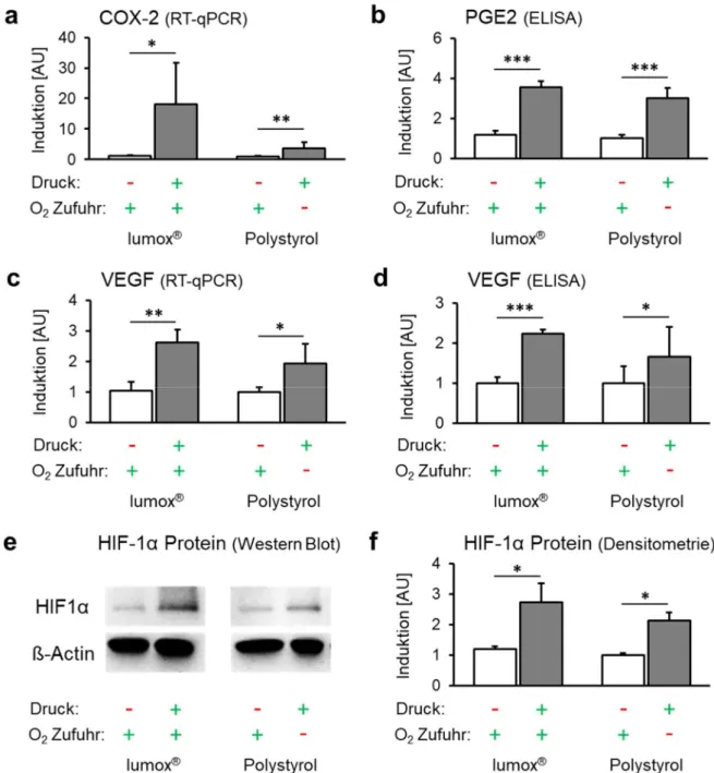
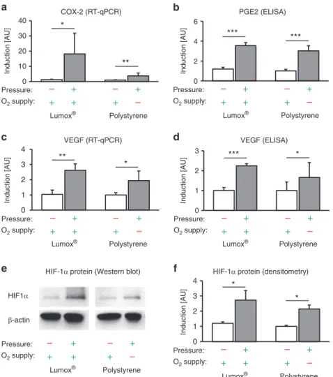
Die Expression des proinflammatorischen Gens COX-2 (Zyklooxygenase 2) wurde durch
Druckapplikation (Mechanotransduktion) unabhängig von der O 2 -Versorgung signifikant
erhöht (p ≤ 0,024; Abb. 4a). Dies resultierte in einer ebenfalls O 2 -unabhängigen Steigerung der
Proteinexpression von PGE2 nach Druckkraftanwendung (p < 0,001; Abb. 4b).
14
Abb. 4. Auswirkungen der Mechanotransduktion vs. Sauerstoffversorgung auf das Expressionsmuster der hPDLF und die Stabilisierung von HIF-1α. (a) COX-2-mRNA, (b) PGE2 (Translationsebene), (c) VEGF-mRNA und (d) VEGF-Proteinexpression mit Druckapplikation unter normoxischen (Lumox ® ) und hypoxischen (Polystyrol) Bedingungen (N=3, n=9). (e) Repräsentativer Immunoblot der HIF-1α-Proteinexpression. (f) Densitometrische Immunoblot-Analyse der HIF-1α-Proteinexpression (N=4). Balken zeigen arithmetische Mittelwerte ± Standardabweichung. * p ≤ 0,05; ** p ≤ 0,01; *** p ≤ 0,001. AU
= arbiträre Einheiten.
Die mechanische Kompression erhöhte die Expression von VEGF (Vascular Endothelial
Growth Factor) signifikant unabhängig von der O 2 -Versorgung (p ≤ 0,015; Abb. 4c). Eine
reduzierte O 2 -Zufuhr während der Kompression hatte dagegen keinen zusätzlichen
15
signifikanten Effekt auf die VEGF-Genexpression (p = 0,415). Auf Proteinebene wurde VEGF ebenfalls durch mechanische Krafteinleitung unabhängig von der O 2 -Versorgung hochreguliert (p ≤ 0,032; Abb. 4d).
HIF-1α (Hypoxie-induzierter Faktor 1α) wurde durch mechanische Druckkräfte sowohl unter normoxischen (p = 0,045; Lumox ® ) als auch unter hypoxischen Bedingungen (p = 0,007;
Polystyrol) signifikant stabilisiert (Abb. 4e/f), wogegen das Ausmaß der O 2 -Versorgung keine signifikante zusätzliche stabilisierende Wirkung auf HIF-1α hatte (p = 0,416).
3.3 Effekt der Mechanotransduktion vs. Hypoxie auf die Expression von RANK-L/OPG durch hPDLF
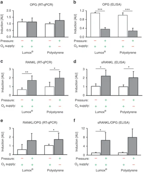
Lumox ® - und Polystyrol-Kontrollgruppen (beide Normoxie, kein Druck) zeigten keine signifikanten RANK-L- oder OPG-Expressionsunterschiede (p ≥ 0,271).
Die OPG-Genexpression durch hPDLF wurde weder unter Normoxie noch unter Hypoxie durch mechanische Druckbelastung signifikant beeinflusst (p ≥ 0,502; Abb. 5a). Im Gegensatz dazu wurde die OPG-Proteinsekretion durch hPDLF während der Druckanwendung (Mechanotransduktion) ohne Einfluss der O 2 -Zufuhr signifikant reduziert (p ≤ 0,001; Abb. 5b).
Die RANK-L-Genexpression (Abb. 5c) und Proteinsekretion (Abb. 5d) wurden hingegen durch
Druckanwendung bei Normoxie (p ≤ 0,036; Lumox ® ) und Hypoxie (Polystyrol) (p ≤ 0,052)
verstärkt. Das Ausmaß der Sauerstoffversorgung während der Kompression hatte keinen
signifikanten Effekt (p ≥ 0,813). Das RANK-L/OPG-Verhältnis auf Transkriptionsebene zeigte
nach mechanischer Kompression und eingeschränkter O 2 -Zufuhr einen signifikanten Anstieg
(p = 0,013; Abb. 5e), während die Druckapplikation unter Normoxie (Lumox ® ) nur tendenziell
(p = 0,200) ein erhöhtes RANK-L/OPG-Verhältnis verursachte. Die Änderung der
Sauerstoffversorgung bei Druckanwendung hatte ebenfalls keinen Einfluss auf das RANK-
L/OPG-mRNA-Verhältnis (p = 0,986). Auf Proteinebene zeigte das sRANK-L/OPG-Verhältnis
einen signifikanten Anstieg nach Druckanwendung ohne Sauerstoffbeschränkung (p = 0,016)
und einen Trend mit Sauerstoffbeschränkung (p = 0,052; Abb. 5f). Eine Änderung der
Sauerstoffversorgung zeigte keine signifikante Wirkung (p = 0,928).
16
Abb. 5. Wirkung der Mechanotransduktion vs. Sauerstoffversorgung auf die Expression von RANK-L/OPG durch hPDLF. (a) Gen- und (b) Proteinexpression von OPG mit Druckapplikation unter normoxischen (Lumox ® ) und hypoxischen (Polystyrol) Bedingungen (N=3, n=9). (c) Gen- und entsprechende (d) Proteinsekretion von sRANK-L durch hPDLF (N=2, n=6). Berechnetes RANK-L/OPG-Verhältnis auf (e) transkriptioneller und (f) Proteinebene. Balken zeigen arithmetische Mittelwerte ± Standardabweichung. * p ≤ 0,05; **
p ≤ 0,01; *** p ≤ 0,001. AU = arbiträre Einheiten.
17
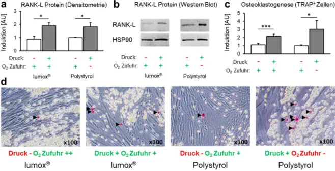
3.4 Effekt der Mechanotransduktion vs. Hypoxie auf die Proteinexpression von membrangebundenen RANK-L und die hPDLF-vermittelte Osteoklastogenese
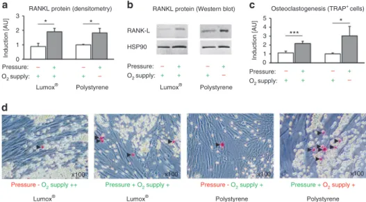
Die densitometrische Analyse der Immunoblots zeigte eine signifikante Induktion der RANK- L-Proteinexpression durch Druckkraftapplikation (Abb. 6a/b) bei Normoxie (p = 0,001;
Lumox ® ) und Hypoxie (p = 0,014; Polystyrol) unabhängig von der O 2 -Zufuhr (p = 0,964). Dies führte zu einer signifikant höheren Osteoklastogenese in der Kokultur nach mechanischer Kompression (Mechanotransduktion) unabhängig von der O 2 -Versorgung (p ≤ 0,019; Abb.
6c/d). Hypoxische Zustände während der Kompression hatten eine leichte zusätzliche osteoklastogene Wirkung, die jedoch nicht signifikant war (p = 0,298). Lumox ® - und Polystyrol-Kontrollgruppen (beide Normoxie, kein Druck) zeigten keine signifikanten Unterschiede in der Osteoklastogenese (p = 0,775).
Abb. 6. Wirkung der Mechanotransduktion vs. Sauerstoffversorgung auf die hPDLF- vermittelte Osteoklastogenese. (a) Densitometrische Immunoblotanalyse der Proteinexpression von membrangebundenen RANK-L (N=5); (b) repräsentativer Immunoblot der Proteinexpression von membrangebundenen RANK-L; (c) Quantifizierung von TRAP- positiven osteoklastenähnlichen Zellen pro Kokulturwell (N=3, n=9); (d) repräsentative Bilder (x100) der Kokultur mit TRAP-Färbung. TRAP-positive Zellen erscheinen rot (schwarze Pfeile), sphärische RAW264.7-Osteoklastenvorläuferzellen gelb und spindelförmige hPDLF transparent. Balken zeigen arithmetische Mittelwerte ± Standardabweichung. * p ≤ 0,05; ** p
≤ 0,01; *** p ≤ 0,001. AU = arbiträre Einheiten.
18 4 Diskussion
In dieser Arbeit wurde die relative Bedeutung der Mechanotransduktion gegenüber reduzierter Sauerstoffzufuhr (Hypoxie) auf die hPDLF-vermittelte Osteoklastogenese in Druckzonen des Parodontalligamentes (PDL) während einer simulierten kieferorthopädischen Zahnbewegung untersucht. Die Ergebnisse zeigten, dass die mechanische Deformation der hPDLF selbst scheinbar eine wichtigere Funktion in der Vermittlung der kieferorthopädischen Zahnbewegung einnimmt als die begleitende Restriktion der Sauerstoffversorgung.
Die Bildung von Kollagen ist dabei ein wichtiger Prozess in der Umstrukturierung des PDL.
Sowohl die Menge an Gesamtkollagen, als auch die Expression des für Kollagen Typ I kodierenden Gens COL1A2 konnten sauerstoffunabhängig durch die mechanische Druckbelastung erhöht werden, was für eine primär mechanische Steuerung dieses Prozesses spricht. Eine Vermehrung von Typ-I-Kollagen durch mechanische Belastung wird bereits durch Kook et al. über die Extracellular-signal-Regulated-Kinasen und c-Jun-N-terminal-Kinasen beschrieben. 17
Die Alkalische Phosphatase (ALPL) wird von reifen Osteoblasten während des Knochenumbaus sezerniert, welche ultrastrukturelle und funktionelle Gemeinsamkeiten mit hPDLF aufweisen. 18-20 Sowohl auf transkriptioneller, als auch auf Proteinebene wurde ALPL unabhängig von der Sauerstoffversorgung durch mechanische Kompression von hPDLF verstärkt sezerniert, was ebenfalls für einen vorwiegend mechanische Regulation spricht. Eine erhöhte Genexpression von ALPL nach Kompression von hPDLF konnte bereits durch Nettelhoff et al. beobachtet werden. 22
Eine primäre Reaktion der hPDLF auf kieferorthopädische Kräfte ist die Synthese und Sekretion von Prostaglandinen durch die Zyklooxygenase 2 (COX-2), welche wiederum die Osteoklastogenese über das RANK-L/OPG-System anregen. 1 Wie bereits in der Literatur beschrieben, 1,7,22 wurde die COX-2-Genexpression nach Druckbelastung der hPDLF hochreguliert. Wie die mechanische Hochregulation von PGE2, ein Produkt der COX-2, 23 erfolgte auch die Hochregulation von COX-2 unabhängig vom bestehenden Sauerstoffangebot.
Es scheint somit, dass auch die pseudoinflammatorische Vermittlung der kieferorthopädischen Zahnbewegung rein mechanisch erfolgt.
VEGF ist ein Wachstumsfaktor, der an der Neubildung und Vasodilatation von Blutgefäßen
beteiligt ist. 24 Während der kieferorthopädischen Zahnbewegung wird im PDL die Neubildung
und Umstrukturierung bestehender Blutgefäße induziert. 25 Bisher wurde angenommen, dass im
Kontext der kieferorthopädischen Zahnbewegung VEGF aufgrund einer Kompression von
19
Blutgefäßen innerhalb des PDL primär durch Sauerstoffmangel hochreguliert wird. 13 Da VEGF ein Zielgen von HIF-1α ist, lässt sich eine Hochregulation von VEGF über die hypoxische Stabilisation von HIF-1α erklären. 26 Die Ergebnisse der vorliegenden Arbeit zeigen allerdings eine mechanische Stabilisation von HIF-1α. Le Li et al. beobachteten eine erhöhte Expression von VEGF durch hPDLF unter Kompression oder Hypoxie und einen additiven Effekt unter Kombination beider Stimuli, 29 während eine Hochregulation von VEGF durch reine Kompression von hPDLF von Miyagawa et al. berichtet wurde. 25 Auch von einer inflammatorischen Regulation von VEGF durch Lipopolysaccharide wurde berichtet. 30 Somit ist davon auszugehen, dass die Expression von VEGF sowohl auf mRNA- als auch auf Proteinebene und der damit einhergehende Prozess der Neoformation und Vasodilatation von Blutgefäßen nicht nur durch einen Sauerstoffmangel im Gewebe, sondern auch maßgeblich durch die Mechanotransduktion und die damit verbundenen pseudoinflammatorischen Prozesse während der Zahnbewegung reguliert wird.
Der Hypoxie-induzierte Faktor 1α (HIF-1α) wird unter hypoxischen Bedingungen stabilisert 31,32 und gilt daher allgemein als Marker für Zellen unter Sauerstoffmangel. 13,27 Es gibt allerdings auch Berichte über eine nicht-kanonische Stabilisierung von HIF1α über bakterielle Lipopolysaccharide 30,33 oder eine mechanische Beanspruchung der Zelle. 31 So konnte auch in dieser Arbeit gezeigt werden, dass eine mechanische Deformation der hPDLF (Mechanotransduktion) zu einer signifikanten sauerstoffunabhängigen Stabilisierung von HIF- 1α führte, während die Verringerung der Sauerstoffzufuhr keinen Effekt zeigte. Dies deutet darauf hin, dass im Zuge der kieferorthopädischen Zahnbewegung die Stabilisierung von HIF- 1α ebenfalls vorwiegend mechanisch erfolgt. Durch die enge Beziehung von HIF-1α zu seinen Zielgenen VEGF und COX-2, und damit über den proinflammatorischen Signalweg zur Osteoklastogenese, 9,30,35,36 nimmt HIF-1α dabei eine Schlüsselstellung in der Vermittlung der kieferorthopädischen Zahnbewegung ein. Feng et al. beschrieben eine mechanische Stabilisierung von HIF-1α in Endothelzellen von Blutgefäßen über das deubiquitierende Enzym Cezanne. 31 Dieser Weg könnte möglicherweise auch für die mechanische Stabilisierung von HIF-1α in hPDLF während der kieferorthopädischen Zahnbewegung verantwortlich sein und bedarf weiterer Forschung.
RANK-L (Receptor Activator of NFκ-b Ligand) und OPG (Osteoprotegerin) sind Regulatoren
des Knochenumbaus 37 und beeinflussen somit die kieferorthopädische Zahnbewegung und die
Wurzelresorption. 38 RANK-L ist verantwortlich für die Aktivierung von Osteoklasten und die
Differenzierung von Osteoklastenvorläuferzellen. 38 Eine erhöhte Expression beider Subtypen
von RANK-L, löslich und membrangebunden, wurde nach der mechanischen Druckapplikation
20
beobachtet, was auf eine erhöhte Osteoklastenbildung und Knochenresorption an den Kompressionszonen des parodontalen Ligaments hinweist. Eine Reduktion der O 2 -Versorgung änderte hingegen nichts an der kraftinduzierten Expression von RANK-L. Dementsprechend wurde auch die OPG-Sekretion nur durch Druckkraftapplikation reduziert ohne Wirkung einer eingeschränkten O 2 -Versorgung. Bei der Regulation von RANK-L und OPG besteht über den proinflammatorischen Weg von COX-2 und PGE2 eine Verbindung zu HIF-1α. 36 Dies könnte einer von mehreren Gründen sein, warum auch das RANK-L/OPG-System hauptsächlich durch mechanische Belastungen und nicht durch reduzierte Sauerstoffwerte beeinflusst wird.
Das erhöhte RANK-L/OPG-Verhältnis führte unter Druckbelastung der hPDLF zu einer ebenfalls erhöhten Osteoklastogenese. Dieser mechanische Effekt wurde in der Literatur bereits mehrfach beschrieben, 7,24,25 und scheint ebenfalls von einer Veränderung der O 2 -Zufuhr unabhängig zu sein. Le Li et al. 29 beobachteten eine erhöhte Osteoklastogenese sowohl unter mechanischer Belastung von hPDLF, als auch unter Hypoxie. Allerdings überwiegte in der Untersuchung von Le Li et al. der mechanische Effekt nach 6h der Inkubation, wogegen der hypoxische Effekt erst nach 24h bzw. 72h signifikant wurde. Dies lässt vermuten, dass Hypoxie die durch hPDLF vermittelte Osteoklastogenese langsamer, aber nachhaltiger als die mechanische Belastung induzieren kann. Le Li et al. verwendeten jedoch viel höhere Druckkräfte von 25 g/cm 2 und eine ausgeprägtere Form der Hypoxie mit einer Restsauerstoffkonzentration von nur 2%, was die Vergleichbarkeit der Ergebnisse einschränkt.
Zellzahl und Zellviabilität wurden durch Druckkräfte reduziert. Dieser Effekt, der auch in anderen Studien berichtet wurde 22,43 , trat unter hypoxischen Bedingungen verstärkt auf und ist wahrscheinlich ein künstlicher Nebeneffekt des angewandten in-vitro-Modells, welcher bereits zuvor berichtet wurde. 16
Abschließend lässt sich festhalten, dass die zelluläre und molekulare Vermittlung der
Osteoklastogenese im Kontext der kieferorthopädischen Zahnbewegung durch hPDLF
maßgeblich durch die Kraftapplikation (Mechanotransduktion) reguliert wird, während
hypoxische Effekte nur eine geringe Rolle zu spielen scheinen. Der Hypoxiemarker HIF-1α mit
seinen vielen Zielgenen wie COX-2 und VEGF scheint ebenfalls vor allem durch mechanische
Belastung und nicht durch Hypoxie im Rahmen der kieferorthopädischen Zahnbewegung
stabilisiert zu werden.
ARTICLE OPEN
The role of mechanotransduction versus hypoxia during
simulated orthodontic compressive strain — an in vitro study of human periodontal ligament fi broblasts
Niklas Ullrich
1, Agnes Schröder
2, Jonathan Jantsch
3, Gerrit Spanier
4, Peter Proff
2and Christian Kirschneck
2During orthodontic tooth movement (OTM) mechanical forces trigger pseudo-in fl ammatory, osteoclastogenic and remodelling processes in the periodontal ligament (PDL) that are mediated by PDL fi broblasts via the expression of various signalling molecules.
Thus far, it is unknown whether these processes are mainly induced by mechanical cellular deformation (mechanotransduction) or by concomitant hypoxic conditions via the compression of periodontal blood vessels. Human primary PDL fi broblasts were randomly seeded in conventional six-well cell culture plates with O
2-impermeable polystyrene membranes and in special plates with gas-permeable membranes (Lumox ® , Sarstedt), enabling the experimental separation of mechanotransducive and hypoxic effects that occur concomitantly during OTM. To simulate physiological orthodontic compressive forces, PDL fi broblasts were stimulated mechanically at 2 g·cm
−2for 48 h after 24 h of pre-incubation. We quanti fi ed the cell viability by MTT assay, gene expression by quantitative real-time polymerase chain reaction (RT-qPCR) and protein expression by western blot/enzyme-linked immunosorbent assays (ELISA). In addition, PDL-fibroblast-mediated osteoclastogenesis (TRAP
+cells) was measured in a 72-h coculture with RAW264.7 cells. The expression of HIF-1 α , COX-2, PGE2, VEGF, COL1A2, collagen and ALPL, and the RANKL/OPG ratios at the mRNA/protein levels during PDL-fibroblast-mediated osteoclastogenesis were significantly elevated by mechanical loading irrespective of the oxygen supply, whereas hypoxic conditions had no signi fi cant additional effects. The cellular – molecular mediation of OTM by PDL fi broblasts via the expression of various signalling molecules is expected to be predominantly controlled by the application of force (mechanotransduction), whereas hypoxic effects seem to play only a minor role. In the context of OTM, the hypoxic marker HIF-1 α does not appear to be primarily stabilized by a reduced O
2supply but is rather stabilised mechanically.
International Journal of Oral Science (2019) 11:33 ; https://doi.org/10.1038/s41368-019-0066-x
INTRODUCTION
In the dental specialty of orthodontics, removable or fi xed orthodontic appliances are used for the treatment of malocclu- sions to move malpositioned teeth to the correct position.
Mechanical orthodontic forces create compression and tension areas in different regions of the periodontal ligament.
1Whereas tension areas are characterized by increased bone formation, bone resorption processes take place in pressure areas.
1Human periodontal ligament (hPDL) fibroblasts are the predominant cells within the periodontal ligament.
2They are responsible for the regulation of tissue homoeostasis and the formation of collagenous structural proteins, and play a regulatory role in innate immune defence.
1,2These cells also play an important mediating role during orthodontic tooth movement (OTM)
1,2and have thus been intensively investigated in basic orthodontic research,
3–7especially with regard to their responses to compressive or tensile orthodontic forces or periodontal pathogens and their toxins.
8–10In compression areas of the periodontal ligament during orthodontic force application, hPDL fi broblasts become
mechanically deformed (mechanotransduction), and thus mechanosensitive receptors and ion channels in the cell membrane are predicted to be stimulated.
11A hPDL-mediated sterile pseudo-in fl ammatory reaction induces increased expres- sion of IL-6, IL-8 and COX-2, followed by enhanced extracellular matrix remodelling and bone resorption triggered by increased receptor activator of NF- κ B ligand (RANKL) and reduced osteo- protegerin (OPG) expression.
1,7On the other hand, the concomi- tant compression of blood vessels during OTM disturbs circulation in compressive areas of the periodontal ligament,
12reducing the local oxygen supply within the PDL (hypoxia).
13It has been postulated that this local reduction of the O
2supply may play a signi fi cant role in the cellular regulation of orthodontic tooth movement.
13Although the molecular and cellular processes enabling OTM that are mediated by hPDL fi broblasts under mechanical load have been studied before,
3,7,14it is still unclear whether these processes are mainly triggered by the mechanical deformation of hPDL fi broblasts (mechanotransduction) during orthodontic force application or by concomitant hypoxia resulting from the
Received: 31 January 2019 Revised: 3 July 2019 Accepted: 18 August 2019
1
University Medical Centre of Regensburg, Franz-Josef-Strauß-Allee 11, D-93053 Regensburg, Germany;
2Department of Orthodontics, University Medical Centre of Regensburg, Franz-Josef-Strauß-Allee 11, D-93053 Regensburg, Germany;
3Department of Medical Microbiology and Hygiene, University Medical Centre of Regensburg, Franz-Josef-Strauß- Allee 11, D-93053 Regensburg, Germany and
4Department of Cranio-Maxillo-Facial Surgery, University Medical Centre of Regensburg, Franz-Josef-Strauß-Allee 11, D-93053 Regensburg, Germany
Correspondence: Niklas Ullrich (niklas.ullrich@ukr.de) or Christian Kirschneck (christian.kirschneck@ukr.de)
www.nature.com/ijos
International Journal of Oral Science
1234567890();,:
compression of periodontal blood vessels and a disturbance in the local blood fl ow. Understanding the impact of mechanotransduc- tion vs. that of hypoxia on the molecular processes enabling OTM could, in the long term, lead to the development of new therapeutic and preventive options for orthodontic treatment.
In this study, we thus investigated the expression patterns of genes and proteins that were previously identi fi ed to be associated with OTM
1,15,16after compressive force application in the presence of a normal or reduced oxygen supply by using conventional six-well cell culture plates or special cell culture dishes with gas-permeable membranes to gain a better under- standing of the respective roles of mechanotransduction and oxygen supply in the molecular and cellular processes occurring in compressive areas of the periodontal ligament during OTM.
RESULTS
Effects of mechanotransduction vs. hypoxia on hPDL cell number and viability
Mechanotransduction (pressure) caused a signi fi cant reduction both in cell number per cm
2(P ≤ 0.007, Fig. 1a) and in cell viability (P ≤ 0.001, Fig. 1b), which was more pronounced in the presence of reduced O
2supply, particularly regarding cell viability.
Effects of mechanotransduction vs. hypoxia on the hPDL expression pattern and HIF-1 α stabilization
The Lumox® and polystyrene control groups (under normoxia and no pressure) did not show significant expression differences for any of the evaluated genes/proteins (P ≥ 0.086) or for HIF-1α stabilization (P = 0.081).
The expression of COL1A2 (collagen 1 alpha-2) was signi fi cantly enhanced by compressive force application, independent of the O
2supply (P ≤ 0.003, Fig. 2a). The level of O
2supply during compression, however, had no signi fi cant effect on COL1A2 gene expression (P = 0.348). Increased levels of COL1A2 gene expres- sion also resulted in an increased quantity of total collagen with (P
= 0.019) or without O
2restriction (P < 0.001, Fig. 2b). Altering the O
2supply during pressure application did not affect this observation (P = 0.906).
Alkaline phosphatase (ALPL) gene expression was also upregu- lated by compressive forces during normoxia (P = 0.013) and hypoxia (P = 0.02, Fig. 2c). In addition, a reduced O
2supply during cell compression seemed to attenuate pressure-induced ALPL upregulation (P = 0.110) at the transcriptional level. ALPL at the protein level showed O
2-independent accumulation after mechan- ical compression (P < 0.001, Fig. 2d). The apparent attenuation of pressure-induced ALPL expression by a reduced O
2supply was not
0.0 0.5 1.0 1.5
0.0 0.5 1.0
1.5 ***
**
a Cell number / cm
2b
Induction [AU]
Pressure: – + – +
O
2supply:
Pressure:
O
2supply:
Polystyrene
+ + + –
**
Cell viability (MTT)
Induction [AU]
– + – +
Lumox
®Lumox
®Polystyrene
+ + + –
** ***
***
Fig. 1 Cell number per cm
2(a) and cell viability of hPDL fi broblasts (b, MTT assays) after 72 h of incubation. N = 2, n = 6. Bars indicate mean values ± standard deviation. **P ≤ 0.01, ***P ≤ 0.001
0 1 2 3 4
*** **
a COL1A2 (RT-qPCR)
Induction [AU]
Pressure: – + – +
O
2supply:
Lumox
®Polystyrene Lumox
®Polystyrene
+ + + –
Pressure: – + – +
O
2supply:
Lumox
®Polystyrene
+ + + –
Pressure: – + – +
O
2supply:
Lumox
®Polystyrene
+ + + –
0 1 2 3
Induction [AU]
Pressure: + – +
O
2supply:
–
+ + + –
b Total collagen
*** *
0 25 50 75
100 *
*
c ALPL (RT-qPCR)
Induction [AU]
0 1 2 3
Induction [AU]
d ALPL (ELISA)
*** ***
Fig. 2 Effects of mechanotransduction vs. oxygen supply on the hPDL fi broblast expression pattern. a COL1A2 mRNA, b total collagen, c ALPL mRNA and d ALPL protein expression in the presence of pressure under normoxic (Lumox ® ) and hypoxic (polystyrene) conditions (N = 3, n = 9).
Bars indicate mean values ± standard deviation. *P ≤ 0.05, **P ≤ 0.01, ***P ≤ 0.001. AU = arbitrary units Effects of stress and hypoxia on hPDL fi broblasts
Ullrich et al.
2
International Journal of Oral Science (2019) 11:33
re fl ected at the protein level, and a reduction in the O
2supply did not alter the ALPL protein accumulation (P = 0.384).
The expression of the proin fl ammatory gene cyclooxygenase 2 (COX-2) was signi fi cantly upregulated by compressive force application (mechanotransduction) independent of the O
2supply (P ≤ 0.024, Fig. 3a). However, the upregulation of COX-2 by pressure in combination with a reduced O
2supply was less pronounced compared with the change in the relative COX-2 gene expression during pressure application during normoxia (P
= 0.052). This mechanically induced COX-2 gene expression was also associated with the signi fi cantly increased protein expression of PGE2 after compressive force application regardless of the level of O
2supply (P < 0.001, Fig. 3b). Reducing the O
2supply during compression had no effect on PGE2 expression (P = 0.223).
Compressive force application increased the expression of vascular endothelial growth factor (VEGF) signi fi cantly, indepen- dently of the O
2supply (P ≤ 0.015, Fig. 3c). A reduced O
2supply during compression, by contrast, had no additional signi fi cant effect on VEGF gene expression (P = 0.415). At the protein level, VEGF was also upregulated by mechanical force application, independent of the O
2supply (P ≤ 0.032, Fig. 3d). Similar to VEGF gene expression, VEGF protein expression during pressure application showed no additional changes after O
2restriction (P = 0.330).
Finally, hypoxia-inducible factor 1α (HIF-1α) was significantly stabilized by mechanical compressive forces (mechanotransduction) during both normoxic (P = 0.045, Lumox®) and hypoxic conditions
(P = 0.007, polystyrene) (Fig. 3e, f), whereas the level of the O
2supply had no signi fi cant additional stabilizing effect on HIF-1 α (P = 0.416).
Effects of mechanotransduction vs. hypoxia on RANKL/OPG expression in hPDL fi broblasts
The Lumox® and polystyrene control groups (in normoxia and no pressure) did not show significant RANKL or OPG expression differences (P ≥ 0.271). OPG gene expression in hPDL fibroblasts was not significantly affected by compressive mechanical strain, both in normoxic and hypoxic conditions (P ≥ 0.502, Fig. 4a). In contrast, OPG protein secretion from hPDL fi broblasts was signi fi cantly reduced during pressure application (mechanotrans- duction), and no effect of reduced O
2supply was observed (P ≤ 0.001, Fig. 4b). RANKL gene expression (Fig. 4c) and protein secretion (Fig. 4d) were enhanced by pressure application during normoxia (P ≤ 0.036, Lumox ® ) and hypoxia (polystyrene) (P ≤ 0.052). The level of the oxygen supply during compression, however, had no signi fi cant effect (P ≥ 0.813). The RANKL/OPG ratio at the transcriptional level showed a signi fi cant increase in the presence of mechanical compression and a restricted O
2supply (P = 0.013, Fig. 4e), whereas pressure application under normoxia (Lumox ® ) resulted in a tendency towards an increase in the RANKL/OPG ratio (P = 0.200). Altering the oxygen supply during pressure application did not show any effect on the RANKL/OPG mRNA ratio (P = 0.986). At the protein level, the sRANKL/OPG ratio showed a significant increase without oxygen
0 1 2 3
4 *
* a
0 10 20 30
40 *
**
COX-2 (RT-qPCR)
Induction [AU]
HIF1 α β -actin 0 1 2 3 4
** *
c VEGF (RT-qPCR)
Induction [AU]
f HIF-1α protein (densitometry)
Induction [AU]
e HIF-1α protein (Western blot)
0 2 4 6
Induction [AU]
b PGE2 (ELISA)
*** ***
0 1 2 3
Induction [AU]
d VEGF (ELISA)
*** *
– –
–
+ +
+ +
+
Pressure:
O
2supply:
Lumox
®Polystyrene
– –
–
+ +
+ +
+
Pressure:
O
2supply:
Lumox
®Polystyrene
– –
–
+ +
+ +
+
Pressure:
O
2supply:
Lumox
®Polystyrene
– –
–
+ +
+ +
+
Pressure:
O
2supply:
Lumox
®Polystyrene
– –
–
+ +
+ +
+
Pressure:
O
2supply:
Lumox
®Polystyrene
– –
–
+ +
+ +
+
Pressure:
O
2supply:
Lumox
®Polystyrene
Fig. 3 Effects of mechanotransduction vs. oxygen supply on hPDL fi broblast expression patterns and HIF-1 α stabilization. a COX-2 mRNA, b PGE2 protein, c VEGF mRNA and d VEGF protein expression in the presence of pressure under normoxic (Lumox ® ) and hypoxic (polystyrene) conditions (N = 3, n = 9). e Representative immunoblot of HIF-1 α protein expression. f Densitometric immunoblot analysis of HIF-1 α protein expression (N = 4). Bars indicate mean values ± standard deviation. *P ≤ 0.05, **P ≤ 0.01, ***P ≤ 0.001. AU = arbitrary units
Effects of stress and hypoxia on hPDL fi broblasts Ullrich et al.
3
International Journal of Oral Science (2019) 11:33
restriction (P = 0.016) and a tendency towards increase during oxygen restriction (P = 0.052) (Fig. 4f). A change in the oxygen supply did not affect this observation (P = 0.928).
Effects of mechanotransduction vs. hypoxia on membrane-bound RANKL protein expression and hPDL-mediated osteoclastogenesis The densitometric immunoblot (Western blot) analysis of membrane-bound RANKL protein expression showed a signi fi cant induction of protein expression by compressive force application (Fig. 5a, b) during normoxia (P = 0.001, Lumox ® ) and hypoxia (P = 0.014, polystyrene), which did not depend on the O
2supply (P = 0.964). This resulted in signi fi cantly increased hPDL- fi broblast- mediated osteoclastogenesis in the coculture after compressive force application (mechanotransduction), independently of the O
2supply (P ≤ 0.019, Fig. 5c, d). Hypoxic conditions during compres- sion resulted in a slight additional increase in osteoclastogenesis, however, this was not signi fi cant (P = 0.298). The Lumox ® and polystyrene control groups (in normoxia and no pressure) did not show signi fi cant differences in osteoclastogenesis (P = 0.775).
DISCUSSION
In this study, we investigated the relative importance of mechanotransduction and reduced O
2supply (hypoxia) to hPDL-
fi broblast-mediated osteoclastogenesis during orthodontic tooth movement. The results from our in vitro experiments showed that the mechanical deformation of hPDL fi broblasts seemed to play a much more important role in the mediation of orthodontic tooth movement by hPDL fibroblasts at a cellular–molecular level than the concomitant reduction in the O
2supply.
The gene expression of COL1A2, which encodes the alpha-2 chain of collagen type I and is thus indicative of collagen synthesis, is very important for orthodontic tooth movement, considering that Type I collagen is the predominant collagen in the extracellular matrix of the periodontal ligament.
17COL1A2 was much more strongly expressed during mechanical loading, whereas a reduced O
2supply had no signi fi cant additional effect during loading. The same results were found for total collagen, which was also increased after the mechanical compression of hPDL fi broblasts independent of the oxygen supply. This indicates that the process of collagen synthesis might also be predomi- nantly controlled by mechanotransduction. This fi nding is substantiated by the fi ndings of Kook et al.
17, who described the mechanical upregulation of Type I collagen by extracellular signal-regulated kinase and c-Jun N-terminal kinase, which transmitted mechanical signals into the nuclei of hPDL fibroblasts.
Orthodontic tooth movement involves the alteration of not only the collagen network but also the surrounding bone architecture.
OPG (RT-qPCR)
a
c
e
b
d
f
RANKL (RT-qPCR) sRANKL (ELISA)
sRANKL/OPG (ELISA) RANKL/OPG (RT-qPCR)
2.0
3 2 1 0
1.2 ***
**
*
* *
* *
***
0.8 0.4 0.0 1.5
1.0
Induction [A U]
Induction [A U] Induction [A U]
3 2 1 0
Induction [A U]
12 8 4 0
Induction [A U]
3 2 1 0
Induction [A U]
0.5 0.0
OPG (ELISA)
– –
–
+ +
+ +
+
Pressure:
O
2supply:
– –
–
+ +
+ +
+
Pressure:
O
2supply:
– –
–
+ +
+ +
+
Pressure:
O
2supply:
– –
–
+ +
+ +
+
Pressure:
O
2supply:
Lumox
®Polystyrene
Lumox
®Polystyrene
– –
–
+ +
+ +
+
Pressure:
O
2supply:
Lumox
®Polystyrene
– –
–
+ +
+ +
+
Pressure:
O
2supply:
Lumox
®Polystyrene Lumox
®Polystyrene Lumox
®Polystyrene
Fig. 4 Effects of mechanotransduction vs. oxygen supply on hPDL fi broblast RANKL/OPG expression. a Gene and b protein expression of OPG in the presence of pressure under normoxic (Lumox ® ) and hypoxic (polystyrene) conditions (N = 3, n = 9). c Gene expression and the corresponding d protein secretion of (soluble) RANKL by hPDL fi broblasts (N = 2, n = 6). Calculated RANKL/OPG ratio for the e transcriptional and f protein levels. Bars indicate mean values ± standard deviation. *P ≤ 0.05, **P ≤ 0.01, ***P ≤ 0.001. AU = arbitrary units
Effects of stress and hypoxia on hPDL fi broblasts Ullrich et al.
4
International Journal of Oral Science (2019) 11:33
ALPL is highly expressed in and secreted by mature osteoblasts during bone formation; osteoblasts share ultrastructural and functional similarities with hPDL fibroblasts, and these can transform into osteoblasts.
18–20Osteoblast activity in the period- ontal ligament is much higher than in other connective tissues.
21In our study, we observed increased ALPL expression at both the mRNA and protein levels after orthodontic compressive force application, which is similar to the fi ndings of Nettelhoff et al.
22The restriction of the oxygen supply did not alter the force- induced expression of ALPL at all, indicating that ALPL is mainly mechanically regulated as well.
The primary response of hPDL fi broblasts to orthodontic forces is the synthesis and secretion of prostaglandins by COX-2, which in turn triggers osteoclastogenesis via the RANKL/OPG pathway.
1As already reported,
1,7,22orthodontic compressive force applica- tion led to enhanced COX-2 expression in our model of orthodontic tooth movement, whereas reduced oxygen levels did not alter this proin fl ammatory effect. PGE2, a product of COX- 2,
23was also upregulated by the mechanical compression of hPDL fi broblasts independent of the O
2supply. This indicates the presence of a primarily mechanically triggered pathway causing inflammation in the context of orthodontic tooth movement.
VEGF is a growth factor involved in the neoformation and vasodilation of blood vessels.
24During orthodontic tooth move- ment, the formation of new blood vessels and the reshaping of existing blood vessels is induced in the periodontal ligament.
25Until now, it has been assumed that in the context of orthodontic tooth movement, VEGF is primarily upregulated by low oxygen levels due to a compression of blood vessels within the PDL.
13This mechanism is often explained by the dependence of VEGF expression on HIF-1 α , as HIF-1 α is stabilised by hypoxic conditions and VEGF is a target gene of HIF-1 α .
26HIF-1 α is of major importance for the adaptation of tissues to a reduced O
2supply.
27However, it has been proposed that HIF-1 α can also be stabilised under normoxic conditions,
28as was the case in our study in the context of OTM, which would thus explain the mechanically induced upregulation of VEGF expression independent of the O
2supply. Our results con fi rm this assumption and indicate that the upregulation of VEGF is mainly mechanical. Li et al.
29described the enhancement of the expression of VEGF by either compres- sion or hypoxia and the additive effect resulting from the combination of both stimuli; however, the upregulation of VEGF
resulted only from compression according to Miyagawa.
25An in fl ammatory elevation of VEGF by lipopolysaccharides has been reported as well.
30Considering all these findings, one must assume that the expression of VEGF at both the mRNA and protein levels, and especially the processes of the neoformation and vasodilation of blood vessels, are not only regulated by oxygen levels but also predominantly by mechanical forces and the resulting pseudo-in fl ammatory processes during OTM.
The signalling molecule HIF-1 α has often been used as a marker for cells under hypoxic conditions.
13,27Whereas the canonical method of HIF-1 α stabilisation is via hypoxia,
31,32there have been reports that HIF-1 α stabilisation is also possible by non-canonical methods,
32including Toll-like receptor activation by bacterial lipopolysaccharides,
30,33which occurs during periodontitis,
8,9,34and by mechanotransduction, which has been reported in endothelial cells.
31Our model of simulated orthodontic compres- sive forces showed the predominantly mechanical stabilisation of HIF-1 α , indicating that in the context of orthodontic tooth movement, HIF-1 α might be stabilised mechanically rather than by hypoxia. Considering that many of the investigated genes, such as COX-2 and VEGF, are target genes of HIF-1 α ,
9,30,35and that even the RANKL/OPG system and osteoclastogenesis are influenced by HIF-1α via the proinflammatory pathway consisting of COX-2, PGE2 and RANKL,
36HIF-1α seems to be a key factor involved in the complex regulation of orthodontic tooth movement. Feng et al.
31observed the mechanical stabilisation of HIF-1 α in endothelial cells in blood vessels via the deubiquitinating enzyme Cezanne. This pathway could possibly be responsible for the mechanical stabilisation of HIF-1 α in hPDL fi broblasts during orthodontic tooth movement, which merits further investigation.
The TNF-related ligand RANKL and its decoy receptor osteo- protegerin (OPG) both play important roles in the regulation of bone remodelling,
37orthodontic tooth movement and root resorption.
38RANKL is responsible for the differentiation of osteoclast-precursor cells and the activation of premature osteoclasts.
38The enhanced expression of both the soluble and membrane-bound subtypes of RANKL was observed after orthodontic compressive force application, indicating that increased osteoclastogenesis and bone resorption occurred in the compression areas of the periodontal ligament. A reduced O
2supply, on the other hand, did not alter the force-induced expression of RANKL of either subtype. In line with that finding, b
Polystyrene Lumox®
Lumox®
Lumox® Polystyrene
Polystyrene Lumox® Polystyrene
Pressure - O2 supply ++ Pressure + O2 supply + Pressure - O2 supply + Pressure + O2 supply +
x100 x100 x100 x100
c a
d
RANKL protein (densitometry)
RANK-L HSP90
RANKL protein (Western blot) Osteoclastogenesis (TRAP+ cells) 3
* *
***
*
2
Induction [AU]
1 0
4 5 3 2 Induction [AU] 1 – 0
–
–
+ +
+ +
+
– +
+ +
– – +
+ – +
+ +
– – + + Pressure:
O2 supply:
Pressure:
O2 supply: Pressure:
O2 supply: