of Early and Late Checkpoints in the Quality Control of MHC Class I-Restricted Antigen Presentation
Inaugural-Dissertation zur
Erlangung des Doktorgrades
der Mathematisch-Naturwissenschaftlichen Fakultät der Universität zu Köln
vorgelegt von Ralf Max Leonhardt
aus Köln
Köln, 2005
Berichterstatter: Priv.-Doz. Dr. Michael R. Knittler
Prof. Dr. Thomas Langer
Tag der mündlichen Prüfung: 08.12.2005
Print Point Werbeagentur, Köln
Danksagungen
Ich bedanke mich bei Priv.-Doz. Dr. Michael R. Knittler für all die Unterstützung in den zurückliegenden Jahren, sowie für die vielen großen und kleinen Momente der Hilfsbereitschaft, ohne die dieses Projekt so nicht hätte durchgeführt werden können.
Seine Offenheit für Diskussionen und seine schier unbegrenzte Bereitschaft jeder Zeit für Fragen oder auch Probleme zur Verfügung zu stehen, haben entscheidend zum Erfolg dieser Arbeit beigetragen. Die Vielzahl molekularbiologischer, biochemischer und zellbiologischer Methoden und Techniken, die ich von ihm erlernt habe, ermöglichen mir einen weiteren Weg in der Wissenschaft. Neben seinem herausragenden Einsatz für mich als Arbeitsgruppenleiter und Promotionsbetreuer, möchte ich mich aber auch ausdrücklich für die Herzlichkeit und Freundschaft bedanken, die ich erfahren durfte und die weit über den beruflichen Alltag hinausging.
Ich bedanke mich ferner bei Professor Dr. Jonathan C. Howard für das große in mich gesetzte Vertrauen. Ich den zurückliegenden Jahren habe ich mich stets als Teil seiner Arbeitsgruppe fühlen dürfen und unendlich von den gemeinsamen Veranstaltungen profitiert. Nicht zuletzt die schöne Zeit in Thoires wird mir in Erinnerung bleiben. Neben seinem großen Interesse an meiner Arbeit und den vielen wichtigen Anregungen zu meinem Projekt, möchte ich mich aber auch für die enorme finanzielle Unterstützung bedanken, die mir zuteil geworden ist. Sie hat mir den Rücken freigehalten, die Existenz gesichert und somit ermöglicht mich auf meine Forschung zu konzentrieren. Ohne dieses Engagement hätte ich meine Doktorarbeit vermutlich nicht beenden können.
Ich möchte mich an dieser Stelle auch nochmals bei Priv.-Doz. Dr. Christine Kocks für die Einführung in das wissenschaftliche Arbeiten bedanken. Die hervorragende Ausbildung, während meiner Diplomarbeit, die ich in ihrer Gruppe genossen habe, hat für mich eine Basis geschaffen, auf der ich die im Folgenden beschriebene Arbeit aufbauen konnte. Ihre Arbeitsweise hat mich nachhaltig geprägt und somit den Erfolg meiner Arbeit auch tiefgreifend beeinflußt.
Bei Prof. Dr. Thomas Langer bedanke ich mich für die zeitweise Finanzierung meiner Stelle und für die Übernahme des Co-Referats.
Prof. Dr. Helmut Klein danke ich für die Übernahme des Vorsitzes der Prüfungskommission.
Ich möchte mich bei Dr. Matthias Cramer für die große Hilfsbereitschaft in den letzten Jahren bedanken. Seine hervorragende Beratung hat so manchen Prozeß beschleunigt und die Ausfüllung so manchen Formulars erheblich erleichtert. Nicht zuletzt danke ich ihm dafür, daß er sich als Beisitzer für meine Disputation zur Verfügung gestellt hat.
Ich danke Cemalettin Bekpen für das zur Verfügung stellen von mRNA aus diversen humanen Zellinien. Darüber hinaus möchte ich mich bei ihm aber auch für die schöne und äußerst produktive Zeit im gemeinsam genutzten Labor bedanken, sowie für die umfangreiche Beratung in Fragestellungen betreffend RT-PCR.
Ferner bedanke ich mich bei Kirstin Keusekotten für das Überlassen von pSP64- TAP2 Plasmid-DNA. An die angenehme Atmosphäre im Labor während der Zeit ihrer Diplomarbeit werde ich mich noch lange erinnern.
Natasa Papic und Julia Hunn danke ich für ihre freundliche Hilfe am Fluoreszenzmikroskop.
Dr. Sascha Martens danke ich für das Bereitstellen von TIB-75 Hepatozyten, sowie für die Einführung in das Arbeiten mit adherenten Zellen.
Bei allen aktuellen und ehemaligen Mitgliedern der Arbeitsgruppen Knittler, Howard und Praefcke möchte ich mich für die schöne Zeit in den zurückliegenden Jahren bedanken. Die gute und herzliche Atmosphäre auf unserer Etage hat erheblich dazu beigetragen, so manchen Tiefpunkt zu überwinden. Ich danke auch für die ständige Diskussionsbereitschaft, das Interesse an meiner Arbeit sowie die unzähligen Anregungen, die ich erhalten habe.
Professor Dr. Roberto Testi, Professor Dr. Peter Cresswell und Dr. Barbara Tomassini danke ich für das zur Verfügung stellen von CEM- bzw. CEM-NKR-Zellen.
Professor Dr. Wolfgang Garten danke ich für die Bereitstellung des anti-Furin Antiserums.
Professor Dr. John Creemers danke ich für das Bereitstellen der beiden anti-PC7- Antiseren KP-1 und MP-1.
Professor Dr. R. Urade danke ich für das Bereitstellen des gegen ERp57 gerichteten anti-ER60 Antiserums.
Dr. Bodo Ortmann danke ich für das Bereitstellen des anti-Tapasin Antiserums R.gp48N.
Ich bedanke mich bei der Deutschen Forschungsgemeinschaft (DFG) für die großzügige Förderung (KN541/1-1).
Ich danke meinen Eltern, Ingrid und Walter Leonhardt, sowie meiner Großmutter Anna Leonhardt für die liebevolle Unterstützung, die mir das Studium der Biologie ermöglicht hat.
Zuletzt bedanke ich mich bei Dipl.-Ing. Thorsten Eitelgörge für das Lösen diverser Computer- bzw. Softwareprobleme, welches die zeitgerechte Abgabe dieser Arbeit erheblich erleichtert hat.
Table of contents
1. Introduction 1
1.1 The organization of the mammalian immune system 1 1.2 The processing of MHC class I-presented antigenic peptide
ligands
4 1.3 The transporter associated with antigen processing (TAP) 7
1.4 MHC class I molecules 10
1.5 Lectin chaperones, ERp57 and the oxidative folding pathway in
the ER 12
1.6 The assembly and function of the MHC class I peptide-loading complex
13
2. Description of the project 24
3. Abbreviations 28
4. Materials and Methods 30
4.1 Cell lines and cell culture 30
4.2 Cloning and stable expression of TAP chain variants 31
4.3 Antibodies 33
4.4 Immunoprecipitation and Western blot analysis 34 4.5 Inhibition of proteasomal activity with ALLN 35
4.6 Pulse-chase analysis 36
4.7 Determination of MHC class I thermostability in detergent extracts
36
4.8 Immunofluorescence 37
4.9 Deglycosylation of MHC class I molecules with endoglycosidase H (EndoH)
37 4.10 Flow cytometry and determination of MHC class I thermostability
in intact cells 38
4.11 Nucleotide-binding assay 39
4.12 Radioiodination of peptides 39
4.13 Peptide cross-linking 40
4.14 Peptide transport assay 40
4.15 Determination of proprotein convertase expression by RT-PCR 41 4.16 Examination of proprotein convertase regulation by interferons 42 4.17 Preparation of microsomes from T2 cells by sucrose gradient
fractionation
44
4.18 In vitro translation 45
4.19 Immunodepletion 45
5. Results 47
5.1 ER-export and surface expression of MHC class I does not depend on the number or position of tapasin-docking sites in TAP
47
5.2 Head-to-tail-fusion of TAP chains allows stable expression of transporters lacking the N-domains in both subunits
52 5.3 Assembled and non-assembled TAP1 chains use different TMD-
subregions for tapasin binding 57
5.4 TAP variants 1-2∆N and 2-1∆N show different effects on the quality control of antigen presentation
61
5.5 The N-domain of TAP2 is essential for the structural integrity of
the PLC 65
5.6 Calnexin is part of the peptide loading complex in T2 cells expressing TAP
67 5.7 Calnexin is not essential for recruitment of ERp57 into the
loading complex or for the quality control of antigen presentation 74 5.8 High level MHC class I surface expression in the cell line T2(1-
2∆N) depends on the proteolytical activity of proprotein convertases
76
5.9 Proprotein convertase expression is differentially regulated by interferons at the transcriptional level
81
6. Discussion 85
6.1 At steady-state TAP does not simultaneously associate with MHC class I via both N-domains
85 6.2 Calnexin is a component of the mature human peptide loading
complex
88 6.3 The N-region of TAP2 is essential for the structural and
functional integrity of the peptide loading complex
93 6.4 Functional expression of TAP lacking both N-regions can be
achieved by head-to-tail fusion of the two chains linked to one another the connector region of MDR1b
98
6.5 Proprotein convertases rescue suboptimally loaded MHC class I molecules in late secretory compartments
100
7. Summary 108
8. Zusammenfassung 109
9. References 111
10. Erklärung 146
11. Lebenslauf 147
1. Introduction
1.1 The organization of the mammalian immune system
The mammalian immune response can generally be subdivided into three interdependent limbs, namely the innate immune response, the humoral response and the cell-mediated response. The innate immune system is believed to provide a first barrier against the spread of pathogens within a newly infected host. Thereby it counteracts the establishing infection and contributes to an early clearance of the pathogen. However, the term “innate immune response” describes a very diverse repertoire of mechanisms, among them complement-mediated killing of bacteria (Gasque et al, 2004), secretion of peptide antibiotics (Boman et al, 1995), elimination of extracellular pathogens through phagocytic uptake by macrophages (Hingley- Wilson et al, 2000) and NK-cell mediated cytotoxicity (Ljunggren et al, 1990). The innate immune system identifies its microbial targets mainly by the recognition of relatively invariable so-called pathogen-associated molecular patterns (PAMPs) (Janeway et al, 2002) through a huge number of specialized receptors, including the Toll-like and NOD family of receptors (Janeway et al, 2002; Tschopp et al, 2003).
Numerous cell types including macrophages, NK cells and epithelial cells contribute effector functions to this limb of the immune system and in addition many (perhaps all) non-immune cells can acquire an antiviral state upon γ-interferon treatment, a phenomenon commonly referred to as cell-autonomous resistance (MacMicking et al, 2004; Weber et al, 2004). The humoral immune response is mediated by the B cell population, which secretes an enormously diverse repertoire of antibodies (Calame et al, 2003). Antibodies neutralize toxins (Little et al, 1988; Wild et al, 2003), block the adhesion of pathogens to mucosal surfaces (Kunisawa et al, 2005) or opsonize pathogens to mark them for phagocytic uptake (or immediate destruction) (Stuart et
al, 2005). Thus, an antibody-mediated immune response is predominantly directed against extracellular pathogens. According to the theory of clonal selection each individual B cell produces only a single type of antibody. The mature gene that encodes an antibody is generated by rearrangement of DNA (Sadofsky et al, 2001) and its expression gives either rise to the B cell receptor (BCR), which is anchored in the plasma membrane or to the secreted antibody, which is its soluble analog (Cambier et al, 1992). Antibodies can recognize the antigens they are targeted against in their native conformation. This distinguishes them fundamentally from the related T cell receptors (TCRs) which require both processing of their epitopes and presentation of their peptide antigens in the context of a major histocompatibility (MHC) class I or II molecule (Myers et al, 1991). TCR diversity is generated by a site- directed DNA recombination process largely similar to the mechanism that generates the high variety within the antibody repertoire (Sadofsky et al, 2001). T cells originate from precursor stem cells in the bone marrow and migrate during their early development to the thymus where they differentiate (Bommhardt et al, 2004). There they undergo both positive as well as negative selection with the consequence that only MHC-restricted cells survive that do not recognize self-antigen with high affinity (Sebzda et al, 1999). MHC class II-restricted T cells become selected to the CD4+ lineage and henceforth recognize peptide antigens that (largely) derive from extracellular proteins ingested by professional phagocytic cells. These include dendritic cells, macrophages and B cells. Accordingly, these cell types express high levels of MHC class II. The proteolytic processing of MHC class II-presented antigens occurs in compartments along the endocytic route (e.g. endosomes or lysosomes) and requires the activity of a diverse set of enzymes, among them cysteine proteases (e.g. cathepsins S, L, B, H and F) and the γ-interferon-induced lysosomal thiol reductase (GILT) (Watts et al, 2004). Two major CD4+ lineages exist: inflammatory
TH1 cells and TH2 helper cells. The former secrete γ-interferon and activate macrophages to become bactericidal whereas the latter activate B cells to secrete antibodies (Dong et al, 2000). Thus, CD4+ T-lymphocytes represent an important connection between cell-mediated immunity on the one hand and innate or humoral immune response on the other hand. In contrast, MHC class I-restricted T cells become selected to the CD8+ lineage. The vast majority of antigenic peptides that are recognized by CD8-positive T cells in combination with MHC class I are breakdown products of the proteolytic degradation machinery in the cytosol (Leonhardt et al, 2003; Pamer et al, 1998). Accordingly, the cell-mediated immune response exerted by cytotoxic (CD8+) T lymphocytes (CTLs) is mainly directed against intracellular pathogens, primarily viruses and some bacteria. In addition CTLs make a major contribution to the elimination of tumor cells. In line with this, MHC class I molecules (in contrast to their class II counterparts) are expressed on the surface of all nucleated cells. The recognition of non-self peptide by a CTL at the surface of an infected (or malignant) cell leads to the release by the T cell of granules containing perforin and granzymes that have membrane-disrupting and apoptosis- inducing activity (Trapani et al, 2002). Using this pathway and/or a FasL-induced mechanism the CTL activates the apoptotic program of the infected (or malignant) target cell (Trapani et al, 2002). Therefore it is not surprising that many viral (Hewitt et al, 2003; Lybarger et al, 2005) but also bacterial (Neumeister et al, 2005; Qimron et al, 2004) pathogens have developed a huge number of immune evasion strategies that subvert the class I antigen presentation pathway. In addition, tumor cells also frequently down-modulate class I surface levels to avoid their recognition by CTLs (Bubenik et al, 2003). However, during an infection (or the onset of cancer) the three limbs of the immune system synergistically attack intruding microbes (or malignant cells) and in most cases efficiently protect the host organism from disease.
1.2 The processing of MHC class I-presented antigenic peptide ligands
Fig. 1 The MHC class I antigen presentation pathway
Intracellular proteins (1) are degraded by the proteasome in the cytosol (2) and the resulting oligopeptides are subsequently translocated into the ER by the peptide transporter TAP (3). There they are loaded onto empty MHC class I molecules (3). These are at that stage part of a multi- protein complex, the so-called MHC class I peptide loading complex (PLC). Within this complex TAP is bridged to MHC class I by tapasin. Additionally the PLC comprises the lectin chaperone calreticulin and the oxidoreductase ERp57. Upon binding of a peptide ligand the loaded MHC class I molecule dissociates from the PLC (4) and migrates to the plasma membrane (7). On this way it passes the Golgi apparatus (5) and the trans-Golgi network (6).
As mentioned above antigenic peptide ligands for MHC class I mostly derive from the degradation of intracellular proteins in the cytosol (Pamer et al, 1998). The major endoprotease that contributes oligopeptides to this pathway is the proteasome (Fig. 1) (Goldberg et al, 2002; Kloetzel et al, 2001). Moreover, in the context of some MHC class I alleles the tripeptidyl peptidase II (TPP II) is supposed to play an
important role for the endoproteolytic generation of antigens (Seifert et al, 2003).
However, proteasomal activity yields only a small fraction of peptides that have the
appropriate length of 8 to 10 amino acids to bind an MHC class I molecule. Namely, more than 70% of the oligopeptides generated by the proteasome are too short, whereas 15% exceed the size of a suitable class I ligand (Kisselev et al, 1999).
Aminopeptidases can trim the latter population to a fitting length (Goldberg et al, 2002). Currently, for several cytosolic aminopeptidases a role in the MHC class I antigen presentation pathway is discussed, among them puromycin-sensitive aminopeptidase (Levy et al, 2002; Stoltze et al, 2000), bleomycin hydrolase (Stoltze et al, 2000), TPP II (Levy et al, 2002) and γ-interferon inducible leucine aminopeptidase (LAP) (Beninga et al, 1998). Furthermore, upon induction with γ- interferon the proteolytic machinery in the cytosol becomes modified (presumably) for adjusting it to the requirements of the immune system in the context of an infection.
Particularly, the composition of the proteasome changes by the incorporation of three new γ-interferon-induced subunits and the regulator PA28. The resulting so-called immunoproteasome is distinct from the regular housekeeping proteasome with respect to its cleavage specificity and hence the peptide repertoire that it generates (Cascio et al, 2001). Consistently, the production of several important viral or tumor- associated peptide antigens specifically depends on the activity of this modified protease (Cerundolo et al, 1995; Lautscham et al, 2003; Schultz et al, 2002) or the presence of PA28 (Sijts et al, 2002). Interestingly, the activity of the immunoproteasome was shown to yield more N-terminally extended precursors of an intensively investigated model peptide ligand for MHC class I when compared to its constitutive counterpart (Cascio et al, 2001). However, the finding that the incorporation of the immuno-subunits into the proteasome conversely abrogates the production of some epitopes (Morel et al, 2000) queries the view that immunoproteasomes are always the “better” proteasomes under all circumstances.
During the early phase (approx. one week) of a viral or bacterial infection constitutive
proteasomes were reported to become gradually replaced by their immuno- counterparts (Khan et al, 2001). Thus, the combined presence of both in the beginning of the pathogenic challenge may broaden the spectrum of antigenic peptide ligands produced, thereby increasing the efficiency of an immune response.
After their generation in the cytosol oligopeptides have to traverse the endoplasmic reticulum (ER) membrane before they can be loaded onto class I (Fig. 1). The major route used for entry into the ER is via the peptide transporter associated with antigen processing (TAP) which resides in the ER membrane (Leonhardt et al, 2003; Schmitt et al, 2000). Although some very hydrophobic class I ligands were reported to get access to class I without the need for this transporter (Lautscham et al, 2001;
Lautscham et al, 2003), the vast majority of cytosolic peptides essentially depends on the activity of TAP for their translocation into the ER. This becomes evident by markedly decreased MHC class I surface levels on cells of TAP-deficient individuals as a consequence of an almost stagnant peptide supply into the endoplasmic reticulum (Gadola et al, 2000). However, some MHC class I alleles as HLA-A2 can efficiently associate with peptides derived from signal sequences in a TAP- independent manner (Wölfel et al, 2000). After transport into the ER N-terminally extended class I ligands can be further trimmed by the γ-interferon-inducible aminopeptidase ERAP1 until a suitable length for loading onto class I is reached (Saric et al, 2002; Serwold et al, 2002). In the ER peptide-receptive MHC class I molecules associate with TAP and several chaperones to form the so-called MHC class I peptide loading complex (PLC) (Fig. 1 and 4) (Leonhardt et al, 2003; Wright et al, 2004). This complex retains class I in the ER as long as the loading with a high affinity peptide ligand has occurred (see below). Upon binding of such a ligand the MHC class I molecule dissociates from the PLC and migrates along the standard secretory route to the cell surface, where it presents its peptide to CTLs (Fig. 1)
(Yewdell et al, 2003). An unexpected surprise for the field was the finding that some antigenic peptides for class I can also be generated in the trans-Golgi network (TGN) by furin, a member of the proprotein convertase family (Gil-Torregrosa et al, 1998;
Gil-Torregrosa et al, 2000). However, to what extent this pathway contributes to the overall repertoire of surface-presented peptides and how these specific ligands are loaded onto class I has remained elusive.
Taken together, MHC class I molecules present peptides derived from the degradation of intracellular proteins on the cell surface for perusal by CD8+ T cells.
This process allows CTLs to monitor the current protein content of a host cell and to identify (and kill) cells that harbor abnormal or non-self (e.g. of viral origin) proteins.
To sacrifice a virally infected or malignant cell is a necessary evil that protects the host from spreading of the infection to other cells or the development of a life- threatening tumor.
1.3 The transporter associated with antigen processing (TAP)
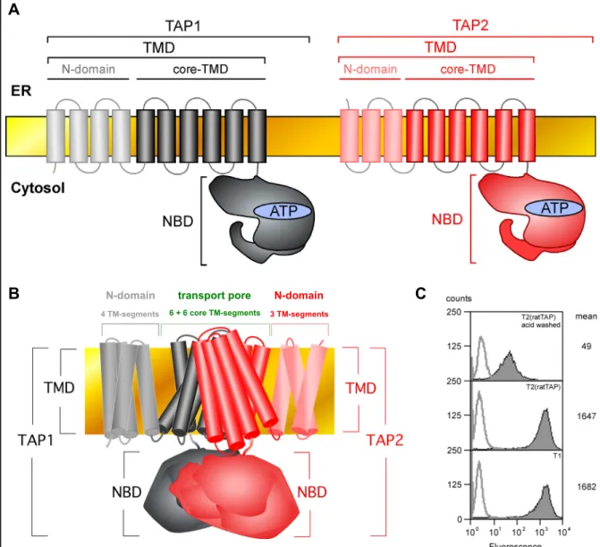
The peptide transporter TAP (Fig. 2) is a member of the ATP binding cassette (ABC) transporter family (Schmitt et al, 2000). ABC transporters have been isolated from all three kingdoms of life, where they translocate diverse substances including vitamins, drugs, ions, amino acids, peptides, sugars or lipids across biological membranes (Garmory et al, 2004; Van der Does et al, 2004). Irrespective of the substrate specificity, which is clearly distinct among individual members of this protein family all ABC transporters share an identical four domain organization comprising two nucleotide binding domains (NBDs) and two transmembrane domains (TMDs) (Stefkova et al, 2004). TAP also possesses this characteristic four domain composition (Fig. 2A and Fig. 2B) (Schmitt et al, 2000). Its NBDs bind and hydrolyze ATP (Chen et al, 2003; Knittler et al, 1999; Saveanu et al, 2001; Schmitt et al, 2000)
A
Fig. 2 The structural organization of the ABC transporter TAP
(A) The TAP transporter is a heterodimer that consists of the two subunits TAP1 (depicted in black) and TAP2 (depicted in red). Either chain contributes an N-terminal transmembrane domain (TMD) and a C-terminal nucleotide binding domain (NBD) to the assembled transporter. In both, TAP1 and TAP2, the TMD can be further subdivided into the N-domain at the extreme N-terminus (involved in the tapasin interaction) and an inner core TMD, comprising 6 α-helices (involved in peptide binding and the formation of the translocation pore). The NBD binds and hydrolyzes ATP to energize the peptide translocation into the ER. (B)Model of the TAP transport pore. The closed conformation of the lipid A-transporter MsbA from Vibrio cholera(Chang et al, 2003) served as a template to predict the arrangement of the putative six transmembrane segments of the TAP core TMD. C) MHC class I surface expression on T1 cells expressing human TAP and their TAP- deficient derivative T2, reconstituted with rat TAPa. T1 cells or T2 transfectants were incubated with the monoclonal anti-MHC class I-antibody 4E followed by FITC-labeled secondary antibody to determine the surface expression of HLA-B5 (filled histograms). To control for the specificity of the primary antibody T2(ratTAP) cells were briefly exposed to acidic buffer for removal of assembled surface MHC class I molecules before processing the cells for flow cytometry. Background staining was analyzed by incubating with secondary antibody alone (non-filled histograms).
B
4 TM-segments 6 + 6 core TM-segments 3 TM-segments
N-domain transport pore N-domain C
to energize the peptide translocation process, whereas its TMDs contain the substrate binding site and form the translocation pore (Fig. 2B; Nijenhuis et al, 1996).
TAP is a heterodimer consisting of two homologous polypeptides, TAP1 and TAP2.
Each of these subunits contributes one NBD and one TMD to the assembled transporter (Fig. 2A). Both TAP chains are essential for peptide binding and transport (Arora et al, 2001; Meyer et al, 1994; Momburg et al, 1992). Sequential hydrolysis of ATP by TAP1 and TAP2 has been proposed to drive the translocation of peptide across the ER membrane and to restore the ground state of the transporter, which is characterized by high affinity for cytosolic substrates (Alberts et al, 2001). The TMDs of TAP1 and TAP2 are predicted to comprise 10 and 9 transmembrane helices, respectively (Schmitt et al, 2000). The 6 inner membrane spanning helices of both TAP subunits are designated as the core TMD, since they are sufficient to allow for the formation of a functional substrate binding site and translocation pore in the assembled transporter (Koch et al, 2004). The N-terminal extensions (N-domains) of the TMDs, (presumably) comprising 4 transmembrane helices in TAP1 and 3 transmembrane helices in TAP2 function as docking sites for tapasin (Koch et al, 2004). The preferred substrates for the transporter are peptides with a length of 8 to 12 amino acids (Abele et al, 2004). Thus, TAP most efficiently delivers those peptides into the ER that have an optimal size for binding to MHC class I. With respect to the amino acid sequence of its substrates TAP has been reported to be highly promiscuous. However, the nature of the C-terminal residue and to a minor extent of the first three N-terminal residues plays a role for peptide binding (and most likely transport) by TAP (Schmitt et al, 2000). Importantly, human TAP and rat TAPa were both shown to be permissive for peptides with any C-terminus with the exception of proline (Schmitt et al, 2000), suggesting that both transporters translocate a similar set of peptides (Momburg et al, 1994; Pamer et al, 1998). In line with this, the human T1 cell line, which expresses the human TAP transporter and its derivative T2(ratTAP), which expresses the rat TAPa transporter have equal levels of MHC class I at the cell surface (Fig. 2C), indicating that human TAP and rat TAPa are
functionally equivalent. The major function of TAP is to supply ER-resident MHC class I molecules with antigenic ligands mostly generated by the proteasome in the cytosol. Since its inhibition (or absence) results in a dramatic loss of MHC class I surface levels (Ahn et al, 1997; Radosevich et al, 2003) TAP must be considered a bottleneck of the class I antigen presentation pathway in most cells. Therefore, the peptide transporter may be expected to be a major target for viral evasion strategies and indeed several viral effectors have been identified that interfere in one or the other way with the function of TAP (Lybarger et al, 2005; Momburg et al, 2002b).
Taken together, TAP is a central component of the cell-mediated immune response that provides an (almost) essential connection between the proteolytic degradation machinery in the cytosol and peptide-receptive MHC class I molecules in the ER.
1.4 MHC class I molecules
An MHC class I molecule is defined as a non-covalently associated trimer consisting of the membrane-anchored MHC class I heavy chain, the soluble light chain called β2-microglobulin and a peptide ligand, usually of 8 to 10 amino acids (Pamer et al, 1998). The heavy chain is a 43 kDa transmembrane glycoprotein and consists of three domains: α1, α2 and α3. The membrane-proximal α3-domain, as well as the β2- microglobulin adopts a standard immunoglobulin-like fold and both comprise the conserved disulfide bond classically found in these domains (Dick et al, 2004). The peptide binding groove is part of the α1- and the α2-domain. The floor of this pocket consists of a β-sheet whereas either side is lined by an α-helix (Dick et al, 2004). The α2-domain comprises the third disulfide-bridge of an MHC class I molecule (Dick et al, 2004). This disulfide-bridge lies in close proximity to the N-terminus of the bound peptide ligand (see below). The MHC class I heavy chain is both polygenic and extremely polymorphic (Adams et al, 2000). The human MHC locus on chromosome
6 encodes three different heavy chain genes, namely HLA-A, HLA-B and HLA-C (Adams et al, 2000). The human genome is diploid and hence the extensive polymorphism among the heavy chain genes promotes the expression of 6 different MHC class I alleles per individual. The association of peptide ligands with an MHC class I molecule occurs mainly through the insertion of so-called anchor residues into specific pockets in the peptide binding groove (Achour et al, 1998; Kjer-Nielsen et al, 2002). The presence of the correct anchor residues within a ligand (e.g. frequently the C-terminus) is a prerequisite for a peptide antigen to bind a given MHC class I allele. Since the polymorphism in the MHC class I heavy chain largely concentrates on the peptide binding groove, each class I allele has its particular requirements for specific anchor residues. The consequence is that different MHC class I alleles bind different sets of peptides. In this regard the expression of 6 dissimilar alleles by most individuals (and the expression of numberless alleles within a human population) increases the number of pathogenic epitopes to that the cell-mediated immune system can respond. Thus, both MHC class I polymorphism and the existence of multiple class I loci in the genome create an advantage for the organism (or in a broader sense for the population) that improves the host defense against microbial intruders as viruses or intracellular bacteria. Indeed, the presence of some class I alleles was reported to predispose individuals to (or protect individuals from) specific diseases (Jeffery et al, 2000; Schrier et al, 1995) whereas others appear to affect the course of the disease (Hendel et al, 1999). However, the class I polymorphism does not only expand the antigenic peptide repertoire presented at the cell surface for immuno-surveillance by CTLs. Additionally, MHC class I alleles were reported to substantially differ in their requirements for the presence of a functional PLC to acquire stable binding peptides (Peh et al, 1998). In this regard HLA-B5 is an example for an allele that essentially depends on the presence of tapasin for
establishing high class I surface levels (Gao et al, 2004; Grandea et al, 1997), whereas HLA-B27 appears to be virtually tapasin-independent in this regard (Peh et al, 1998). Since some viruses are known to interfere with the proper formation of a PLC (Lybarger et al, 2005), the class I polymorphism may also be part of a cellular counter strategy in providing alleles that stay functional even under these specific circumstances when viruses attempt to subvert the standard antigen presentation pathway.
1.5 Lectin chaperones, ERp57 and the oxidative folding pathway in the ER
The two lectin chaperones calnexin and calreticulin act together with the oxidoreductase ERp57 in an oxidative folding pathway for newly synthesized glycoproteins in the ER (Fig. 3) (Kleizen et al, 2004). After the initial transfer of a sugar tree to an acceptor asparagine within a nascent substrate protein the first step is that α-glucosidases Ι and ΙΙ sequentially trim two glucose residues from the tree to generate a monoglucosylated glycan. This immature carbohydrate structure, which is typically found on newly synthesized and therefore not yet completely folded glycoproteins is subsequently recognized by either calnexin or calreticulin (High et al, 2000; Kleizen et al, 2004). Both of these chaperones associate and act in synergy together with the thiol-disulfide oxidoreductase ERp57, which facilitates the coupling of substrate oxidation and folding (Molinari et al, 1999; Oliver et al, 1999). After eventual ERp57-mediated introduction of disulfide bonds the folding cycle is completed with the action of α-glucosidase ΙΙ on the substrate protein, which removes the terminal glucose on the glycan. Subsequently, the folded glycoprotein is released from calnexin or calreticulin. In the case that folding is not yet completed the glycoprotein substrate is recognized by UGGT (UDP glucose:glycoprotein glucosyltransferase), which acts as a folding sensor in the pathway and can
reglucosylate the substrate to make it reenter a new calnexin/calreticulin folding cycle (High et al, 2000; Kleizen et al, 2004).
Fig. 3 The oxidative folding cycle for glycoproteins in the ER
Newly synthesized glycoproteins initially carry glycan precursors with the carbohydrate structure GlcNAc2Man9Glc3 (1). This sugar tree is trimmed by the sequential action of α-glucosidases Ιand ΙΙ, thereby generating a mono-glucosylated oligosaccharide (2). This oligosaccharide is recognized by the membrane-anchored lectin chaperone calnexin (CNX) (3a) or its soluble homolog calreticulin (CRT) (3b). Both chaperones share the capability to recruit the oxidoreductase ERp57 into the resulting folding complexes (3a/3b) that promotes the proper formation of disulfide bridges within its substrate proteins (4a/4b). The removal of the terminal glucose residue from the sugar tree by α-glucosidase ΙΙ releases the polypeptide from the respective lectin chaperone (5).
Reglucosylation can occur (dashed line) if the folding sensor UGGT determines that the glycoprotein is not yet properly folded (6). Subsequently, the latter can enter a new CNX/CRT- mediated folding cycle.
1.6 The assembly and function of the MHC class I peptide-loading complex During the sequential assembly of the PLC MHC class I molecules interact at multiple stages with lectin chaperones and the oxidoreductase ERp57 (Fig. 4). The first contact occurs between calnexin and the nascent class I heavy chain, presumably to facilitate proper folding of the latter during co-translational protein translocation into the ER (Dick et al, 2004; Tector et al, 1995; Vassilakos et al, 1996).
The fact that ERp57 is found in these early assembly complexes (Farmery et al,
Fig. 4 Hypothetical model on the assembly of the MHC class I-peptide loading complex Glycosylated MHC class I heavy chains (HC) (1) associate with calnexin and ERp57 in the ER (2).
Both chaperones together promote the proper oxidative folding of the HC. The subsequent interaction with β2-microglobulin (3) leads to the disassembly of this initial folding complex through the release of calnexin (and probably also ERp57). Concomitantly with (or rapidly upon) the association of β2-microglobulin with the HC (3), calreticulin (CRT) is incorporated (4). The resulting trimer (4) is inserted into a so-called precursor complex (5) that comprises the TAP transporter, tapasin, calnexin (CNX) and ERp57. This final fusion yields the mature peptide loading complex (6) and is (according to the standard model) believed to be accompanied by the release of calnexin.
2000; Lindquist et al, 2001) together with the finding that heavy chains are already partially oxidized at this stage (Dick et al, 2004; Tector et al, 1995; Farmery et al, 2000) makes it tempting to speculate that ERp57 is the oxidase, responsible for the formation of the respective disulfide bonds, although a contribution of other ER- resident oxidases as e.g. PDI cannot be excluded. Consistent with this view disulfide- linked dimers of free MHC class I heavy chains and ERp57 could be isolated from human cells (Lindquist et al, 2001) and rat cells expressing human HLA-B27 (Antoniou et al, 2002). Full oxidation of MHC class I heavy chains, which is believed to precede their incorporation into the PLC (Dick et al, 2004) is a prerequisite for an MHC class I molecule to become peptide-receptive as is the association with β2- microglobulin (Neefjes et al, 1993; Sugita et al, 1994). Binding of β2-microglobulin to the MHC class I heavy chain is accompanied by the subsequent release of calnexin and thereby dissociates the initial assembly complex (at least in human cells) (Sugita et al, 1994). Since ERp57 on its own has no peptide-binding activity (Ellgaard et al, 2004) and is believed to be recruited to its substrate proteins solely through
interactions with the lectin chaperones it is plausible to assume that ERp57 is also released at this point. Rapidly upon (or concomitantly with) the binding of β2- microglobulin, calreticulin associates with the assembled MHC class I molecule, which at this stage is likely to harbor monoglucosylated N-linked glycans (Wearsch et al, 2004; Radcliffe et al, 2002). Calreticulin recognizes MHC class I via one of its three exposed sugar trees (Harris et al, 2001). Consistently, treatment with castanospermine, a drug that inhibits the deglucosylation of N-linked glycans prevents the formation of a complex between MHC class I and the lectin chaperone in vivo (Sadasivan et al, 1996). In the past it had been postulated that calreticulin tethers ERp57 back into the assembling PLC (Harris et al, 2001). However, this view was challenged by the finding that the oxidoreductase co-precipitates at normal levels with TAP in calreticulin-deficient cells (Gao et al, 2002). So far the exact function that calreticulin exerts within the PLC has not been determined, although diverse roles as e.g. cooperative stabilization of the PLC, chaperoned peptide- delivery from TAP to MHC class I or assisted folding of MHC class I have been proposed (Gao et al, 2002; Culina et al, 2004). One attractive hypothesis postulates that calreticulin tethers empty (or suboptimally loaded) MHC class I molecules that are about to escape the ER back into the PLC (Gao et al, 2002). Such a role in MHC class I retention is in keeping with the observation that cells deficient for calreticulin display accelerated export of unstable, peptide-receptive class I that largely dissociates during (or upon) the migration to the plasma membrane (Gao et al, 2002). It is important to note that the selective retrieval of empty (or suboptimally loaded) MHC class I to the PLC would place calreticulin in the focus of a quality control pathway that ensures the egress from the ER of only highly stable MHC- peptide-complexes. Indeed, this safeguard mechanism is the central function exerted by the PLC (Wright et al, 2004). However, the retention of MHC class I, which is (for
whatever reason) inappropriate for ER export, represents a rather passive contribution to the quality control of peptide loading. A more active role is believed to be played by another chaperone: the 48 kDa transmembrane glycoprotein tapasin.
Prior to its incorporation into the PLC tapasin presumably forms a so-called precursor complex that additionally contains the peptide transporter TAP, ERp57 and calnexin (Diedrich et al, 2001). The biogenesis of this early complex is not yet well described.
However, a recent report suggests that the assembled PLC finally results from the tapasin-mediated introduction of calreticulin-associated MHC class I into this precursor complex (Diedrich et al, 2001). It was stated that this last step in the assembly of the PLC leads to the release of calnexin (at least in human cells) (Diedrich et al, 2001), so that the PLC in the end consists of TAP, tapasin, calreticulin, ERp57 and MHC class I (Wright et al, 2004). Tapasin binds at the same time MHC class I molecules via its ER-luminal N-terminus (Bangia et al, 1999) and TAP via its C-terminal transmembrane domain (Raghuraman et al, 2002; Lehner et al, 1998; Tan et al, 2002), thereby acting as a bridge between peptide transporter (TAP) and peptide receptor (MHC class I) (Fig. 4). The transmembrane domains of both TAP subunits can bind tapasin independently from each other (Raghuraman et al, 2002). Thus, it was hypothesized that TAP has the potential to form a PLC via TAP1 and TAP2 in a symmetrical manner (Fig. 4). Soon after the discovery of tapasin it was noticed that cells that are deficient for this chaperone display substantially reduced (or complete loss of) MHC class I surface levels, albeit some allelic variation is observed (Garbi et al, 2000; Grandea et al, 1997; Ortmann et al, 1997; Peh et al, 1998). This is a consequence of a series of events that all together have a negative impact on proper loading of MHC class I with optimal high affinity peptide ligands. First, tapasin is required for stabilization of TAP and is therefore needed to maintain high expression levels of the transporter (Garbi et al, 2003;
Lehner et al, 1998; Tan et al, 2002). Additionally, tapasin stimulates the binding of cytosolic peptides to TAP (Li et al, 2000). Thus, in its absence the overall amount of peptides being translocated into the ER is drastically reduced (Abele et al, 2004;
Lehner et al, 1998; Li et al, 2000; Momburg et al, 2002). Second, by the above mentioned bridging function a close proximity between TAP and MHC class I is achieved, which may allow for a high local peptide concentration in the immediate vicinity of MHC class I (Abele et al, 2004). In this context, it is important to note that diminished peptide binding by MHC class I in a situation where tapasin is missing has been reported (Dick et al, 2002). It should additionally be mentioned that the close proximity between TAP and MHC class I may ensure that the latter is supplied with that specific pool of peptides that has most recently been generated in the cytosol. The benefit for the immune system could be that viral peptides would efficiently become accessible to class I as early as possible before the antigen processing machinery may be subverted by viral evasion strategies at a later time point during infection (Wright et al, 2004). Third, tapasin is similarly to calreticulin required for the retention of empty or suboptimally loaded class I molecules in the ER (Barnden et al, 2000; Schoenhals et al, 1999). Hence, the absence of tapasin results in a premature release into the secretory route of unstable MHC class I molecules, that later at the cell surface show a drastically increased tendency to acquire exogenously applied peptide (Barnden et al, 2000). Interestingly, one group observed accelerated egress from the ER of MHC class I also in human cells expressing a tapasin variant that fails to interact with TAP (Lehner et al, 1998), pointing towards a role for the TAP-tapasin interaction in the retention of class I. Although, these results are controversially discussed (Tan et al, 2002), they beautifully fit into a model where the PLC acts in the ER-membrane as an anchor that retains yet improperly loaded MHC class I until optimization of its peptide cargo has occurred. In line with this idea
TAP-associated MHC class I displays a dramatically decelerated lateral diffusion within the ER membrane when compared to free peptide-loaded molecules (Marguet et al, 1999). Furthermore, a mutant MHC class I molecule lacking both, tapasin- and calreticulin-interaction was reported to leave the ER loaded with TAP-dependent low- affinity peptides at a highly accelerated rate (Lewis et al, 1996; Lewis et al, 1998;
Paulsson et al, 2004; Peace-Brewer et al, 1996). Interestingly, an artificial retention of this MHC class I variant in the ER by transient application of the drug brefeldin A let to a significant improvement in the stability of MHC-peptide complexes (Lewis et al, 1998). This demonstrates that increasing the time that an MHC class I molecule spends in the ER as such has a significant positive effect on its stability and suggests that optimization of peptide cargo is time-dependent. Taken together, it is attractive to speculate that TAP-bound tapasin acts as a docking station in the PLC for calreticulin that functions in the recruitment of empty (or suboptimally loaded) MHC class I molecules for allowing them the acquisition of high affinity ligands. However, the major contribution of tapasin to the quality control of peptide loading does presumably not simply lie in the passive retention of suboptimally loaded MHC class I molecules in the ER. Rather, in the past years a role for tapasin as a peptide editor for MHC class I analogous to the function of the chaperone HLA-DM for MHC class II (Brocke et al, 2002) has been described and is despite one conflicting report (Zarling et al, 2003) widely accepted in the field (Howarth et al, 2004; Sesma et al, 2005;
Williams et al, 2002). As a consequence of this peptide editing (or filtering) function exerted by tapasin highly stable MHC-peptide complexes are formed through selective loading of peptide ligands with low off-rates (Howarth et al, 2004; Sesma et al, 2005; Williams et al, 2002). Consistently, the peptide repertoire presented on the surface of tapasin-deficient cells is altered when compared to tapasin-proficient cells (Garbi et al, 2000; Howarth et al, 2004; Purcell et al, 2001; Zarling et al, 2003).
However, the corresponding mechanism is still not clear. A recent review proposes that tapasin deforms the peptide binding pocket of a bound MHC class I molecule in a way that some stabilizing contacts to the peptide cannot be formed (Wright et al, 2004). Under such conditions, which disfavor peptide binding only high affinity ligands are thought to resist the dissociation from MHC class I. It should be noted that this effect may counterbalance the function of tapasin as a facilitator of class I peptide loading (see above) and that some variation with regard to the nature of the peptide ligands would be expected. Further elucidating the role of tapasin in the PLC, several independent studies have shown that tapasin is essential for the recruitment of ERp57 into the complex (Dick et al, 2002; Diedrich et al, 2001; Harris et al, 2001;
Hughes et al, 1998; Tan et al, 2002) where both polypeptides are disulfide-linked with each another (Dick et al, 2002). The exact function that the oxidoreductase exerts within the PLC is not known yet. However, in cells expressing a tapasin variant that fails to recruit ERp57 PLC-associated MHC class I molecules were found to be partially reduced (Dick et al, 2002). Crystal structures of class I suggest that the disulfide-bridge in the α2-domain, which is located at the fringe of the peptide binding pocket may be rather exposed (and thereby vulnerable to reduction) in the absence of a peptide ligand (Dick et al, 2004). Therefore, ERp57 may passively shield this disulfide-bridge against reduction by glutathione as long as no (optimal) peptide is bound (Dick et al, 2004). Another model argues in a similar direction by proposing a more active role for ERp57 as an oxidase in the PLC that regenerates partially reduced MHC class I (Dick et al, 2002). ERp57 is further believed to act as a clamp between calreticulin and tapasin, thereby increasing the overall stability of the PLC by cooperative interactions (Wright et al, 2004). Moreover, a periodical resolution of the disulfide-bridge between ERp57 and tapasin was recently proposed to act as a timer for the release of loaded class I from the PLC (Wright et al, 2004). It was
speculated that when this bond is broken the cooperativity of the tapasin-calreticulin- interaction would be disrupted with the consequence that the affinity of tapasin for MHC class I would decrease (Wright et al, 2004). At this stage a bound high affinity peptide could induce the closure of the peptide binding pocket and thereby displace tapasin from class I (Wright et al, 2004). Subsequently, the stably loaded MHC class I molecule could be deglucosylated followed by its release from calreticulin and the PLC (Wright et al, 2004). However, this model is yet speculative and remains to be proven. Another recent report presented evidence that the recruitment of ERp57 to the PLC is essential for the optimization of the peptide cargo loaded onto class I (Dick et al, 2002). Since ERp57 was reported to possess cysteine-protease activity (Urade et al, 1992) this enzyme may directly shape the MHC class I-associated peptide repertoire. However, another study could not validate a role for the oxidoreductase in peptide editing (Howarth et al, 2004). Nevertheless, ERp57 could play a role in the degradation of suboptimally loaded (or empty) MHC class I, thereby helping to selectively remove unstable molecules that otherwise would enter the secretory route. Class I molecules that (for whatever reason) cannot fold or acquire peptide properly are believed to become retrotranslocated into the cytosol, where they are deglycosylated and subsequently degraded by the proteasome (Hughes et al, 1997). Accordingly, inhibitors that interfere with the availability of peptides within the ER like lactacystin (proteasome inhibitor), ALLN (proteasome inhibitor) or ICP47 (TAP inhibitor) induce (or drastically enhance) the transfer of class I back into the cytosol (Hughes et al, 1997), probably using the Sec61 channel (Pilon et al, 1997;
Wiertz et al, 1996). The resolution of disulfide-bridges within such molecules is believed to precede their delivery to the cytosol (Tortorella et al, 1998) and may be a prerequisite for the complete unfolding of the protein prior to dislocation. Interestingly, ERp57 was found to possess reductase (in addition to disulfide isomerase and
oxidase) activity in vitro (Antoniou et al, 2002; Frickel et al, 2004). Partially folded MHC class I is a substrate for this reductase activity, whereas fully folded molecules are resistant (Antoniou et al, 2002). Although the oxidoreductase does not resolve disulfide bonds within suboptimally loaded MHC class I it acts on peptide-free molecules (Antoniou et al, 2002). Since no biological process works with 100%
fidelity, even peptide-loading within the PLC would be expected to yield a small fraction of MHC class I molecules associated with low-affinity ligands. ERp57- mediated reduction of this population after loss of their peptide cargo would prevent the resulting empty class I molecules from binding new peptide outside the PLC and its associated peptide-editing machinery. Regarding this, it is important to note that class I molecules that have dissociated from the PLC are probably deglucosylated (Van Leeuwen et al, 1996) and therefore unlikely to be a substrate for calreticulin- mediated retention. Interestingly, loaded MHC class I molecules were reported to be retained in the ER for a while until export occurs (Marguet et al, 1999). This would provide time for such an additional quality control step. In this context it should be mentioned that calnexin was also reported to be involved in the degradation of glycoproteins. The lectin chaperone was shown to hand over terminally misfolded polypeptides to the acceptor EDEM prior to their retrotranslocation into the cytosol (Molinari et al, 2003; Oda et al, 2003; Sifers et al, 2003). EDEM is an ER stress- inducible membrane protein with homology to α-mannosidase that specifically binds to mannose-N-acetylglucosamine Man8GlcNAc2 isomer B and has been implicated in the degradation of ER glycoproteins (Molinari et al, 2003; Oda et al, 2003; Sifers et al, 2003). Upon application of proteasome inhibitors a population of MHC class I was reported to redistribute into a subcellular compartment, closely associated with the ER that additionally contains high amounts of calnexin (Kamhi-Nesher et al, 2001). It was speculated that this compartment is involved in the degradation of misfolded
polypeptides (Kamhi-Nesher et al, 2001). The predicted role of ERp57 in the degradation of MHC class I molecules (Antoniou et al, 2002) makes it is tempting to speculate that the oxidoreductase will also localize to the same subregions within the ER. Taken together, it is likely that protein complexes that are associated with MHC class I assembly share a very similar composition with complexes that are involved in MHC class I degradation. Both would be predicted to contain calnexin and ERp57 along with class I.
Interestingly, the amount of PLC-bound ERp57 has been reported to be significantly diminished in the TAP-deficient cell lines .174 (Hughes et al, 1998) and T2 (Momburg et al, 2002) or LCL .220 cells expressing a tapasin variant that fails to associate with TAP (Tan et al, 2002). This shows that TAP is required in human cells for stable integration of ERp57 into the PLC and underscores the importance of cooperative interactions among PLC components for the structural integrity of the complex.
Strikingly, the ability of tapasin to bind the peptide transporter was reported to be essential for optimal peptide loading onto MHC class I, but not for high class I surface levels (Lehner et al, 1998; Tan et al, 2002). This becomes evident by an abnormally high thermolability (Gao et al, 2004; Momburg et al, 2002; Tan et al, 2002; Williams et al, 2002) and reduced surface half-life of MHC class I molecules (Tan et al, 2002;
Williams et al, 2002) in cells that express tapasin variants, which fail to interact with TAP. However, none of these studies rules out that the low TAP-steady state expression level (as a consequence of lacking transporter stabilization by tapasin (see above)) and therefore the low peptide translocation rate into the ER is the reason for the obvious defect in class I loading. Additionally, given that TAP contributes to the peptide editing process, it is not clear, whether the peptide transporter acts by recruiting ERp57 into the PLC, by cooperatively stabilizing the
PLC, by increasing the local peptide concentration for MHC class I or by other yet unknown mechanisms.
2. Description of the project
Both TAP subunits, TAP1 and TAP2, efficiently associate with tapasin, MHC class I and the accessory chaperones of the PLC when expressed as isolated subunits (Antoniou et al, 2002b; Daumke et al, 2001; Raghuraman et al, 2002). From these studies it had been concluded that cells expressing only one or the other TAP chain can serve as a model system to analyze the protein composition and biogenesis of the PLC. Years ago, it had been postulated that the N-domains that precede the core TMD in both TAP subunits (see above) may act as docking modules for tapasin. This speculation founded on the observation that other ABC proteins as e.g. the sulfonurea receptor (SUR) use their N-terminal extensions to assemble with additional components into higher order complexes (Babenko et al, 2003; Chan et al, 2003). Similarly, the cytosolic N-terminal domain preceding the core TMD in the ABC ion channel CFTR functionally interacts with the SNARE protein syntaxin 1A to regulate the chloride transport (Naren et al, 1998). Thus, to determine whether the N- termini of the TAP chains play a role in the recruitment of tapasin, Koch and co- workers had recently generated a truncation-variant of human TAP, lacking both N- domains. The respective transporter was over-expressed in insect cells either alone or in combination with tapasin. These studies convincingly showed that the mutant transporter could efficiently bind and translocate peptides (Koch et al, 2004), demonstrating that the core TMD is sufficient to allow for the basic functions of the transporter. However, in contrast to wild-type TAP this transporter variant showed no detectable interaction with co-expressed tapasin (Koch et al, 2004), suggesting a key role for the N-domains in the organization of the PLC. In combination with the above mentioned finding that both TAP chains have an intrinsic capability to bind tapasin it was concluded that both N-domains simultaneously serve as tapasin docking sites.
However, this conclusion is mainly based on the hypothesis that the interaction of
isolated TAP subunits with tapasin mirrors the respective interactions in the assembled transporter. This hypothesis has never been proven. Moreover, the weak point of the studies undertaken by Koch and co-workers is that they could not analyze the consequences of a possibly deficient PLC formation by N-terminal truncated TAP for MHC class I peptide loading, since they expressed the transporter variant in the absence of class I (Koch et al, 2004). Furthermore, effects of alterations within the PLC on the quality control of peptide loading can anyhow not be satisfactory analyzed in an insect cell system, where MHC class I-mediated antigen presentation does normally not take place. However, these questions are of high significance as in the absence of a proper working quality control instable MHC class I molecules are generated that tend to lose their weakly bound peptide cargo as they migrate along the secretory route. As a consequence they can end up at the cell surface as empty molecules (or even worse they might be reloaded by extracellular peptides and subsequently become recognized by CTLs). Yet, to elucidate the mechanisms that allow the formation of stable MHC class I molecules, presenting high affinity ligands is of fundamental importance to understand how an appropriate cellular immune response against intracellular pathogens is raised.
Hence, to get insight into the function of the N-domains for the loading of MHC class I molecules with peptide ligands, N-terminally truncated TAP chains were expressed in the human TAP-deficient cell line T2 alone, in combination with the corresponding wild-type chain or as head-to-tail fusion proteins. The TAP variants were characterized in different functional assays for their ability to translocate class I ligands across the ER membrane. Moreover, the established cell lines were used to investigate, whether indeed both N-domains of the transporter simultaneously interact with tapasin (see above). Although recent reports have demonstrated that the insertion of TAP into the PLC is essential to allow MHC class I acquiring full
stability (Gao et al, 2004; Momburg et al, 2002; Tan et al, 2002; Williams et al, 2002) the underlying mechanism has not yet been elucidated. Moreover, all studies cited above analyzed cell lines expressing tapasin variants that fully fail to interact with TAP. Thus, it is impossible in the respective experimental systems to distinguish between effects caused by the loss of the TAP1-tapasin-interaction and effects due to the loss of the TAP2-tapasin interaction. In contrast, the cell lines established during this project provided a powerful means to experimentally separate tapasin- binding in the two TAP chains. Thereby they allowed for the first time to investigate the molecular composition and function of PLCs wherein the transporter is incorporated solely via TAP1 or TAP2. Using the respective cell lines it became possible to examine, whether the bridging function of tapasin as such is sufficient to ensure the loading of optimal high affinity ligands as proposed by several groups. In this case bridging of the transporter to MHC class I by either TAP chain would be expected to equally improve the stability of the peptide-loaded molecules.
Alternatively, the N-domains of TAP could play a more specific role beyond passively increasing the local peptide concentration for MHC class I during the loading process. To answer the question whether both N-domains are essential for the correct assembly of the PLC and its proper quality control function of MHC class I- loading, the transient association of MHC class I molecules with the PLC, the kinetics of their maturation and finally the characteristics of their surface expression in the established cell lines was investigated. Furthermore, to probe for the stability of intracellular and surface exposed MHC class I molecules in the transfectants, their thermoresistance as well as the time-course of their decay from the cell surface was determined. The findings of these studies demonstrate the important role of the N- domains of TAP for the structural and functional integrity of the PLC. Furthermore, they show that in a situation where the quality control function of the PLC fails, a
post-ER mechanism can rescue the surface expression of stable MHC class I molecules. Thus, the outcome of this work extends the knowledge on the biogenesis of the PLC and provides new insights into the early and late events of antigen presentation.
3. Abbreviations
ADP adenosine diphosphate
ALLN N-acetyl-leucyl-leucyl-norleucinal ATP adenosine triphosphate
BFA brefeldin A
BSA bovine serum albumin
CFTR cystic fibrosis transmembrane conductance regulator
CNX calnexin
CR connector region
CRT calreticulin
CTL cytotoxic T-lymphocyte
DMEM Dulbecco´s modified Eagle´s medium DTT 1,4-dithio-DL-threitol
ECL enhanced chemiluminescence Endo H endoglycosidase H
FBS fetal bovine serum
FITC fluorescein isothiocyanate HBV hepatitis B virus
HCMV human cytomegalovirus
HCV hepatitis C virus
HDA hexa-D-arginine
HEPES N-[2-hydroxyethyl]-piperazine-N’-[ethanesulfonic acid]
HIV human immunodeficiency virus HKV influenza A Hong Kong virus
HLA human leukocyte antigen HRP horseradish peroxidase
HRSV human respiratory syncytial virus
HSAB N-hydroxysulfosuccinimidyl-4-azidobenzoate HV68 γ-herpesvirus 68
IMDM Iscove´s modified Dulbecco´s medium MDR1 multidrug resistance transporter 1 MFI mean fluorescence intensity MHC major histocompatibility NBD nucleotide binding domain
ND N-domain
NSF N-ethylmaleimide-sensitive fusion protein PACE4 paired amino acid converting enzyme 4
PAGE polyacrylamide gel electrophoresis PBS phosphate-buffered saline
PC proprotein convertase
PLC MHC class I peptide loading complex RPMI Roswell Park Memorial Institute
SARS severe acute respiratory syndrome coronavirus SDS sodium dodecyl sulphate
SLO streptolysin O
SNARE soluble NSF attachment protein receptor
TAP transporter associated with antigen processing TBS Tris-buffered saline
TGN trans-Golgi network
TMD transmembrane domain
Tris tris-(hydroxymethyl)-aminomethane VZV varicella-zoster virus
4. Materials and Methods
Molecular cloning, polymerase chain reaction (PCR), sodium dodecyl sulphate (SDS) polyacrylamide gel electrophoresis (PAGE) and Western blotting were carried out according to standard procedures (Ausubel et al, 1990; Coligan et al, 1995;
Sambrook et al, 1989) using enzymes purchased from New England Biolabs, Promega and Amersham. SDS-PAGE was performed under reducing conditions if not otherwise indicated. Immunoblots were developed according to the ECL system (Amersham). Chemicals used were graded p.A. and purchased from Merck or Sigma unless otherwise specified. All solutions and culture media were prepared with ultrapure water derived from a combined reverse osmosis / ultrapure water system (Seral) equipped with UV and ultrafiltration.
4.1 Cell lines and cell culture
T2 is a human lymphoblastoid cell line deficient for the TAP subunits and expresses HLA-A2 and HLA-B5 MHC class I molecules (DeMars et al, 1984; Salter et al, 1985).
Transfectants of T2 expressing rat wild-type TAPa (Momburg et al, 1992) or truncated variants of rat TAP1a and/or rat TAP2a were cultured in Iscove´s modified Dulbecco´s medium (IMDM) (Gibco/Invitrogen) supplemented with 10% fetal bovine serum (FBS) and 1 mg/ml G418 (PAA). CEM (Foley et al, 1965) and its calnexin-deficient derivative CEM-NKR (Howell et al, 1985) are T cell leukaemia cell lines that express the MHC class I alleles HLA-A1, HLA-A30, HLA-B21, HLA-Bw6 and HLA-Cw2 (Scott et al, 1995). Both were cultured in IMDM (Gibco/Invitrogen) supplemented with 10%
FBS. T1 cells (Salter et al, 1985) were cultured in IMDM (Gibco/Invitrogen) supplemented with 10% FBS. The cervical carcinoma cell line HeLa (Scherer et al, 1953) was grown in IMDM (Gibco/Invitrogen) supplemented with 10% FBS. The monocyte-derived cell line THP-1 (Tsuchiya et al, 1980) was cultured in Roswell Park
Memorial Institute (RPMI) + L-glutamine medium (Gibco/Invitrogen) supplemented with 10% FBS. Murine chemically transformed hepatocytes (TIB-75) (Patek et al, 1978) and the human hepatoma cell line HepG2 (Knowles et al, 1980) were cultured in Dulbecco´s modified Eagle´s medium (DMEM) (Gibco/Invitrogen) supplemented with 10% FBS, 1 mM sodium pyruvate (Gibco/Invitrogen), 2 mM L-glutamine (Gibco/Invitrogen) and non-essential amino acids. All culture media contained 100 U/ml penicillin (Gibco/Invitrogen) and 100 µg/ml streptomycin (Gibco/Invitrogen) to inhibit bacterial growth.
4.2 Cloning and stable expression of TAP chain variants
The vector pBluescript KS ΙΙ (+) (Stratagene) containing full-length rat TAP1a cDNA lacking the internal EcoRΙ-site (Deverson et al, 1990) or the full-length rat TAP2a cDNA (Powis et al, 1991) was used as template in the QuikChangeTM site directed mutagenesis procedure (Stratagene) to generate N-terminal truncated variants of TAP1 and TAP2. The complementary primers 5´-GCGCACGCTCGATGCCCTCCGG ACACAAGGGCGCTGG-3´ and 5´-CCAGCGCCCTTGTGTCCGGAGGGCATCGAGC GTGCGC-3´ were used to delete nucleotides 4-417 of rat TAP1a (position 1 is the A of the start codon) resulting in a TAP1 variant that lacks the residues 2-139 (1∆N).
Primers 5´-CGCAGACCCCACCATGCCCGGGCAGGAGAATAACAGAGC-3´ and 5´- GCTCTGTTATTCTCCTGCCCGGGCATGGTGGGGTCTGCG-3´ were used to delete nucleotides 4-384 from rat TAP2a resulting in a TAP2 variant that lacks the residues 2-128 (2∆N). All primers were purchased from Invitrogen. From the resulting TAP constructs an EcoRΙ-fragment was cloned into the EcoRΙ-site of the expression vector pHßApr1neo (Gunning et al, 1987). The cDNA-constructs of TAP1 and TAP2 were co-transfected in different combinations into TAP-negative T2 cells by
electroporation using a BioRad gene pulser (BioRad) at 270 V and 500 µF.
Additionally, cell lines expressing single TAP chain variants were generated. For cloning of the two tandem transporters precursor plasmids were generated in a first step where the sequence encoding either wild-type TAP1 or 1∆N was followed by the sequence encoding 2∆N. Therefore, the pBluescript-derivative comprising 2∆N was digested with ScaΙ and Acc65Ι. The resulting 3269 bp-fragment was either ligated to the 4385 bp-fragment from a ScaΙ/BsrGΙ-digest of the pBluescript-derivative containing wild-type TAP1 or to the 3971 bp-fragment from a ScaΙ/BsrGΙ-digest of the pBluescript-derivative comprising 1∆N. Both plasmids were digested with AgeΙ and BbvCΙ resulting in a 7088 bp- or a 6674 bp-fragment, respectively. The fragments were ligated to the AgeΙ/BbvCΙ-fragments containing the linker region of murine MDR1b (Gros et al, 1986) (see below). The generated plasmids encoded tandem transporters containing (TD1/2∆N) or lacking (TD1∆N/2∆N) the N-domain in TAP1. The TAP-tandem variants were cloned as EcoRΙ-fragments into the expression vector pHβApr1neo (Gunning et al, 1987) and then transfected into T2 cells. The murine MDR1b linker region was cloned by RT-PCR. Total RNA was collected from TIB-75 hepatocytes (Patek et al, 1978) using the RNeasy-Kit (Qiagen).
Subsequently, mRNA was prepared using the Oligotex-Kit (Qiagen). An RT-PCR was performed using the SuperScript first-strand synthesis Kit (Invitrogen) in combination with the MDR1b-specific primer 5´-CCCCTACAATCCTTGAAAATACTATGGC-3´ at a concentration of 2 µM. The resulting cDNA served as a template in a standard PCR (the conditions used were identical to that chosen for the determination of the proprotein convertase expression in T2 cells (see below)) , in which primers 5´- CCCCTACAATCCTTGAAAATACTATGGC-3´ and 5´-GGTGGTGTCATTGTGGAGC AAGG-3´ were used to amplify a fragment of 403 bp. This fragment was digested by
SacΙ and HinP1Ι yielding a 303 bp-fragment that was ligated with pBluescript KS ΙΙ (+) cleaved by SacΙ and AccΙ. The resulting vector served as a template in a standard Pfu-Polymerase-driven PCR using the TAP-MDR1b-chimeric primers 5´- TGTTACCGGTCCATGGTGGAGGCTCTTGCGGCTCCTTCAGACCCAGGAAATAA TGCTTATGGATCCC-3´ and 5´-ATCCGGCTTGGACAGCCTCAGCAACCGCCAAAA GGAAACCAGAGGC-3´. Thereby a 252 bp-fragment was amplified, which was subsequently digested with AgeΙ and BbvCΙ. The resulting 227 bp-fragment encoded the last 12 residues of rat TAP1a followed by MDR1b-sequence 640-701 and the residues 140-141 of rat TAP2a. This AgeΙ/BbvCΙ-fragment was ligated with the AgeΙ/BbvCΙ-fragments that were derived from the tandem precursor plasmids (see above). All TAP constructs were fully sequenced from both directions. All cell lines expressing TAP single chains or heterodimeric TAP variants were subcloned. Cells expressing high MHC class I surface levels were enriched by magnetic activated cell sorting (MACS) using the MiniMACS-Separator (Miltenyi Biotec) in combination with the anti-MHC class I-antibody 4E and magnetically labelled rat-anti-mouse IgG2a+b (Miltenyi Biotec). Recovered cells were subcloned for a second time.
4.3 Antibodies
116/5 and D90 are polyclonal rabbit antisera recognizing the C-terminus of rat TAP2 (Momburg et al, 1992) or rat TAP1 (Powis et al, 1991). 148.3 is a monoclonal antibody directed against the C-terminus of human TAP1 (Meyer et al, 1994). 3B10.7 is a monoclonal rat anti-human MHC class I antibody binding the heavy chains of HLA-A and -B (Lutz et al, 1987). 4E is a β2m-dependent mouse monoclonal antibody that recognizes an epitope common to all assembled HLA-B molecules (Yang et al, 1984; Trapani et al, 1989; Tector et al, 1995). w6/32 is a mouse monoclonal antibody