PROF.DR.DR. H.C.M.LANDTHALER
DER FAKULTÄT FÜR MEDIZIN DER UNIVERSITÄT REGENSBURG
S TUDY OF P OTENTIAL P HOTOSENSITIZERS FOR S ELF -D ISINFECTING S URFACES
U SING THE M ETHOD OF
A NTIMICROBIAL P HOTODYNAMIC A CTION
INAUGURAL –DISSERTATION ZUR ERLANGUNG DES DOKTORGRADES DER BIOMEDIZINISCHEN WISSENSCHAFTEN
DER
FAKULTÄT FÜR MEDIZIN DER UNIVERSITÄT REGENSBURG
VORGELEGT VON
A RIANE F ELGENTRÄGER
AUS KÖTHEN
2013
Dekan: Prof. Dr. Dr. Torsten E. Reichert
Betreuer: Prof. Dr. Wolfgang Bäumler
Tag der mündlichen Prüfung: 01.07.2013
Meinen Lieben:
Il mio caro Andrea und meiner Familie.
„Das tiefste und erhabenste Gefühl, dessen wir fähig sind, ist das Erlebnis des Mystischen. Aus ihm allein keimt wahre Wissenschaft. Wem dieses Gefühl fremd ist, wer sich nicht mehr wundern und in Ehrfurcht verlieren kann, der ist seelisch bereits tot.“
Albert Einstein
„In der Wissenschaft gleichen wir alle nur den Kindern, die am Rande des Wissens hier und da einen Kiesel aufheben, während sich der weite Ozean des Unbekannten vor unseren Augen erstreckt.“
Isaac Newton
Zu danken habe ich natürlich weitaus mehr Personen, als ich hier je aufzählen könnte, aber besonders herausheben möchte ich folgende Kollegen, Wegbegleiter und Freunde:
Ein großes Dankeschön geht an meinen Betreuer Prof. Dr. Wolfgang Bäumler für die Annahme als Doktorandin und die Begleitung in den letzten Jahren. Seine fachlichen Diskussionen schätzte ich sehr. Wolfgang ermöglichte mir Freiheiten in der Schwerpunktsetzung meiner Projekte, die Teilnahme an internationalen Konferenzen, auf denen ich mich wissenschaftlich austauschen konnte und unterstützte mich bei all meinen Vorhaben stets unvoreingenommen und uneingeschränkt.
Darüber hinaus danke ich meinem Mentor PD Dr.Tim Maisch für seine wertvollen Tipps im Doktorandenalltag, speziell in Bezug auf Konferenzen und für seine fachliche Unterstützung insbesondere in mikrobiologischen Angelegenheiten.
Danken will ich ausdrücklich…
…Anita, für ihre überlegte und ausgleichende Art, ihre fachlichen Ratschläge und dafür, einfach eine tolle Freundin zu sein!
…Andy, für die Kooperationsmöglichkeiten mit dem Institut für Organische Chemie an der Uni Regensburg und die aus seinen Synthesen resultierenden Phenothiazinium-Derivate und Photosensibilisatoren für die PU-Oberflächen, die dann in meinem Messaufbau landeten. Mit der Hilfe bei den NMR-Messungen stand er mir in chemischen Angelegenheiten immer beratend zur Seite.
…Johannes, für die seinerseits jederzeit zur Verfügung gestellt Hilfe bei allen technischen Fragen am Experimentalaufbau.
…Anja, für die Antworten auf meine biologischen Fragen und die gute Zeit zusammen im Büro – in allen Lebenslagen.
…Fernanda, für die Herstellung der Biofilme und planktonischen Suspensionen von C.
albicans.
…Lisa, für die Herstellung der Zelluloseazetat-Oberflächen und der guten Kooperation bei diesem Projekt.
…Micaela, für die helfende Hand bei den Phototoxizitätstests auf den PU-Oberflächen während ihres Praktikums bei mir.
…Friederike, für die auf meinen Voruntersuchungen zu Phenothiazinen basierenden, aufbauenden Untersuchungen während ihrer Masterarbeit.
zwischen Tür und Angel und der positiven Grundeinstellung im Laboralltag: dazu gehören Alena, Andi, Christoph, Eva, Ewa, Fabian, Francesco, Freddy, Judith, Laura, Manni, Manu, Michl, Petra, Tatjana, Sebastian und alle anderen H4‘lern…
…der Sensorik Bayern GmbH für die finanzielle Unterstützung zu Beginn meiner Doktorarbeit, die möglichen Firmenkooperationen, um einige Untersuchungen aus dieser Dissertation in der Praxis zu erproben, und dem ganzen Sensorik-Team für die freundschaftliche Aufnahme und köstliche Versorgung bei den Veranstaltungen im Biopark.
…all meinen Freunden, nah und fern, die ich gerade in der Endphase der Dissertation viel zu selten sehen konnte.
Special thanks to all of my favourite musicians, who know that playing music is much more than only a distraction… it’s essential
Aber zuletzt – doch sicher an erster Stelle – gilt mein besonderer Dank …
…meiner Familie für ihre Unterstützung, ihre Zuversicht in schwierigeren Zeiten, ihr Vertrauen in meine Entscheidungen,
…und Andrea, für seine Liebe, und dafür, dass er mir durch sein Wesen vermittelt, dass es gut ist, nicht alles messen und in Einheiten ausdrücken zu können und müssen. Unsere Ausdauer die letzten Jahre, sowie der Konsens über die wichtigen Dinge im Leben sind etwas ganz besonderes.
This thesis addresses the principle of antimicrobial photodynamic inactivation (PDI) as a complementary method to antibiotics or disinfection in order to deactivate pathogenic microorganisms. Herein, the prevention of settlement and growth of microorganisms on surfaces using the PDI method is a main issue with focus on the development and improvement of such surfaces. Therefore, different photosensitizers were investigated first in solution in regard to their ability to generate singlet oxygen and their phototoxic effect.
Some of these photosensitizers were polymerized into different surface structures, their physical properties were clarified with spectroscopic methods and the phototoxic efficacy on the surfaces was determined.
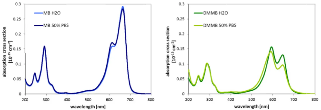
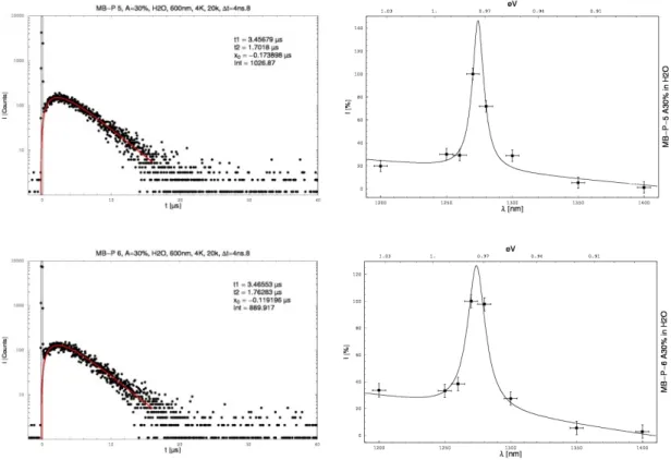
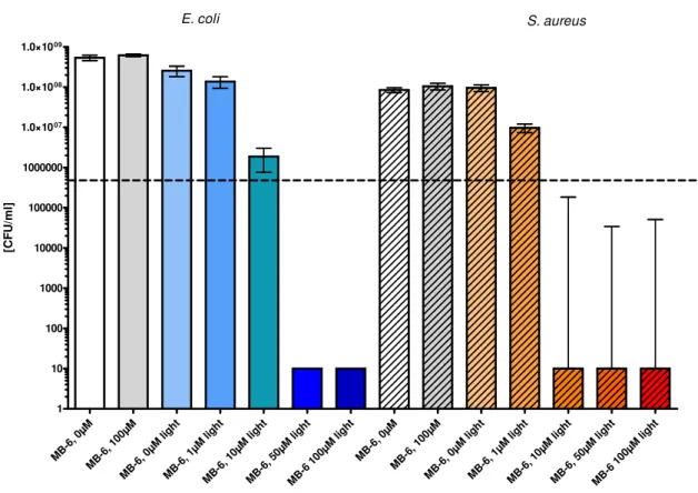
Chapter 3: In literature a rather high dark toxicity component of the photosensitizer class of phenothiazines was described and the systematics of the toxic processes were controversially discussed. Due to their wide use in PDI, the phenothiazine Methylene blue (MB) and common derivatives of this photosensitizer class were investigated in solution of H2O with direct spectroscopic methods in regard to their singlet oxygen generation and thus to clarify the singlet oxygen quantum yield. For all photosensitizers substantially smaller quantum yields as compared to values in literature were obtained via direct monitoring of singlet oxygen. This is considered as a probable effect of dimerisation or the use of indirect methods to monitor singlet oxygen without the explicit exclusion of the participation of other reactive species. Further, the attachment to Gram(+) and Gram(-) bacteria was determined quantitatively. In a second study, newly synthesized derivatives of MB were investigated in order to systemize the influence of side chains on the spectroscopic characteristics, especially the ability to generate singlet oxygen, the photostability and the phototoxic effectivity. In contrast to the commercially available dimerisation could be suppressed in some derivatives and achieving a higher photostability whilst comparable phototoxicity.
Chapter 4: The phototoxic properties of the porphyrin XF73 were reported to be rather high due to good cell attachment or penetration abilities with allowing a therapeutic window. Therefore, the photophysical characteristics of this photosensitizer were investigated in H2O and in cell physiological solution with regard to aggregation and singlet oxygen generation. The results were compared to findings with TMPyP. Aggregation was enhanced in XF73, most probably due to the flexible side chains, and a stacking was further
like PBS. The singlet oxygen generation was dramatically influenced by this stacking effect.
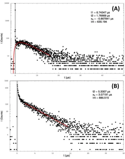
Chapter 5: The porphyrin TMPyP was incubated in C. albicans planktonic and biofilm growing samples. Thereafter, the singlet oxygen luminescence was directly detected at 1270 being the first report of the involvement of singlet oxygen in these cell types with this highly sensitive, non-invasive monitoring technique. Biofilm samples were considered as complex surfaces, exhibiting different decay kinetics for singlet oxygen than their planktonic counterparts. Different experimental laser setups were used for the measurements, thus allowing irradiation of the Soret- or Q-band of the photosensitizer with different irradiation power. With an enhanced sensitivity when irradiating at 420 , very low photosensitizer concentrations were necessary to monitor the singlet oxygen luminescence with a good signal-to-noise ratio. A dependency of the rise and decay times of the singlet oxygen luminescence on the photosensitizer concentration used for incubation was detected. Based on these findings a model was proposed, suggesting different surroundings for TMPyP within or adherent to the cell.
Chapter 6: Cellulose acetate surfaces and polyurethane-coated PMMA surfaces doped with different photosensitizers were produced and compared with regard to singlet oxygen generation and phototoxic efficacy. The generation of singlet oxygen by irradiation of the photosensitizer was proven within direct spectrally and time-resolved single-photon counting. The assignment of the rise and decay time was challenging, because the bi- exponential fit model was not sufficient for the complex surface system, which showed a multi-exponential decay manner. The presence of singlet oxygen was also monitored via an indirect method using the reaction with potassium iodide. Nevertheless, the PU-surfaces showed an effect without photosensitizer inherent, which indicates ROS generation of the material itself. The phototoxic efficacy against S. aureus was dependent on the irradiation time and the photosensitizer concentration in the material. Also, a difference in the phototoxic efficacy of the porphyrin-based and phenalenone-based photosensitizer doped in the surface material was detected, resulting in 2 reduction (> 99%) with the first and 3 reduction (> 99.9%) with the latter sample ( 50 , irradiation time 30 ).
Die vorliegende Doktorarbeit behandelt die antimikrobielle photodynamische Inaktivierung (PDI) als eine ergänzende Methode zur Verwendung von Antibiotika oder Desinfektionsmitteln, um pathogene Mikroorganismen zu deaktivieren. Prävention von Ansiedlung und Wachstum dieser Mikroorganismen auf Oberflächen mit Hilfe der PDI ist dabei das Hauptthema; dabei liegt der besondere Fokus auf der Herstellung und Verbesserung solcher Oberflächen. Dafür wurden verschiedene photoaktive Substanzen (Photosensibilisatoren) erst in Lösung in Hinblick auf ihre Fähigkeit zur Singulett- Sauerstoff-Generierung untersucht. Einige dieser Photosensibilisatoren wurden anschließend in verschiedene Oberflächenstrukturen einpolymerisiert und im Anschluss mit Hilfe spektroskopischer Methoden hinsichtlich ihrer physikalischen Eigenschaften und ihrer phototoxischen Effektivität untersucht.
Kapitel 3: In der Literatur ist eine Dunkeltoxizität der Photosensibilisatorklasse der Phenothiazine beschrieben und die Systematik des toxischen Prozesses wird kontrovers diskutiert. Aufgrund ihres breiten Anwendungsspektrums im Bereich PDI, wurden die Phenothiazine Methylen blau (MB) und gebräuchliche Derivate dieser Photosensibilisator- klasse in wässriger Lösung mit spektroskopischen Methoden untersucht und dabei die Singulett-Sauerstoff Generierung durch Bestimmung der Quantenausbeute für Singulett- Sauerstoff ermittelt. Für alle Photosensibilisatoren wurden mit direktem Monitoring von Singulett-Sauerstoff deutlich niedrigere Quantenausbeuten bestimmt, als in der Literatur angegeben. Als Grund dafür werden ein Dimerisierungseffekt oder die in der Literatur verwendeten indirekten Messmethoden für Singulett-Sauerstoff, welche nicht explizit die Beteiligung anderer reaktiver Spezies ausschließen, verantwortlich gemacht. Desweiteren wurde die Anlagerung oder Aufnahme der Photosensibilisatoren in Gram(+) und Gram(-) Bakterien quantitativ bestimmt. In einer zweiten Studie wurden neu synthetisierte MB- Derivate hinsichtlich des Einflusses der Seitenketten am jeweiligen Molekül auf die spektroskopische Charakteristik hin untersucht, insbesondere bezüglich der Fähigkeit zur Singulett-Sauerstoff Generierung, der Photostabilität und der phototoxischen Effektivität.
Im Gegensatz zu den käuflich erhältlichen Derivaten aus dem ersten Teil konnte Dimerisierungsverhalten bei einigen Exemplaren unterdrückt werden, bei gleichzeitig hoher Photostabilität und mit MB vergleichbarer Phototoxizität.
eingestuft aufgrund einer guten Anlagerung bzw. Aufnahme in Zellen, unter gleichzeitigem Vorhandensein eines therapeutischen Fensters. Deswegen wurden die photophysikalischen Eigenschaften dieses Photosensibilisators in und in zellphysiologischer Lösung hinsichtlich Aggregationsverhalten und Singulett-Sauerstoff Generierung untersucht. Die Ergebnisse wurden mit denen des Referenzphotosensibilisators TMPyP verglichen.
Aggregation war mit XF73 sehr wahrscheinlich durch dessen flexible Seitenketten erhöht und wurde zusätzlich konzentrationsabhängig getrieben in Gegenwart von , welches in zellphysiologischer Lösung wie PBS zu finden ist. Aggregation von XF73 beeinflusst deutlich negativ die Generierung von Singulett-Sauerstoff.
Kapitel 5: Das Porphyrin TMPyP wurde in C. albicans inkubiert, welche sowohl planktonisch als auch als Biofilm wuchsen. Anschließend wurde die Lumineszenz von Singulett-Sauerstoff direkt bei 1270 detektiert und in dieser Arbeit erstmalig als beteiligtes toxisches Agens in diesem Mikroorganismus mit Hilfe der hochsensitiven und nicht-invasiven Monitoring-Technik beschrieben. Die Biofilm-Proben werden als komplexe Oberflächen angesehen, die unterschiedliche Abklingkinetik für Singulett-Sauerstoff aufweisen, im Vergleich zu ihren planktonischen Entsprechungen. Verschiedene Laseraufbauten wurden für die Messungen verwendet und ermöglichten Bestrahlung in der Soret- oder Q-Bande von TMPyP mit unterschiedlicher Leistung. Mit der erzielten erhöhten Sensitivität für die Bestrahlung bei 420 (Soret) waren nur sehr niedrige Photosensibilisatorkonzentrationen nötig, um die Singulett-Sauerstoff-Lumineszenz mit einem guten Signal-Rausch-Verhältnis zu aufzunehmen. Eine Abhängigkeit der Anstiegs- und Abklingzeiten der Singulett-Sauerstoff-Lumineszenz von der Konzentration des Photosensibilisators wurde bestimmt. Basierend auf diesen Ergebnissen wird hier ein Modell vorgeschlagen, welches verschiedene Umgebungen für TMPyP innerhalb oder anhaftend an die Zelle vorschlägt.
Kapitel 6: Oberflächen aus Zelluloseazetat oder mit Polyurethan überzogenes PMMA wurden mit unterschiedlichen Photosensibilisatoren dotiert und miteinander verglichen hinsichtlich Generierung von Singulett-Sauerstoff und phototoxischer Effektivität. Die Generierung von Singulett-Sauerstoff konnte mit Hilfe von spektral- und zeitaufgelöster Einzelphotonenzählung nachgewiesen werden. Die Zuordnung von Anstiegs- und Abklingzeiten war eine Herausforderung, weil das bi-exponentielle Fitmodell nicht ausreichte, um das komplexe Oberflächensystem zu beschreiben, welches ein multi- exponentielles Verhalten an den Tag legte. Das Vorhandensein von Singulett-Sauerstoff wurde zudem mit einer indirekten Methode überprüft, die sich die Reaktion von
dieser Methode auch das PU-Material ohne inhärenten Photosensibilisator die Ausbildung reaktiver Spezies. Die Phototoxizität gegen S. aureus war abhängig von der Bestrahlungszeit und der Konzentration des Photosensibilisators im Material. Zudem wurde ein Unterschied zwischen der phototoxischen Effektivität der Oberfläche mit einpolymerisiertem Photosensibilisator auf Porphyrin-Basis und Phenalenone-Basis festgestellt. Ersterer führte zu einer Reduktion von 2 -Schritten (> 99%), letzterer zu einer Reduktion von 3 -Schritten (> 99.9%) mit einer Bestrahlungsleistung von 50 und einer Bestrahlungszeit von 30 .
aPDT antimicrobial photodynamic treatment
CA cellulose acetate
C. albicans Candida albicans
CFU colony forming units
DMMB Di-methyl methylene blue
DNA deoxyribonucleic acid
E. coli Escherichia coli
EM electromagnetic
F fluorescence
water
hydrogen peroxide
IC internal conversion
IR infrared
ISC intersystem crossing
quenching rate constant
LPS lipopolysaccharides
micro
MH Müller-Hinton
MB 3,7-Bis(dimethylamino)-phenothiaziniumchlorid, Methylene blue
MB-1 3-[(2-Ammoniumethyl)(methyl)amino]-7-(dimethylamino) phenothiazin-5-ium dichloride
MB-2 3-{methyl[2-(methylammonio)ethyl]amino}-7-(dimethylamino) phenothiazin-5- ium dichloride
MB-3 3-(piperazin-4-ium-1-yl)-7-(dimethylamino)phenothiazin-5-ium dichloride MB-4 3-[(2-Hydroxyethyl)(methyl)amino]-7-(dimethylamino) phenothiazin-5-ium
chloride
MB-5 3-(morpholin-4-yl)-7-(dimethylamino)phenothiazin-5-ium chloride
millilitre
MRSA Methicillin-resistant Staphylococcus aureus molecular weight
! " sodium azide
NIR near infrared
NMB New methylene blue N
molecular oxygen
, #! ∆ %& singlet molecular oxygen, singlet oxygen
∙ superoxide radical
OPO optical parametric oscillator
P phosphorescence
PACT photodynamic antimicrobial chemotherapy
PBS phosphate buffered saline
PDI photodynamic inactivation
PDT photodynamic therapy
PIB photodynamic inactivation of bacteria
PMMA poly-methylmethacrylate
PMT photomultiplier tube
PN Phenalenone
PN-O-A N,N-Dimethyl-N-(2-hydroxyethyl)-2-ammoniummethyl-phenalen-1-one chloride PN-O-B N-(3-Hydroxypropyl)-N-(2-methylen-phenalen-1-one)acetamid
PNS 1h-Phenalen-1-One-2-Sulfonic Acid
PS photosensitizer
PU polyurethane
PUR polyurethane containing no photosensitizer
ROS reactive oxygen species
S. aureus Staphylococcus aureus
(∆ singlet oxygen decay time
TBO Toluidine blue O
TCSPC time-correlated single photon counting
TMPyP 5,10,15,20-Tetrakis(1-methyl-4-pyridinio)-porphyrin tetra(p-toluenesulfonate) TPP 5,10,15,20-Tetraphenyl-21H,23H-porphine, meso-Tetraphenylporphyrin TPP-O-A 5-(4-Hydroxyphenyl)-10,15,20-triphenylporphyrin
TPP-O-B 5-[4-(6-Hydroxyhexoxy)-phenyl]-10,15,20-triphenylporphyrin TRPD time-resolved phosphorescence detection
()* photosensitizer triplet state decay time
UV ultraviolet
VIS visible
VR vibrational relaxation
XF73 5,15-bis-[4-(3-Trimethylammonio-propyloxy)-phenyl]-porphyrin
1. CHAPTER 1: Introduction . . . . . . . . 27
1.1 Problematics of antibiotics and escaping microorganisms . . . . 29
1.2 Antibacterial photodynamic treatment and basic principles . . . . 30
1.2.1 Generation and mode of action of molecular singlet oxygen . . . 31
1.2.2 Photosensitizers . . . 33
1.2.3 Cells . . . 34
1.3 Self-disinfecting surfaces and current state of research . . . . 36
1.3.1 Reported phototoxic efficacy of some aPDT-surfaces . . . . 36
1.3.2 Singlet oxygen detection and decay of photosensitizer triplet state in polymer materials . . . 37
1.4 Objectives . . . 39
1.5 References . . . 40
2. CHAPTER 2: Material and Methods . . . . . . 43
2.1 Chemicals: Photosensitizers, Solvents, Quencher, and substances for chemical analyses . . . 45
2.1.1 Photosensitizers . . . 45
2.1.1.1 Porphyrins . . . 45
2.1.1.2 Perinaphthenons . . . 46
2.1.1.3 Phenothiaziniums . . . 47
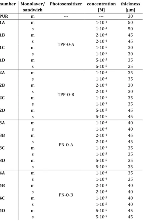
2.1.1.4 Photosensitizers doped into polyurethane . . . 49
2.1.2 Solvents and salts . . . 53
2.1.3 Quencher . . . 53
2.1.4 Potassium iodide . . . 54
2.2 Surfaces and their manufacturing . . . 55
2.2.1 PS in Cellulose acetate (CA) . . . 55
2.2.2 PS in Polyurethane (PU) covering poly-methylmethacrylate (PMMA) . . . 55
2.3 Basic techniques . . . 58
2.3.1 Absorption spectroscopy . . . 58
2.3.2 NMR spectroscopy . . . 58
2.4.1 Time-correlated single photon counting . . . 59 2.4.2 Time-resolved NIR phosphorescence detection . . . 59
2.5 Microscopy techniques . . . 63
2.5.1 Fluorescence microscopy . . . 63
2.5.2 Transmission electron microscopy . . . 63
2.6 Emission spectra of the lamps . . . 64
2.6.1 Waldmann UV236: Irradiation of XF73 and TMPyP with C. albicans . . 64 2.6.2 Waldmann PDT1200: Irradiation of MB derivatives with S. aureus and E. coli . 65 2.6.3 Waldmann PIB 3000 (full spectrum > 400 nm): Toxicity tests on PU-surfaces . 65
2.7 Microbiological techniques . . . 67
2.7.1 Microbial strains . . . 67
2.7.2 Culture conditions . . . 67
2.7.3 Spectroscopic measurements with cells . . . 68 2.7.4 aPDT phototoxicity experiments in vitro and on surfaces . . . 68
2.8 References . . . 70
3. CHAPTER 3: Spectroscopic and Phototoxicity Studies of Phenothiazinium Derivatives 71
3.1 Introduction . . . 73
3.2 Experimental section . . . 76
3.2.1 Time- and spectrally resolved singlet oxygen luminescence . . . 76 3.2.2 Quantum yield of singlet oxygen formation . . . 76
3.2.3 Photostability . . . 77
3.2.4 Bacterial strains . . . 77
3.2.5 Light source . . . 77
3.2.6 Phototoxicity assay of the bacteria . . . 78 3.2.7 Data analysis and statistics for cell experiments . . . . 78
3.3 Results & Discussion . . . 79
3.3.1 Investigation of MB, NMB, DMMB, and TBO . . . 80 3.3.1.1 Absorption spectra for different photosensitizer concentrations . . 80
3.3.1.2 Photostability . . . 84
3.3.1.3 Time- and spectral resolved direct detection of singlet oxygen . . . 85 3.3.1.4 Determination of the singlet oxygen quantum yields by direct methods. . 87
3.3.1.5 Phenothiaziniums and bacteria: attachment and influence on the
photosensitizer absorption . . . 90
3.3.2 New MB-derivatives . . . 94
3.3.2.1 Absorption spectra for different photosensitizer concentrations . . 94
3.3.2.2 Photostability . . . 96
3.3.2.3 Time- and spectral resolved direct detection of singlet oxygen . . 99 3.3.2.4 Determination of the singlet oxygen quantum yields by direct methods . 101
3.3.2.5 Photobiological activity . . . 102
3.4 Conclusions . . . 108
3.5 References . . . 111
4. CHAPTER 4: Spectroscopic Studies of the Porphyrin XF73 . . . . 115
4.1 Introduction . . . 117
4.2 Experimental section . . . 119
4.2.1 Chemicals and solvents . . . 119
4.2.2 Absorption spectrum . . . 119
4.2.3 Photostability . . . 119
4.2.4 Cell experiments . . . 119
4.2.5 Fluorescence spectrophotometer . . . 120
4.2.6 Singlet oxygen luminescence and quantum yield of singlet oxygen
formation (ФΔ) . . . 120
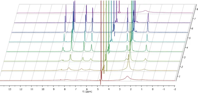
4.2.7 NMR-spectra . . . 121
4.3 Results & Discussion . . . 122
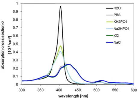
4.3.1 Absorption spectroscopy in aqueous photosensitizer solution . . . 123 4.3.2 NMR spectra of XF73 and TMPyP in D2O solution . . . . 124 4.3.3 Absorption spectroscopy in aqueous XF73 solution with PBS or PBS
constituents. . . 127
4.3.4 Photostability of XF73 . . . 129
4.3.5 Singlet oxygen luminescence experiments without PBS . . . 130 4.3.6 Singlet oxygen luminescence experiments with PBS . . . . 133 4.3.7 Fluorescence microscopy of C. albicans incubated with XF73 . . . 135
4.4 Conclusions . . . 136
5. CHAPTER 5: Spectroscopic Studies of Porphyrins in Planktonic Cells and Biofilms . 141
5.1 Introduction . . . 143
5.2 Experimental section . . . 145
5.2.1 Chemicals . . . 145
5.2.2 Fungi cells . . . 145
5.2.3 Singlet oxygen luminescence detection by direct spectroscopic methods . 146
5.3 Results & Discussion . . . 148
5.3.1 Fluorescence imaging of TMPyP in C. albicans planktonic cells and biofilm . 148 5.3.2 Direct spectroscopic detection of singlet oxygen in TMPyP dissolved in H2O . 150 5.3.3 Direct spectroscopic detection of singlet oxygen in planktonic C. albicans
cells with the Nd:YAG laser . . . 150
5.3.4 Detection of singlet oxygen in biofilms with the Nd:YAG laser . . . 156 5.3.5 Detection of singlet oxygen in planktonic C. albicans cells with the tunable
laser system and a gas flow unit . . . 159
5.3.5.1 Irradiation with 520 nm in the first Q-band of TMPyP . . . . 160 5.3.5.2 Irradiation with 420 nm in the Soret band of TMPyP . . . . 163
5.4 Conclusions . . . 171
5.5 References . . . 174
6. CHAPTER 6: Spectroscopic and Phototoxicity Studies of Photosensitizers in Surfaces 175
6.1 Introduction . . . 177
6.2 Experimental section . . . 179
6.2.1 Chemicals . . . 179
6.2.2 Surfaces . . . 179
6.2.2.1 Cellulose acetate (CA) doped with photosensitizer . . . . 179 6.2.2.2 Polyurethane (PU) doped with photosensitizer on PMMA polymer plate . 179 6.2.3 Singlet oxygen luminescence detection by direct spectroscopic methods . 180
6.2.4 Photostability . . . 180
6.2.5 Indirect detection of singlet oxygen with potassium iodide (KI) . . 180 6.2.6 Transmission electron microscopy (TEM) . . . 180
6.3 Results & Discussion . . . 182 6.3.1 Cellulose acetate (CA) with MB, TMPyP and TPP . . . . 182 6.3.1.1 Absorption spectra of MB, TMPyP, and TPP in CA . . . . 183 6.3.1.2 Direct detection of the singlet oxygen luminescence at 1270 nm . . 186 6.3.1.3 Indirect detection of the generation of singlet oxygen with potassium iodide . 194 6.3.2 Polyurethane (PU) with TPP-O-A/B and PN-O-A/B . . . . 195 6.3.2.1 Absorption spectra and photostability . . . 196 6.3.2.2 Direct detection of the singlet oxygen luminescence . . . . 202 6.3.2.3 Indirect detection of the generation of singlet oxygen with potassium iodide . 213
6.3.2.4 Fluorescence of TPP-O-A in PU . . . 215
6.3.2.5 Transmission electron microscopy . . . 216
6.3.3 Phototoxicity experiments on surfaces . . . 219 6.3.3.1 Recovery from the PU-surface and fluctuation of the dark control . . 221 6.3.3.2 Phototoxicity efficacy of TPP-O-A in PU . . . 222 6.3.3.3 Toxicity efficacy of PN-O-A in PU . . . 223
6.4 Conclusions . . . 225
6.5 References . . . 230
7. CHAPTER 7: Conclusions . . . . . . . . 233
PUBLICATIONS . . . . . . . . . 239
CONTRIBUTIONS TO CONFERENCES . . . . . . . 241
DISCLAIMER / SELBSTSTÄNDIGKEITSERKLÄRUNG . . . . . 245
Chapter 1
Introduction
In this chapter a short introduction to the overall topic of this doctoral thesis is given, ranging from the complex of problems of antibiotics to efficiently kill pathogen microorganisms to describing antimicrobial photodynamic inactivation as an alternative treatment modality. The historical development is described in short, as well as the basic physical principles of photosensitizer and singlet oxygen. The development and studies on surfaces is outlined in regard to effective photosensitizing efficacy and their photophysical properties, and the objectives of the thesis are presented.
29
1.1 PROBLEMATICS OF ANTIBIOTICS and ESCAPING MICRO- ORGANISMS
The widespread and inappropriate use of antibiotics in health system for diseases of man and the over-use in food industry for maintaining a low infection level and a fast animal growth in industrial livestock farming has provoked resistance patterns of both fungal and bacterial pathogens against these [1-6]. It might be no exaggeration to call it one of the main challenges for health care in the 21st century. Darwin's theory of selection pressure, in form of antibiotics having highly specific mechanisms of antimicrobial action, again gives credit to this genetic flexibility of these human pathogenic microorganisms [7, 8]. The World Economic Forum (WEF) concluded in their report on global risks that “arguably the greatest risk ... to human health comes in the form of antibiotic-resistant bacteria. We live in a bacterial world where we will never be able to stay ahead of the mutation curve” [9].
The most serious infections are caused by a group of microorganisms which are merged under the acronym “ESKAPE”, because this group of microorganisms has the ability to escape the effects of antimicrobial agents. These important pathogens are Enterococcus faecium, Staphylococcus aureus, Klebsiella pneumoniae, Acinetobacter baumannii, Pseudomonas aeruginosa, and Enterobacter species [10]. This group of superbugs was extended by Peterson including Clostridium difficile as one of the most common hospital- acquired infections [11] and especially pointing out that infections by Enterobacter E. coli currently account for ≈ 20% of all cases of bacteraemia in the United Kingdom [12]. Not only methicillin-resistant S. aureus (MRSA), but also the Enterobacter species have developed a broad-spectrum resistance against many classes of antibiotics. The eukaryotic fungus Candida albicans as the main agent for superficial skin mycosis has of course to be added to that list of 'escaping' microorganisms, since also within this pathogen resistance patterns became an increasing problem, especially in patients with immune deficiency [13].
The production and admission of new classes of antibiotics is a slow process outdistanced at the moment by the faster resistance development. Therefore, new alternative treatments are developed amongst which photodynamic treatment is one approach that has become important and prevalent within the past years [14-19].
30
1.2 ANTIBACTERIAL PHOTODYNAMIC TREATMENT and BASIC PRINCIPLES
As an alternative treatment against microorganisms photodynamic inactivation (PDI) is described in this work. The pathogen is treated with a photoactive dye or photosensitizer in combination with light and in the presence of oxygen, which results in the formation of reactive oxygen species (ROS), such as singlet oxygen. Singlet oxygen is described to be one of the key agents in PDI [20-25] and in contrast to antibiotics, which act toxically specific as a single drug, in PDI not the photosensitizer drug, but singlet oxygen or other ROS react by oxidising the surrounding unspecifically.
In the 1970's first treatments against cancer cells were reported with this method, but reports of the photodynamic action go back more than 100 years to the laboratories of Raab and Tappeiner, who discovered PDI by staining Paramecium caudatum with the photosensitizer Acridine orange and detected their death upon light exposure under involvement of oxygen [26, 27]. The physical background of photosensitizer activation with light and deactivation in the presence of oxygen was offered by Kautsky et al. [28, 29].
Overviews of the basic PDI principle in regard to historical development and physical, chemical and biological aspects can be found to a great extent in literature [30] and Denis et al. give an outline of its development by additionally describing the main problems that PDI still has - despite its use as an anti-cancer agent - of gaining ground as a medical treatment against microorganisms [31]. The treatment or prevention of infectious diseases caused by microorganisms with PDI uses different acronyms, mainly aPDT for antimicrobial photodynamic therapy, PDI for photodynamic inactivation or PACT for photodynamic antimicrobial chemotherapy. The treatment is applied so far in dentistry as treatment of oral cancer, bacterial and fungal infections, or for the photodynamic diagnosis (PDD) of the malignant transformation of oral lesions [32-34]. PDI also represents a novel therapeutic approach in the controlling of biofilms, which are able to grow on variable surfaces, organic or non-organic [35-37].
The usage of PDI in general might be split in therapy and prevention of infection with pathogens. As a therapeutic method for the decolonisation of infected wounds PDI might be a powerful medical treatment, if the structure of the photosensitizers is chosen selective enough to distinguish between human cells and pathogen with unique chemical binding features and therefore offer a therapeutic window.
31 In contrast to that, the preventive character of PDI gets increasingly in the focus of research, helpful against hospital-acquired infections but also in food industry [38-41]. The prevention of microorganisms from settling and growing on surfaces is a matter of great concern, since disinfection as such might then be obsolete. Therefore, a photosensitizer is linked or doped into a polymer and with the condition of sufficient oxygen permeability of the material singlet oxygen will be generated under light irradiation, can diffuse from the surface and induce the death of adherent microorganisms [37, 42-48].
1.2.1 Generation and mode of action of molecular singlet oxygen
The ground state of di-oxygen, , is a triplet state, with two unpaired electrons with parallel spins in a non-binding orbital. Thus, oxygen has paramagnetic properties and is a bi-radical. The bi-radical character and the double bond of the two oxygen atoms are illustrated best with according to and extending the Lewis formulae [49]. Since the ground state is a triplet state the radical properties of oxygen are limited due to the spin- conservation for chemical reactions. Singlet molecular oxygen, , which is molecular oxygen in its lowest excited singlet state, therefore is highly reactive. This state has two electrons with antiparallel spin in one of its non-binding orbital. The existence of was experimentally confirmed with its emission band 1270 in the earth atmosphere and with oxygen in the liquid phase 5 years after the theoretical description by Mulliken [50].
The first direct spectroscopic detection of the decay of dissolved singlet molecular oxygen into its ground state in a solution of was done by Krasnovsky et al. [51]. The detection of the decay of singlet oxygen is still challenging for low quantum yields and in some liquids due to the low signal-to-noise ratio of the luminescence signal at 1270 . According to Kasha the relaxation from singlet to triplet molecular oxygen is quantum mechanically forbidden, due to a violation of the conservation of the spin, the symmetry and the orbital angular momentum, which results in a metastable state and a relatively long lifetime of singlet oxygen on a typical time scale for chemical reactions [49, 52]. In solution a shorter lifetime of singlet oxygen is yielded due to perturbation.
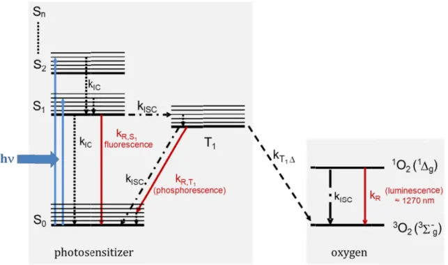
After the photoactivation of a photosensitizer with a light source with photons of an appropriate wavelength and thus energy (), the photosensitizer is promoted in an electronically excited state . The photosensitizer can then release the additional energy via radiative or non-radiative processes, which are indicated in figure 1.1.
32
Fig. 1.1: Jablonski diagram showing the process of the generation of singlet oxygen via direct energy transfe photosensitizer to oxygen; energy levels of the photosensitizer, PS (left) and energy levels of molecular oxygen, O
excited singlet state
first excited singlet state of molecular oxygen, respectively. Possible relaxation rates of the excited PS are internal conversion (k
energy transfer from the excited T the generation of singlet oxygen
After relaxation into the first excited singlet state relax in
internal conversion lived triplet
vibrational relaxation named occur via radiative relaxat multiplicity) or phosp transferred from
resulting in the formation of singlet oxygen After formation of the T
relaxation distinguished
triplet state with other molecules
to the generation of free radicals. In the type
Jablonski diagram showing the process of the generation of singlet oxygen via direct energy transfe photosensitizer to oxygen; energy levels of the photosensitizer, PS (left) and energy levels of molecular oxygen, O2 (right) are shown. S
excited singlet state
first excited singlet state of molecular oxygen, respectively. Possible relaxation rates of the excited PS are internal conversion (k
energy transfer from the excited T the generation of singlet oxygen
After relaxation into the first excited singlet state into the ground state via non
internal conversion
lived triplet-T1-state, distinguished vibrational relaxation named occur via radiative relaxat
tiplicity) or phosp transferred from the
resulting in the formation of singlet oxygen After formation of the T
relaxation to the ground state. In general, t distinguished: in the type
triplet state with other molecules
to the generation of free radicals. In the type
Jablonski diagram showing the process of the generation of singlet oxygen via direct energy transfe photosensitizer to oxygen; energy levels of the photosensitizer, PS (left) and energy levels of molecular
(right) are shown. S excited singlet states; T1 is the
first excited singlet state of molecular oxygen, respectively. Possible relaxation rates of the excited PS are internal conversion (kIC), fluorescence (k
energy transfer from the excited T the generation of singlet oxygen
After relaxation into the first excited singlet state ground state via non
internal conversion (); when state, distinguished
vibrational relaxation named intersystem crossing (
occur via radiative relaxation resulting in fluorescence ( tiplicity) or phosphorescence (
the triplet-T1
resulting in the formation of singlet oxygen After formation of the T1-state o
the ground state. In general, t
n the type-I-reaction the photosensitizer reacts in its excited singlet or triplet state with other molecules
to the generation of free radicals. In the type
Jablonski diagram showing the process of the generation of singlet oxygen via direct energy transfe photosensitizer to oxygen; energy levels of the photosensitizer, PS (left) and energy levels of molecular
(right) are shown. S0 is the ground state of the PS, S is the first excited triplet state of the PS; O
first excited singlet state of molecular oxygen, respectively. Possible relaxation rates of the excited PS are ), fluorescence (kR,S1
energy transfer from the excited T1-state of the PS to molecular oxygen the generation of singlet oxygen 1O2.
After relaxation into the first excited singlet state ground state via non-radiative
); when energy is transferred to a different electronic state, state, distinguished from the
intersystem crossing (
ion resulting in fluorescence ( horescence (→
1-state to adjacent molecular oxygen resulting in the formation of singlet oxygen
state other photoc the ground state. In general, t
reaction the photosensitizer reacts in its excited singlet or triplet state with other molecules by electron transfer or hydrogen abstraction, which le to the generation of free radicals. In the type
Jablonski diagram showing the process of the generation of singlet oxygen via direct energy transfe photosensitizer to oxygen; energy levels of the photosensitizer, PS (left) and energy levels of molecular
is the ground state of the PS, S first excited triplet state of the PS; O
first excited singlet state of molecular oxygen, respectively. Possible relaxation rates of the excited PS are
R,S1), intersystem
state of the PS to molecular oxygen
After relaxation into the first excited singlet state
radiative mechanisms like
energy is transferred to a different electronic state, from the state by its spin multiplicity intersystem crossing (
ion resulting in fluorescence (
, changing spin multiplicity) state to adjacent molecular oxygen
resulting in the formation of singlet oxygen [30].
ther photochemical reactions might occur
the ground state. In general, two different types of reaction pathways reaction the photosensitizer reacts in its excited singlet or
electron transfer or hydrogen abstraction, which le to the generation of free radicals. In the type-II-reaction singlet oxygen is generated by
Jablonski diagram showing the process of the generation of singlet oxygen via direct energy transfe photosensitizer to oxygen; energy levels of the photosensitizer, PS (left) and energy levels of molecular
is the ground state of the PS, S1 the first excited singlet state, S first excited triplet state of the PS; O2 and
first excited singlet state of molecular oxygen, respectively. Possible relaxation rates of the excited PS are ), intersystem crossing (k
state of the PS to molecular oxygen
, the excited state can spontaneously mechanisms like
energy is transferred to a different electronic state, state by its spin multiplicity
) can occur ion resulting in fluorescence (→
, changing spin multiplicity) state to adjacent molecular oxygen
hemical reactions might occur
wo different types of reaction pathways reaction the photosensitizer reacts in its excited singlet or
electron transfer or hydrogen abstraction, which le reaction singlet oxygen is generated by
Jablonski diagram showing the process of the generation of singlet oxygen via direct energy transfe photosensitizer to oxygen; energy levels of the photosensitizer, PS (left) and energy levels of molecular
the first excited singlet state, S and 1O2 are the ground state and the first excited singlet state of molecular oxygen, respectively. Possible relaxation rates of the excited PS are
crossing (kISC), phosphorescence (k
state of the PS to molecular oxygen (kT1∆). The energy transfer yields
ited state can spontaneously mechanisms like vibrational relaxation, energy is transferred to a different electronic state,
state by its spin multiplicity
can occur. Energy loss might also
→ , conservation of spin , changing spin multiplicity). Energy can be state to adjacent molecular oxygen in its triplet ground state
hemical reactions might occur
wo different types of reaction pathways reaction the photosensitizer reacts in its excited singlet or
electron transfer or hydrogen abstraction, which le reaction singlet oxygen is generated by
Jablonski diagram showing the process of the generation of singlet oxygen via direct energy transfer from photosensitizer to oxygen; energy levels of the photosensitizer, PS (left) and energy levels of molecular the first excited singlet state, Sn higher are the ground state and the first excited singlet state of molecular oxygen, respectively. Possible relaxation rates of the excited PS are ), phosphorescence (kR,T1) and . The energy transfer yields
ited state can spontaneously vibrational relaxation, energy is transferred to a different electronic state, a long
state by its spin multiplicity another Energy loss might also , conservation of spin . Energy can be in its triplet ground state
hemical reactions might occur, leading to a wo different types of reaction pathways reaction the photosensitizer reacts in its excited singlet or
electron transfer or hydrogen abstraction, which le reaction singlet oxygen is generated by
r from photosensitizer to oxygen; energy levels of the photosensitizer, PS (left) and energy levels of molecular higher are the ground state and the first excited singlet state of molecular oxygen, respectively. Possible relaxation rates of the excited PS are ) and . The energy transfer yields
ited state can spontaneously vibrational relaxation, long- another Energy loss might also , conservation of spin . Energy can be in its triplet ground state
leading to a wo different types of reaction pathways are reaction the photosensitizer reacts in its excited singlet or electron transfer or hydrogen abstraction, which leads reaction singlet oxygen is generated by
direct energy t oxygen in its
probability depends mainly on the photosensitizer properties, the substrate, or the properties of the surrounding medium,
The 1980s by kind of
photosensitizer
origin of singlet oxygen and the microorganisms
1.2.2
Effective
quantum yield of singlet oxygen formation. In regard to differences in the outer cell membrane
cationic photosensitizers in comparison therapeutic window should possible.
with the first discovered photosensitizers, but photosensitizer
direct energy t oxygen in its triplet
probability depends mainly on the photosensitizer properties, the substrate, or the properties of the surrounding medium,
The direct microbi 1980s by Dahl kind of other photosensitizer
origin of singlet oxygen and the microorganisms
1.2.2 Photosensitizers
Effective photosensitizers should generate singlet oxygen
quantum yield of singlet oxygen formation. In regard to differences in the outer cell membrane of microorganisms and thus different uptake features of neutral, anionic or
ationic photosensitizers in comparison therapeutic window should possible.
with the first discovered photosensitizers, but photosensitizer
direct energy transfer from the excited triplet triplet ground state. Type
probability depends mainly on the photosensitizer properties, the substrate, or the properties of the surrounding medium,
microbicidal efficacy of singlet oxygen was proven by an experiment i Dahl et al. who
other ROS. In this case photosensitizer and microorganism
origin of singlet oxygen and the microorganisms
Photosensitizers
photosensitizers should generate singlet oxygen
quantum yield of singlet oxygen formation. In regard to differences in the outer cell microorganisms and thus different uptake features of neutral, anionic or ationic photosensitizers in comparison
therapeutic window should possible.
with the first discovered photosensitizers, but photosensitizer enhance the
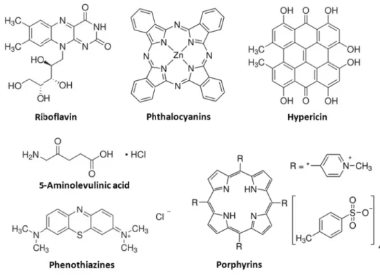
Fig. 1.2: Chemical structures of frequently used classes of photosensitizers.
ransfer from the excited triplet ground state. Type
probability depends mainly on the photosensitizer properties, the substrate, or the properties of the surrounding medium,
cidal efficacy of singlet oxygen was proven by an experiment i exposed bacteria to
In this case, a possible type
microorganism was excluded by increasing the origin of singlet oxygen and the microorganisms
photosensitizers should generate singlet oxygen
quantum yield of singlet oxygen formation. In regard to differences in the outer cell microorganisms and thus different uptake features of neutral, anionic or ationic photosensitizers in comparison
therapeutic window should possible.
with the first discovered photosensitizers, but enhance the antibacterial
: Chemical structures of frequently used classes of photosensitizers.
ransfer from the excited triplet-state of the photosensitizer to molecular ground state. Type-I and type
probability depends mainly on the photosensitizer properties, the substrate, or the properties of the surrounding medium, such as the oxygen concentration.
cidal efficacy of singlet oxygen was proven by an experiment i bacteria to pure singlet oxygen
, a possible type
was excluded by increasing the origin of singlet oxygen and the microorganisms [
photosensitizers should generate singlet oxygen
quantum yield of singlet oxygen formation. In regard to differences in the outer cell microorganisms and thus different uptake features of neutral, anionic or ationic photosensitizers in comparison to human eukaryotic cells furthermore a
Especially Gram(
with the first discovered photosensitizers, but it was found that antibacterial efficacy
: Chemical structures of frequently used classes of photosensitizers.
state of the photosensitizer to molecular I and type-II can occur simult
probability depends mainly on the photosensitizer properties, the substrate, or the the oxygen concentration.
cidal efficacy of singlet oxygen was proven by an experiment i pure singlet oxygen
, a possible type-I-mechanism was excluded by increasing the
53].
photosensitizers should generate singlet oxygen to a high extent
quantum yield of singlet oxygen formation. In regard to differences in the outer cell microorganisms and thus different uptake features of neutral, anionic or human eukaryotic cells furthermore a Especially Gram(-) bacteria responded poorly to
it was found that efficacy [54].
: Chemical structures of frequently used classes of photosensitizers.
state of the photosensitizer to molecular II can occur simult
probability depends mainly on the photosensitizer properties, the substrate, or the the oxygen concentration.
cidal efficacy of singlet oxygen was proven by an experiment i pure singlet oxygen under exclusion of
mechanism due to
was excluded by increasing the distance between the
to a high extent
quantum yield of singlet oxygen formation. In regard to differences in the outer cell microorganisms and thus different uptake features of neutral, anionic or human eukaryotic cells furthermore a
) bacteria responded poorly to it was found that positive charges
: Chemical structures of frequently used classes of photosensitizers.
state of the photosensitizer to molecular II can occur simultaneously and their probability depends mainly on the photosensitizer properties, the substrate, or the
the oxygen concentration.
cidal efficacy of singlet oxygen was proven by an experiment in the late under exclusion of due to direct contact of
distance between the
to a high extent, i.e. have a high quantum yield of singlet oxygen formation. In regard to differences in the outer cell microorganisms and thus different uptake features of neutral, anionic or human eukaryotic cells furthermore a
) bacteria responded poorly to positive charges
: Chemical structures of frequently used classes of photosensitizers.
33 state of the photosensitizer to molecular aneously and their probability depends mainly on the photosensitizer properties, the substrate, or the
n the late under exclusion of any direct contact of distance between the
have a high quantum yield of singlet oxygen formation. In regard to differences in the outer cell microorganisms and thus different uptake features of neutral, anionic or human eukaryotic cells furthermore a ) bacteria responded poorly to PDI positive charges in the
34
A lot of different photosensitizer types,
were investigated in the last decades in regard to the generation of singlet oxygen phototoxic effect against pathogen microorganisms
PDI, are displayed in electrons in their binding
change of the spin multiplicity from singlet to triplet excited state (
mechanically forbidden, but the probability therefore is enhanced if electronic ring currents due to spin
atom effects
1.2.3
In this work bo
facultative pathogens and can cause severe illness their settlement
Candida
considered to be t
in both immunocompromised and immunocompetent epithelia
of internal organs cell wall and contains mitochondria, and several thus grow
A lot of different photosensitizer types,
were investigated in the last decades in regard to the generation of singlet oxygen phototoxic effect against pathogen microorganisms
, are displayed in electrons in their binding
change of the spin multiplicity from singlet to triplet excited state (
mechanically forbidden, but the probability therefore is enhanced if electronic ring currents due to spin-orbit-coupling are possible, due to collisions with the solvent or due to heavy atom effects [49].
Cells
In this work both, prokaryotic and eukaryotic facultative pathogens and can cause severe illness
settlement and growth
andida albicans is a eukaryotic cell considered to be the most c
both immunocompromised and immunocompetent epithelia, in severe cases
of internal organs [ cell wall and contains mitochondria, and several
grows vegetatively
A lot of different photosensitizer types,
were investigated in the last decades in regard to the generation of singlet oxygen phototoxic effect against pathogen microorganisms
, are displayed in figure
electrons in their binding -orbitals. In these orbitals the spins typically are antiparallel; a change of the spin multiplicity from singlet to triplet excited state (
mechanically forbidden, but the probability therefore is enhanced if electronic ring currents coupling are possible, due to collisions with the solvent or due to heavy
th, prokaryotic and eukaryotic facultative pathogens and can cause severe illness
and growth on surfaces is a eukaryotic cell
he most common fungal pathogen both immunocompromised and immunocompetent
, in severe cases also
[55-58]. The cytoplasm of
cell wall and contains for eukaryotes typical organelles like a nucleus mitochondria, and several cytoplasmic inclusion bodies
vegetatively in either
Fig. 1.
A lot of different photosensitizer types,
were investigated in the last decades in regard to the generation of singlet oxygen phototoxic effect against pathogen microorganisms
1.2. Many of these photoactive dyes exhibit de
orbitals. In these orbitals the spins typically are antiparallel; a change of the spin multiplicity from singlet to triplet excited state (
mechanically forbidden, but the probability therefore is enhanced if electronic ring currents coupling are possible, due to collisions with the solvent or due to heavy
th, prokaryotic and eukaryotic facultative pathogens and can cause severe illness
on surfaces, and thus a distribution through humans is a eukaryotic cell symbiotically present in
ommon fungal pathogen both immunocompromised and immunocompetent
also followed by dissemination The cytoplasm of
for eukaryotes typical organelles like a nucleus cytoplasmic inclusion bodies
either ovoid yeast, pseudohyphae
Fig. 1.3: C. albicans cell wall structure
A lot of different photosensitizer types, occurring naturally or having been synthesized, were investigated in the last decades in regard to the generation of singlet oxygen
phototoxic effect against pathogen microorganisms. Some examples, frequently tested in Many of these photoactive dyes exhibit de
orbitals. In these orbitals the spins typically are antiparallel; a change of the spin multiplicity from singlet to triplet excited state (
mechanically forbidden, but the probability therefore is enhanced if electronic ring currents coupling are possible, due to collisions with the solvent or due to heavy
th, prokaryotic and eukaryotic, microorganisms facultative pathogens and can cause severe illness. T
, and thus a distribution through humans symbiotically present in
ommon fungal pathogen.
both immunocompromised and immunocompetent followed by dissemination The cytoplasm of C. albicans
for eukaryotes typical organelles like a nucleus cytoplasmic inclusion bodies
ovoid yeast, pseudohyphae
: C. albicans cell wall structure
occurring naturally or having been synthesized, were investigated in the last decades in regard to the generation of singlet oxygen
. Some examples, frequently tested in Many of these photoactive dyes exhibit de
orbitals. In these orbitals the spins typically are antiparallel; a change of the spin multiplicity from singlet to triplet excited state (
mechanically forbidden, but the probability therefore is enhanced if electronic ring currents coupling are possible, due to collisions with the solvent or due to heavy
microorganisms
he risk of infection is , and thus a distribution through humans symbiotically present in
. This yeast organism both immunocompromised and immunocompetent humans and
followed by dissemination via the bloodstream and infection C. albicans is separated from t
for eukaryotes typical organelles like a nucleus cytoplasmic inclusion bodies [59]. The cell type
ovoid yeast, pseudohyphae or hyphae (
: C. albicans cell wall structure [60].
occurring naturally or having been synthesized, were investigated in the last decades in regard to the generation of singlet oxygen
. Some examples, frequently tested in Many of these photoactive dyes exhibit de
orbitals. In these orbitals the spins typically are antiparallel; a change of the spin multiplicity from singlet to triplet excited state (see 1.2.1
mechanically forbidden, but the probability therefore is enhanced if electronic ring currents coupling are possible, due to collisions with the solvent or due to heavy
microorganisms were used, which are he risk of infection is enhanced
, and thus a distribution through humans healthy individuals
This yeast organism causes infections and may affect
the bloodstream and infection is separated from the exterior by a for eukaryotes typical organelles like a nucleus, capsules,
. The cell type is polyphenic and hyphae (filamentous
occurring naturally or having been synthesized, were investigated in the last decades in regard to the generation of singlet oxygen and to a . Some examples, frequently tested in Many of these photoactive dyes exhibit de-localised orbitals. In these orbitals the spins typically are antiparallel; a 1.2.1) is quantum mechanically forbidden, but the probability therefore is enhanced if electronic ring currents coupling are possible, due to collisions with the solvent or due to heavy
used, which are enhanced due to , and thus a distribution through humans.
healthy individuals and causes infections affect the mucosal the bloodstream and infection he exterior by a capsules, vacuoles, is polyphenic and filamentous) form occurring naturally or having been synthesized,
and to a . Some examples, frequently tested in localised orbitals. In these orbitals the spins typically are antiparallel; a ) is quantum mechanically forbidden, but the probability therefore is enhanced if electronic ring currents coupling are possible, due to collisions with the solvent or due to heavy
used, which are due to
and is causes infections the mucosal the bloodstream and infection he exterior by a vacuoles, is polyphenic and form.