of inhibitors of bacterial hyaluronidase:
An approach to obtain compounds with drug-like properties
Dissertation
Zur Erlangung des Doktorgrades der Naturwissenschaften (Dr. rer. nat.) der Fakultät für Pharmazie und Chemie
der Universität Regensburg
vorgelegt von Carolin Meyer aus Bad Soden-Salmünster
2014
Leitung von Herrn Prof. Dr. A. Buschauer am Institut für Pharmazie der Naturwissenschaftlichen Fakultät IV – Chemie und Pharmazie – der Universität Regensburg.
Das Promotionsgesuch wurde eingereicht im März 2014.
Tag der mündlichen Prüfung: 17.04.2014
Prüfungsausschuss: Prof. Dr. A. Slenczka (Vorsitzender) Prof. Dr. A. Buschauer (Erstgutachter) Prof. Dr. G. Bernhardt (Zweitgutachter) Prof. Dr. F.-M. Matysik (Prüfer)
Für meinen Vater Stefan Meyer (* 1945 – † 2002)
Danksagungen
An dieser Stelle möchte ich mich bedanken bei:
Herrn Prof. Dr. Armin Buschauer für die Möglichkeit an diesem interessanten Projekt zu arbeiten, seine wissenschaftlichen Anregungen und seine konstruktive Kritik bei der Durchsicht dieser Arbeit,
Herrn Prof. Dr. Günther Bernhardt für seine stete Hilfsbereitschaft, fachliche Unterstützung bei der Lösung experimenteller Probleme und seine konstruktive Kritik bei der Durchsicht dieser Arbeit,
Herrn Prof. Dr. Franz Bracher (Ludwigs-Maximilian-Universität München) und seinem Mitarbeiter Dr. Nikolaus Hilz für die erfolgreiche Zusammenarbeit und Bereitstellung der 6,7-Dichloro-1H-indole als Testsubstanzen,
Herrn Dr. Marc Kunze für die Bereitstellung der beiden Schlangengifte,
Herrn Dr. Michael Thormann und Frau Laure Bourbon (Origenis GmbH Martinsried) für die Durchführung der Tests zur Plasmaproteinbindung,
Frau Dr. Janina Hamberger für die gute Zusammenarbeit und die Testung von Substanzen an SpnHyl,
Frau Lydia Schneider für die große Hilfe bei der Testung der Substanzen,
Frau Dita Fritsch für die Hilfe bei der Charakterisierung der Schlangengifte und die Testung von Substanzen an SpnHyl,
Frau Uta Hasselmann, Frau Karin Reindl und Herrn Peter Richthammer für die Unterstützung bei technischen und organisatorischen Problemen,
allen Mitarbeitern der analytischen Abteilung der Universität Regensburg für die Aufnahme und Hilfestellung bei der Interpretation der NMR- und Massenspektren,
den Mitstreitern auf dem Gebiet der Hyaluronidasen, Dr. Janina Hamberger, Dr. Martin Rothenhöfer und Dr. Christian Textor für die gute Zusammenarbeit und zahlreichen Diskussionen,
allen aktuellen und ehemaligen Mitgliedern des Lehrstuhls für die gute Kollegialität, Arbeitsatmosphäre und die schönen Zeiten auch außerhalb des Labors,
meinen Laborkollegen Dr. Roland Geyer, Dr. Melanie Kaske, Kilian Kuhn und Xueke She (Coco) für die schöne Zeit im Labor,
den aktuellen und ehemaligen Mitarbeitern für die unvergessliche Zeit in Regensburg sowie auf der Hütte und beim Skifahren in Ochsengarten, insbesondere: Steffi, Paul, Johannes (Felix), Roland, Stefan, Niki und Meli,
Uta für die schönen Mittagspausen, ihr offenes Ohr und ihre zahlreichen Ratschläge in jeglichen Belangen,
Coco für gemeinsame Abende, an denen sie mir beigebracht hat chinesisch zu kochen,
meinen Freunden Pia, Petra und Stefan für ihre tollen Besuche bei mir in Regensburg sowie Christina, Patricia, Michi und Theresa für die schöne Zeit beim TC Rot Blau und außerhalb.
Ganz besonderer Dank gilt meiner Familie, allen voran meinen Eltern Anne und Jochen, auf deren Hilfe und Unterstützung ich mich immer verlassen kann,
meinen Geschwistern Sascha (mit Helga und Simon), Vanessa (mit Sebastian und Maxi), Christopher und Constantin und vor allem Christian für ihre Geduld, ihr Verständnis und ihren Rückhalt.
Poster presentations:
Meyer, C. S.; Textor, C. S.; Bernhardt, G.; Buschauer, A.; „Analogs of Diflunisal as Inhibitors of Bacterial Hyaluronidase“, 22nd International Symposium on Medicinal Chemistry; Berlin, September 2 – 6, 2012
Meyer, C. S.; Textor, C. S.; Bernhardt, G.; Buschauer, A.; „Analogs of Diflunisal as Inhibitors of Bacterial Hyaluronidase“, 6th Summer School Medicinal Chemistry, University of Regensburg, Germany, September 26 – 28, 2012
Meyer, C. S.; Textor, C. S.; Bernhardt, G.; Bracher, F.; Buschauer, A.; „Indole Derivatives as Inhibitors of Bacterial Hyaluronidase“; Frontiers in Medicinal Chemistry; Munich, March 17 - 20, 2013
Contents
1 Introduction... 1
1.1 Hyaluronic acid ... 2
1.1.1 Structure and physicochemical properties ... 2
1.1.2 Hyaluronan metabolism and physiological functions ... 3
1.2 Hyaluronidases ... 3
1.2.1 Occurrence and classification ... 3
1.2.2 Hyaluronidases from eukaryotes ... 5
1.2.3 Hyaluronidases from prokaryotes ... 6
1.2.4 Hyaluronidase inhibitors ... 8
1.3 Methods for the determination of hyaluronidase activity ... 9
1.3.1 Morgan-Elson assay ...10
1.3.2 Turbidimetric assay ...12
1.4 Influence of the pH value on enzymatic activity ...12
1.5 References ...14
2 Scope and Objectives ... 23
3 Screening of commercially available drugs for hyaluronidase inhibition ... 25
3.1 Introduction ...26
3.2 Materials and methods ...27
3.2.1 Materials and methods ...27
3.2.2 Morgan-Elson assay ...27
3.2.2.1 General procedures ...27
3.2.2.2 Calculation of enzyme inhibition and IC50 values ...29
3.2.3 Turbidimetric assay ...29
3.2.3.1 General procedures ...29
3.2.3.2 Cuvette assay ...30
3.2.3.3 96-well microtiter plate assay ...30
3.2.3.4 Compound solubility ...31
3.2.3.5 Calculation of enzyme inhibition and IC50 values ...31
3.2.3.6 Determination of turbidity in the incubation mixture the presence of plasma proteins ...32
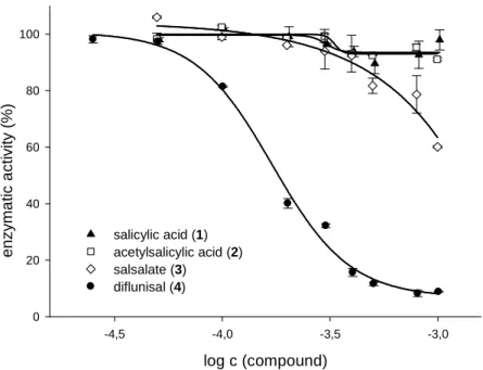
3.3 Inhibitory activity of selected non-steroidal anti-inflammatory drugs ...32
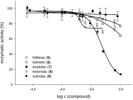
3.3.1 Salicylates ...32
3.3.2 Acetic acid derivatives ...34
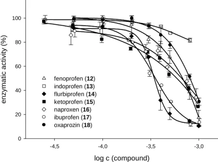
3.3.3 Propionic acid derivatives ...36
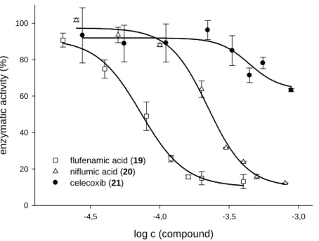
3.3.4 N-Anthranilic acids and celecoxib ...38
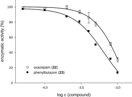
3.3.5 Miscellaneous compounds ...39
3.4 Inhibitory activity of diflunisal in the presence of plasma proteins ...41
3.5 Peak plasma concentrations of commercial drugs ...46
3.6 Summary and conclusion ...48
3.7 References ...50
4 Diflunisal analogs as inhibitors of bacterial hyaluronidase ... 53
4.1 Introduction ...54
4.2 Chemistry ...56
4.3 Pharmacological results and discussion ...58
4.3.1 General conditions ...58
4.3.2 Inhibitory activities of biphenyl-2-carboxylic acid derivatives ...59
4.3.3 Inhibitory activities of biphenyl-3-carboxylic acid derivatives ...60
4.3.4 Inhibitory activities of biphenyl-4-carboxylic acid derivatives ...63
4.3.5 Inhibitory activities of 4-hydroxybiphenyl-3-carboxylic acid analogs ...66
4.4 Inhibitory activities of selected compounds on SpnHyl ...69
4.5 Inhibitory activities of vitamin C palmitate in the presence of palladium catalyst ...70
4.6 Summary and conclusion ...71
4.7 Experimental section ...73
4.7.1 General conditions ...73
4.7.2 Chemistry ...75
4.7.2.1 Preparation of compounds 4.24-4.46 ...75
4.8 References ...92
5 Indole-2-carboxylic acids and 2-(6,7-dichloro-1H-indol-2-yl)-1,3,4- oxadiazoles as inhibitors of bacterial hyaluronidases ... 95
5.1 Introduction ...96
5.2 Chemistry ...99
5.2.1 Synthesis of 2-(6,7-dichloro-1H-indol-2-yl)-1,3,4-oxadiazoles ...99
5.2.2 Synthesis of 1H-indole-2-carboxylic acid compounds ... 100
5.3 Pharmacological results and discussion ... 102
5.3.1 General conditions ... 102
5.3.2 Inhibition of hyaluronidases by 2-(1H-indol-2-yl)-1,3,4-oxadiazoles ... 102
5.3.3 Inhibition of hyaluronidases by 6,7-dichloro-1H-indole-2-carboxylic acid derivatives . ... 103
5.3.4 Inhibition of hyaluronidases by 5-benzyloxy- and 5-hydroxy-1H-indole-2-carboxylic acid derivatives ... 106
5.4 Inhibitory activities of selected compounds on SpnHyl ... 108
5.5 Summary ... 109
5.6 Experimental section ... 110
5.6.1 General conditions ... 110
5.6.2 Chemistry ... 110
5.6.2.1 Preparation of compounds 5.1, 5.32 ... 110
5.6.2.2 Preparation of ethyl 2-acetylpentanoate (5.3) ... 111
5.6.2.3 Preparation of compounds 5.4-5.5, 5.34-5.36 ... 112
5.6.2.4 Preparation of compounds 5.6, 5.7, 5.37-5.39 ... 113
5.6.2.5 Preparation of compounds 5.9-5.11 ... 114
5.6.2.6 Preparation of compounds 5.12-5.14 ... 115
5.6.2.7 Preparation of tert-butyl 4-(bromomethyl)benzoate (5.15) ... 116
5.6.2.8 Preparation of compounds 5.16-5.21, 5.40-5.44 ... 117
5.6.2.9 Preparation of compounds 5.22-5.31, 5.45-5.52, 5.60 ... 120
5.6.2.10 Preparation of compounds 5.53-5.59 ... 125
5.7 References ... 128
6 2-Phenylindolizines as hyaluronidase inhibitors ... 131
6.1 Introduction ... 132
6.2 Chemistry ... 133
6.3 Pharmacological results and discussion ... 135
6.3.1 General conditions ... 135
6.3.2 Inhibitory activity of 2-phenylindolizine compounds ... 136
6.4 Summary ... 138
6.5 Experimental section ... 139
6.5.1 General conditions ... 139
6.5.2 Chemistry ... 139
6.5.2.1 Preparation of compounds 6.4-6.6 ... 139
6.5.2.2 Preparation of ethyl 2-methylisonicotinoate30 6.8 ... 140
6.5.2.3 Preparation of compounds 6.15-6.19 ... 140
6.5.2.4 Preparation of compounds 6.20-6.22 ... 142
6.5.2.5 Preparation of compounds 6.25, 6.26 ... 144
6.6 References ... 145
7 Characterization of snake venoms regarding hyaluronidase activity 147 7.1 Introduction ... 148
7.2 Materials and methods ... 150
7.2.1 Determination of protein content... 150
7.2.2 SDS-Polyacrylamide gel electrophoresis (SDS-PAGE) ... 150
7.2.3 Zymography ... 151
7.2.4 Colorimetric hyaluronidase activity assay (Morgan-Elson assay) ... 152
7.2.4.1 General procedures ... 152
7.2.4.2 Determination of hyaluronidase activity ... 152
7.2.5 Turbidimetric hyaluronidase activity assay ... 153
7.2.6 Plasma protein binding ... 153
7.3 Results and discussion ... 154
7.3.1 Protein content and determination of molecular mass ... 154
7.3.2 Hyaluronidase activity ... 155
7.3.3 Influence of pH on enzymatic activity ... 157
7.3.4 Inhibitory activities of selected compounds on snake venom hyaluronidases ... 159
7.3.5 Plasma protein binding of selected compounds ... 161
7.4 Summary and conclusion ... 163
7.5 References ... 164
8 Summary ... 167
9 Appendix ... 171
9.1 Abbreviations ... 172
1 Introduction
1.1 Hyaluronic acid
1.1.1 Structure and physicochemical properties
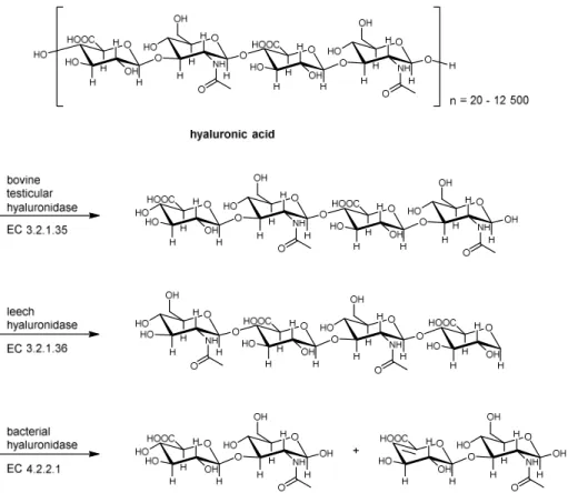
Hyaluronic acid (hyaluronan) was discovered in the vitreous humor of the eye by Meyer and Palmer in 1934 and subsequently found in most parts of the body, including the synovial fluid of joints and the skin. The proposed name “hyaluronic acid” derives from hyaloid (vitreous) and the constituent uronic acid.1 The structure of hyaluronan was also determined by Meyer and coworkers, but not before the 1950s.2 Hyaluronan is a linear high molecular weight polysaccharide and belongs to the family of glycosaminoglycans (GAG). It consists of repeating disaccharides comprised of β-1,3 linked N-acetyl-D-glucosamine (GlcNAc) and glucuronic acid (GlcUA) which are connected by β-1,4 glycosidic bonds.3 The structure of hyaluronic acid is displayed in Figure 1.1.
Figure 1.1 Chemical structure of hyaluronic acid (HA).
Hyaluronan consists of 20-25,000 disaccharide units, corresponding to a polymer length of 2- 25 µm. The hyaluronic acid (HA) polymers are among the largest matrix molecules with molecular weights between 106 to 107 Da.4 The apparent size of hyaluronan is even greater because the polymer is able to incorporate a large volume of water into its solvent domain that is 1000 times greater than the original volume.5 In the body, hyaluronan occurs in salt form and of highly negative charge because the carboxylic groups of the glucuronic acid moieties are deprotonated under physiological conditions (pKa 3-4).6 Hence, this macromolecule is most frequently referred to as “hyaluronan”.
X-ray diffraction and NMR experiments in dimethyl sulfoxide (DMSO) confirmed that the secondary structure of hyaluronan is a tape-like, two-fold helix.7-10 Recent NMR and computer modeling studies by Almondet al. suggest a dynamic local confirmation, which is on average a contracted 4-fold helix.11 In general, the backbone of the hyaluronan molecule is stiffened by a combination of the chemical structure of the disaccharide, internal hydrogen bonds, and interaction with the solvent.12 Therefore, in solution, the hyaluronan polymer
adopts the shape of an expanded, random coil with reversible formation and breakdown of interactions between the hyaluronan strains.6,13,14
1.1.2 Hyaluronan metabolism and physiological functions
Compared to other members of the glycosaminoglycan family, hyaluronan differs in several respects: it is the only GAG that is non-sulfated, it is synthesized at the cytoplasmic surface of the plasma membrane as a free linear polymer, not in the Golgi apparatus and, therefore, is not covalently linked to a protein core.3,4,15,16 Hyaluronan is synthesized at the inner face of the plasma membrane by one of the three membrane-bound hyaluronan synthases, namely HAS1, HAS2 or HAS3.17 The cytoplasmic product is extruded through the plasma membrane into the extracellular matrix (ECM) permitting unconstrained polymer growth.18,19
Hyaluronic acid is a major constituent of the extracellular matrix (ECM) of most tissues. It is found at high concentrations in rooster comb and several soft connective tissues, including skin, umbilical cord, synovial fluid, and vitreous humor.3,20 Hyaluronan can form highly viscous solutions and, thereby, influence the properties of this matrix.21 It plays an important role in various physiological and pathophysiological processes, such as embryonic development and morphogenesis22, wound healing23-25, repair and regeneration, inflammation26-28 and tumorigenesis29-31. Moreover, hyaluronan interacts with cell-surface receptors, such as CD44 (cluster of differentiation 44) and RHAMM (receptor for hyaluronic- acid-mediated motility) that are associated with signal transduction and may help with cell motility and adhesion.32,33
1.2 Hyaluronidases
1.2.1 Occurrence and classification
Hyaluronidases are a group of enzymes that degrade hyaluronan. They were first discovered in an extract of mammalian testes and other tissues by Duran-Reynals at the beginning of the last century. This so called “spreading factor”, which facilitated the diffusion of dyes and antiviral vaccines, was later named hyaluronidase by Karl Meyer to denote enzymes that degrade hyaluronan.34,35 The hyaluronidases are widely distributed in the animal kingdom and can be found in mammals, invertebrate animals (crustaceans, leeches, and insects),
animal venoms, bacteria (e.g. Streptomyces) and bacteriophages, and in pathogenic fungi (e.g. Candida).21,35
Based on their catalytic mechanism, the hyaluronidases can be divided into three groups (Figure 1.2).36,37
Figure 1.2 Classification of hyaluronidase according to Meyer.36 Adopted from Muckenschnabel.38
The hyaluronidases from the first group (EC 3.2.1.35) are endo-β-N-acetyl-D- hexosaminidases that cleave the β-1,4-glycosidic linkages in hyaluronan to yield tetra- and hexasaccharides as the major final products with N-acetylglucosamine at the reducing end.
Enzymes of this class can be found in mammalian spermatozoa, lysosomes, and the venoms of snakes and hymenoptera.21,39,40 The second group (EC 3.2.1.36) includes the endo-β- glucuronidases, which are present in leeches and in certain crustaceans.41,42 These enzymes cleave the β-1,3-glycosidic bond in hyaluronan and yield tetra- and hexasaccharides as the main end-products with glucuronic acid at the reducing end.35 The third group (EC 4.2.2.1) includes the microbial hyaluronidases, which act as endo-N-acetyl-D-hexosaminidases.
These enzymes cleave the β-1,4-glycosidic linkage by a β-elimination resulting in unsaturated disaccharides as products of exhaustive degradation of hyaluronan. Based on their catalytic mechanism, these enzymes were also termed hyaluronate lyases. According to
their catalytic mechanism, the latter must be clearly distinguished from the other two groups.35,43-45
1.2.2 Hyaluronidases from eukaryotes
In humans, there are six hyaluronidase-like sequences, five of them encoding functionally active enzymes, the hyaluronidases Hyal-1, Hyal-2, Hyal-3, Hyal-4 and PH-20 (SPAM1), whereas the pseudogene hyalp1 is transcribed, but not translated.46,47 The sequence similarity between the six subtypes varies from 33 % to 42 %.37 Hyal-1 was detected in mammalian plasma and urine, as well as at high levels in the liver, kidney, spleen, and heart and is localized in the lysosomes.48 Hyal-2 is present in many tissues, except the adult brain.
It is either localized in the lysosome or anchored to the plasma membrane by a glycosylphosphatidylinositol (GPI) link.49 Hyal-1 and Hyal-2 are the major hyaluronidases and have a pronounced activity optimum at pH 4. 48,50,51 However, the expression of Hyal-2 is extremely low and difficult to purify.52 The GPI-anchored PH-20 protein (SPAM1 sperm adhesion molecule) is located on the surface of mammalian sperm to facilitate penetration of the sperm through the hyaluronan-rich matrix of the oocyte.53
The major soluble hyaluronidase present in bull testes extract, the bovine testicular hyaluronidase (BTH), which has been therapeutically applied as a spreading factor in several medical fields for many years, has been shown to be a fragment of the membrane bound PH-20 enzyme.54,55
Hyaluronidase activity has been detected in the venom of many animals, such as snakes56,57, stonefish58, scorpions59,60, spiders61, lizards,62 and caterpillars63, in which they predominantly serve as “spreading factor” but have no intrinsic toxic activity.45 By degrading the hyaluronan of the extracellular matrix of the prey, local tissue damage is caused, which facilitates the distribution of other venom constituents. The hyaluronidase from bee venom was the first to be characterized in detail.64 Hyaluronidases in snake venom have been recognized as possible targets for the treatment of snake bites. Therefore, the venoms from two different snake species will be characterized in chapter 7.
1.2.3 Hyaluronidases from prokaryotes
Hyaluronidases are produced by many different genera of bacteria. Proteins or enzymes found on the surface of gram-positive organisms significantly contribute to pathogenesis and might be involved in the disease process caused by these pathogens. Human infections caused by gram-positive bacteria are increasingly difficult to treat, predominantly due to the emergence of antibiotic-resistant strains not only against penicillin but also against novel antibiotics such as vancomycin.65,66 The HA lyases produced by gram-positive bacteria are considered to act as virulence factors facilitating the spread of pathogens or toxins by breaking down the components of the extracellular matrix of the host.67 The enhanced tissue- permeability, caused by hyaluronidase-mediated degradation of HA, appears to play a role in many health problems such as gangrene, meningitis, synovitis, hyperplasia, nephritis, mycoplasmosis, periodontal disease, mastitis, pneumonia, septicemia, syphilis, toxic shock syndrome, and wound infections.68-72
Gram-positive microorganisms capable of producing HA lyases include various species of Spreptococcus, Staphylococcus, Peptostreptococcus, Propionibacterium, Streptomyces, and Clostridium.73-77 The HA lyases produced by gram-negative bacteria are not excreted to the extracellular matrix and, therefore, are less likely to play a role in pathogenesis.35,67,78
By reducing the spreading of the pathogen, the inhibition of the hyaluronate lyases could be an approach to the treatment of bacterial infections.79 In this context, the HA lyases from S. agalactiae strain 4755 (SagHyal4755) and S. pneumoniae (SpnHyl) are the best characterized among the microbial hyaluronidases.80-82 S. pneumoniae predominantly colonizes the upper respiratory tract of humans and is a major human pathogenic bacterium.
It causes life-threatening diseases, examples of which are pneumonia, bacteremia, and meningitis, as well as less threatening diseases, such as otitis media and sinusitis. Young children and the elderly are particularly prone to these diseases.82 The currently licensed pneumococcal vaccine is only moderately effective and it is not prescribed for children younger than two years.65,83 The bacterium S. agalactiae (group B streptococcus, GBS) is part of the normal human flora but has been an important cause of infection in newborn and young infants.84,85 Human infections by this gram-positive bacterium are meningitis and septicemia, which result in considerable morbidity and mortality.86 In cultures of S. pneumoniae and S. agalactiae the HA lyase is found both in the culture and in cell- associated fractions, which suggests that at least part of the enzyme is released by the pathogen to surrounding host tissues during infection in order to facilitate bacterial invasion.74,81
The native hyaluronidases from both S. agalactiae and S. pneumoniae have molecular masses of 111 kDa and 107 kDa, respectively, but undergo autocatalytic conversion to smaller enzymatically active 92 kDa and 82 kDa forms.81,87 X-ray crystallography revealed that the structures of the HA lyases from S. agalactiae and S. pneumoniae are similar regarding the architecture of the entire enzyme as well as the geometry of the active site, sharing a similarity in the range of 65 % to 80 %.37,44,65 The active site of the lyases are composed of two main parts, a catalytic group responsible for substrate degradation and an aromatic patch responsible for the selection of cleavage sites on the substrate chains.44 In the catalytic cleft three catalytic residues, Asn429, His476, and Tyr488 (SagHyal4755
numbering88), are located, which degrade the substrate through a proton acceptance and donation (PAD) mechanism (Figure 1.3).44,71,82
Figure 1.3 Mechanism of hyaluronan degradation by hyaluronate lyase from S. agalactiae (SagHyal4755);
schematic presentation of hyaluronan with HA1, HA2 and HA3 as disaccharide units and the position of the side chains Tyr488, His479 and Asn429 relative to the substrate (modified from Li and Jedrzejas).71,82
The degradation process includes five different steps: (1) enzyme binding to negatively charged hyaluronan; (2) cleaving of β-1,4-glycosidic bond; (3) hydrogen exchange between enzyme and microenvironment; (4) release of disaccharide product; (5) translocation of the remaining HA by one disaccharide unit towards the reducing end.65,89,90 The truncated polymeric hyaluronan stays in the cleft after the release of the disaccharide and undergoes a processive mode of action until the chain is fully degraded into disaccharides.91 However, Kühn et al. proposed a nonprocessive mode of action for SagHyal4755, based upon the observation that hyaluronan degradation by SagHyal4755 yields a mixture of oligosaccharides and hyaluronan fragments of different sizes.92
1.2.4 Hyaluronidase inhibitors
In general, the role of hyaluronan and hyaluronan degrading enzymes, the hyaluronidases, is far from being understood. Potent and selective inhibitors for the hyaluronidases are required as pharmacological tools to further characterize and understand the physiological and pathophysiological role of hyaluronidases. Furthermore, from a therapeutic point of view, inhibitors might be useful in developing anti-bacterial67, anti-venom/toxin35, anti-tumor93, anti- allergic agents,93 or even as contraceptives.94 An overview of all previously reported putative inhibitors of hyaluronidases was given by Girish in 2009.77 Documented hyaluronidase inhibitors are of different chemical nature. The presence of proteins as hyaluronidase inhibitors in human serum was thought to be a defense mechanism against venoms and pathogenic bacteria.95-97 A purified high molecular mass, thermolabile magnesium dependent glycoprotein from mouse serum, probably belonging to the inter-α-inhibitor family, was reported to be an inhibitor of bovine, testicular, snake and bee venom hyaluronidase.98 The inhibitory activity of diverse metal ions, among them being iron, cadmium, copper, or zinc salts was reported long ago.99 Based on substrate similarity, several glycosaminoglycans (GAGs) and polysaccharides were identified as hyaluronidase inhibitors. Heparin and heparan sulfate were observed to inhibit hyaluronidase, but only at concentrations much higher than physiological levels and in a non-competitive manner.100-102 Other described structurally related compounds were oxygen-sulfated GAGs and oxygen-sulfated fragments of hyaluronan.103 Toida et al. discovered that the O-sulfated hyaluronan fragments showed stronger inhibition than other O-sulfated GAGs. Furthermore, not only did the inhibition increase with the chain length of the hyaluronan oligomer, but these compounds also showed both competitive and non-competitive inhibition, in contrast to heparin.104 Chitosan was also characterized as an inhibitor with direct correlation between molecular weight and inhibitory activity.105 Alginic acid, which is also structurally related to hyaluronan, consists of L-glucuronic and D-mannuronic acid and shows molecular weight dependent inhibition of hyaluronidase.106 The synthetic condensation polymer of mandelic acid is reported to inhibit hyaluronidase.94
A lot of low molecular weight natural products, such as: alkaloids107,108, flavones and flavone- analogs60,109-114, and terpenes115,116 have been investigated for hyaluronidase inhibition. Other substances reported to show weak inhibition of hyaluronidase activity are antioxidants79,117 and polyphenols118, non-saturated fatty acids119, antibiotics120, and lanostanoids.121 Moreover, anti-allergic drugs (e.g. disodium cromoglycate, DSCG)111,122 and anti-inflammatory drugs (cf. chapter 3.1) are claimed to possess hyaluronidase inhibitory activities. The research in our laboratories has led to the discovery and validation of potent inhibitors of hyaluronidases.
For example, lipophilic derivatives of L-ascorbic acid (vitamin C) were developed, which are
among the most potent inhibitors of the bacterial hyaluronidase SagHyal4755 known today.123-126 A variety of heterocycles, such as benzimidazoles, benzoxazoles, indoles, phenylindoles and gluconolactones have been identified as potent hyaluronidase inhibitors.127-136 The aforementioned compounds are not drug-like mainly due to very high plasma protein binding, high molecular weights (> 500 g/mol), low bioavailability, or bioincompatibility (e.g. heparin). Because of the important role of hyaluronan in biological systems, potent inhibitors of hyaluronidases are needed to further investigate the role of hyaluronan and hyaluronidases in physiological and pathophysiological processes.
1.3 Methods for the determination of hyaluronidase activity
Since the discovery of the hyaluronidases by Duran-Reynals in 1928137, various methods for the determination of the enzymatic activity have been described. In 1994 Hynes and Ferretti138 summarized previously described test systems and divided them into groups based upon the assay performed, namely spectrophotometric139,140, radiochemical141, fluorogenic142, enzymoimmunological143, plate (solid media)144, chemical145,146, physicochemical145,147,148, and zymographic137,149 methods. Although many different methods for the determination of hyaluronidase activity can be found in the literature, only few can be deployed for routine analyses. Many of the early developed assays lack sensitivity or the workup procedures are rather laborious and time-consuming. M. Stern and R. Stern150 discussed the advantages and limitations of the conventional methods in an article about a new ELISA-like assay for hyaluronidase and hyaluronidase inhibitors. A lot of assay techniques are based upon changes in viscosity151 and turbidity152. In 1944 the turbidimetric assay was described by Kass and Seastone153, based upon the observation of Meyer and Palmer154 that hyaluronan forms insoluble complexes with acidified blood serum. It was further developed by Dorfman and Ott152, as well as Di Ferrante.148 In this thesis, a colorimetric assay, which was developed by Reissig et al.146, and the turbidimetric assay are routinely exploited to determine the inhibitory effect of the synthesized compounds. Inhibitory activity was determined for the bacterial hyaluronate lyases from S. agalactiae strain 4755 (SagHyal4755), S. pneumoniae (SpnHyl), and the bovine testicular enzyme BTH (formerly an approved drug; e.g. under the trade name Neopermease®). Both assay systems are briefly explained in the following sections.
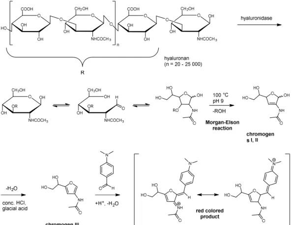
1.3.1 Morgan-Elson assay
The Morgan-Elson assay is a colorimetric assay and has been classified as a chemical assay by Hynes and Ferretti.138 The reducing ends of N-acetylhexosamine moieties form a red colored product after reaction with p-(dimethylamino)benzaldehyde (Ehrlich’s reagent;
DMAB), as described by Reissig146 and Gacessa.155 This so called Morgan-Elson reaction is often used to determine the activity of hyaluronidases that liberate hyaluronan fragments with N-acetyl-D-glucosamine at the reducing end. The photometrical detection of the red-colored product at a wavelength of 586 nm enables the determination of the enzymatic activity. The classes of hyaluronidases that can be assayed by the Morgan-Elson reaction are the hyaluronate-4-glycanohydrolases (EC 3.2.35) and hyaluronate lyases (EC 4.2.2.1), as described in section 1.2.
The reaction, which was investigated and described in detail by Muckenschnabel et al.,140 is shown in Figure 1.4. After incubation of the hyaluronidase at 37 °C the cleaved hyaluronan is heated to 100 °C under basic conditions (pH 9). The furanose form of N-acetyl-D- glucosamine residues is converted to the chromogens I (α-configuration) and II (β-configuration) after elimination of ROH (cf. Figure 1.4).156,157 Subsequent treatment with concentrated hydrochloric acid (HCl) and glacial acetic acid results in elimination of water yielding chromogen III. The final red colored product, mesomeric forms of N3-protonated 3-acetylimino-2-(4-dimethylaminophenyl)-methylidene-5-(1,2-dihydroxyethyl)furane, is obtained after the chromogen III reacts with p-(dimethylamino)benzaldehyde (Ehrlich’s reagent).
Figure 1.4 Mechanism of the Morgan-Elson reaction resulting in a red colored product according to Muckenschnabel et al.140
The Morgan-Elson assay represents a useful, widely applicable and well reproducible method for the determination of hyaluronidase activity in the presence of inhibitors. However, in some cases the Morgan-Elson assay cannot be operated. If the putative inhibitor is colored or undergoes side-reactions with DMAB the photometric detection produces false-negative results. Heterocycles, such as indoles or phenylindoles, which were identified to be potent inhibitors of SagHyal4755, often form colored products under assay conditions.
Pindur and coworkers extensively investigated the reaction of 3-methyl-1H-indole with p-(dimethylamino)benzaldehyde. They were able to show that the reaction follows the mechanism proposed by van Urk, resulting in intensively colored products.136,158,159 The yellow-colored indole derivative indomethacin, which is a non-steroidal anti-inflammatory drug, is also known as an inhibitor of SagHyal4755. It undergoes the van Urk-reaction providing colored products, rendering the application of the Morgan-Elson assay impossible.
The Morgan-Elson assay can be used to quantify hyaluronidase activity according to the definition of the International Union of Biochemistry by defining 1 unit (U) as the amount of enzyme that catalyzes the liberation of 1 µmol N-acetyl-D-glucosamine at the reducing ends of sugars per minute under specified conditions.160
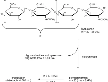
1.3.2 Turbidimetric assay
According to the classification by Hynes and Ferretti138, the turbidimetric assay represents a physicochemical assay. This method, as described by Di Ferrante148, is based upon the formation of insoluble complexes between non-degraded, high-molecular hyaluronan (mw > 8 kDa) and cetrimonium bromide (cetyltrimethylammonium bromide, CTAB). In contrast, a solution of oligosaccharides and smaller hyaluronan fragments (mw < 6-8 kDa) remains clear under the same conditions. Since the formed turbidity is proportional to the amount of high molecular weight hyaluronan fragments, the enzymatic activity can be quantified by photometric detection at 580 nm and 600 nm by means of reference samples (Figure 1.5).
Figure 1.5 Principle of the turbidimetric assay based on the method of Di Ferrante148. Figure adopted from Braun.131
This assay is fast, highly reproducible, easy to perform in cuvettes or microtiter plates and can be considered as the standard method for the determination of hyaluronidase activity and enzyme inhibition of the synthesized compounds in this thesis.
1.4 Influence of the pH value on enzymatic activity
The activities of the different hyaluronidases are dependent on the pH value. Hoechstetter160 and Hofinger161 studied the pH dependent activity profiles of BTH and SagHyal4755. It is not possible to find a pH value, where all investigated enzymes show high activity. The bacterial
hyaluronate lyase shows highest activity at pH 5.0. Jedrzejas87 reported a pH 6 as the optimum for SpnHyl. Figure 1.6 shows the pH dependent activity of SpnHyl as determined in the turbidimetric and Morgan-Elson assay.
pH
2 4 6 8 10
relative enzyme activity in %
0 20 40 60 80 100
Colorimetric assay Turbidimetric assay
Figure 1.6 pH dependent activity of bacterial hyaluronate lyase from Streptococcus pneumoniae (SpnHyl) as determined in the turbidimetric assay (red) and the Morgan-Elson assay (black).
The results differ from those reported by Jedrzejas. The pH values between 4.5 and 5.0 were determined to be the optimum for hyaluronidase activity. To obtain comparable IC50 values on BTH and SpnHyl the inhibitors were investigated at pH 5.0 as these enzymes show considerably high activity under these conditions in the turbidimetric assay. Thus, the obtained IC50 values are comparable to previously gained data from our workgroup (cf. investigations by Binder126, Braun131, Salmen127, Spickenreither124, and Textor136).
Hyaluronan and hyaluronan degrading enzymes, the hyaluronidases, are widespread in nature and were both described in the middle of the last century. However, the role of both is far from being understood and the academic research remains a challenging field.1,147 The hyaluronidases have been a group of less extensively studied glycosidases.21,77 Inhibitors of hyaluronidases are required as pharmacological tools to investigate their complex functions in hyaluronan metabolism. Moreover, such substances are of potential therapeutic value, e. g. for the treatment of bacterial infections or snake bites.79,108
The research in our laboratory has led to the discovery of potent inhibitors with a preference for the bacterial enzymes. For example, lipophilic derivatives of L-ascorbic acid (vitamin C) were developed, which are among the most potent inhibitors of the bacterial hyaluronidase SagHyal4755 known today.123-126 Moreover, potent hyaluronidase inhibitors were identified among a variety of heterocycles (cf. section 0).127-136 Generally, the potency of hyaluronidase
inhibition correlates with the lipophilicity of the compounds, which strongly affects the drug- like properties, due to very high plasma protein binding. Although (weak) inhibitors of the bacterial hyaluronidases have been discovered among natural products and synthetic molecules, small drug-like molecules with sufficient inhibitory potency are not available at present.77,162,163 To cope with this problem, the focus of this thesis was set on the design, synthesis and characterization of inhibitors for the bacterial hyaluronidase SagHyal4755 with drug-like properties.
1.5 References
1. Meyer, K.; Palmer, J. W. The polysaccharide of the vitreous humor. J. Biol. Chem.
1934, 107, 629-634.
2. Weissmann, B.; Meyer, K. The structure of hyalobiuronic acid and of hyaluronic acid from umbilical cord. J. Am. Chem. Soc. 1954, 76, 1753-1757.
3. Laurent, T. C.; Fraser, J. R. E. Hyaluronan. FASEB J. 1992, 6, 2397-2404.
4. Toole, B. P. Hyaluronan: From extracellular glue to pericellular cue. Nat. Rev. Cancer 2004, 4, 528-539.
5. Stern, R.; Asari, A. A.; Sugahara, K. N. Hyaluronan fragments: An information-rich system. Eur. J. Cell Biol. 2006, 85, 699-715.
6. Hascall, V. C.; Laurent, T. C. Hyaluronan: structure and physical properties.
http://www.glycoforum.gr.jp/science/hyaluronan/HA01/HA01E.html
7. Heatley, F.; Scott, J. E. A Water Molecule Participates in the Secondary Structure of Hyaluronan. Biochem. J. 1988, 254, 489-493.
8. Atkins, E. D. T.; Meader, D.; Scott, J. E. Model for Hyaluronic-Acid Incorporating 4 Intramolecular Hydrogen-Bonds. Int. J. Biol. Macromol. 1980, 2, 318-319.
9. Heatley, F.; Scott, J. E.; Jeanloz, R. W.; Walkernasir, E. Secondary Structure in Glycosaminoglycuronans - Nmr-Spectra in Dimethylsulfoxide of Disaccharides Related to Hyaluronic-Acid and Chondroitin Sulfate. Carbohydr. Res. 1982, 99, 1-11.
10. Scott, J. E. Supramolecular Organization of Extracellular-Matrix Glycosaminoglycans, Invitro and in the Tissues. FASEB J. 1992, 6, 2639-2645.
11. Almond, A.; DeAngelis, P. L.; Blundell, C. D. Hyaluronan: The local solution conformation determined by NMR and computer modeling is close to a contracted left-handed 4-fold helix. J. Mol. Biol. 2006, 358, 1256-1269.
12. Scott, J. E. Secondary and Tertiary Structure of Hyaluronan in Aqueous Solution.
Some Biological Consequences.
http://www.glycoforum.gr.jp/science/hyaluronan/HA02/HA02E.html
13. Sheehan, J. K.; Almond, A. Hyaluronan: Static, Hydrodynamic and Molecular Dynamic Views. http://www.glycoforum.gr.jp/science/hyaluronan/HA21/HA21E.html 14. Scott, J. E.; Heatley, F. Biological properties of hyaluronan in aqueous solution are
controlled and sequestered by reversible tertiary structures, defined by NMR spectroscopy. Biomacromolecules 2002, 3, 547-553.
15. Lee, J. Y.; Spicer, A. P. Hyaluronan: a multifunctional, megaDalton, stealth molecule.
Curr. Opin. Cell Biol. 2000, 12, 581-586.
16. Prehm, P. Hyaluronate Is Synthesized at Plasma-Membranes. Biochem. J. 1984, 220, 597-600.
17. Itano, N.; Sawai, T.; Yoshida, M.; Lenas, P.; Yamada, Y.; Imagawa, M.; Shinomura, T.; Hamaguchi, M.; Yoshida, Y.; Ohnuki, Y.; Miyauchi, S.; Spicer, A. P.; McDonald, J.
A.; Kimata, K. Three isoforms of mammalian hyaluronan synthases have distinct enzymatic properties. J. Biol. Chem. 1999, 274, 25085-25092.
18. Weigel, P. H.; Hascall, V. C.; Tammi, M. Hyaluronan synthases. J. Biol. Chem. 1997, 272, 13997-14000.
19. Stern, R. Hyaluronan catabolism: a new metabolic pathway. Eur. J. Cell Biol. 2004, 83, 317-325.
20. Fraser, J. R. E.; Laurent, T. C.; Laurent, U. B. G. Hyaluronan: its nature, distribution, functions and turnover. J. Intern. Med. 1997, 242, 27-33.
21. Kreil, G. Hyaluronidases - a Group of Neglected Enzymes. Protein Sci. 1995, 4, 1666- 1669.
22. Toole, B. P. Hyaluronan in morphogenesis. Semin. Cell Dev. Biol. 2001, 12, 79-87.
23. Weigel, P. H.; Fuller, G. M.; LeBoeuf, R. D. A model for the role of hyaluronic acid and fibrin in the early events during the inflammatory response and wound healing. J.
Theor. Biol. 1986, 119, 219-234.
24. Longaker, M. T.; Chiu, E. S.; Adzick, N. S.; Stern, M.; Harrison, M. R.; Stern, R.
Studies in fetal wound healing. V. A prolonged presence of hyaluronic acid characterizes fetal wound fluid. Ann. Surg. 1991, 213, 292-296.
25. Chen, W. Y. J.; Abatangelo, G. Functions of hyaluronan in wound repair. Wound Repair Regen. 1999, 7, 79-89.
26. de la Motte, C. A.; Hascall, V. C.; Drazba, J.; Bandyopadhyay, S. K.; Strong, S. A.
Mononuclear Leukocytes Bind to Specific Hyaluronan Structures on Colon Mucosal Smooth Muscle Cells Treated with Polyinosinic Acid:Polycytidylic Acid: Inter-α-Trypsin Inhibitor Is Crucial to Structure and Function. Am. J. Pathol. 2003, 163, 121-133.
27. Noble, P. W. Hyaluronan and its catabolic products in tissue injury and repair. Matrix Biol. 2002, 21, 25-29.
28. Majors, A. K.; Austin, R. C.; De la Motte, C. A.; Pyeritz, R. E.; Hascall, V. C.; Kessler, S. P.; Sen, G.; Strong, S. A. Endoplasmic Reticulum Stress Induces Hyaluronan Deposition and Leukocyte Adhesion. J. Biol. Chem. 2003, 278, 47223-47231.
29. Toole, B. P.; Hascall, V. C. Hyaluronan and Tumor Growth. Am. J. Pathol. 2002, 161, 745-747.
30. Toole, B. P. Hyaluronan promotes the malignant phenotype. Glycobiology 2002, 12, 37R-42R.
31. Sherman, L.; Sleeman, J.; Herrlich, P.; Ponta, H. Hyaluronate receptors: key players in growth, differentiation, migration and tumor progression. Curr. Opin. Cell Biol. 1994, 6, 726-733.
32. Turley, E. A.; Noble, P. W.; Bourguignon, L. Y. W. Signaling properties of hyaluronan receptors. J. Biol. Chem. 2002, 277, 4589-4592.
33. Lesley, J.; English, N.; Hascall, V. C.; Tammi, M.; Hyman, R. Hyaluronan binding by cell surface CD44. J. Biol. Chem. 2002, 1, 341-348.
34. Duran-Reynals, F. Studies on a certain spreading factor existing in bacteria and its significance for bacterial invasiveness. J. Exp. Med. 1933, 58, 161-181.
35. Girish, K. S.; Kemparaju, K. The magic glue hyaluronan and its eraser hyaluronidase:
A biological overview. Life Sci. 2007, 80, 1921-1943.
36. Meyer, K. Hyaluronidases. In The Enzymes, Boyer, P. D., Ed. Academic Press: New York, 1971.
37. Stern, R.; Jedrzejas, M. J. Hyaluronidases: Their genomics, structures, and mechanisms of action. Chem. Rev. 2006, 106, 818-839.
38. Muckenschnabel, I. Analytische Untersuchungen zum Einsatz von Hyaluronidase als Adjuvans in der Krebschemotherapie. Doctoral thesis, University of Regensburg, Regensburg, 1997.
39. Cramer, J. A.; Bailey, L. C.; Bailey, C. A.; Miller, R. T. Kinetic and mechanistic studies with bovine testicular hyaluronidase. Biochim. Biophys. Acta 1994, 1200, 315-321.
40. Takagaki, K.; Nakamura, T.; Izumi, J.; Saitoh, H.; Endo, M.; Kojima, K.; Kato, I.;
Majima, M. Characterization of Hydrolysis and Transglycosylation by Testicular Hyaluronidase Using Ion-Spray Mass-Spectrometry. Biochemistry 1994, 33, 6503- 6507.
41. Yuki, H.; Fishman, W. H. Purification and characterization of leech hyaluronic acid- endo-β-glucuronidase. J. Biol. Chem. 1963, 238, 1877-1879.
42. Karlstam, B.; Ljunglof, A. Purification and Partial Characterization of a Novel Hyaluronic Acid-Degrading Enzyme from Antarctic Krill (Euphausia-Superba). Polar Biol. 1991, 11, 501-507.
43. Suzuki, S. Microbial hyaluronan lyases.
http://www.glycoforum.gr.jp/science/hyaluronan/HA14/HA14E.html
44. Jedrzejas, M. J. Structural and functional comparison of polysaccharide-degrading enzymes. Crit. Rev. Biochem. Mol. Biol. 2000, 35, 221-251.
45. Frost, G. L.; Csoka, T.; Stern, R. The hyaluronidases: A chemical, biological and clinical overview. Trends Glycosci. Glyc. 1996, 8, 419-434.
46. Csoka, A. B.; Frost, G. I.; Stern, R. The six hyaluronidase-like genes in the human and mouse genomes. Matrix Biol. 2001, 20, 499-508.
47. Csoka, A. B.; Scherer, S. W.; Stern, R. Expression analysis of six paralogous human hyaluronidase genes clustered on chromosomes 3p21 and 7q31. Genomics 1999, 60, 356-361.
48. Frost, G. I.; Csóka, T. B.; Wong, T.; Stern, R. Purification, Cloning, and Expression of Human Plasma Hyaluronidase. Biochem. Biophys. Res. Commun. 1997, 236, 10-15.
49. Lepperdinger, G.; Strobl, B.; Kreil, G. HYAL2, a human gene expressed in many cells, encodes a lysosomal hyaluronidase with a novel type of specificity. J. Biol. Chem.
1998, 273, 22466-22470.
50. Lepperdinger, G.; Müllegger, J.; Kreil, G. Hyal2 — less active, but more versatile?
Matrix Biol. 2001, 20, 509-514.
51. El-Safory, N. S.; Fazary, A. E.; Lee, C. K. Hyaluronidases, a group of glycosidases:
Current and future perspectives. Carbohydr. Polym. 2010, 81, 165-181.
52. Hamberger, J. Characterization of mammalian hyaluronidase-2 activity and identification of inhibitors of Streptococcal hyaluronan lyase. Doctoral thesis, University of Regensburg, Regensburg, 2012.
53. Cherr, G. N.; Yudin, A. I.; Overstreet, J. W. The dual functions of GPI-anchored PH- 20: hyaluronidase and intracellular signaling. Matrix Biol. 2001, 20, 515-525.
54. Menzel, E. J.; Farr, C. Hyaluronidase and its substrate hyaluronan: biochemistry, biological activities and therapeutic uses. Cancer Lett. 1998, 131, 3-11.
55. Meyer, M. F.; Kreil, G.; Aschauer, H. The soluble hyaluronidase from bull testes is a fragment of the membrane-bound PH-20 enzyme. FEBS Lett. 1997, 413, 385-388.
56. Girish, K. S.; Shashidharamurthy, R.; Nagaraju, S.; Gowda, T. V.; Kemparaju, K.
Isolation and characterization of hyaluronidase a "spreading factor" from Indian cobra (Naja naja) venom. Biochimie 2004, 86, 193-202.
57. Pukrittayakamee, S.; Warrell, D. A.; Desakorn, V.; McMichael, A. J.; White, N. J.;
Bunnag, D. The hyaluronidase activities of some Southeast Asian snake venoms.
Toxicon 1988, 26, 629-637.
58. Poh, C. H.; Yuen, R.; Chung, M. C. M.; Khoo, H. E. Purification and Partial Characterization of Hyaluronidase from Stonefish (Synanceja-Horrida) Venom. Comp.
Biochem. Phys. B 1992, 101, 159-163.
59. Morey, S. S.; Kiran, K. M.; Gadag, J. R. Purification and properties of hyaluronidase from Palamneus gravimanus (Indian black scorpion) venom. Toxicon 2006, 47, 188- 195.
60. Pessini, A. C.; Takao, T. T.; Cavalheiro, E. C.; Vichnewski, W.; Sampaio, S. V.; Giglio, J. R.; Arantes, E. C. A hyaluronidase from Tityus serrulatus scorpion venom: isolation, characterization and inhibition by flavonoids. Toxicon 2001, 39, 1495-1504.
61. Young, A. R.; Pincus, S. J. Comparison of enzymatic activity from three species of necrotising arachnids in Australia: Loxosceles rufescens, Badumna insignis and Lampona cylindrata. Toxicon 2001, 39, 391-400.
62. Tu, A. T.; Hendon, R. R. Characterization of lizard venom hyaluronidase and evidence for its action as a spreading factor. Comp. Biochem. Physiol. B Comp.
Biochem. 1983, 76, 377-383.
63. da C.B. Gouveia, A. I.; da Silveira, R. B.; Nader, H. B.; Dietrich, C. P.; Gremski, W.;
Veiga, S. S. Identification and partial characterisation of hyaluronidases in Lonomia obliqua venom. Toxicon 2005, 45, 403-410.
64. Kemeny, D. M.; Dalton, N.; Lawrence, A. J.; Pearce, F. L.; Vernon, C. A. The purification and characterisation of hyaluronidase from the venom of the honey bee, Apis mellifera. Eur. J. Biochem. 1984, 139, 217-223.
65. Jedrzejas, M. J. Pneumococcal virulence factors: Structure and function. Microbiol.
Mol. Biol. Rev. 2001, 65, 187-207.
66. Novak, R.; Henriques, B.; Charpentier, E.; Normark, S.; Tuomanen, E. Emergence of vancomycin tolerance in Streptococcus pneumoniae. Nature 1999, 399, 590-593.
67. Hynes, W. L.; Walton, S. L. Hyaluronidases of Gram-positive bacteria. FEMS Microbiol. Lett. 2000, 183, 201-207.
68. Matsushita, O.; Okabe, A. Clostridial hydrolytic enzymes degrading extracellular components. Toxicon 2001, 39, 1769-1780.
69. Makris, G.; Wright, J. D.; Ingham, E.; Holland, K. T. The hyaluronate lyase of Staphylococcus aureus - A virulence factor? Microbiology 2004, 150, 2005-2013.
70. Sutherland, I. W. Polysaccharide lyases. FEMS Microbiol. Rev. 1995, 16, 323-347.
71. Li, S.; Kelly, S. J.; Lamani, E.; Ferraroni, M.; Jedrzejas, M. J. Structural basis of hyaluronan degradation by Streptococcus pneumoniae hyaluronate lyase. EMBO J.
2000, 19, 1228-1240.
72. Spellerberg, B. Pathogenesis of neonatal Streptococcusagalactiae infections. Microb.
Infect. 2000, 2, 1733-1742.
73. Günther, E.; Ozegowski, J.-H.; Köhler, W. Occurrence of Extracellular Hyaluronic Acid and Hyaluronatlyase in Streptococci of Groups A, B, C, and G. Zbl. Bakt. 1996, 285, 64-73.
74. Berry, A. M.; Lock, R. A.; Thomas, S. M.; Rajan, D. P.; Hansman, D.; Paton, J. C.
Cloning and Nucleotide-Sequence of the Streptococcus-Pneumoniae Hyaluronidase Gene and Purification of the Enzyme from Recombinant Escherichia-Coli. Infect.
Immun. 1994, 62, 1101-1108.
75. Fitzgerald, T. J.; Gannon, E. M. Further Evidence for Hyaluronidase Activity of Treponema-Pallidum. Can. J. Microbiol. 1983, 29, 1507-1513.
76. Canard, B.; Garnier, T.; Saint-Joanis, B.; Cole, S. Molecular genetic analysis of the nagH gene encoding a hyaluronidase of Clostridium perfringens. Molec. Gen. Genet.
1994, 243, 215-224.
77. Girish, K. S.; Kemparaju, K.; Nagaraju, S.; Vishwanath, B. S. Hyaluronidase Inhibitors: A Biological and Therapeutic Perspective. Curr. Med. Chem. 2009, 16, 2261-2288.
78. Linhardt, R. J.; Galliher, P. M.; Cooney, C. L. Polysaccharide lyases. Appl. Biochem.
Biotechnol. 1987, 12, 135-176.
79. Li, S. L.; Taylor, K. B.; Kelly, S. J.; Jedrzejas, M. J. Vitamin C inhibits the enzymatic activity of Streptococcus pneumoniae hyaluronate lyase. J. Biol. Chem. 2001, 276, 15125-15130.
80. Pritchard, D. G.; Trent, J. O.; Li, X.; Zhang, P.; Egan, M. L.; Baker, J. R.
Characterization of the active site of group B streptococcal hyaluronan lyase.
Proteins: Struct. Funct. Bioinform. 2000, 40, 675-675.
81. Jedrzejas, M. J.; Chantalat, L. Structural studies of Streptococcus agalactiae hyaluronate lyase. Acta Crystallogr. Sect. D. Biol. Crystallogr. 2000, D56, 460-463.
82. Jedrzejas, M. J.; Mello, L. V.; De Groot, B. L.; Li, S. Mechanism of hyaluronan degradation by Streptococcus pneumoniae hyaluronate lyase: structures of complexes with the substrate. J. Biol. Chem. 2002, 277, 28287-28297.
83. Walsh, C. T. Vancomycin resistance: decoding the molecular logic. Science 1993, 261, 308-309.
84. Baker, C. J.; Edwards, M. S. Group B Streptococcal Infections. Ann. N. Y. Acad. Sci.
1988, 549, 193-202.
85. Dillon Jr, H. C.; Khare, S.; Gray, B. M. Group B streptococcal carriage and disease: A 6-year prospective study. J. Pediatr. 1987, 110, 31-36.
86. Musser, J. M.; Mattingly, S. J.; Quentin, R.; Goudeau, A.; Selander, R. K.
Identification of a high-virulence clone of type III Streptococcus agalactiae (group B Streptococcus) causing invasive neonatal disease. Proc. Natl. Acad. Sci. U. S. A.
1989, 86, 4731-4735.
87. Jedrzejas, M. J.; Mewbourne, R. B.; Chantalat, L.; McPherson, D. T. Expression and purification of Streptococcus pneumoniae hyaluronate lyase from Escherichia coli.
Protein Expression Purif. 1998, 13, 83-89.
88. Mello, L. V.; de Groot, B. L.; Li, S.; Jedrzejas, M. J. Structure and Flexibility of Streptococcus agalactiae Hyaluronate Lyase Complex with Its Substrate. Insights into the Mechanism of Processive Degradation of Hyaluronan. J. Biol. Chem. 2002, 277, 36678-36688.
89. Ponnuraj, K.; Jedrzejas, M. J. Mechanism of hyaluronan binding and degradation:
structure of Streptococcus pneumoniae hyaluronate lyase in complex with hyaluronic acid disaccharide at 1.7 Å resolution. J. Mol. Biol. 2000, 299, 885-895.
90. Rigden, D. J.; Littlejohn, J. E.; Joshi, H. V.; de Groot, B. L.; Jedrzejas, M. J. Alternate Structural Conformations of Streptococcus pneumoniae Hyaluronan Lyase: Insights into Enzyme Flexibility and Underlying Molecular Mechanism of Action. J. Mol. Biol.
2006, 358, 1165-1178.
91. Jedrzejas, M. J. Three-dimensional Structure of Hyaluronate Lyase from Streptococcus Species and Their Mechanism of Hyaluronan Degradation.
http://www.glycoforum.gr.jp/science/hyaluronan/HA24/HA24E.html
92. Kühn, A. V.; Ozegowski, J.-H.; Peschel, G.; Neubert, R. H. H. Complementary exploration of the action pattern of hyaluronate lyase from Streptococcus agalactiae using capillary electrophoresis, gel-permeation chromatography and viscosimetric measurements. Carbohydr. Res. 2004, 339, 2541-2547.
93. Isoyama, T.; Thwaites, D.; Selzer, M. G.; Carey, R. I.; Barbucci, R.; Lokeshwar, V. B.
Differential selectivity of hyaluronidase inhibitors toward acidic and basic hyaluronidases. Glycobiology 2006, 16, 11-21.
94. Zaneveld, L. J. D.; Anderson, R. A.; Diao, X. H.; Waller, D. P.; Chany, C.; Feathergill, K.; Doncel, G.; Cooper, M. D.; Herold, B. Use of mandelic acid condensation polymer (SAMMA), a new antimicrobial contraceptive agent, for vaginal prophylaxis. Fertil.
Steril. 2002, 78, 1107-1115.
95. Haas, E. On the mechanism of invasion. I. Antinvasin I, an enzyme in plasma. J. Biol.
Chem. 1946, 163, 63-88.
96. Dorfman, A.; Ott, M. L.; Whitney, R. The hyaluronidase inhibitor of human blood. J.
Biol. Chem. 1948, 174, 621-629.
97. Moore, D. H.; Harris, T. N. Occurrence of hyaluronidase inhibitors in fractions of electrophoretically separated serum. J. Biol. Chem. 1949, 179, 377-381.
98. Mio, K.; Carrette, O.; Maibach, H. I.; Stern, R. Evidence that the serum inhibitor of hyaluronidase may be a member of the inter-alpha-inhibitor family. J. Biol. Chem.
2000, 275, 32413-32421.
99. Meyer, K.; Rapport, M. M. The inhibition of testicular hyaluronidase by heavy metals.
J. Biol. Chem. 1951, 188, 485-490.
100. Wolf, R. A.; Glogar, D.; Chaung, L. Y.; Garrett, P. E.; Ertl, G.; Tumas, J.; Braunwald, E.; Kloner, R. A.; Feldstein, M. L.; Muller, J. E. Heparin inhibits bovine testicular hyaluronidase activity in myocardium of dogs with coronary artery occlusion. Am. J.
Cardiol. 1984, 53, 941-944.
101. Mio, K.; Stern, R. Inhibitors of the hyaluronidases. Matrix Biol. 2002, 21, 31-37.
102. Houck, J. C. The competitive inhibition of hyaluronidase. Arch. Biochem. Biophys.
1957, 71, 336-341.
103. Suzuki, A.; Toyoda, H.; Toida, T.; Imanari, T. Preparation and inhibitory activity on hyaluronidase of fully O-sulfated hyaluro-oligosaccharides. Glycobiology 2001, 11, 57-64.
104. Toida, T.; Ogita, Y.; Suzuki, A.; Toyoda, H.; Imanari, T. Inhibition of hyaluronidase by fully O-sulfonated glycosaminoglycans. Arch. Biochem. Biophys. 1999, 370, 176-182.
105. Girish, K. S.; Kemparaju, K. A low molecular weight isoform of hyaluronidase:
Purification from Indian cobra (Naja naja) venom and partial characterization.
Biochemistry (Moscow) 2005, 70, 708-712.
106. Asada, M.; Sugie, M.; Inoue, M.; Nakagomi, K.; Hongo, S.; Murata, K.; Irie, S.;
Takeuchi, T.; Tomizuka, N.; Oka, S. Inhibitory effect of alginic acids on hyaluronidase and on histamine release from mast cells. Biosci. Biotechnol. Biochem. 1997, 61, 1030-1032.
107. Girish, K. S.; Kemparaju, K. Inhibition of Naja naja venom hyaluronidase by plant- derived bioactive components and polysaccharides. Biochemistry (Moscow) 2005, 70, 948-952.
108. Girish, K. S.; Kemparaju, K. Inhibition of Naja naja venom hyaluronidase: Role in the management of poisonous bite. Life Sci. 2006, 78, 1433-1440.
109. Kakegawa, H.; Matsumoto, H.; Satoh, T. Inhibitory Effects of Hydrangenol Derivatives on the Activation of Hyaluronidase and Their Antiallergic Activities. Planta Med. 1988, 385-389.
110. Kim, M. Y.; Kim, Y. C.; Chung, S. K. Identification and in vitro biological activities of flavonols in garlic leaf and shoot: inhibition of soybean lipoxygenase and hyaluronidase activities and scavenging of free radicals. J. Sci. Food Agric. 2005, 85, 633-640.
111. Kakegawa, H.; Matsumoto, H.; Satoh, T. Inhibitory Effects of Some Natural-Products on the Activation of Hyaluronidase and Their Antiallergic Actions. Chem. Pharm. Bull.
(Tokyo) 1992, 40, 1439-1442.
112. Kuppusamy, U. R.; Das, N. P. Inhibitory Effects of Flavonoids on Several Venom Hyaluronidases. Experientia 1991, 47, 1196-1200.
113. Garg, A.; Anderson, R. A.; Zaneveld, L. J. D.; Garg, S. Biological activity assessment of a novel contraceptive antimicrobial agent. J. Androl. 2005, 26, 414-421.
114. Li, M. W.; Yudin, A. I.; VandeVoort, C. A.; Sabeur, K.; Primakoff, P.; Overstreet, J. W.
Inhibition of monkey sperm hyaluronidase activity and heterologous cumulus penetration by flavonoids. Biol. Reprod. 1997, 56, 1383-1389.
115. Furuya, T.; Yamagata, S.; Shimoyama, Y.; Fujihara, M.; Morishima, N.; Ohtsuki, K.
Biochemical characterization of glycyrrhizin as an effective inhibitor for hyaluronidases from bovine testis. Biol. Pharm. Bull. 1997, 20, 973-977.
116. Hertel, W.; Peschel, G.; Ozegowski, J. H.; Willer, P. J. Inhibitory effects of triterpenes and flavonoids on the enzymatic activity of hyaluronic acid-splitting enzymes. Arch.
Pharm. 2006, 339, 313-318.
117. Okorukwu, O. N.; Vercruysse, K. P. Effects of ascorbic acid and analogs on the activity of testicular hyaluronidase and hyaluronan lyase on hyaluronan. J. Enzym.
Inhib. Med. Chem. 2003, 18, 377-382.
118. Tatemoto, H.; Tokeshi, I.; Nakamura, S.; Muto, N.; Nakada, T. Inhibition of boar sperm hyaluronidase activity by tannic acid reduces polyspermy during in vitro fertilization of porcine oocytes. Zygote 2006, 14, 275-285.
119. Suzuki, K.; Terasaki, Y.; Uyeda, M. Inhibition of hyaluronidases and chondroitinases by fatty acids. J. Enzym. Inhib. Med. Chem. 2002, 17, 183-186.
120. Tanyildizi, S.; Bozkurt, T. The effects of lincomycin-spectinomycin and sulfamethoxazole-trimethoprim on hyaluronidase activities and sperm characteristics of rams. J. Vet. Med. Sci. 2003, 65, 775-780.
121. Wangun, H. V. K.; Berg, A.; Hertel, W.; Nkengfack, A. E.; Hertweck, C. Anti- inflammatory and anti-hyaluronate lyase activities of lanostanoids from Piptoporus betulinus. J. Antibiot. 2004, 57, 755-758.
122. Yingprasertchai, S.; Bunyasrisawat, S.; Ratanabanangkoon, K. Hyaluronidase inhibitors (sodium cromoglycate and sodium auro-thiomalate) reduce the local tissue damage and prolong the survival time of mice injected with Naja kaouthia and Calloselasma rhodostoma venoms. Toxicon 2003, 42, 635-646.
123. Spickenreither, M.; Braun, S.; Bernhardt, G.; Dove, S.; Buschauer, A. Novel 6-O- acylated vitamin C derivatives as hyaluronidase inhibitors with selectivity for bacterial lyases. Bioorg. Med. Chem. Lett. 2006, 16, 5313-5316.
124. Spickenreither, M. Inhibitors of bacterial and mammalian hyaluronidases: design, synthesis and structure-activity relationships with focus on human enzymes. Doctoral thesis, University of Regensburg, Regensburg, 2007.
125. Botzki, A.; Rigden, D. J.; Braun, S.; Nukui, M.; Salmen, S.; Hoechstetter, J.;
Bernhardt, G.; Dove, S.; Jedrzejas, M. J.; Buschauer, A. L-ascorbic acid 6- hexadecanoate, a potent hyaluronidase inhibitor - X-ray structure and molecular modeling of enzyme-inhibitor complexes. J. Biol. Chem. 2004, 279, 45990-45997.
126. Binder, F. Hemmstoffe humaner Hyaluronidasen: Synthese und Untersuchung an rekombinanten Enzymen. Diploma thesis, University of Regensburg, Regensburg, 2007.
127. Salmen, S. Inhibitors of bacterial and mammalian hyaluronidase. Synthesis and structure-activity relationships. Doctoral thesis, University of Regensburg, Regensburg, 2003.
128. Botzki, A. Structure-based design of hyaluronidase ihibitors. Doctoral thesis, University of Regensburg, Regensburg, 2004.
129. Spickenreither, M. Hemmstoffe bakterieller Hyaluronat Lyasen: Synthese und Struktur-Wirkungsbeziehungen von N-Acylindolen. Diploma thesis, University of Regensburg, Regensburg, 2004.
130. Botzki, A.; Salmen, S.; Bernhardt, G.; Buschauer, A.; Dove, S. Structure-based design of bacterial hyaluronan lyase inhibitors. Qsar Comb. Sci. 2005, 24, 458-469.
131. Braun, S. New Inhibitors of bacterial hyaluronidase - Synthesis and structure activity relationships. Doctoral thesis, University of Regensburg, Regensburg, 2005.
132. Salmen, S.; Hoechstetter, J.; Kasbauer, C.; Paper, D. H.; Bernhardt, G.; Buschauer, A. Sulphated oligosaccharides as inhibitors of hyaluronidases from bovine testis, bee venom and Streptococcus agalactiae. Planta Med. 2005, 71, 727-732.
133. Rigden, D. J.; Botzki, A.; Nukui, M.; Mewbourne, R. B.; Lamani, E.; Braun, S.; von Angerer, E.; Bernhardt, G.; Dove, S.; Buschauer, A.; Jedrzejas, M. J. Design of new benzoxazole-2-thione-derived inhibitors of Streptococcus pneumoniae hyaluronan lyase: structure of a complex with a 2-phenylindole. Glycobiology 2006, 16, 757-765.
134. Textor, C. Hemmstoffe humaner und bakterieller Hyaluronidasen: Synthese und Struktur-Wirkungsbeziehungen von N-Acylindolen. Diploma thesis, University of Regensburg, Regensburg, 2008.