Niphargus dancaui sp. nov. (Amphipoda, Niphargidae) – a new species thriving in sulfi dic groundwaters in southeastern Romania
Traian BRAD1,*, Cene FIŠER2, Jean-François FLOT3, Serban M. SARBU4
1 “Emil Racoviţă” Institute of Speleology, str. Clinicilor 5, 400006 Cluj-Napoca, Romania
2 Department of Biology, Biotechnical Faculty, University of Ljubljana, Večna pot 111, 1000 Ljubljana, Slovenia.
3 Université Libre de Bruxelles (ULB), Evolutionary Biology & Ecology, C.P. 160/12, Avenue F.D. Roosevelt 50, 1050 Brussels, Belgium.
4 ‟Emil Racoviţă” Institute of Speleology, str. Frumoasă 31, 010986 Bucureşti, Romania.
* corresponding author: traian.brad@iser.ro
2 Email: cene.fi ser@bf.uni-lj.si
3 Email: jfl ot@ulb.ac.be
4 Email: iserbansarbu@yahoo.com
1 urn:lsid:zoobank.org:author:A2A9B356-4C75-4C61-9724-99538B8DF3CA
2 urn:lsid:zoobank.org:author:292536DA-ADC2-4C50-8E6F-DF5CBA2ED4F5
3 urn:lsid:zoobank.org:author:D171C848-B6B3-46F8-B422-EBD205C5F347
4 urn:lsid:zoobank.org:author:3A7EFBE9-5004-4BFE-A36A-8F54D6E65E74
Abstract. Niphargus dancaui sp. nov., previously referred to as Niphargus cf. stygius, was sampled from various groundwater sites in and near the town of Mangalia (SE Romania) and described with Movile Cave (a sulfi dic, chemoautotrophically based ecosystem) as type locality. A short comparison with Niphargus stygius specimens from Slovenia was made, together with a morphological analysis of interpopulational variability. Males of N. dancaui sp. nov. were relatively large (17 mm), with long antennae, pereiopods and uropod III. Females were slightly smaller, with shorter antennae, pereiopods and uropod III. Interpopulational variability was noticed in the chaetotaxy of the telson lobes and uropod III. N. dancaui sp. nov. is morphologically very close to N. lessiniensis and N. tridentinus, two species present in northern Italy, but distinct genetically from them based on 28S rRNA sequences. Instead, the closest relative of N. dancaui sp. nov. sequenced so far for this marker is N. montanarius, which inhabits a sulfi dic cave system in central Italy. The work presented here contributes to our knowledge of groundwater crustacean biodiversity in general and of the systematics of the genus Niphargus in particular.
Keywords. 28S phylogeny, hypogenic cave, interpopulational diversity, Movile Cave.
Brad T., FišerC., Flot J.-F. & Sarbu S.M. 2015. Niphargus dancaui sp. nov. (Amphipoda, Niphargidae) – a new species thriving in sulfi dic groundwaters in southeastern Romania. European Journal of Taxonomy 164: 1–28.
http://dx.doi.org/10.5852/ejt.2015.164
http://dx.doi.org/10.5852/ejt.2015.164 www.europeanjournaloftaxonomy.eu 2015 · Brad T. et al.
This work is licensed under a Creative Commons Attribution 3.0 License.
R e s e a r c h a r t i c l e
urn:lsid:zoobank.org:pub:6EC6F517-B4A2-4221-B8F0-678A59557896
Introduction
The amphipod genus Niphargus Schiödte, 1849 is widely distributed in the western Palearctic and represents the most speciose genus of freshwater amphipods in the world (Väinölä et al. 2008). This genus is taxonomically demanding and its species richness is far from being completely described.
The morphology of niphargids is not a very good proxy for the phylogenetic relationships among these species (Fišer et al. 2008; Trontelj et al. 2012; Flot et al. 2014), and cryptic species (i.e., species that are nearly indistinguishable from a morphological point of view but possess distinctive DNA sequences) appear common in this group (Trontelj et al. 2009).
Most Niphargus species are confi ned to subterranean waters (Fišer 2012), where they represent the dominant group of macroinvertebrates. Niphargus thrives in all types of subterranean waters, from shallow hypotelminorheic ones (i.e., shallow seeps fi lled with groundwater; Fišer et al. 2010) to epikarst (Sket 1981), sinking streams and phreatic lakes (Trontelj et al. 2012). Niphargid amphipods have also been noted for their dominant presence in hypogenic caves, i.e., caves that result from limestone dissolution by hydrogen sulfi de arising from deep reservoirs (Latella et al. 1999; Sarbu et al. 2000; Forti et al. 2002;
Flot et al. 2010, 2014). These sulfi dic caves are of particular interest from an ecological viewpoint:
instead of sunlight (photoautotrophy) or allochthonous organic material (organoheterotrophy), they derive their energy from inorganic chemical compounds (chemoautotrophy; Sarbu et al. 1996, 2000).
Although the fauna of these unique ecosystems should be highly protected, not all Niphargus species present in hypogenic caves have been described. As incomplete taxonomy is a major impediment to conservation biology, the present study aims to contribute to bridging this gap.
The two best-studied hypogenic cave ecosystems in Europe are the Frasassi cave system in Italy and Movile Cave in Romania. The niphargid fauna of Frasassi was recently studied molecularly, revealing the presence of four species (Flot et al. 2010; Karaman et al. 2010). A similar molecular study conducted in Romania revealed the presence of seven niphargid species in and around Movile Cave: Niphargus cf.
stygius (Schiödte, 1847), N. decui Karaman & Sarbu, 1995, N. dobrogicus Dancău, 1964, N. gallicus Schellenberg, 1935, N. hrabei Karaman, 1932, Pontoniphargus racovitzai Dancău, 1970, and P. ruffoi Karaman & Sarbu, 1993 (Flot et al. 2014; Fišer et al. 2015). The sulfi dic waters of Movile Cave are host to two of these species. One of them, P. racovitzai, was originally described as belonging to the distinct genus Pontoniphargus Dancău, 1970, although molecular analyses show it clearly nested within the genus Niphargus (Flot et al. 2014). The second species was called Niphargus cf. stygius by Dan Dancău in the 1980s (in an unpublished determination) as it presented morphological similarities with Niphargus stygius (Schiödte, 1847). Sarbu & Popa (1992) considered it as a putative new species, and a recent molecular analysis provided additional support for this hypothesis (Flot et al. 2014). However, this species was never named nor properly described.
Material and methods
Morphological analysis
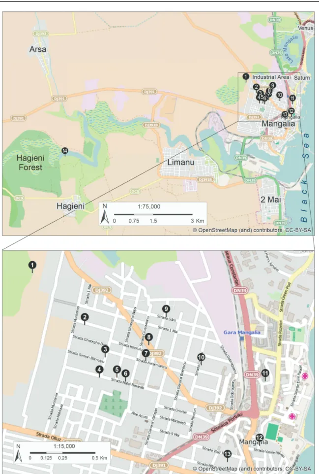
Niphargus cf. stygius specimens were sampled from existing wells in the town of Mangalia (SE Romania), from Movile Cave and from a spring in Hagieni Forest (Table 2, Fig. 1). In Mangalia, we either descended in the wells and picked specimens directly from the walls; pulled and dragged a planktonic net through the mass of water in the well; or examined large amounts of water (typically 100-150 liters) brought up to the surface using a rope and a bucket; in Movile Cave, we picked specimens directly from the edges of the cave lake; in Hagieni Forest spring, we collected them under the rocks and in detritus.
Niphargids were immediately transferred to 70% or 96% ethanol to allow for both morphological and genetic analyses. Morphological inspection, drawings and measurements were performed using an MBS- 1 stereo microscope (Lytkarino, USSR) and a Carl Zeiss microscope (Jena, Germany). The terminology used for body parts and the choice of appendages taken into consideration for measurements were as in
Fig. 1. Map of the research area with indication of sampling locations
Fišer et al. (2009). True spines, i.e., extrusions of cuticle, are not known in Niphargus (Fišer et al. 2009).
Species of this genus have appendages armed with fl exible thin setae, fl exible plumose setae and stout spiniform setae. To simplify descriptions, we refer to the thin fl exible setae as ‘setae’ and stout spiniform setae as ‘spines’ as in Švara et al. 2015.
Molecular analyses
To shed light on the relationships of Niphargus cf. stygius with other species of the genus Niphargus, all nuclear ribosomal 28S sequences of members of the Niphargidae available on 28 Jul. 2015 were downloaded from GenBank (Benson et al. 2015). This dataset of 494 sequences was then completed with unpublished 28S sequences of topotypes of N. montellianus Stoch, 1998 and N. tridentinus Stoch, 1998 (sequenced using the same protocol as in Flot et al. 2010; GenBank accession numbers KT878856 and KT878857) and pre-aligned in MAFFT using the FFT-NS-i mode to identify unsuitable sequences (originating from a different 28S region than our target region). After fi ltering out these sequences, the remaining dataset of 422 sequences from Lefébure et al. (2006, 2007), Fišer et al. (2008), Trontelj et al.
(2009), Flot (2010), Flot et al. (2010, 2014), Hartke et al. (2011), Trontelj et al. (2012), Hekmatara et al. (2013), Fišer et al. (2013), McInerney et al. (2014), Altermatt et al. (2014), Ntakis et al. (2015) and Esmaeili-Rineh et al. (2015) were screened for perfect duplicates using FaBox (Villesen 2007), resulting in a dataset a 260 unique sequences, to which two outgroup sequences from the genera Synurella Wrzesniowski, 1877 and Gammarus Fabricius, 1775 were added as in Flot et al. (2014). The sequences were aligned in MAFFT version 7 (Katoh & Standley 2013) using the E-INS-i option and analyzed in FastTree 2 (Price et al. 2010) using the GTR model (Lanave et al. 1984; Tavaré 1986) with 1000 bootstrap replicates (Felsenstein 1985). The resulting Newick tree was turned into PDF using MEGA6 (Tamura et al. 2013), then beautifi ed using Inkscape (Bah 2011).
The holotype and paratypes are deposited in the collection of the Department of Biology, Biotechnical Faculty, University of Ljubljana.
Results
Order Amphipoda Latreille, 1816 Family Niphargidae Bousfi eld, 1977
Genus Niphargus Schiödte, 1849 Niphargus dancaui sp. nov.
urn:lsid:zoobank.org:act:01A72B96-94E1-401C-A6D0-4B4DD9C8D554 Figs 1–15
Niphargus cf. stygius – Sarbu & Popa 1992: 651.
Diagnosis
Mid to large-sized Niphargus of robust appearance, with acute to right postero-ventral angle of epimeral plates. Pleon with at most 4 setae along the posterior margin of each segment and a single tiny seta in postero-lateral position on urosomite I. Antenna I shorter than half of the total body length. The outer lobe of maxilla I has 7 spines with 1–3 teeth. The inner lobe of maxilliped has 9 spines. The propodus of both gnathopods are almost square-shaped, with 5 setae along the outer margins of the dactyli. The pereopods are shorter than half of the total body length, with one spine at the nail base. The uropods III are sexually dimorphic and elongated in males. The male telson bears 3 apical spines, 1 lateral and 1 dorsal spine on each lobe, plus 1 subapical spine per lobe for the female telson.
Etymology
The specifi c name is derived from the name of the late Dan Dancău (1933–1994), who fi rst studied the amphipod fauna in the Dobrogea region and described some of its species, notably Pontoniphargus racovitzai from Doi Mai and Mangalia (Dancău 1970) and Niphargus dobrogicus from Doi Mai, Schitu and Vama Veche (Dancău 1964).
Material examined Holotype
ROMANIA: ♂, Movile Cave, Mangalia (Fig. 1, Table 2). The holotype specimen collected from location 1 in Fig. 1 was not dissected, but was deposited intact in 75% ethanol.
Paratypes
ROMANIA: Mangalia, 1 ♂, 3 ♀♀, well 12, Aleea Cetăţii 1; 1 ♂, well 7, str. Avram Iancu 26; 1 ♀, well 10, str. Crinului 34; 1 ♀, well 3, str. General Dragalina; 1♂, 1 ♀, well 8, str. Horia, Cloşca şi Crişan 13.
Remark
The description was performed and species variability was examined on the basis of several paratypes collected from various hand-dug wells in the town of Mangalia (SE Romania) (Fig. 1, Table 2). The male and female described here were sampled from a well on Aleea Cetăţii 1. The other specimens were included in order to examine the variability.
Description (male)
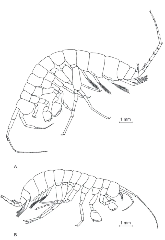
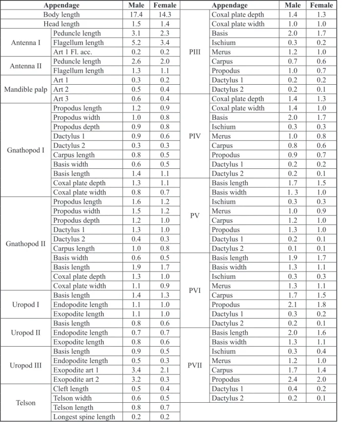
The total male body length is 17.4 mm (Fig. 2A). A detailed chart containing the measurements of all diagnosis-relevant body appendages for both male and female is presented in Table 1.
Head
The head (Fig. 3A) represents 8.5 % of total body length, no rostrum was observed.
Antennae
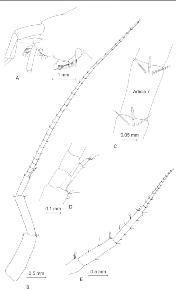
Antenna I (Fig. 3B) almost half of total body length (Table 1), with a fl agellum formed of 32 articles.
Most fl agellum articles bear one short aesthetasc (Fig. 3C). Length of peduncle slightly more than one third of the total length of antenna I. Accessory fl agellum (Fig. 3D) biarticulated, the proximal article exceeds half of second article of main fl agellum, distal article is approximately one fi fth of total length of accessory fl agellum. Antenna II (Fig. 3E) with fl agellum formed of 13 articles, half as long as antenna I. Peduncle almost twice as long as fl agellum.
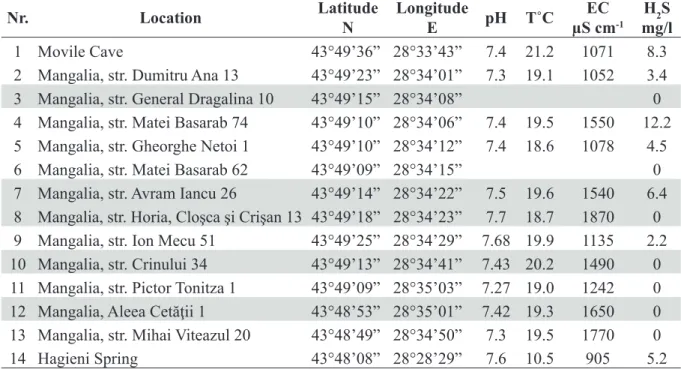
Mouthparts
Labium (Fig. 4A) bilobate; length of inner lobes half length of outer lobes. Both inner and outer lobes with distally fi ne setae. Labium displayed in Fig. 4A from female sampled from str. Horia, Cloşca şi Crişan 13; identical to all inspected labia from other specimens, males and females.
Left mandible (Fig. 4B) with fi ve teeth on incisor process, four teeth on lacinia mobilis and a row of eight serrate spines between lacinia mobilis and molar process (Fig. 4C).
Right mandible (Fig. 4D) with four teeth on incisor process, several small denticles on lacinia mobilis and a row of fi ve denticulate setae between lacinia mobilis and molar process (Fig. 4E).
Two mandibular palps (Fig. 4B and Fig. 4D), highly similar and of same length. The three articles represent 21% (article 1), 37% (article 2) and 42% (article 3) of total palp length (Table 1). Proximal article without setae, article 2 with 8–11 ventral setae and article 3 with one group of 5–6 A setae, three groups of 3–4 B setae, approximately 36 D setae and 5 E setae (Fišer et al. 2009).
Fig. 2. General appearance of Niphargus dancaui sp. nov. A. ♂. B. ♀.
Fig. 3. ♂. A. Head. B. Antenna I. C. Aesthetascs of antenna I. D. Accessory fl agellum of antenna I.
E. Antenna II.
Appendage Male Female Appendage Male Female
Body length 17.4 14.3
PIII
Coxal plate depth 1.4 1.3
Head length 1.5 1.4 Coxal plate width 1.0 1.0
Antenna I
Peduncle length 3.1 2.3 Basis 2.0 1.7
Flagellum length 5.2 3.4 Ischium 0.3 0.2
Art 1 Fl. acc. 0.2 0.2 Merus 1.2 1.0
Antenna II Peduncle length 2.6 2.0 Carpus 0.7 0.6
Flagellum length 1.3 1.1 Propodus 1.0 0.7
Mandible palp
Art 1 0.3 0.2 Dactylus 1 0.2 0.2
Art 2 0.5 0.4 Dactylus 2 0.2 0.1
Art 3 0.6 0.4
PIV
Coxal plate depth 1.4 1.3
Gnathopod I
Propodus length 1.2 0.9 Coxal plate width 1.4 1.0
Propodus width 1.0 0.8 Basis 2.0 1.7
Propodus depth 0.9 0.8 Ischium 0.3 0.3
Dactylus 1 0.9 0.6 Merus 1.0 0.8
Dactylus 2 0.3 0.3 Carpus 0.8 0.6
Carpus length 0.8 0.5 Propodus 0.9 0.7
Basis width 0.6 0.5 Dactylus 1 0.2 0.2
Basis length 1.4 1.1 Dactylus 2 0.2 0.1
Coxal plate depth 1.3 1.1
PV
Basis length 1.7 1.5
Coxal plate width 0.8 0.7 Basis width 1. 3 1.0
Gnathopod II
Propodus length 1.6 1.2 Ischium 0.3 0.3
Propodus width 1.5 1.2 Merus 1.0 0.9
Propodus depth 1.2 1.0 Carpus 1.2 1.0
Dactylus 1 1.3 1.0 Propodus 1.3 1.0
Dactylus 2 0.4 0.3 Dactylus 1 0.2 0.1
Carpus length 1.0 0.8 Dactylus 2 0.1 0.1
Basis width 0.6 0.5
PVI
Basis length 1.9 1.7
Basis length 1.9 1.7 Basis width 1.3 1.1
Coxal plate depth 1.3 1.0 Ischium 0.3 0.3
Coxal plate width 1.1 0.9 Merus 1.3 1.1
Uropod I
Basis length 1.4 1.3 Carpus 1.7 1.5
Endopodite length 1.1 1.0 Propodus 2.1 1.8
Exopodite length 1.1 1.0 Dactylus 1 0.3 0.2
Uropod II
Basis length 0.8 0.6 Dactylus 2 0.2 0.1
Endopodite length 0.7 0.7
PVII
Basis length 2.0 1.6
Exopodite length 0.8 0.6 Basis width 1.3 1.1
Uropod III
Basis length 0.9 0.5 Ischium 0.3 0.4
Endopodite length 0.5 0.3 Merus 1.2 1.0
Exopodite art 1 3.4 2.1 Carpus 1.7 1.4
Exopodite art 2 3.2 0.3 Propodus 2.4 2.0
Telson
Cleft length 0.5 0.4 Dactylus 1 0.4 0.2
Telson width 0.6 0.5 Dactylus 2 0.2 0.1
Telson length 0.8 0.7
Longest spine length 0.2 0.2
Table 1. Measurements in mm of the various appendages of male and female Niphargus dancaui sp. nov.
sampled from a well located on Aleea Cetăţii 1 in the town of Mangalia (SE Romania).
Maxilla I (Fig. 4F) with 7 apical setae on distal palp article. Outer lobe with 7 spines with 1–3 teeth, inner lobe with 3 apical setae.
Maxilla II (Fig. 4G) with inner lobe slightly shorter than outer lobe; both lobes with numerous apical setae.
Maxilliped (Fig. 4H) with palp formed of four articles. Article 2 with numerous setae in approximately 10 groups aligned along inner margin. Article 3 with three groups of 4–5 setae on inner margin, one group of 3 setae on dorsal margin and one apical group with 5 setae. Article 4 without setae. Outer lobe of maxilliped with 6 shorter, fl attened spines and 7 longer, slightly hairy, apical spines. Inner lobe with 9 setae-like spines.
Gnathopod I
Gnathopod I (Fig. 5A) with relatively ovoid coxal plate with depth greater than its width (ratio depth:width 1.0:0.6). Basis length:width ratio 1.0:0.4. Ischium with one posteroventral group of 4 setae.
Basis length:carpus length 1.0:0.6. Carpus with two groups of 8–10 setae on ventral margin, and one group of 4 setae located anterodorsally. Length:width ratio of propodus 1.0:0.8. Propodus with 7 groups of 3–4 setae on ventral margin, one anterodorsal group with 6 setae and one antero-apical group of 4 setae. Two groups with 2–3 setae on lateral surface of propodus close to its ventral side,and two groups of 4–5 setae closer to propodus dorsal margin. One group of 3 long setae present close to palmar spine.
Strong palmar spine and 4 outer denticulate spines in palmar corner. Dactylus (Fig. 5B) strong, with claw representing one quarter of total dactylus length and with fi ve setae along outer margin.
Nr. Location Latitude
N
Longitude
E pH T˚C EC
μS cm-1 H2S mg/l
1 Movile Cave 43°49’36” 28°33’43” 7.4 21.2 1071 8.3
2 Mangalia, str. Dumitru Ana 13 43°49’23” 28°34’01” 7.3 19.1 1052 3.4
3 Mangalia, str. General Dragalina 10 43°49’15” 28°34’08” 0
4 Mangalia, str. Matei Basarab 74 43°49’10” 28°34’06” 7.4 19.5 1550 12.2 5 Mangalia, str. Gheorghe Netoi 1 43°49’10” 28°34’12” 7.4 18.6 1078 4.5
6 Mangalia, str. Matei Basarab 62 43°49’09” 28°34’15” 0
7 Mangalia, str. Avram Iancu 26 43°49’14” 28°34’22” 7.5 19.6 1540 6.4 8 Mangalia, str. Horia, Cloşca şi Crişan 13 43°49’18” 28°34’23” 7.7 18.7 1870 0 9 Mangalia, str. Ion Mecu 51 43°49’25” 28°34’29” 7.68 19.9 1135 2.2 10 Mangalia, str. Crinului 34 43°49’13” 28°34’41” 7.43 20.2 1490 0 11 Mangalia, str. Pictor Tonitza 1 43°49’09” 28°35’03” 7.27 19.0 1242 0 12 Mangalia, Aleea Cetăţii 1 43°48’53” 28°35’01” 7.42 19.3 1650 0 13 Mangalia, str. Mihai Viteazul 20 43°48’49” 28°34’50” 7.3 19.5 1770 0
14 Hagieni Spring 43°48’08” 28°28’29” 7.6 10.5 905 5.2
Table 2. List of sampling locations with their geographic position and physico-chemical characteristics.
The numerals in the fi rst column relate to the location numbers in Fig. 1. The specimens dissected and analyzed for this study were selected from the locations highlighted in grey.
Fig. 4. ♂, mouthparts. A. Labium. B. Left mandible. C. Detail (the incisor process and lacinia mobilis) of left mandible. D. Right mandible. E. Detail (the incisor process and lacinia mobilis) of right mandible.
F. Maxilla I. G. Maxilla II. H. Maxilliped.
Gnathopod II
Coxal plate (Fig. 5C) with rectangular shape, deeper than wide (ratio depth:width 1.0:0.8). Basis length:width ratio 1.0:0.3. Ischium with one anteroventral group of 2 setae. Basis length:carpus length 1.0:0.6. Carpus has with two groups of 8–10 setae on ventral margin and one group of 2 setae located anterodorsally. Propodus almost square-shaped, with length:width ratio of 1.0:0.96. Propodus with 7 groups of 2–4 setae on ventral margin, one anterodorsal group of 2 setae, and one apical group of 4 setae.
Lateral surface of gnathopod II propodus without setae, with only two long setae close to palmar spine.
Fig. 5. ♂. A. Gnathopod I. B. Propodus of gnathopod I. C. Gnathopod II. D. Propodus of gnathopod II.
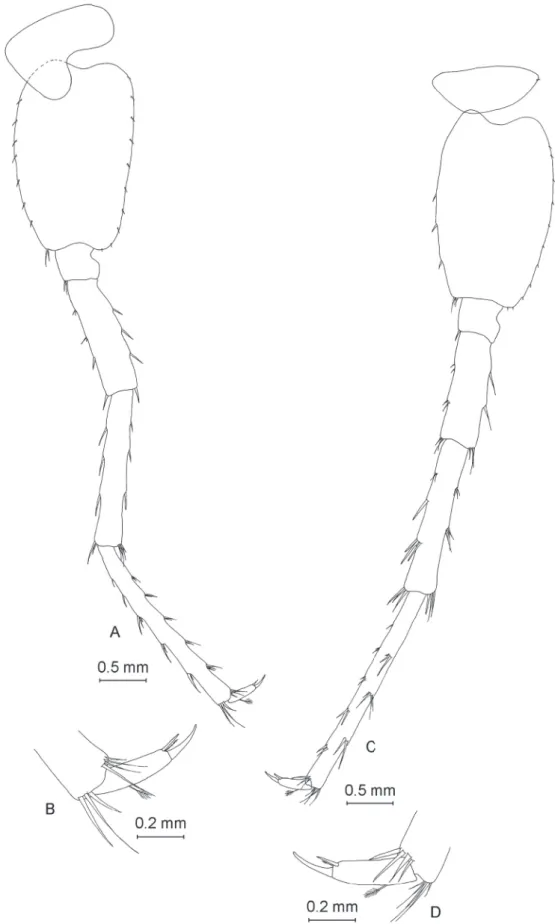
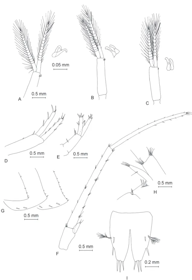
Fig. 6. ♂. A. Pereopod III. B. Dactylus of pereopod III. C. Pereopod IV. D. Dactylus of pereopod IV.
E. Pereopod V. F. Dactylus of pereopod V.
One strong spine and one outer, smaller spine on palmar corner. Dactylus (Fig. 5D) strong, with claw representing one quarter of total dactylus length and with fi ve setae along outer margin.
Pereopod III
Coxal plate of pereopod III (Fig. 6A) with rectangular shape, with depth:width ratio of 1.0:0.7. Posterior margin concave, with three setae. Gill irregularly ovoid. Dactylus (Fig. 6B) robust, with a nail measuring half of total dactylus length; with one dorsal seta with plumose tip and one spine at nail base. Propodus length:dactylus length ratio 1.0:0.34. Pereopod III nearly equal in length to pereopod IV (pereopod III length:pereopod IV length ratio 1.0:0.95).
Pereopod IV
Coxal plate of pereopod IV (Fig. 6C) almost square-like, depth:width ratio 1.0:0.96. Posterior margin concave, with four setae. Gill irregularly ovoid. Dactylus (Fig. 6D) robust, with nail slightly longer than half of total dactylus length; with one dorsal seta with plumose tip, one spine and one seta with plumose tip at nail base. Propodus length:dactylus length ratio 1.0:0.35.
Pereopod V
Coxal plate of pereopod V (Fig. 6E) shape of heart, with one small seta on anterior lobe. Basis with ovoid-trapezoidal shape, with length:width ratio of 1.0:0.68. Basis with 9 spine-like setae on anterior margin and 9 small setae on posterior margin. Dactylus (Fig. 6F) with one seta with plumose end on outer margin and one spine and one smaller seta with plumose end at base of nail. Nail represents 41%
of total dactylus length.
Pereopod VI
Coxal plate of pereopod VI (Fig. 7A) highly similar to that of pereopod V. Basis with ovoid-trapezoidal shape, with length:width ratio of 1.0:0.68. Basis with 9 spine-like setae on anterior margin and 9 small setae on posterior margin. Dactylus (Fig. 7B) with one seta with plumose end on outer margin and one spine and one smaller seta with plumose end at base of nail. Nail represents 34% of total dactylus length.
Pereopod VII
The pereopod VII (Fig. 7C) is almost half of the total body length. The coxal plate pereopod VII is half- ovoid, with one small seta on its posterior margin. The basis has a ovoid-trapezoidal shape, with a ratio length:width of 1.0:0.66. The basis presents 6 spine-like setae on the anterior margin and 11 small setae on the posterior margin. The dactylus (Fig. 7D) has one seta with a plumose end on the outer margin, one spine and one smaller seta with a plumose end at the base of the nail. The nail represents 33% of the total dactylus length.
Pereopods V:VI:VII equal 1.0:1.35:1.40.
Pleopods
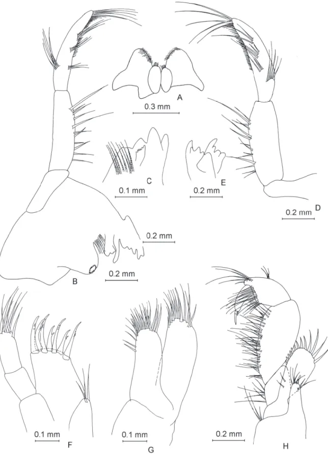
Pleopods I–III (Fig. 8A, Fig. 8B and Fig. 8C) highly similar, with rami of unequal length and 2 retinacles each.
Uropod I (Fig. 8D) with two dorsolateral spines onto peduncle. Length of endopodite equal to that of exopodite, segments with a low number of spines. One strong spine at base of uropod I.
Uropod II (Fig. 8E) with three dorsolateral spines onto peduncle. Exopodite slightly longer than endopodite, exopodite length:endopodite length ratio 1.0:0.88, both rami with a low number of spines.
Uropod III (Fig. 8F) long (43% of body length) and sexually differentiated. Protopodite with 4–5 small apical spines. Endopodite as long as protopodite with two apical setae. Proximal segment of exopodite
Fig. 7. ♂. A. Pereopod VI. B. Dactylus of pereopod VI. C. Pereopod VII. D. Dactylus of pereopod VII.
Fig. 8. ♂. — A–C. Pleopods with detail of retinacles A. Pleopod I. B. Pleopod II. C. Pleopod III. — D. Uropod I. E. Uropod II. F. Uropod III. G. Epimeral plates. H. Urosome of ♂ sampled from str. Horia, Cloşca şi Crişan13. I. Telson.
almost equal to distal segment (ratio 1.0:0.94). Proximal segment with seven groups of 1–3 spines on inner margin, and six groups with 1–3 spines on outer margin. Distal segment of exopodite with 8 spines on inner margin, 6 spines on outer margin and 5 apical setae.
Epimeral plates
Epimeral plate I (Fig. 8G) with acute postero-ventral angle, convex ventral margin with no spines and straight posterior margin with fi ve setae.
Epimeral plate II (Fig. 8G) with right postero-ventral angle, straight posterior margin and convex ventral margin. Two spines present along ventral margin, one strong spine in postero-ventral angle and two shorter setae along posterior margin.
Epimeral plate III (Fig. 8G) slightly different compared to epimeral plate II; postero-ventral angle rather acute, posterior margin slightly concave, ventral margin convex. Three spines present along ventral margin and nine setae along posterior margin.
Urosomite I with two dorsolateral spines, whereas urosomite II with four dorsolateral spines of various lengths. Dorsal margin of urosomite III spineless (Fig. 8H). Urosome on Fig. 8H belonged to ♂ analyzed from str. Horia, Cloşca şi Crişan 13. Number of setae on urosomites identical on all inspected specimens.
Telson
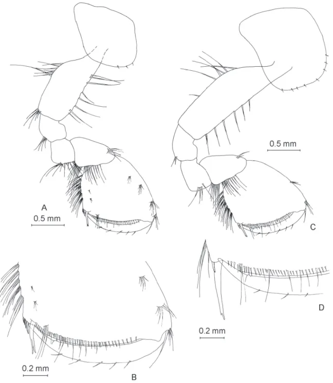
Telson (Fig. 8I) slightly longer than wide (width:length ratio 1.0:1.2). Three apical spines relatively short, approximately one fi fth of telson length. Telson with two fragile, plumose-ended setae along each side, as well as one lateral spine and one dorsal spine per lobe.
Sexual dimorphism
The female (Fig. 1B) is smaller (body length 14.3 mm) compared to the male. The female appendages (Figs 9–14) are highly similar to those of males, with a few exceptions. Antenna I, with 22 articles, reaches only one third of the total body length (Fig. 9B). Antenna II has just 9 articles (Fig. 9E). The female gnathopods I and II (Fig. 11) are similar to those of the male, except that the female propodus has a more inclined palmar margin, conferring it a rather trapezoidal shape in comparison to the more rectangular shape of the male propodus (Fig. 5). The female telson (Fig. 14H) is slightly different from the male one, with one sub-apical spine on each lobe. The uropod III (Fig. 14F) presents a distal segment of the exopodite shorter than that of the male. For the female, the proximal segment:distal segment ratio of the exopodite is 1.0:0.17, vs. 1.0:0.94 for the male.
Intraspecifi c variability
The intraspecifi c variability of Niphargus dancaui sp. nov. appears to be relatively low. The individuals sampled from various locations differed mainly in age-related size. The male and female described here were fully grown adults. These two specimens sampled from the well on Aleea Cetăţii 1 were larger and probably older than the other inspected specimens. However, their appendages, including their mouth parts, gnathopods, pereopods, pleopods and uropods, were largely similar with a few exceptions.
The number of articles in the fl agellum of antennae I and II appears variable, with numbers of articles ranging from 17 to 24 and from 9 to 11, respectively. Although the telson has always three apical spines and a pair of lateral spines its number of subapical and dorsal spines is variable (Fig. 14I–J). The shape of the epimeral plates is largely similar but the number of spines along their ventral margins ranges from 1 to 2 and from 1 to 3 in epimeral plates II and III, respectively.
Fig. 9. ♀. A. Head. B. Antenna I. C. Aesthetascs of antenna I. D. Accessory fl agellum of antenna I.
E. Antenna II.
Fig. 10. Female mouthparts. A. Left mandible. B. Right mandible. C. Maxilla I. D. Maxilla II.
E. Maxilliped.
Fig. 11. ♀. A. Gnathopod I. B. Propodus teeth of gnathopod I. C. Gnathopod II. D. Propodus teeth of gnathopod II.
Fig. 12. ♀. A. Pereopod III. B. Dactylus of pereopod III. C. Pereopod IV. D. Dactylus of pereopod IV.
E. Pereopod V. F. Dactylus of pereopod V.
Fig. 13. ♀. A. Pereopod VI. B. Dactylus of pereopod VI. C. Pereopod VII. D. Dactylus of pereopod VII.
Fig. 14. — A–H. ♀. A. Pleopod I (with detail of retinacles). B. Pleopod II (with detail of retinacles).
C. Pleopod III (with detail of retinacles). D. Uropod I. E. Uropod II. F. Uropod III. G. Epimeral plates. H. Telson. — I. Telson of ♂ sampled from str. Avram Iancu 26. J. Telson of ♀ sampled from str.
Crinului 34.
Discussion
Morphological affi nities of Niphargus dancaui sp. nov.
Niphargus dancaui sp. nov. shares several similarities with N. stygius (Sket 1974; personal observations).
Both species are mid- to large-sized, robust and have similar lengths of appendages (e.g., pereopods, antennae) compared to their body lengths. Moreover, both species present similar mouthparts, gnathopods (notably the shape of the propods), spine patterns on pereopod dactyli, sexually non-dimorphic rami of uropod I, as well as similar sexually dimorphic traits such as elongated uropod III in males.
Yet, several details help distinguish N. dancaui sp. nov. from N. stygius. As pointed out by S. Karaman (1952), N. stygius and some related species (N. novomestanus Karaman, 1952, N. likanus Karaman, 1952, N. podpecanus Karaman, 1952, N. kenki Karaman, 1952, N. karamani Schellenberg, 1935) share four main traits: the outer margins of the dactyls of both gnathopods are armed with groups of setae, at most 4 setae are present along the posterior margin of the pleon segments, the fi rst urosomite segment has a single tiny seta in a postero-lateral position, and the uropod III is sexually dimorphic. Some of these characters are also present in N. dancaui sp. nov. (namely, its sexually dimorphic uropod III and the fact that the dorso-posterior margin of its pleonites presents less than 4 setae). However, the setae along the gnathopod dactyls of N. dancaui sp. nov. are not in groups, and the urosomite I of this species has more than one seta, which sets N. dancaui sp. nov. apart from N. stygius and related species.
In this aspect, N. dancaui sp. nov. resembles some other species from the N. costozzae group living in northern Italy (N. costozzae Stoch, 1998, N. lessiniensis Stoch, 1998, N. montellianus and N. tridentinus;
see Stoch 1998 for a revision) or the N. sphagnicolus-N. plurispinosus group from Slovenia and Slovakia (Rejic 1956; Hudec & Mock 2014). The latter group differs from N. dancaui sp. nov. by having more than three dorsal spines per telson lobe. Niphargus costozzae and N. montellianus differ from N. dancaui sp. nov. by having their setae along the gnathopod dactyli arranged in groups and by having more than one dorsal spine per telson lobe. Niphargus lessiniensis and N. tridentinus, however, show striking similarities to N. dancaui sp. nov. as described here. The only difference we found is the number of setal groups along the proximal exopodite article of uropod III in males: in both Italian species up to fi ve setal groups can be found along the inner or outer margin of this article, whereas this number is slightly higher in N. dancaui sp. nov. (up to seven groups of setae).
Position of N. dancaui sp. nov. in the niphargid tree of life
The updated 28S rRNA phylogeny of Niphargidae presented here (Fig. 15) is consistent with the ones in Flot et al. (2014) and McInerney et al. (2014): notably, the position of the two species Niphargus glenniei and N. irlandicus as a sister group to the rest of the genus is confi rmed with a very strong bootstrap support. This phylogeny shows that N. dancaui sp. nov. (black arrow) is genetically very distinct from all the species to which it is morphologically similar (white arrows): instead, it may be related to Niphargus montanarius and Niphargus sp. 4 from the Frasassi Cave system (Flot et al. 2010), a relationship that only receives low bootstrap support.
Ecological data
Individuals of N. dancaui sp. nov. collected from various wells in the town of Mangalia, in Hagieni Spring and in Movile Cave were largely similar regardless of the concentration of hydrogen sulfi de in the waters where they were collected (Table 2). These niphargids seem to be highly tolerant of the presence of hydrogen sulfi de in water, but are not dependent on it for their survival.
Niphargus dancaui sp. nov. is probably more widely distributed in the area than our 14 sampled locations since it was found to occur in Hagieni Spring (Fig. 1), which is located at an aerial distance of 8 km from the town of Mangalia. However, N. dancaui sp. nov. is probably endemic to the area of Mangalia, given
Niphargus tatrensis Niphargus aggtelekiensis
Niphargus cf. tatrensis Niphargus pectinicauda Niphargus bajuvaricus
Niphargus multipennatus Niphargus aberrans
Niphargus labacensis Niphargus sp. 2 VZ-2014
Niphargus strouhali alpinus Niphargus longidactylus Niphargus cf. aquilex Niphargus grandii
Niphargus tamanini Niphargus pupetta
Niphargus decui Niphargus transsylvanicus Niphargus sp. 2 INM-2013 Niphargus sp. 3 INM-2013 Niphargus andropus
Niphargus cf. tauri Niphargus wolfi Niphargus carniolicus Niphargus cf. tauri Niphargus cf. aquilex
Niphargus aquilex dobati Niphargus cf. tauri
Niphargus sp. 4 INM-2013 Niphargus bihorensis Niphargus cf. tauri Niphargus cf. aquilex Niphargus ambulator
Niphargus sp. 12 SER-2013 Pontoniphargus racovitzai/ruffoi Niphargus dobrogicus
Niphargus gallicus
Niphargus kieferi Niphargus aquilex
Niphargus schellenbergi Niphargus schellenbergi Niphargus cf. aquilex Niphargus cf. longidactylus Niphargus aquilex
Niphargus pachypus Niphargus delamarei
Niphargus virei Niphargus laisi Niphargus ladmiraulti
Niphargus kochianus/dimorphopus Niphargus irlandicus
Niphargus glenniei Synurella sp.
Gammarus sp.
96
99
52 79
89 100 94 100
56 85 80 61
99
94
55
98 99
86 100 82
92 50
100 79 57
69
54
75
83
98 84 99
55 95
97
100 99
100 92
100 62
0.01
Niphargus rhenorhodanensis Niphargus rhenorhodanensis Niphargus puteanus
Niphargus thienemanni Niphargus cf. fontanus Niphargopsis casparyi
Niphargus rhenorhodanensis Niphargus rhenorhodanensis Niphargus rhenorhodanensis Niphargus rhenorhodanensis Niphargus rhenorhodanensis Niphargus foreli
Niphargus sp. 4 JFF-2010 Niphargus montanarius Niphargus dancaui
Niphargus sp. 1 VZ-2014 Niphargus frasassianus
Niphargus pasquinii Niphargus cf. longicaudatus Niphargus cvijici Niphargus longicaudatus
Niphargus cf. longicaudatus Niphargus cf. longicaudatus Niphargus timavi Niphargus hrabei
Niphargus plateaui Niphargus tridentinus
Niphargus costozzae Niphargus montellianus
Niphargus sphagnicolus Niphargus dolenianensis Niphargus thuringius
Niphargus sp. ROM Niphargus sp. Djevojacka Niphargus tridentinus Niphargus lessiniensis
Niphargus rhenorhodanensis Niphargus cf. longicaudatus Niphargus vinodolensis
Niphargus illidzensis dalmaticus Niphargus elegans elegans Niphargus spoeckeri Niphargus hadzii Niphargus stygius likanus
Niphargus novomestanus Niphargus sp. N0098
Niphargus brachytelson Niphargus podpecanus Niphargus elegans zagrebensis Niphargus zagrebensis
Niphargus illidzensis Niphargus illidzensis Niphargus slovenicus Niphargus cf. tauri Niphargus laticaudatus Niphargus sp. 1 INM-2013
Iranian clade (Esmaeili-Rineh et al. 2015) Niphargus sanctinaumi Niphargus maximus Niphargus cf. aquilex Niphargus sp. 3 VZ-2014 Niphargus cf. aquilex Niphargus auerbachi Niphargus rhenorhodanensis
Niphargus lourensis Niphargobates orophobata Niphargus pachytelson
Niphargus subtypicus Niphargus steueri kolombatovici Niphargus dolichopus Niphargus stenopus Niphargus rejici
Niphargus cf. arbiter Niphargus arbiter Niphargus ictus
Niphargus salonitanus Niphargus cf. salonitanus Niphargus longiflagellum Niphargus stochi Niphargus spinulifemur Niphargus foreli
Niphargus sp. N0115 Niphargus stygius Niphargus fontanus Niphargus fontanus
Niphargus vjeternicensis bilecanus Niphargus vjetrenicensis
Niphargus balcanicus Niphargus dabarensis
Niphargus hercegovinensis Niphargus vjetrenicensis kusceri Niphargus trullipes Niphargus polymorphus
Niphargus factor Niphargus lunaris
Niphargus hvarensis Niphargus boskovici Niphargus krameri Niphargus cf. krameri Niphargus karamani Niphargus orcinus
88 99 100 95
68 100
56
55
100 87 99
100
57 100 99
74 61 63
68 90
100
79
50
94 99
100 52
100 100 99 98 99 100
100 78 99 99 93 88
73 99
100 96 85 96
100 98
87 100 100
97 100
98
85
73 99
99
80
100
60 100 97 60 92 100 90 Niphargus kenki
99 95
99 99
96
Fig. 15. (previous page) Maximum-likelihood phylogenetic tree of all published 28S niphargid sequences. This tree was obtained using FastTree2 under the GTR model; support values >50%
(from 1000 bootstrap replicates) are indicated next to the nodes. The black arrow shows the location of Niphargus dancaui sp. nov. in the niphargid phylogeny, whereas white arrows point at niphargid species morphologically similar to N. dancaui sp. nov. The two black boxes highlight the positions of the topotypes that were newly sequenced for the present article.
that it was never found in the wells sampled in the neighboring villages (i.e., Limanu, Vama Veche, Doi Mai, Arsa, Albeşti, Vânători, Coroana, Pecineaga, Dulceşti, 23 August - data not shown).
Groundwater crustaceans are in general stenobiontic: they do not tolerate large fl uctuations of the abiotic conditions in their environment (Gibert 2001). In places inhabited by humans, groundwater ecosystems are in general polluted with various anthropogenic wastes produced by industries, agricultural practices or household activities. Niphargus dancaui sp. nov. therefore appears vulnerable to extinction according to IUCN Red List categories and criteria. Apart from the occurrence of this species in Hagieni Spring, all other recorded sampling locations (Movile Cave and in 12 old hand-dug wells in the town of Mangalia) are spread over approximately 2 km2. The wells were used in the past as drinking water sources but have now been replaced with a modern water supply system. As a result, most of these wells are abandoned or are even being used for dumping various wastes, with potentially severe consequences for groundwater crustaceans inhabiting the aquifer. It is our hope that the description of N. dancaui sp. nov. and its recognition as a species endemic to Mangalia and its vicinity will lead to conservation measures to protect the fauna of this unique sulfi dic ecosystem.
Acknowledgements
Thanks to M. Baciu, D. Bianco, A. Cohn, S. Dattagupta, A. Hillebrand-Voiculescu, E. Piva and F. Stoch for assisting with fi eld work and/or collecting some of the specimens that were analyzed in this article.
Cene Fišer was funded by the Slovenian Research Agency, Program P1-0184.
References
Altermatt F., Alther R., Fišer C., Jokela J., Konec M., Küry D., Mächler E., Stucki P. & Westram A. 2014.
Diversity and distribution of freshwater amphipods in Switzerland (Crustacea: Amphipoda). PLOS ONE 9: e110328. http://dx.doi/org/10.1371/journal.pone.0110328
Bah T. 2011. Inkscape: Guide to a Vector Drawing Program (4th ed.). Prentice Hall, Boston, MA.
Benson D.A., Clark K., Karsch-Mizrachi I., Lipman, D.J., Ostell J. & Sayers E.W. 2015. GenBank.
Nucleic Acids Research. 43: D30–D35. http://dx.doi.org/10.1093/nar/gku1216
Dancău D. 1964. Noi contribuţii la studiul amfi podelor subterane Niphargus dobrogicus n. sp. Lucrările Institutului de Speologie “Emil Racoviţă” 3: 397–403.
Dancău D. 1970. Sur un nouvel amphipode souterrain de Roumanie, Pontoniphargus racovitzai n.g., n.sp. In: Orghidan T. & Dumitresco M. (eds) Livre du Centenaire. Emile G. Racovitza 1868–1968:
275–285. Académie de la République Socialiste de Roumanie, Bucarest.
Esmaeili-Rineh S., Sari A., Delić T., Moškrič A. & Fišer C. 2015. Molecular phylogeny of the subterranean genus Niphargus (Crustacea: Amphipoda) in the Middle East: a comparison with European niphargids.
Zoological Journal of the Linnean Society 175 (4): 812–826. http://dx.doi.org/10.1111/zoj.12296 Felsenstein J. 1985. Confi dence limits on phylogenies: an approach using the bootstrap. Evolution 39 (4): 783–791.
Fišer C. 2012. Niphargus: a model system for evolution and ecology. In: Culver D.C. & White W.B.
(eds) Encyclopedia of Caves: 555–564. Academic Press, Amsterdam.
Fišer C., Sket B. & Trontelj P. 2008. A phylogenetic perspective on 160 years of troubled taxonomy of Niphargus (Crustacea: Amphipoda). Zoologica Scripta 37: 665–680. http://dx.doi/org/10.1111/j.1463- 6409.2008.00347.x
Fišer C., Trontelj P., Luštrik R. & Sket B. 2009. Toward a unifi ed taxonomy of Niphargus (Crustacea:
Amphipoda): a review of morphological variability. Zootaxa 2061: 1–22.
Fišer C., Konec M., Kobe Z., Osanič M., Gruden P. & Potočnik H. 2010. Conservation problems with hypothelminorheic Niphargus spe cies (Amphipoda: Niphargidae). Aquatic Conservation: Marine and Freshwater Ecosystems 20 (5): 602–604. http://dx.doi.org/10.1002/aqc.1119
Fišer C., Zagmajster M. & Zakšek V. 2013. Coevolution of life history traits and morphology in female subterranean amphipods. Oikos 122 (5): 770–778. http://dx.doi.org/10.1111/j.1600-0706.2012.20644.x Fišer C., Luštrik R., Sarbu S., Flot J.-F., Trontelj P. 2015. Morphological evolution of coexisting amphipod species pairs from sulfi dic caves suggests competitive interactions and character displacement, but no environmental fi ltering and convergence. PLOS ONE 10: 1–13. http://dx.doi.org/10.1371/journal.
pone.0123535
Flot J.-F. 2010. Vers une taxonomie moléculaire des amphipodes du genre Niphargus : exemples d’utilisation de séquences d’ADN pour l’identifi cation des espèces. Bulletin de la Société des Sciences Naturelles de l’Ouest de la France 32: 62–68.
Flot J.-F., Wörheide G. & Dattagupta S. 2010. Unsuspected diversity of Niphargus amphipods in the chemoautotrophic cave ecosystem of Frasassi, central Italy. BMC Evolutionary Biology 10: 171.
http://dx.doi.org/10.1186/1471-2148-10-171
Flot J.-F., Bauermeister J., Brad T., Hillebrand-Voiculescu A., Sarbu S.M. & Dattagupta S. 2014.
Niphargus-Thiothrix associations may be widespread in sulphidic groundwater ecosystems: evidence from southeastern Romania. Molecular Ecology 23 (6): 1405–1417. http://dx.doi.org/10.1111/mec.12461 Forti P., Galdenzi S. & Sarbu S.M. 2002. The hypogenic caves: a powerful tool for the study of seeps and their environmental effects. Continental Shelf Research 22: 2373–2386.
Gibert J. 2001. Basic attributes of groundwater ecosystems. In: Griebler C., Danielopol D.L. Gibert J., Nachtnebel H.P. & Notenboom J. (eds). Groundwater Ecology. A Tool for Management of Water Resources: 39–54. Offi ce for Offi cial Publications of the European Communities, Luxembourg.
Hartke T.R., Fišer C., Hohagen J., Kleber S., Hartmann R. & Koenemann S. 2011. Morphological and molecular analyses of closely related species in the stygobiontic genus Niphargus (Amphipoda). Journal of Crustacean Biology 31: 701–709. http://dx.doi/org/10.1651/10-3434.1
Hekmatara M., Zakšek V., Heidari Baladehi M. & Fišer, C. 2013. Two new species of Niphargus (Crus- tacea: Amphipoda) from Iran. Journal of Natural History 47 (21 –22): 1421–1449. http://dx.doi.org/10.
1080/00222933.2012.743616
Hudec I. & Mock A. 2014. Niphargus plurispinosus sp. n. (Crustacea, Amphipoda), a stygophile and hypotelminorheic representative from Central Europe. Subterranean Biology 13: 65–87. http://dx.doi.
org/10.3897/subtbiol.13.6531
Karaman S. 1952. Podrod Stygoniphargus u Sloveniji i Hrvatskoj. Prirodoslovna Istraživanja 25: 5–38.
Karaman G.S., Borowsky B. & Dattagupta S. 2010. Two new species of the genus Niphargus Schiödte, 1849 (Amphipoda, fam. Niphargidae) from the Frasassi cave system in Central Italy. Zootaxa 2439:
35–52.
Katoh K. & Standley D.M. 2013. MAFFT multiple sequence alignment software version 7: improvements in performance and usability. Molecular Biology and Evolution 30: 772–780. http://dx.doi.org/10.1093/
molbev/mst010
Lanave C., Preparata G., Saccone C. & Serio G. 1984. A new method for calculating evolutionary substitution rates. Journal of Molecular Evolution 20: 86–93. http://dx.doi.org/10.1007/BF02101990 Latella L., Di Russo C., De Pasquale L., Dell’Anna L., Nardi G. & Rampini M. 1999. Preliminary investigations on a new sulfurous cave in Central Italy. Mémoires de Biospéologie 26: 131–135.
Lefébure T., Douady C.J., Gouy M., Trontelj P., Briolay J. & Gibert J. 2006 Phylogeography of a subterranean amphipod reveals cryptic diversity and dynamic evolution in extreme environments.
Molecular Ecology 15 (7): 1797–1806. http://dx.doi.org/10.1111/j.1365-294X.2006.02888.x
Lefébure T., Douady C.J., Malard F. & Gibert J. 2007 Testing dispersal and cryptic diversity in a widely distributed groundwater amphipod (Niphargus rhenorhodanensis). Molecular Phylogenetics and Evolution 42 (3): 676–686. http://dx.doi.org/10.1016/j.ympev.2006.08.020
McInerney C.E., Maurice L., Robertson A.L., Lee R.F.D.K., Arnscheidt J., Venditti C., Dooley J.S.G., Mathers T., Matthijs S., Eriksson K., Proudlove G.S. & Hänfl ing B. 2014. The ancient Britons:
groundwater fauna survived extreme climate change over tens of millions of years across NW Europe.
Molecular Ecology 23 (5): 1153–1166. http://dx.doi.org/10.1111/mec.12664
Ntakis A., Anastasiadou C., Zakšek V. & Fišer C. 2015. Phylogeny and biogeography of three new species of Niphargus (Crustacea: Amphipoda) from Greece. Zoologischer Anzeiger 255: 32–46. http://
dx.doi.org/10.1016/j.jcz.2015.02.002
Price M.N., Dehal P.S. & Arkin AP. 2010. FastTree 2 – Approximately maximum-likelihood trees for large alignments. PLoS ONE 5: e9490. http://dx.doi.org/10.1371/journal.pone.0009490
Rejic M. 1956. Dve novi vrsti nifargid iz Slovenije. Biološki Vestnik 5: 79–84.
Sarbu S.M. & Popa R. 1992. A unique chemoautotrophically based cave ecosystem. In: Camacho A.I.
(ed.) The Natural History of Biospeleology: 637–666. Monograph of the National Museum of Natural Sciences 7, Madrid, Spain.
Sarbu S.M., Kane T.C. & Kinkle B.K. 1996. A chemoautotrophically based cave ecosystem. Science 272: 1953–1955. http://dx.doi.org/10.1126/science.272.5270.1953
Sarbu S.M., Galdenzi S., Menichetti M. & Gentile G. 2000. Geology and biology of the Frasassi caves in central Italy: an ecological multi-disciplinary study of a hypogenic underground karst system.
In: Wilkens H., Culver D.C. & Humphreys W.F. (eds) Subterranean Ecosystems: 359–378. Elsevier Academic Press, Amsterdam.
Sket B. 1974. Niphargus stygius (Schiödte) (Amphipoda, Gammaridae) — die Neubeschreibung des Renerotypus, Variabilität, Verbreitung und Biologie der Art, I. Biološki Vestnik 22: 91–103.
Sket B. & Velkovrh F. 1981. Postojnsko-Planinski jamski sistem kot model za preučevanje onesnaženja podzemeljskih voda. Naše Jame 22: 27–44.
Stoch F. 1998. Revision of the Niphargus stygius-group in Venetia and Trentino (northeastern Italy), with description of three new species (Crustacea, Amphipoda, Niphargidae). Bollettino del Museo Civico di Storia Naturale di Verona 22: 229–274.
Švara V., Delić T., Raða T. & Fišer C. 2015. Molecular phylogeny of Niphargus boskovici (Crustacea:
Amphipoda) reveals a new species from epikarst. Zootaxa 3994: 354–376. http://dx.doi.org/10.11646/
zootaxa.3994.3.2
Tamura K., Stecher G., Peterson D., Filipski A. & Kumar S. 2013. MEGA6: Molecular evolutionary genetics analysis version 6.0. Molecular Biology and Evolution 30: 2725–2729. http://dx.doi.
org/10.1093/molbev/mst197
Tavaré S. 1986. Some probabilistic and statistical problems in the analysis of DNA sequences. Lectures on Mathematics in the Life Sciences 17: 57–86.
Trontelj P., Douady C.J., Fišer C., Gibert J., Gorički Š., Lefébure T., Sket B. & Zakšek V. 2009. A molecular test for cryptic diversity in ground water: how large are the ranges of macrostygobionts?
Freshwater Biology 54 (4): 727–744. http://dx.doi.org/10.1111/j.1365-2427.2007.01877.x
Trontelj P., Blejec A. & Fišer C. 2012 Ecomorphological convergence of cave communities. Evolution 66 (12): 3852–3865. http://dx.doi.org/10.1111/j.1558-5646.2012.01734.x
Väinölä R., Witt J., Grabowski M., Bradbury J., Jazdzewski K. & Sket B. 2008. Global diversity of amphipods (Amphipoda; Crustacea) in freshwater. Hydrobiologia 595: 241–255. http://dx.doi.
org/10.1007/s10750-007-9020-6
Villesen P. 2007. FaBox: an online toolbox for FASTA sequences. Molecular Ecology Notes 7: 965–968.
http://dx.doi.org/10.1111/j.1471-8286.2007.01821.x
Manuscript received: 31 July 2015 Manuscript accepted: 22 October 2015 Published on: 16 December 2015 Topic editor: Rudy Jocqué
Desk editor: Kristiaan Hoedemakers
Printed versions of all papers are also deposited in the libraries of the institutes that are members of the EJT consortium: Muséum national d’Histoire naturelle, Paris, France; Botanic Garden Meise, Belgium;
Royal Museum for Central Africa, Tervuren, Belgium; Natural History Museum, London, United Kingdom; Royal Belgian Institute of Natural Sciences, Brussels, Belgium; Natural History Museum of Denmark, Copenhagen, Denmark.