A study of mutual antagonism between EDS1 and transcription factor MYC2
in Arabidopsis immunity
Inaugural-Dissertation zur
Erlangung des Doktorgrades
der Mathematisch-Naturwissenschaftlichen Fakultät der Universität zu Köln
vorgelegt von Jingde Qiu aus Neijiang, China
Köln 2017
Diese Arbeit wurde durchgeführt am Max-Planck-Institut für Pflanzenzüchtungsforschung in Köln in der Abteilung für Pflanze-Mikroben Interaktionen (Direktor: Prof. Dr. P. Schulze-Lefert),Arbeitsgruppe Prof. Dr. Jane Parker, angefertigt.
The work described in this thesis was conducted under the supervision of Prof. Dr.
Jane Parker at the Max Planck Institute for Plant Breeding Research (Department of Plant-microbe interactions, Director: Prof. Dr. P. Schulze-Lefert)
Berichterstatter: Prof. Dr. Jane E. Parker Prof. Dr. Alga Zuccaro
Prüfungsvorsitzender: Prof. Dr. Gunther Döhlemann
Tag der Disputation: 09.12.2016
Ph.D. Thesis-Table of contents
Table of Contents
Table of Contents ... I Acknowledgement ... V Publications ... VII Abbreviations ... VIII Summary ... XIII Zusammenfassung... XV
1 Introduction ... 1
1.1 The two major layers of innate immunity in plants ... 1
1.2 The role of NLRs in ETI ... 2
1.2.1 NLRs reside in different cellular compartments ... 2
1.2.2 NLRs recognize specific pathogen effectors ... 3
1.2.3 Certain NLR families function as “helpers” in resistance signaling ... 4
1.2.4 Several nuclear NLRs regulate transcription changes... 5
1.3 Modulation of hormones in plant immunity ... 6
1.3.1 SA biosynthesis, metabolism, accumulation and signaling in immunity ... 6
1.3.2 JA biosynthesis and signaling in defense responses ... 8
1.3.3 MYC2 and the SA-JA crosstalk in stress responses ... 8
1.4 The EDS1 signaling node in immunity ... 11
1.4.1 Nuclear EDS1 accumulation is necessary for immunity ... 11
1.4.2 EDS1 acts as a hub in NLRs signaling and is a virulence target for effectors ... 12
1.4.3 EDS1 functions with its sequence-related partners PAD4 and SAG101 in innate immunity 13 1.4.4 EDS1 modulates SA-JA crosstalk ... 15
1.5 Repression and de-repression in plant immunity ... 16
1.6 Thesis aims ... 16
2 Results ... 18
2.1 Forward genetic screening for suppressors of eds1 (sed mutants) ... 18
2.1.1 Isolation of Arabidopsis sed mutants in an EMS mutagenized M2 population ... 18
2.1.2 A mutation in CPR5 leads to autoimmune phenotype of sed3 ... 21
2.1.3 eds1-2 pathogen hypersusceptibility is reduced in sed1 and sed2 ... 21
Ph.D. Thesis-Table of contents
II
2.1.5 A defect in COR perception causes sed4 resistance against Pst DC3000 avrRps4 ... 26
2.2 Mutual antagonism between EDS1 and MYC2 in defense responses ... 31
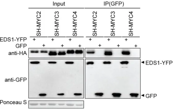
2.2.1 EDS1, PAD4 and SAG101 interact with MYC2 family TFs in plants ... 31
2.2.2 EDS1, PAD4 and SAG101 each form complexes with MYC2 in Arabidopsis ... 34
2.2.3 N-terminal and C-terminal parts of MYC2 associate with PAD4 and SAG101 ... 36
2.2.4 MYC2ΔN suppresses pEDS1 driven EDS1 transcription ... 38
2.2.5 MYC2 specifically represses pEDS1 promoter activity ... 42
2.2.6 MYC2 DNA-binding is essential for MYC2 suppression of pEDS1 activity ... 45
2.2.7 Suppression of pEDS1 activity by MYC2 is G-box-independent ... 47
2.2.8 MYC2-family TFs function redundantly to negatively regulate EDS1 expression ... 49
2.2.9 MYC2-family TFs delay induction of EDS1 in basal resistance ... 51
3 Discussion ... 54
3.1 Forward genetic screening for suppressors of eds1 ... 55
3.1.1 Truncated CPR5 restores TNL (RRS1/RPS4) resistance to Pst DC3000 avrRps4 in sed3 . 55 3.1.2 Disrupted BR signaling by BSU1P650L might restore basal and TNL resistance in eds1-2 . 56 3.1.3 DMR6 acts as negative regulator in plant immunity ... 57
3.1.4 Disrupted JA signaling partially relieves eds1-2 defects in TNL and basal immunity ... 57
3.1.5 EDS1-dependent resistance signaling suppresses bacterial COR virulence ... 59
3.2 Mutual antagonism between EDS1 and MYC2 in defense responses ... 60
3.2.1 Arabidopsis EDS1, PAD4 and SAG101 each forms complexes with MYC2 in planta ... 60
3.2.2 EDS1-family proteins modulate MYC2-family TF transcriptional activity ... 62
3.2.3 MYC2 suppresses pEDS1 activity independently of G-box and G-box variant motifs ... 64
3.2.4 MYC2, without direct DNA-binding, promotes protein post-transcriptional accumulation 65 3.2.5 MYC2-family TFs redundantly negatively regulate EDS1 expression ... 66
3.2.6 MYC2-family TFs delay induction of EDS1 in basal resistance ... 67
3.3 Summary and Perspectives ... 70
4 Materials and methods ... 72
4.1 Materials ... 72
4.1.1 Plant materials ... 72
4.1.2 Pathogens ... 73
4.1.3 Bacterial strains ... 73
4.1.4 Saccharomyces cerevisiae (yeast) strain ... 74
Ph.D. Thesis-Table of contents
4.1.5 Vectors ... 74
4.1.6 Oligonucleotides ... 79
4.1.7 Enzymes ... 81
4.1.8 Chemicals ... 81
4.1.9 Antibiotics (stock solutions) ... 81
4.1.10 Media ... 82
4.1.11 Antibodies ... 83
4.1.12 Buffers and solutions ... 84
4.2 Methods... 86
4.2.1 Maintenance and cultivation of Arabidopsis plants ... 86
4.2.2 Generation of Arabidopsis F1 and F2 progeny ... 86
4.2.3 Maintenance of P. syringae pv. Tomato cultures ... 86
4.2.4 P. syringae pv. tomato spraying treatment ... 87
4.2.5 P. syringae pv. tomato growth assay ... 87
4.2.6 Transient protein expression in N. benthamiana ... 87
4.2.7 Transient expression assays using Arabidopsis mesophyll protoplasts ... 87
4.2.8 Biochemical methods ... 88
4.2.9 Molecular biological methods ... 90
References ... 98
Erklärung... 117
Curriculum Vitae ... 118
IV
Ph.D. Thesis- Acknowledgement
Acknowledgement
Foremost, I would like to express my great gratitude to Prof. Dr. Jane Parker for giving me the great chance of working in the group. Thanks for your faith and support, for your friendly and patient supervision, and for teaching me how to think, write and express scientifically and critically.
A special thanks to Dr. Haitao Cui who is my direct tutor for guiding and teaching me essential and small techniques. I am truly grateful to work and collaborate with you on different projects, to discuss scientific issues and talk about normal life during the past four years. Thanks a lot.
I would also like to thank Dr. Imre Somssich for being my second supervisor, for your advice and critical comments on my projects, and for your reminding that how urgent it is for me to improve my scientific writing.
Thanks to Prof. Dr. Alga Zuccaro for kindly accepting to examine my thesis and Prof. Dr. Gunther Döhlemann for chairing my examination committee. Thanks for your time, efforts, coments, and scientific inputs.
Many thanks to all the JP members, past and present, for a great, friendly and scientific working environment. Jaqueline, without your endless support in the lab, I must need more time to finish my projects. Deepak the original G in JP and Dmitry (driven by 35S promoter), it is my pleasure to work and discuss with you guys, and thanks very much for your efforts and help to improve my scientific writing. Thanks to Thomas, Marcel, Anne and Shachi for always being available for helping me and giving me advice. Thanks to Clementine, Friederike, Xinhua and Patrick for all the chats and talks.
I should also thank Prof. Dr. P. Schluze-Lefert and the PSL department for unhindered use of all the facilities and always willing to share resources.
Last, I reserve my thanks and gratitude to my family. I am so sorry that I only spent a few days with you during the past four years. I would not make this without your constantly support, encouragement and confidence.
VI
Ph.D. Thesis- Publications
Publications
Cui, H., Gobbato, E., Kracher, B., Qiu, J., Bautor, J. and Parker, J. E. (2017), A core function of EDS1 with PAD4 is to protect the salicylic acid defense sector in Arabidopsis immunity. New Phytol, 213: 1802–1817.
Ph.D. Thesis-Abbreviations
VIII
Abbreviations
°C degree Celsius
aa amino acid
Arabidopsis Arabidopsis thaliana
avr Avirulence
bp base pair
C-terminal carboxy-terminal
CC Coiled-coil
cDNA complementary DNA
cfu colony forming unit(s)
d day(s)
dH2O deionised water
ddH2O distilled, deionized water
DMSO dimethylsulfoxide
DNA deoxyribonucleic acid
DNase deoxyribonuclease
dNTP deoxynucleosidetriphosphate
dpi days post inoculation
EDTA ethylenediaminetetraacetic acid
ETI effector triggered immunity
GFP green fluorescent protein
YFP yellow fluorescent protein
h hour(s)
HA hemagglutinin
hpi hours post inoculation
HRP horseradish peroxidase
JA jasmonic acid
Ph.D. Thesis-Abbreviations
kb kilo base(s)
kDa kilo Dalton
l litre
LRR leucine-rich repeat
M molar (mole/l)
µ micro
MAMP microbe-associated molecular pattern
MAPK mitogen activated protein kinase
mg milligram
min minute(s)
mM millimolar
MPIPZ Max-Planck-Institute for Plant Breeding Research
mRNA messenger RNA
MW molecular weight
NB nucleotide-binding
ng nanogram
NLR nucleotide-binding leucine-rich repeat
nM nanomolar
NOD nucleotide-binding-oligomerization domain
N-terminal amino-terminal
OD optical density
p35S 35S promoter of Cauliflower Mosaic Virus PAGE polyacrylamide gel electrophoresis
PAMP pathogen-associated molecular pattern
PCR polymerase chain reaction
pH negative decimal logarithm of the H+ concentration
Ph.D. Thesis-Abbreviations
X
PRR PAMP/ pattern recognition receptor
Pst Pseudomonas syringae pv. tomato
PTI PAMP/ pattern-triggered immunity
pv. pathovar
qRT-PCR quantitative real-time PCR
R resistance
RNA ribonucleic acid
ROS reactive oxygen species
rpm revolutions per minute
RT room temperature
RT-PCR reverse transcription -PCR
SA salicylic acid
SDS sodium dodecyl sulphate
T3SS type III secretion system
TBS Tris buffered saline
T-DNA transfer-DNA
TF transcription factor
TIR Toll/ interleukin-1 receptor
TNL TIR-NLR
Tris Tris- (hydroxymethyl-) aminomethane
V Volt(s)
v/v volume per volume
WT wild type
w/v weight/volume
Y2H yeast two-hybrid
Ph.D. Thesis-Abbreviations
Amino acids
Amino acid 3-letter code 1-letter code
Alanine Ala A
Arginine Arg R
Isoleucine Ile I
Leucine Leu L
Proline Pro P
Serine Ser S
Threonine Thr T
Tryptophan Trp W
XII
Ph.D. Thesis-Summary
Summary
Intracellular immune signaling plays an important role in modulating plant defense responses against pathogens. Arabidopsis nucleo-cytoplasmic protein EDS1 (Enhanced Disease Susceptibility1), together with its sequence-related signaling partners, PAD4 (Phytoalexin- Deficient4) and SAG101 (Senescence-Associated Gene101), is essential for transcriptional reprogramming during intracellular signaling in basal and TNL (TIR-NB-LRR) receptor-triggered immunity. EDS1 regulates both SA (salicylic acid)-dependent and SA-independent pathways in immune responses. Interactions between TNLs and EDS1 place EDS1 as a bridge between TNLs and induced transcriptional reprogramming in cells. How EDS1 signaling is regulated and which molecular events connect EDS1 to transcriptional defense reprogramming are still unclear.
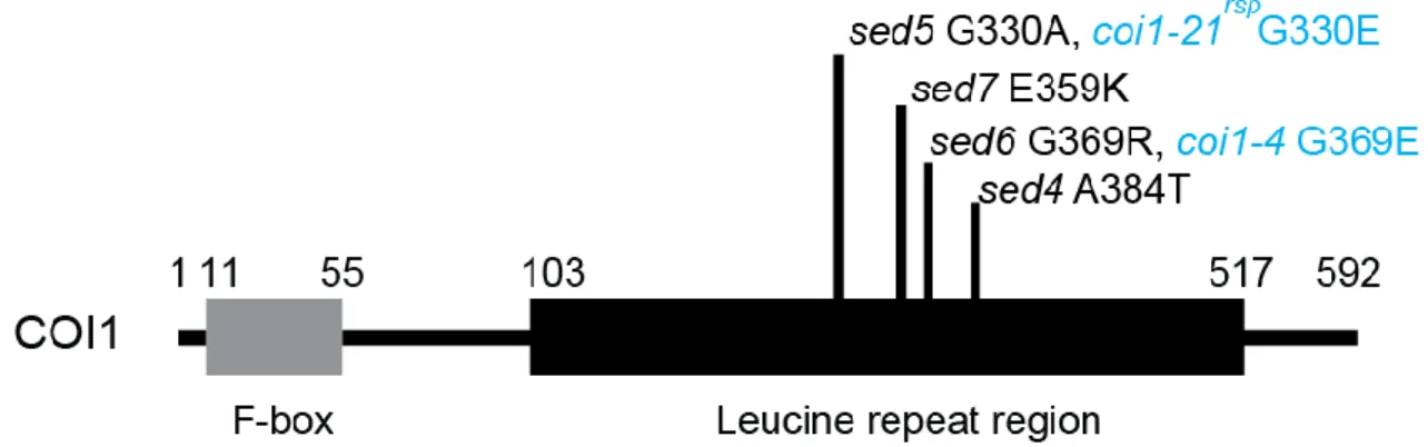
A genetic screen was used to identify suppressors of Arabidopsis eds1-2 hypersusceptibility to Pst (Pseudomonas syringae pv. tomato) DC3000 avrRps4 with the purpose to identify potential components of EDS1 signaling in immunity. Bacterial effector avrRps4 is recognized by the paired Arabidopsis TNL receptors RRS1 (Resistance to Ralstonia solanacearum1)/RPS4 (Resistance to Pseudomonas syringae4). I identified seven mutants with restored resistance to Pst DC3000 avrRps4. Among these, four mutants contain different mutations in COI1 (Coronatine- Insensitive1). Further analysis of several mutants suggests that four different signaling pathways can compensate for defects of eds1-2 in defense responses in Arabidopsis.
Because COI1 is essential for activating JA (jasmonic acid) signaling which antagonizes SA (salicylic acid), I hypothesized that EDS1 negatively regulates JA signaling in order to promote SA resistance. In transient expression assays, EDS1, PAD4 and SAG101 formed complexes with MYC2-family transcription factors (TFs) which regulate an important JA signaling branch. EDS1- MYC2 association was found to interfere with MYC2 transcriptional activity in transient expression assays. This is the first evidence that EDS1 regulates transcriptional reprogramming through association with TFs such as MYC2. Because EDS1 interferes with MYC2 transcriptional activity, I tested whether MYC2 reciprocally affects EDS1 protein and/or gene expression. MYC2 specifically suppressed EDS1 promoter activity independently of MYC2-binding G-box and G- box-related motifs but, surprisingly, requiring MYC2 bHLH domain DNA-binding activity. After exogenous application of a bacterial mimic of bioactive JA, coronatine (COR), MYC2-family TFs
Ph.D. Thesis-Summary
XIV
manifested in Arabidopsis protoplasts transient assays and at an early stage of Pst DC3000 infection and was not detectable at the late infection stage, probably due to activated EDS1 signaling.
In summary, results presented in this thesis reveal a new and important level of regulation of immunity and SA-JA pathway balance by mutual antagonism between EDS1- and MYC2- dependent processes.
Ph.D. Thesis- Zusammenfassung
Zusammenfassung
Intrazelluläre Signalwege spielen eine wesentliche Rolle für die pflanzliche Immunantwort gegen Pathogene. Transkriptionales Umprogrammieren, welches im Zuge der intrazellulären Signaltransduktion in basaler und TNL (TIR-NB-LRR) Rezeptor-abhängiger Immunantwort in Arabidopsis auftritt, erfordert nucleo-zytoplasmatisch lokalisiertes EDS1 (Enhanced Disease Susceptibility1). EDS1 wiederrum bedarf seiner sequenzähnlichen Partnerproteine PAD4 (Phytoalexin Deficient4) und SAG101 (Senescence-Associated Gene101) um voll funktional zu sein. Sowohl SA- (Salicylsäure) abhängige, als auch SA-unabhängige Signalwege werden von EDS1 während einer Immunreaktion reguliert. Die Interaktion von TNLs mit EDS1 platziert EDS1 als Verbindung zwischen TNLs und induzierter Umprogrammierung der Genexpression innerhalb der Zelle. Es ist jedoch noch immer unklar, wie EDS1 reguliert wird und welche molekularen Vorgänge EDS1 mit veränderter Transkription, welche mit der pflanzlichen Immunantwort einhergeht, verbinden.
Um potentielle Komponenten der EDS1 Signaltransduktion zu finden, welche die Hyperanfälligkeit von Arabidopsis eds1-2 Pflanzen gegenüber Pst (Pseudomonas syringae pv.
tomato) DC3000 avrRps4 aufheben, wurde ein genetischer Screen durchgeführt. Das bakterielle Effektorprotein avrRps4 wird von dem Arabidopsis TNL Rezeptorpaar RRS1 (Resistance to Ralstonia solanacearum1) und RPS4 (Resistance to Pseudomonas syringae4) erkannt. Ich konnte sieben Mutanten mit wiederhergestellter Resistenz gegenüber Pst DC3000 avrRPS4 identifizieren.
Von diesen enthielten vier unterschiedliche Mutationen in COI1 (Coronatine Insensitive1). Die tiefergehende Untersuchung diverser Mutanten legt nahe, dass vier eigenständige Signalwege die auftretenden Defekte in der Immunantwort von Arabidopsis eds1-2 Pflanzen kompensieren können.
Da COI1 unabdingbar ist für die Aktivierung des JA (Jasmonsäure) Signalweges, welcher SA entgegenwirkt, stellte ich die Hypothese auf, dass EDS1 negativ auf JA wirkt, um so SA abhängige Pathogenresistenz voranzutreiben. In transienten Expressionsanalysen bildeten EDS1, PAD4 und SAG101 Proteinkomplexe mit Transkriptionsfaktoren (TFs) der MYC2 Familie. Diese TFs regulieren einen wichtigen Teil des JA Signalweges. In transienten Expressionsversuchen beeinträchtigte die Interaktion von EDS1 mit MYC2 die Aktivität von MYC2 als TF. Dies zeigt
Ph.D. Thesis- Zusammenfassung
XVI
wie MYC2 reguliert. Da EDS1 die Aktivität von MYC2 reguliert, testete ich, ob MYC2 auf reziproke Weise EDS1 Genexpression und/oder Proteinlevel beeinflusst. Hierbei inhibierte MYC2 spezifisch EDS1 Promoteraktivität. Dies war unabhängig von G-Box und G-Box-ähnlichen Motiven, an welche MYC2 binden kann, benötigte aber überraschenderweise die MYC2 bHLH Domäne, welche Bindung an DNA vermittelt. Die exogene Zugabe von Coronatine (COR), welches ein bakterieller Imitator bioaktiver JA ist, verdeutlichte, dass TFs der MYC2 Familie EDS1 Genexpression auf redundante Art und Weise inhibieren. Diese hemmende Wirkung auf EDS1 wurde in transienten Arabidopsis Protoplasten Versuchen und in frühen Stadien von Pst DC3000 Infektion weiter bekräftigt. In späteren Infektionsstadien konnte die inhibierende Wirkung jedoch nicht nachgewiesen werden, was wahrscheinlich auf die Aktivierung von EDS1 Signaltransduktion zurückzuführen ist.
Die Ergebnisse dieser Arbeit zeigen eine neue und wichtige Regulationsebene der Immunantwort und der Balance zwischen SA-JA Signalwegen auf, welche von dem gegenseitigen EDS1-MYC2 Antagonismus gesteuert wird.
Ph.D. Thesis- Introduction
1 Introduction
1.1 The two major layers of innate immunity in plants
Plants have evolved a multi-layered immune system to defend themselves from infection by potential pathogenic microorganisms. The first layer of defense takes place on the surface of the plant cell, where PRRs (Pattern-Recognition Receptors) reside. PRRs can specifically detect PAMPs (Pathogen-Associated Molecular Patterns), such as bacteria flagellin, lipopolysaccharides, peptidoglycans, chitin and chitin derivatives, which are conserved across microbial species (Jones and Dangl, 2006; Couto and Zipfel, 2016). PRRs activated by PAMPs initiate a signaling cascade resulting in the onset of PTI (PAMPs-Triggered Immunity) (Jones and Dangl, 2006; Boller and Felix, 2009; Spoel and Dong, 2012). The PTI signal transduction cascade promotes phosphorylation of RBOHD (Respiratory Burst Oxidase Homologue Protein D) which is responsible for ROS (Reactive Oxygen Species) production in the apoplast (Kadota et al., 2014;
Li et al., 2014). Next to this, PTI responses rely on cytosolic Ca2+ signaling which activates CDPKs (Ca2+-dependent Protein Kinases), as well as MAPKs (Mitogen-Activated Protein Kinases) signaling cascades, to induce transcriptional reprogramming (Asai et al., 2002; Couto and Zipfel, 2016). The transcriptional changes translate into diverse cellular responses at the metabolic level:
phytoalexin production, callose deposition at the cell wall and hormone synthesis, which are generally effective to fend off host non-adapted pathogens (Boller and Felix, 2009).
Pathogens that have become adapted to a particular host genotype can disable PTI by secreting effector molecules into the plant apoplast or cytoplasm. Some effectors are delivered by a T3SS (Type III Secretion System) in the case of bacterial pathogens or byfeeding structures (haustoria) in the case of fungal and oomycete pathogens (Buttner and He, 2009; Kemen and Jones, 2012).
Effectors target host cellular processes to aid pathogen colonization, often by dampening PTI (Dangl et al., 2013). In response, host plants have evolved intracellular immune receptors known as R (Resistance) proteins which can specifically recognize the presence or monitor the activity of certain effectors (Cui et al., 2015). Upon perception of effectors, activated R proteins lead to ETI (Effector-Triggered Immunity) involving a boost in PTI-related defenses (Jones and Dangl, 2006;
Cui et al., 2015). The major class of R proteins belongs to a polymorphic family of intracellular
Ph.D. Thesis-Introduction
2
Binding and Leucine-Rich-Repeat) domains (Spoel and Dong, 2012). ETI responses are qualitatively similar to PTI but are generally faster and characterized by stronger and sustained transcriptional changes, which are usually coupled with a local HR (Hypersensitive Response) (Tao et al., 2003; Navarro et al., 2004; Dodds and Rathjen, 2010; Cui et al., 2015).
In this study, a low-level post-infection immune responses against virulent pathogens which are not obviously recognized by NLRs is defined as basal resistance. Plant basal resistance is likely to be “PTI plus weak ETI, minus ETS (Effector-Triggered Susceptibility)”, which allow virulent pathogen slow growth (Jones and Dangl, 2006).
1.2 The role of NLRs in ETI
As in mammals, plant NLR proteins belong to a subclass of the STAND (Signal Transduction ATPases with Numerous Domains) superfamily of proteins (Danot et al., 2009). The molecular function of STAND proteins is modulated by a P-loop motif (GxxxxGKT/S) which can be switched from an ADP-bound “off” position to an ATP-bound “on” state (Leipe et al., 2004). NLR proteins usually contain variable domains at the N-terminus, an LRR domain at the C-terminus and a central conserved NB-ARC [Nucleotide-Binding, shared by Apoptotic protease activating factor1 (Apaf-1), certain R-proteins and Cell death protein 4 (CED-4)] domain (Danot et al., 2009;
Takken and Goverse, 2012). Based on the sequences of the N-terminal domains, plant NLR proteins are generally divided into two groups: TNLs which contain a TIR (Toll/Interleukin1 receptor)-like domain and CNLs which contain a CC (coiled-coil) domain (Maekawa et al., 2011).
1.2.1 NLRs reside in different cellular compartments
NLR proteins localize to and function in diverse cellular compartments, such as at the plasma membrane, endomembrane and/ or nucleus. Upon perception of Pseudomonas syringae effectors avrRpt2, avrRphB and avrRpm1, respectively, the Arabidopsis CNLs, RPS2, RPS5 (Resistance to Pseudomonas syringae2 and 5, respectively) and RPM1 (Resistance to Pseudomonas syringae pv.
maculicola1) are activated at and function on the plasma membrane (Axtell and Staskawicz, 2003;
Gao et al., 2011; Qi et al., 2012). N-termini of some NLR proteins function as membrane-attached domains. The N termini of Flax TNL L6 and M, recognizing fungal effectors AvrL567 and AvrM, respectively, function as anchor domains to attach L6 and M to endomembranes system (Takemoto et al., 2012). Potato CNL protein Rx, conferring resistance to Potato virus X, localizes in both the
Ph.D. Thesis- Introduction
nucleus and cytosol, and the movement of Rx from the nucleus to cytoplasm promotes immune signaling, which is critical for resistance to virus (Slootweg et al., 2010; Tameling et al., 2010). In barley, nuclear localization of the nucleocytoplasmic CNL MLA10 (Polymorphic barley Mildew A10) is essential for resistance to Blumeria graminis expressing AVRA10, while the cytosolic localized MLA10 or its CC-NB1-225 domain is required for cell-death signaling (Shen et al., 2007;
Chang et al., 2013). These results suggest that the subcellular localization of MLA10 is critical for its function. However, a recent study showed that Sr33 (Stem Rust resistance33), an orthology of MLA in wheat, induces disease resistance signaling from the cytosol, suggesting that cytosolic Sr33 activates both cell death and stem rust resistance signaling (Cesari et al., 2016). Thus, the signaling components and regulatory mechanism for cytosolic Sr33-mediated signaling pathways may differ from cytosolic MLA10 although they share ~80% amino acid sequence identity (Cesari et al., 2016). Nuclear accumulation of Arabidopsis TNL RPS4 (Resistance to Pseudomonas syringae4) is necessary for triggering defense response against Pst (Pseudomonas syringae pv.
tomato) DC3000 expressing effector avrRps4 (Wirthmueller et al., 2007). Thus, the localization and spatial dynamics are critical for NLRs activation and function in ETI.
1.2.2 NLRs recognize specific pathogen effectors
NLRs can recognize effectors directly by physical interaction or indirectly by monitoring effector- caused perturbations of host targets, many of which are PTI components (Cui et al., 2015). The direct and indirect recognition are consistent with the gene-for-gene model proposed to address the disease resistance specificity (Flor, 1971). As examples of direct recognition, effector ATR1 secreted by Hpa (Hyaloperonospora arabidopsidis) into host cells is specifically recognized by TNL protein RPP1-WsB [an allele of Recognition of Peronospora parasitica1 in Arabidopsis accession Ws-2 (Wassilewskija)] (Krasileva et al., 2010; Steinbrenner et al., 2015), and the effectors AVR-Pia and AVR1-CO39 from Magnaporthe oryzae can be recognized by resistance protein pair RGA4/RGA5 (R-gene analog4/5) via directly binding in rice (Cesari et al., 2013;
Cesari et al., 2014b). NLRs can also be activated by monitoring pathogen effector-triggered modification of host factors. For example, RPM1 and RPS2 in Arabidopsis sense the phosphorylation or cleavage of RIN4 (RPM1-interacting protein4) caused by Pseudomonas syringae effectors AvrB, AvrRpm1 or AvrRpt2, leading to activation of immune signaling (Axtell
Ph.D. Thesis-Introduction
4
recognition has the potential advantage of increasing NLR perception space for rapidly evolving pathogen effectors (Cui et al., 2015).
Recent studies show that there are variations of NLR direct and indirect recognition, and therefore the distinction is not so clear between these modes of NLR action. For example, some NLRs contain domains resembling effector targets and acting as “integrated decoys” (Cesari et al., 2014a).
Some of these NLRs function as pairs. Crystal structure analysis of homo- and heterodimer forming TIR domains from Arabidopsis TNL receptors RRS1 (Resistance to Ralstonia solanacearum1) and RPS4 suggests that RRS1 and RPS4 act as a heterodimer or oligomer and cooperatively intercept two unrelated effectors, avrRps4 from Pseudomonas syringae and PopP2 from Ralstonia solanacearum (Deslandes et al., 2003; Narusaka et al., 2009; Williams et al., 2014).
In the absence of RRS1/RPS4 recognition, effector PopP2 dislodges WRKY TFs (Transcription Factors) from the DNA and reduces their transcriptional activity by acetylating a conserved lysine within WRKY domains, leading to effector-induced susceptibility (Le Roux et al., 2015; Sarris et al., 2015). A WRKY domain integrated within TNL RRS1-R is also targeted by PopP2 (Le Roux et al., 2015; Sarris et al., 2015). The PopP2-mediated acetylation dislodges RRS1-R from the DNA and thereby activates RPS4-dependent immune responses, including upregulation of defense genes required for disease resistance (Le Roux et al., 2015; Sarris et al., 2015). Therefore, RRS1 acts as an NLR sensor with an integrated effector decoy domain, while RPS4 activates as a signaling NLR, likely via forming signaling heterodimer with RRS1 (Cesari et al., 2014a; Griebel et al., 2014).
1.2.3 Certain NLR families function as “helpers” in resistance signaling
The current view that some NLRs function as “helpers” to relay and amplify resistance signaling is supported by studies on NLR receptors in mammalian immunity. For example, in mouse, NAIPs (NLR family, Apoptosis Inhibitory Proteins), which provide recognition specificity for distinct bacterial pathogens, are required for NLRC4 (NLR family, CARD domain containing4) activation and formation of NLRC4 oligomerization, which in turn regulates downstream signaling (Kofoed and Vance, 2011; Zhao et al., 2011; Hu et al., 2013; Hu et al., 2015; Zhang et al., 2015c). These results suggest that a “sensor-executor” framework, in which NLRs such as NAIPs serving as
“sensor” and the other partner NLRs such as NLRC4 acting as “executor” to regulate downstream signaling (Griebel et al., 2014). In plant ETI, several NLR proteins have no known effector-sensor function, referred to as “helper” NLRs, but assist other immune receptors in transducing signals
Ph.D. Thesis- Introduction
after pathogen recognition to downstream responses (Griebel et al., 2014). Recently, NRC1 (NLR protein required for HR-associated cell death1)-like proteins NRC2a/b and NRC3, without known effector recognition activity, were discovered to be critical for the cell death and resistance mediated by Pto (Pseudomonas syringae. pv. tomato), suggesting NRC2a/b and NRC3 function as helper for intracellular receptor Pto-mediated cell death in N. benthamiana (Wu et al., 2016). In N. benthamiana, the CNL NRG1 (N requirement gene1), acts together with TNL N to trigger resistance against tobacco mosaic virus (Peart et al., 2005). In a genetic screen for lsd1 (lesions simulating disease1) suppressors, mutations in ADR1-L2 (Activated Disease Resistance1-Like2) were isolated (Bonardi et al., 2011). ADR1 and its paralogs, ADR1-L1 and ADR1-L2, are necessary for autoimmune phenotypes of the Arabidopsis typical TNL mutants snc1 (suppressor of npr1-1, constitutive1) and chs2-1 (chilling-sensitive2-1) (Dong et al., 2016). In contrast to canonical NLRs, the helper function of ADR1-L2 does not require an intact P-loop in the NB- ARC domain. (Bonardi et al., 2011). Although these “helper” non-canonical NLRs operate downstream of “sensor” NLRs, it is unclear whether sensor and helper receptors reside in a complex (Griebel et al., 2014).
1.2.4 Several nuclear NLRs regulate transcription changes
Currently, some evidence indicate that nuclear-localized NLRs are able to regulate transcriptional reprogramming via association with transcription regulators. In barley, in the presence of the cognate powdery mildew effector AvrA10, the N-terminal signaling domain of the nucleocytoplasmic MLA10 interacts with TF MYB6 and stimulates its DNA binding activity by releasing TF WRKY1 suppression, which in turn activates defense-related gene expression (Shen et al., 2007; Chang et al., 2013). In rice, CNL protein Pb1 (Panicle blast1) physically interacts with TF WRKY54, a positive regulator of SA signaling, which was proposed to contribute to Pb1- dependent blast resistance (Inoue et al., 2013). In Arabidopsis, the TNL SNC1 associates with TF bHLH84 and transcription co-repressor TPR1 (Topless-Related1) to mediate immune responses (Zhu et al., 2010; Xu et al., 2014). Tobacco TNL receptor N forms a complex with the TF SPL6 [Squamosa Promoter Binding Protein (SBP)-domain] to enable the activation of transcription of defense genes (Padmanabhan et al., 2013). These findings link NLR function with transcriptional regulation in cells.
Ph.D. Thesis-Introduction
6
1.3 Modulation of hormones in plant immunity
Downstream of PTI and ETI activation, a complex network of antagonistic and synergistic crosstalk occurs between phytohormone pathways which play an important role in fine-tuning the strength and dynamics of immune response and maintaining the balance with other stress pathways and growth (Pieterse et al., 2012; Lozano-Duran and Zipfel, 2015). Among the phytohormones, SA (salicylic acid) and JA (jasmonic acid) are two critical defense hormones and the crosstalk between them has been extensively explored in determining plant-microbe interactions (Pieterse et al., 2012). SA is generally considered to be effective against (hemi) biotrophic pathogens, such as Pseudomonas syringae or Hpa, whereas JA is important for plants to defend against necrotrophic pathogens and insect herbivores, as well as serving as a developmental hormone (Pieterse et al., 2012; Wasternack and Hause, 2013).
1.3.1 SA biosynthesis, metabolism, accumulation and signaling in immunity
SA is a phenolic compound, which is synthesized by two distinct enzymatic pathways, the isochorismate and the phenylalanine ammonia-lyase pathway (Dempsey et al., 2011). Isolation and characterization of the ics1 (isochorismate synthase1) mutant have demonstrated an essential role of ICS1 in pathogen-induced SA synthesis in the chloroplast by ICS1 (Wildermuth et al., 2001;
Dempsey et al., 2011). Stress-induced SA appears to be trapped in the chloroplast in the Arabidopsis eds5 (enhanced disease susceptibility5) susceptible mutant with mutation in EDS5 encoding a putative SA-transporter localized on the chloroplast envelope membrane, suggesting that SA export from the chloroplast to different cellular compartments is critical for immune responses (Nawrath et al., 2002; Serrano et al., 2013; Yamasaki et al., 2013). However, SA level is also tightly controlled by further metabolism and regulated by different signaling pathways in plants, since constitutive SA accumulation is often associated with autoimmunity and defects in growth (Jirage et al., 2001; Zhang et al., 2003; Chandran et al., 2014). SA can be chemically modified to other forms of SA, most of which inactivate SA and allow fine-tuning of SA accumulation, function and/ or mobility (Dempsey et al., 2011). For example, SAG (SA glucoside) is produced from accumulating SA after pathogen infection (Dean and Delaney, 2008), and MeSA (methyl salicylate), which is catalyzed by SA methyl transferase such as BSMT1 (Benzoic acid/SA carboxyl Methyltransferase1), negatively interfere with SA-associated defense responses (Chen et al., 2003; Zhu and Park, 2005; Attaran et al., 2009).
Ph.D. Thesis- Introduction
Furthermore, SA biosynthesis and metabolism are finely regulated by different signaling pathways.
In Arabidopsis, mutation of SR1 (Signal Responsive1), also known as CAMTA3 (Ca2+/Calmodulin-binding Transcription Activator3), leads to enhanced spontaneous lesions and resistance to Pseudomonas syringae, suggesting that SR1 acts as a negative regulator of plant immunity (Galon et al., 2008; Du et al., 2009). SR1 represses EDS1 (Enhanced Disease Susceptibility1) (details in section 1.4) via directly binding to a typical CGCG- box (ACGCGT) motif in the EDS1 promoter, resulting also in suppression of SA accumulation (Du et al., 2009).
The finding that TF CBP60g (Calmodulin Binding Protein60g) activates expression of ICS1 via binding directly to its promoter, provides a direct link between Ca2+ signaling and SA-dependent defense responses (Sun et al., 2015). Thus, calcium signaling cascades likely positively and negatively regulate SA-dependent resistance signaling.
Besides calcium signaling cascades, MAPK signaling pathways also modulate SA-dependent immune responses. Pathogen infection of Arabidopsis pretreated with BTH (Benzothiadiazole), a bioactive analog of SA, enhanced MPK3 and MPK6 accumulation and their mRNA levels, which was correlated with increased defense gene expression and induced disease resistance (Beckers et al., 2009). These results suggested that MPK3/6 are important in SA-mediated priming disease resistance, in which plants are able to trigger faster and stronger activation of defense in response to biotic stress (Beckers et al., 2009; Conrath et al., 2015). However, it is unknown how MPK3 and MPK6 modulate the SA signaling pathway because they function redundantly in the same signaling cascade and an mpk3 mpk6 double mutant is embryo-lethal (Wang et al., 2007). In contrast to MPK3 and MPK6, MPK4 were shown to negatively regulate defense responses with the findings that disruption of MPK signaling cascade by loss-of-function mutation in MPK4 leads to activation of a CNL SUMM2 (Suppressor of mkk1 mkk2, 2), which in turn constitutively activates defense responses including increased SA accumulation (Petersen et al., 2000; Brodersen et al., 2006; Zhang et al., 2012). NPR1, a transcriptional co-activator acting downstream of SA accumulation, is stabilized and activated by its homologs NPR3 and NPR4 which function as SA receptors to sense a gradient of SA concentration, which modulates a large set of defense-related genes (Dong, 2004; Fu et al., 2012; Yan and Dong, 2014).
Ph.D. Thesis-Introduction
8
1.3.2 JA biosynthesis and signaling in defense responses
JA is synthesized through a series of enzymatic reactions, and biologically active JA-Ile (jasmonoyl-isoleucine) is rapidly synthesized by JAR1 (Jasmonic acid-Amido Synthetase1) upon necrotrophic pathogens infection, pest attack or wounding (Fonseca et al., 2009; Wasternack and Hause, 2013). JA-Ile, together with an inositol phosphate cofactor, glues the F-box protein COI1 (Coronatine-insensitive1) with JAZ (Jasmonate-ZIM-domain) proteins (Xie et al., 1998; Chini et al., 2007; Thines et al., 2007; Sheard et al., 2010). In the absence of JA-Ile, JAZ proteins repress activation of JA-responsive genes expression via recruitment of chromatin modifying proteins such as HDA6 (Histone Deacetylase6) or transcriptional co-repressors such as TPL (Topless) (Pauwels et al., 2010; Zhu et al., 2011b; Shyu et al., 2012). Enhanced COI1-JAZs association in the presence of JA-Ile leads to degradation of JAZs via 26S proteasome, coupled with releasing TFs from repression of HDA6, TPL or TPR (TPL-Related) proteins, thereby leading to activation of two major branches of signaling pathways involved in regulating diverse JA responses (Chini et al., 2007; Thines et al., 2007; Wasternack and Hause, 2013) (Figure 1.1). Two TFs ERF1 (Ethylene-Responsive transcription Factor1) and ORA59 (Octadecanoid-Responsive Arabidopsis AP2/ERF59) positively regulate one branch signaling pathway upon necrotrophic pathogen attack, which upregulates the expression of JA-responsive marker gene PDF1.2 (Plant Defensin1.2) (Berrocal-Lobo et al., 2002; Lorenzo et al., 2003) (Figure 1.1). Upon the perception of JA-Ile, degradation of JAZs activate MYC2 (MYC-related protein2), a bHLH (basic helix-loop-helix) TF (Chini et al., 2007). The activated MYC2 acts together with its related TFs MYC3 and MYC4 (MYC2-family TFs) to regulate another branch of JA signaling, in which the stress marker genes such as VSP1&2 (Vegetative Storage Proteins1&2), are induced in response to wounding and insect herbivores (Lorenzo et al., 2004; Fernandez-Calvo et al., 2011; Pieterse et al., 2012;
Schweizer et al., 2013). Importantly, MYC2 directly binds to the promoter of ORA59 and represses ORA59 expression, leading to suppression of ERF1/ORA59-controlled genes expression (Zhai et al., 2013). This negative role of MYC2 on ERF1/ORA59 branch signaling make it is possible to fine-tune the expression of a specific subset of JA-responsive genes in different stress responses.
1.3.3 MYC2 and the SA-JA crosstalk in stress responses
MYC2 with its conserved bHLH DNA-binding domain preferentially binds to G-box and G-box- related motifs which are present in the 5’ upstream regions of nearly 30% Arabidopsis genes
Ph.D. Thesis- Introduction
(Dombrecht et al., 2007). Due to the abundance of bHLH domain-containing proteins as well as different variations of the G-box, it is likely that MYC2 does not regulate all G-box-containing genes (Dombrecht et al., 2007; Fernandez-Calvo et al., 2011). A serial of studies revealed that MYC2 positively and negatively regulates a large number of genes involved in variety of JA- regulated functions, such as tryptophan and flavonoid metabolism, hormone biosynthesis and signaling, insect pest resistance and senescence (Abe et al., 2003; Lorenzo et al., 2004; Nickstadt et al., 2004; Laurie-Berry et al., 2006; Chini et al., 2007; Dombrecht et al., 2007; Fernandez-Calvo et al., 2011; Schweizer et al., 2013). Recent emerged evidence has demonstrated that MYC2 serves as a master in modulating crosstalk between the JA signaling pathway and other phytohormone pathways: SA, ABA (abscisic acid), GA (gibberellin) and ethylene signaling pathways (Kazan and Manners, 2013).
Ph.D. Thesis-Introduction
10
Figure 1.1. Current network of MYC2 functions and crosstalk between JA and SA signaling pathways COI1 mediates two branches of JA signaling, including MYC2-mediated branch and ORA59/ERF1-controlled branch.
In response to insect, JA signaling positively regulates insect defense gene VSP1 through MYC2. In contrast, both increased SA accumulation and activated MYC2 negatively regulate necrotrophic pathogen-responsive gene PDF1.2 via repressing TFs ORA59 and ERF1. In response to COR secreted by Pseudomonas syringae, MYC2 activates TFs ANAC019, ANAC055 and ANAC072, which in turn suppress salicylic acid levels by activating the SA metabolism gene BSMT1 and by repressing SA biosynthesis gene ICS1, leading to a reduction of SA accumulation. The increased SA accumulation leads to enhance resistance to biotrophic or hemi-biotrophic pathogens. Arrows indicate positive regulation while the arrows with blunt end denote negative regulation. See text for details.
Through changing cellular redox state, SA modulates modification, sequestering and even degradation of TFs involved in JA signaling, thereby antagonizing the JA pathway (Caarls et al., 2015). Downstream of COI1, accumulation of SA interferes with ORA59 stability, leading to suppression of JA-responsive genes, such as PDF1.2 (Van der Does et al., 2013). (Figure 1.1) Recently, it was shown that insect egg extracts suppress JA signaling-controlled defense via promoting the degradation of TFs MYC2 and its related paralogs MYC3 and MYC2 in an SA- dependent manner (Schmiesing et al., 2016). Thus, in the context of biotic stress, an activated SA signaling pathway targets both the ORA59- and MYC2-mediated JA-responsive signaling pathways to repress JA-related responses.
Reciprocally, JA also antagonizes SA signaling pathways, as demonstrated predominantly through exploring the interactions between pathogens and plants (Pieterse et al., 2012). Effectors secreted by pathogens have been shown to dampen plant defense responses by suppressing SA accumulation or signaling or by activating JA signaling which then interferes with SA outputs.
Two Pseudomonas syringae effectors HopZ1a and HopX1 promote bacterial virulence by directly targeting JAZ proteins for degradation which activates JA signaling (Jiang et al., 2013; Gimenez- Ibanez et al., 2014). The virulence factor, COR (coronatine) secreted by certain Pseudomonas syringae and some other bacterial strains is a potent structural and functional mimic of JA-Ile, which binds to COI1-JAZ complexes (Feys et al., 1994; Sheard et al., 2010). COR promotes bacteria entry into the apoplast by reopening stomata and subsequently increasing bacterial growth by overcoming SA-dependent resistance (Mittal and Davis, 1995; Brooks et al., 2005; Melotto et al., 2006; Sheard et al., 2010; Zheng et al., 2012). Acting downstream of COI1-JAZ release, MYC2
Ph.D. Thesis- Introduction
negatively regulates SA-dependent resistance, because myc2/jin1 mutant displays increased SA- dependent resistance to Pseudomonas syringae strains (Lorenzo et al., 2004; Laurie-Berry et al., 2006). Recently, MYC2 was shown to directly upregulate ANAC [Arabdiopsis NAM (no apical meristem), ATAF, CUC (cup-shaped cotyledon)] TFs, such as ANAC019, ANAC055 and ANAC072 (Zheng et al., 2012). These ANAC TFs act redundantly to reduce SA accumulation via direct repressing the ICS1, a SA biosynthesis gene, and upregulating an SA metabolism gene BMST1, leading to a general reduction in SA-mediated resistance (Zheng et al., 2012) (Figure 1.1). Thus, MYC2 is a master positive regulator of JA-related responses and negative regulator of SA-related defenses.
1.4 The EDS1 signaling node in immunity
EDS1, encoding a protein with a lipase-like domain in its N-terminus, was originally isolated in a genetic screen in Arabidopsis for suppressors of RPP5 (Peronospora parasitica5)- and RPP14- mediated resistance against oomycete Hpa isolates (Parker et al., 1996; Falk et al., 1999).
Subsequently, EDS1 was shown to be an essential components for all tested TNL ETI and certain autoimmune responses (Aarts et al., 1998; Li et al., 2001; Rusterucci et al., 2001; Hu et al., 2005;
Wirthmueller et al., 2007; Heidrich et al., 2013; Xu et al., 2015).
1.4.1 Nuclear EDS1 accumulation is necessary for immunity
The Arabidopsis EDS1 is a nucleocytoplasmic protein (Feys et al., 2005; Garcia et al., 2010).
Based on analysis of EDS1 mislocalized to the cytoplasm or the nucleus in transgenic Arabidopsis plants, it was concluded that a full immune response requires coordinated EDS1 nuclear- cytoplasmic activities (Wirthmueller et al., 2007; Cheng et al., 2009; Garcia et al., 2010). More specifically, nuclear EDS1 is necessary to activate transcriptional reprogramming in defense responses against biotrophic or hemi-biotrophic pathogens in TNL and basal immunity (Garcia et al., 2010). Arabidopsis plants expressing high levels of nuclear EDS1 display TNL autoimmunity- like phenotype and transcriptional reprogramming, which is dependent on a potentially weakly active TNL RPP1-likeLer DM2h (Dangerous mix2), suggesting that the nuclear activity of EDS1 needs to be tightly controlled (Stuttmann et al., 2016). This study also showed that very small amounts of nuclear pool EDS1 are sufficient for mediating basal and TNL immunity deleterious
Ph.D. Thesis-Introduction
12
1.4.2 EDS1 acts as a hub in NLRs signaling and is a virulence target for effectors
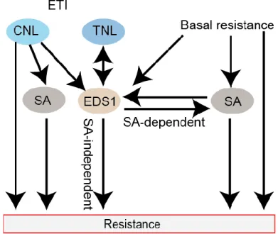
Arabidopsis EDS1 forms complexes with several TNLs: RPS4, RPS6 (Resistance to Pseudomonas syringae 6), SNC1 and VICTR (Variation in Compound Triggered Root growth response), inside nuclei, indicating that EDS1 may act as hub for TNL-triggered immunity and bridge TNLs with downstream resistance pathways (Bhattacharjee et al., 2011; Heidrich et al., 2011; Kim et al., 2012). Apart from its essential role in TNL signaling, EDS1 is also involved in certain CNL receptor-mediated defense responses. EDS1 functions genetically redundantly with SA, potentially compensating for SA defects in resistance mediated by three CNL receptors: RPS2, HRT (HR to TCV) and RPP8 (Recognition of Peronospora parasitica8), recognizing Pst DC3000 avrRpt2, TCV (Turnip Crinkle Virus) and Hpa Emco5, respectively (Venugopal et al., 2009) (Figure 1.2). This has been evidenced by the finding that eds1-22 sid2-1 double mutant showed susceptibility to Pst DC3000 avrRpt2, TCV and Hpa Emco5, whereas eds1-22 and sid2-1 single mutant were resistant (Venugopal et al., 2009). Therefore, EDS1 acts as a core signaling hub involved in basal, CNL- and TNL-triggered immunity (Figure 1.2).
Figure 1.2. Simplified illustration of EDS1 signaling in both basal, CNL- and TNL-triggered resistance.
EDS1 is required for basal resistance, in which SA is also important. EDS1 is essential for all TNL-mediated signaling, while functions redundantly with SA in certain CNLs-mediated resistance. Downstream of EDS1, both SA-dependent and SA-independent responses are activated. Arrows indicate positive regulation.
Ph.D. Thesis- Introduction
EDS1 interacts with unrelated effectors, avrRps4 and HopA1, and EDS1-avrRps4 association alters the complex of EDS1 with RPS4 and a repressor protein SRFR1 (SUPPRESSOR OF RPS4- RLD1) (Bhattacharjee et al., 2011; Heidrich et al., 2011). It was therefore proposed that EDS1 is a probable common virulence target for effectors to dampen the immune signaling. This is supported by the finding that bacterial pathogen effector AvrA1 directly targets soybean GmEDS1 for virulence (Wang et al., 2014a). However, it is ambiguous whether EDS1 is a common virulence target for pathogen effectors because of the finding that rrs1a/rrs1b mutant, defective in recognition Pst DC3000 avrRps4, only displayed partially increased and susceptibility to Pst DC3000 avrRps4 and Pst DC3000 compared to eds1-2 (Saucet et al., 2015). If EDS1 is a virulence target for avrRps4, rrs1a/rrs1b mutant should show similar susceptibility as eds1-2 to Pst DC3000 avrRps4 due to the suppressive target virulent function of EDS1 (Saucet et al., 2015). Thus, more studies are required to examine whether EDS1 is a common virulence target for pathogen effectors.
1.4.3 EDS1 functions with its sequence-related partners PAD4 and SAG101 in innate immunity
Through molecular and genetic analysis in Arabidopsis, EDS1 has been demonstrated to function together with its sequence-related partners, PAD4 (PHYTOALEXIN DEFICIENT4) and SAG101 (SENESCENCE-ASSOCIATED GENE101), in both basal and TNL-mediated resistance (Aarts et al., 1998; Feys et al., 2001; Feys et al., 2005; Rietz et al., 2011; Wagner et al., 2013). The EDS1- family proteins (EDS1, PAD4 and SAG101) contain a lipase-like domain at their N-termini and share a conserved uncharacterized domain referred to EP (EDS1-PAD4) domain (Feys et al., 2001;
Feys et al., 2005). EDS1 physically interacts with PAD4 in both nuclei and the cytoplasm, while the association of EDS1 with SAG101 occurs only in nuclei where SAG101 is restricted to (Feys et al., 2005). Together with the separate complexes among EDS1-family proteins, it was therefore proposed that dynamic interaction of EDS1 with its signaling partners in different cell compartments are important for plant immune signaling relay (Feys et al., 2005). Loss of interaction with EDS1 reduces post-transcriptional accumulation of PAD4 as well as SAG101 (Feys et al., 2005; Rietz et al., 2011). Random mutagenesis of EDS1 established that one variant EDS1L262P disrupts the interaction with PAD4 but still retains a physical association with SAG101 (Rietz et al., 2011). EDS1L262P reduced PAD4 accumulation and compromised basal resistance but did not affect TNL-triggered resistance in the Arabidopsis Ws-2 background (Rietz et al., 2011).
Ph.D. Thesis-Introduction
14
A recently resolved crystal structure of an EDS1-SAG101 heterodimer and a derived EDS1-PAD4 heterodimer model revealed the large interface of EDS1-SAG101 or EDS1-PAD4 proteins in a heterodimers and provided further evidence that EDS1-PAD4 and EDS1-SAG101 complexes are exclusive (Wagner et al., 2013) (Figure 1.3). This is contrary to the finding that EDS1-PAD4- SAG101 formed a ternary complex (Zhu et al., 2011a). Mutations in a hydrophobic helix of EDS1, EDS1LLIF, which is a necessary contact site between the two partner proteins, losses ability to interact with both PAD4 and SAG101 (Wagner et al., 2013). Corresponding contact site mutations in PAD4 and SAG101, PAD4MLF and SAG101LLIY, dramatically reduce association with EDS1 (Wagner et al., 2013). Genetically, EDS1LLIF transgenic plants failed to complement the defect of eds1-2 in resistance to Hpa Cala2 (Wagner et al., 2013). Moreover, the pad4-1 sag101-1 double mutant showed similar hypersusceptibility as eds1-2, a null EDS1 mutant, in RPP2-mediated resistance to Hpa Cala2 (Feys et al., 2005; Wagner et al., 2013). Therefore, these genetic data indicate that formation of EDS1-PAD4 and EDS1-SAG101 heterodimers are essential for EDS1 resistance signaling (Wagner et al., 2013).
Ph.D. Thesis- Introduction
Figure 1.3. Structural features of an EDS1-SAG101 heterodimer
Crystal structure of EDS1-SAG101 heterodimer is represented in cartoon form. EDS1 lipase-like domain (sandy brown)is juxtaposed with the lipase-like domain of SAG101 (dark cyan), while the EP domains of EDS1 (chocolate) and SAG101 (dark turquoise) interact with each other. Red box highlights the amino acids L258, L262, I254, F261A in a hydrophobic helix of EDS1 (in green), which are represented in mesh. The molecular arrangement of the heterodimer is represented by vertical bars. (Adapted from Wagner, et al., 2013)
The crystal structure analysis also indicated that the N-terminal domains contribute dominantly in forming heterodimers EDS1-PAD4 and EDS1-SAG101 (Wagner et al., 2013). Although EDS11-
384, consisting of the EDS1 lipase-like domain, is sufficient and necessary for heterodimer formation with SAG101 in N. benthamiana, and also stable and nucleocytoplasmic in Arabidopsis transgenic plants, it is insufficient to complement the defects of eds1-2 in resistance (Wagner et al., 2013). Therefore, the C-terminal EDS1 consisting of amino acid residues 385-623 is required for EDS1 to modulate defense responses (Wagner et al., 2013).
1.4.4 EDS1 modulates SA-JA crosstalk
Genetic studies in Arabidopsis show that EDS1 and its signaling partner gene PAD4 positively regulate SA accumulation and are essential for SA-mediated basal resistance (Zhou et al., 1998;
Feys et al., 2001). Reciprocally, EDS1 and PAD4 mRNAs are upregulated by exogenous SA application, consistent with EDS1/PAD4 and SA forming a positive feedback loop which amplifies and reinforces resistance against biotrophic pathogens (Zhou et al., 1998; Feys et al., 2001).
Previous observations that constitutive defense responses of snc1 and smg7 (suppressor with morphogenetic effects on genitalia7) are fully dependent on EDS1 and PAD4, but only partially dependent on ICS1 suggests that EDS1/PAD4 signaling node also induces SA-independent pathways to regulate part of the defense responses (Li et al., 2001; Zhang et al., 2003; Gloggnitzer et al., 2014). We recently showed that the autoimmune transgenic Arabidopsis lines with co- overexpressing EDS1 and PAD4 trigger both SA-dependent and SA-independent signaling and EDS1/PAD4 and SA signaling act in parallel in pathogen resistance (Cui et al., in Press).
Arabidopsis microarray analyses of wild-type and signaling-defective mutants, coi1, pad4, and sid2, in which PAD4 not only affected SA signaling pathway but also JA/ET signaling pathway (Glazebrook et al., 2003; Bartsch et al., 2006; Wang et al., 2008). Therefore, as a positive regulator
Ph.D. Thesis-Introduction
16
in SA signaling pathway, EDS1/PAD4 signaling node negatively controls JA/ET signaling pathway via an unknown mechanism.
1.5 Repression and de-repression in plant immunity
To maintain the balance between immune responses and growth fitness, plants employ various strategies to adjust the strength and duration of PTI responses (Couto and Zipfel, 2016). In Arabidopsis, to completely implement BIK1 (Botrytis-Induced Kinase1)-mediated responses upon PAMP perception, BIK1 needs to be released from repression from protein phosphatase type 2C PP2C38 (Couto et al., 2016). PP2C38 regulates phosphorylation stasis of BIK1 via direct interaction, resulting in inhibition of BIK1-mediated immunity (Couto et al., 2016). Upon the flg22 perception, the induced microRNA miR393 removes repression on SA signaling from auxin signaling, leading to certain activation of SA signaling (Navarro et al., 2006; Robert-Seilaniantz et al., 2011). Thus, upon PAMP perception, a process of de-repression is necessary to activate PTI.
ETI is quantitatively stronger than PTI, and it was proposed that this involves the removal of negative regulators upon NLR specific recognition of effector (Cui et al., 2015). In barley ETI, CNL MLA10 interacts with HvWRKY1 and removes its suppression of transcription factor HvMYB6, leading to activation of both basal and MLA-mediated resistance (Shen et al., 2007;
Chang et al., 2013). In rice, CNL Pb1 associates with and protects OsWRKY45, a positive regulator of SA signaling pathway, from ubiquitin-mediated degradation, which is critical for Pb1- mediated resistance (Inoue et al., 2013). In Arabidopsis, SNC1 interacts with TPR1, a transcriptional corepressor, probably leading to downregulation of negative regulators of immunity (Xu et al., 2014). These findings suggest a de-repression model in which activated NLRs remove the suppression of negative immune regulators to trigger robust immunity.
1.6 Thesis aims
Under the pressure of natural selection, plant pathogens have developed diverse strategies, including the secretion of virulence factors, to colonize hosts and circumvent innate immunity.
Reciprocally, plants have evolved to activate robust ETI against pathogen infection. As an early NLR convergence point and immune signaling hub, EDS1 functions as a crucial bridge between activated NLRs and nuclear transcriptional reprogramming (Garcia et al., 2010; Bhattacharjee et al., 2011; Heidrich et al., 2011). However, the molecular mechanism by which EDS1 relays signaling information from an activated NLR to the transcriptional machinery is not yet known.
Ph.D. Thesis- Introduction
Crosstalk between phytohormone pathways is essential for plants to keep a balance between immunity, responsiveness to competing stresses, and growth (Pieterse et al., 2012; Lozano-Duran and Zipfel, 2015). EDS1 with PAD4 promotes SA accumulation to activate SA-dependent resistance but also negatively regulates JA signaling-related responses independently of SA (Falk et al., 1999; Feys et al., 2001; Glazebrook et al., 2003; Brodersen et al., 2006). The question how EDS1 modulates SA-JA crosstalk in immune response remains to be addressed. Also, although EDS1/PAD4 are central immunity components, our knowledge is very limited about how they are themselves are regulated and whether EDS1/PAD4 counteracts the action of negative components to fine-tune immune responses, consistent with a de-repression signaling model.
My aim in this Ph.D. thesis was to test the hypothesis that EDS1 resistance-promoting activity is negatively regulated by repressors which can themselves be overcome by activated EDS1 signaling in TNL-mediated ETI. For this, I started my Ph.D. project with a genetic screen to identify mutants that have recovered EDS1-dependent TNL (RRS1/RPS4) resistance to Pst DC3000 avrRps4 in a hypersusceptible Col-0 eds1-2 mutant background. Specifically, the aims of my Ph.D. thesis were to 1) identify negative regulators that are potentially antagonized by EDS1 resistance signaling, 2) explore how EDS1 activates downstream transcriptional reprogramming, 3) investigate the role of EDS1 in modulating SA-JA crosstalk in stress signaling.
Ph.D. Thesis- Results
18
2 Results
2.1 Forward genetic screening for suppressors of eds1 (sed mutants)
2.1.1 Isolation of Arabidopsis sed mutants in an EMS mutagenized M2 population
Arabidopsis Col-0 recognizes the effector avrRps4 via the TNL RPS4/RRS1 receptors pair and is therefore resistant to Pst DC3000 avrRps4 (Hinsch and Staskawicz, 1996; Narusaka et al., 2009;
Le Roux et al., 2015; Sarris et al., 2015). The Col-0 null (fast-neutron-generated deletion) eds1-2 mutant is hypersusceptible to Pst DC3000 avrRps4 due to defective TNL signaling (Figure 2.1A).
To identify suppressors of eds1-2 (sed), ethyl methanesulfonate (EMS) mutagenesis was initiated by Dr. Haitao Cui with the expectation that sed mutants display restored resistance of eds1-2 to Pst DC3000 avrRps4. The loss-of-function mutations in negative regulators or gain-of-function mutations in positive regulators of different resistance signaling pathways may lead to restored resistance phenotype in eds1-2 (Figure 2.2B). These regulators may be involved in basal resistance or CNL-mediated resistance, which are EDS1-independent. These regulators may also regulate defense responses in EDS1-dependent manners, such as EDS1-triggered SA-dependent and SA- independent resistance signaling.
Ph.D. Thesis- Results
Figure 2.1. Expected signaling defects in the recovery of eds1-2 resistance to Pst DC3000 avrRps4
A. Col-0 eds1-2 is defective in TNL (RRS1/RPS4) resistance to Pst DC3000 avrRps4. Col-0 eds1-2 mutant seedlings, in contrast to the wild type Col-0, do not survive Pst DC3000 avrRps4 infection. Photos were taken 7 d after spray-inoculation with bacteria (108 CFU/mL) of two-week old seedlings.
B. Four possible disrupted signaling pathways may lead to the recovery of eds1-2 resistance to Pst DC3000 avrRps4. The eds1-2 mutant is defective in basal, TNL- and certain CNL-mediated resistance. The loss-of- function mutations in negative regulators or gain-of-function mutations in positive regulators of resistance signaling may restore eds1-2 resistance. These regulators may be involved in EDS1-independent signaling pathway in basal resistance (1) or CNL-mediated resistance (4). It is also possible that these regulators are in EDS1-dependent resistance signaling, such as EDS1-triggered SA-dependent (2) and SA-independent signaling (3). Arrows indicate positive regulation.
Approximately 195000 two-week-old M2 mutant plants from a total of 65-independent M1 pools were spray-inoculated with Pst DC3000 avrRps4 and 75 resistant plants were selected (Figure 2.2).
From the 75 putative M2 plants, 15 plants from 2 independent pools were found to be male-sterile, while 14 plants from two other pools were partially sterile (Details in section 2.2). At the M3
generation, eight of 46 fertile and resistant lines were confirmed to rescue the eds1-2 hypersusceptibility after spray-inoculation of two-week-old seedlings with Pst DC3000 avrRps4.
To rule out the possibility of pollen or seed contamination, the confirmed 8 resistant lines were genotyped with specific EDS1 primers which detect the deletion of parental eds1-2. Three EMS- mutagenized eds1-2 mutants, which originate from different M1 pools and had restored resistance to Pst DC3000 avrRps4, are further referred to here as sed1, sed2 and sed3.
Ph.D. Thesis-Results
20
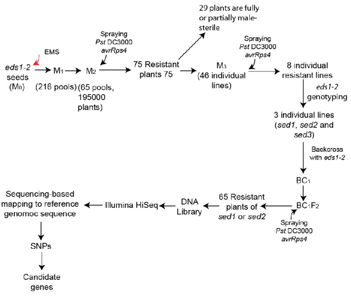
Figure 2.2. Genetic screen for suppressors of eds1-2 hypersusceptibility identifies 3 sed mutants.
Ethyl methanesulfonate (EMS) treated eds1-2 seeds (M0) were sown, each of 216 pools of M2 seeds contained seeds from ~150 M1 plants. Two-week-old M2 seedlings (~195,000) from 65 pools were sprayed with Pst DC3000 avrRps4 at 108 CFU/mL. At 7 d post infection, 75 resistant plants were selected, among which 29 were fully or partially male- sterile. Seeds from the remaining 46 plants were harvested as individual M3 lines. In the M3 generation, three of 46 individual lines were confirmed to be resistant to Pst DC3000 avrRps4 and to contain the parental eds1-2 mutation.
These three lines were referred as sed1, sed2 and sed3. The sed1 and sed5 were backcrossed with Col-0 eds1-2 to generate BC1 seeds. A DNA library was prepared by pooling genomic DNA from 65 BC1F2 resistant plants for each sed mutant. DNA libraries were sequenced on an Illumina Hiseq machine. The number of potentially causative SNPs was reduced by removing SNPs shared by all sed mutants and found in the Arabidopsis line pEDS1:EDS1-YFPNLS
#A3/ eds1-2, which has the same Col-0 eds1-2 genetic background as the sed mutants. After this, only SNPs with G to A nucleotide transition and with an effect on the protein sequence were considered as putative causative SNPs responsible for suppression of eds1-2 hypersusceptibility.