R1-homologous genes
Inaugural-Dissertation zur Erlangung des Doktorgrades der Mathematisch-Naturwissen- schaftlichen Fakultät der Univer-
sität Köln
vorgelegt von Gábor Miklos Gyetvai
aus Gronau
Köln 2010
Die vorliegende Arbeit wurde am Max-Planck-Institut für Pflanzenzüchtungsforschung in Köln angefertigt. Die Arbeiten wurden in der Arbeitsgruppe von Dr. PD Christiane Gebhardt innerhalb der Abteilung Pflanzenzüchtung und Genetik von Prof. Maarten Koornneef durchgeführt.
Teile der Arbeit enstanden in Kooperation mit der Universität Aalborg und dem Max- Planck-Institut für Molekulare Pflanzenphysiologie.
Berichterstatter: Dr. PD Christane Gebhardt
Prof. Dr. Martin Hülskamp
Tag der mündlichen Prüfung: _09.07.2010_
Index of tables and figures 7
Abbreviations and Acronyms 11
Introduction 14
The pathosystem „Solanum tuberosum/Phytophthora infestans” in an agronomical context14 Molecular interaction between Phytophthora infestans and Solanum tuberosum 18 Genetic basis of resistance of Solanum tuberosum against late blight: Qualitative and quanti-
tative resistance 18
The RXLR-Effectors of Phytophthora infestans 20
Types of resistance mediating genes 21
R1 is a member of the CC-NBS-LRR gene family 23
R1 and the resistance hotspot on Chromosome V 23
Organization of the contigs R1 and r1 24
Solanum nigrum and its role as source for resistance 26
Methods for expression analysis 29
Single target driven methods 29
Methods for transcriptome analysis 30
Massive parallel sequencing 31
Small introduction to the relevant statistical tests for this study 32
Motivation for this study 34
Material and Methods 36
Standard conditions for plant and oomycete cultivation 36
Chemicals 36
Infection assays and plant cultivation 37
Confocal microscopy 37
Molecular biological methods 38
Polymerase chain reaction (PCR) 38
Nucleic acid extraction 41
First strand cDNA synthesis 42
Quantitative polymerase chain reaction (qRT-PCR) 42
Transient expression of effector proteins in Solanum nigrum 43
DeepSAGE 45
Biological material and generation of infected tissue 45
Creation of 3´-tag-libraries 47
Bioinformatical methods for the analysis of transcriptome data 47
Creation of the Standard data set 47
General procedures and sample composition for statistics 48
G-test 50
Fisher´s exact test (sage.test) 50
edgeR 51
BaySeq 51
Permu 51
Calculation of the Df and logfoldchange values 52
Cluster analysis 53
Go-term analysis 54
Summary of used Softwares 54
Results 56
Expression analysis of R1-homologous genes of Solanum nigrum 58 Transient expression of effectors from P. infestans in Solanum nigrum 59 Comparative transcriptome analysis of Solanum tuberosum plants during the early phase of
Phytophthora infestans infection 62
Validation of infected Material 62
Technical results 63
Expression levels of indicator-genes 69
Validation of significance statistics 72
Results of global statistical analysis: Cluster and Principal component analysis 81 Results of significance of observation after Fisher´s Exact Test 87 Comparison of DeepSAGE-data with semi-quantitative RT-PCR 98
Discussion 103
Evidence for the expression of predicted R1-homologous genes in Solanum tuberosum 103 Evidence for the expression of genomic predicted ORFs in Solanum nigrum 104 Hypersensitive reaction of Solanum nigrum in response to transient expression of effectors of
Phytophthora infestans 104
Comparative DeepSAGE-analysis of R1- and ORF45-transgenic Solanum tuberosum plants
with wildtype 106
DeepSAGE as method for transcriptome analysis without a reference genome – performance
features and uncertainty factors 106
Evaluation of statistical methods – the normal distributed world 108 Validation and characterization of the early stages of infection as phenotype 110 Using Cluster analysis as tool to find coregulated units 111
Identification of candidate genes for a function in the compatible and incompatible interaction
with Phytophthora infestans 115
Comparability of DeepSAGE-data with qRT-PCR 118
Concluding remarks - the extraordinary role of the plant hormone synthesis pathway (KEGG
ec01070) 119
References 122
Summary (english) 134
Zusammenfassung (deutsch) 135
Protocol for the creation of Ditag-libraries 137
Instructions for the supplementary data 145
Locations of raw data files: 148
Eidesstattliche Erklärung 150
Danksagungen 151
Lebenslauf 154
Index of tables and figures
Figures:
Fig. 1.1.1: Wold map of potato cultivation areas
12 Fig. 1.1.2: The painting:“The King everywhere“
13 Fig. 1.1.3: Alignment of genomic areas of three Phytophthora species 13 Fig. 1.1.4: Scheme of different life stages of Phytophthora infestans 14 Fig. 1.1.5: The life cycle of P. infestans
14 Fig. 1.1.6: Infecting zoospore cysts of Phytophthora cinnamonii
15 Fig. 1.1.7: Symptoms of late blight infections
15 Fig. 1.2.1.1: Race and R-gene nomenclature of Solanum tuberosum
and Phytophthora infestans
16
Fig. 1.2.1.2: Scheme of qualitative and quantitative resistance 17 Fig. 1.2.2.1: Scheme of the interaction zone between pathogen and plants 18 Fig. 1.2.3.1: Illustration of the known classes of R-genes
19 Fig. 1.3.1.1: Picture of the R1-specific phenotype
21 Fig. 1.3.2.1: Map of the contigs R1 and r1
22 Fig. 1.3.2.2: Phenetic tree of the R1-family
23 Fig. 1.3.2.3: Function map of Solanum tuberosum chromosome V 23 Fig. 1.4.1: Taxonomic view of some members of the Solanaceae family 24
Fig. 1.4.2: Picture of Solanum nigrum
24
Fig. 1.4.3: Illustration of a syntheny region between Solanum tuberosum
and Solanum nigrum
25
Fig. 1.4.4: Identification of cosmid clones with R1-homologous sequences
in Solanum nigrum
26
Fig. 1.4.5: Southern blot with a R1-specific probe and several cultivars 26
Fig. 1.5.1.1: Scheme of the bDNA-assay
27
Fig. 1.5.2.1: Scheme of the generation of SAGE-libraries
29
Fig. 1.8.1: Model of the mechanism of R1
32
Fig. 2.6.1.1: Overview of infections for DeepSAGE
44
Fig. 2.6.3.1.1: Density plot of mean expression levels
45
Fig. 2.6.3.8.1: Explaining example of the theory behind the D
f-values 50
Fig. 2.6.3.9.1: Example of the centroid expression view of cluster analysis 51
Fig. 3.1.1: Results of the BAC-assay used for the development of primers for
the R1-family
54
Fig. 3.1.2: Illustration of the used BACs and the position of the R1-family
members
54
Fig. 3.1.3: Verification if transcripts of members of the R1-family 55 Fig. 3.2.1: Verification of expression of R1-homologous genes in Solanum
nigrum
56
Fig. 3.3.1: Summary of results after expression of put. effectors from
Phytophthora infestans in Solanum nigrum
57
Fig. 3.3.2: Alignment of positive effectors
57 Fig. 3.3.3: Phenetic tree of all tested put. effectors
59 Fig. 3.4.1.1: Microscopy picture of an Phytophthora infestans infected
leaf three days after infection
60
Fig. 3.4.2.1: Plot of the number of sequenced tags and the detected unitags
per sample after „Next-Generation-Sequencing“
65 Fig. 3.4.2.2: Pie chart of the proportion of possible target sites 66 Fig. 3.4.2.3: Histogram of the quantity of expression levels
67 Fig. 3.4.3.1: Heat map of tags with a go-annotation „defense-response“ 68 Fig. 3.4.3.2: Histogram of the expression levels of ef-1α
69 Fig. 3.4.4.1: Histogram of the FDR-values of the G-test
70 Fig. 3.4.4.2: Plot of the FDR-values and the logfoldchanges after
G-test
71
Fig. 3.4.4.3: Plot of the FDR-values and the logfoldchanges after
Fisher´s exact test (sage.test)
71
Fig. 3.4.4.4: Plot of the FDR-values and the logfoldchanges after
Student´s t-test
72
Fig. 3.4.4.5: Plot of the FDR-values and the logfoldchanges after
edgeR
73
Fig. 3.4.4.6: Plot of the FDR-values and the logfoldchanges after
Permu
74
Fig. 3.4.4.7: Plot of the logP-values and the logfoldchanges after
BaySeq
75
Fig. 3.4.4.8: Box-plots of the average logfoldchange values of the significant
tags of each test
76 Fig. 3.4.4.9: Histogram of the average D
f-values of the significant tags
of each test
77
Fig. 3.4.4.10: Venn diagram of the top100 tags by each test
78 Fig. 3.4.5.1: Overview of extracted cluster after k-mean cluster analysis 80 Fig. 3.4.5.2: Apertures of of a heat map after hierarchical cluster analysis 82 Fig. 3.4.5.3: Plot of PC1 and PC2 of a principal component analysis 84 Fig. 3.4.6.1: Overrepresented GO-terms in R1 lines 3dpi
91 Fig. 3.4.6.2: Overrepresented GO-terms in wild type lines 1dpi 92 Fig. 3.4.6.3: Overrepresented GO-terms in wild type lines 3 dpi 93 Fig. 3.4.6.4: Overrepresented GO-terms in the ORF45 line 1dpi 94 Fig. 3.4.6.5: Overrepresented GO-terms in the ORF45 line 3dpi 95 Fig. 3.4.7.1: Comparing matrix of DeepSAGE and qRT-PCR
99 Fig. 3.4.7.4: Boxplot of overlapping and not overlapping values 100 Fig. 4.4.3.1: Expression levels of Prb-1b in wildtype
109 Fig. 4.4.4.1: The putative reaction of TC16055
111
Fig. 4.4.5.1: The reaction of ToTAL2
114
Fig. 4.4.5.2: Expression levels of ToTAL2 (StET009648)
114
Fig. 4.4.7.1: Overview of the plant hormone synthesis pathway
119
Tables:
Tab. 1.6.2: Performance features of „Next Generation Sequencing“
platforms
29
Tab. 2.5.1.1: List of used oligonucleotides
37 Tab. 2.6.3.2.1: Definition of replica groups and independent experiments 47 Tab. 2.6.3.11.1: Overview of used softwares
53 Tab. 3.3.1: Similarity scores between positive scored put. effectors and
avrblb2
58
Tab. 3.4.2.1: Overview of the results of „Next Generation Sequencing“ 62 Tab. 3.4.4.1: Summary of the main characteristics of the statistical tests 74 Tab. 3.4.4.2: Correlation matrix of the Df-values if the significant tags by
each test
76
Tab. 3.4.5.1: Summary of relevant clusters after k-mean cluster analysis 79
Tab. 3.4.5.2: Summary of identified tags after hierarchical clustering 83
Tab. 3.4.6.1 Major significant tags of R1 lines
85
Tab. 3.4.6.2 Major significant tags of wild type lines
86
Tab 3.4.6.2: Major significant tags of the ORF45 line
89
Tab. 3.4.7.1: Summary of tags used for qRT-PCR experiments
96
Tab. 3.4.7.2: Expression levels of qRT-PCR and according DeepSAGE data 97
Abbreviations and Acronyms
Abbreviation Full expression
°C degree Celsius
A. lfc. Average logfoldchange
ATP adenosine 5-triphosphate
Avr avirulence
BAC Bacterial artificial chromosome
bp base pair
C.I. Compatible interaction
cDNA complementary DNA
chr. chromosome
Ct cycle threshold
cv. cultivar
D
fDifferentiation factor
DFCI Dana-Farber Cancer Institute
DNA deoxyribonucleic acid
DNase deoxyribonuclease
dNTP deoxynucleosidetriphosphate
dpi days post infection
FDR False discovery rate; also: false discovery rate
corrected p-value
FE Fisher´s exact test
FE Fisher´s exact test
fig. figure
gDNA genomic DNA
GFP green fluorescence protein
Abbreviation Full expression
GO Gene ontology
HR hypersensitive response
In. I. Incompatible interaction
JVCI John Craig Venter Institute
kb kilobase pairs
kDA kilodalton
KEGG Kyoto Encyclopedia of Genes and Genomes
LB lysogeny broth
log logarithm
LRR leucine rich repeat
m mili
m mili
M Molar
ma.lfc maximal logfoldchange
max maximum
MES 2-(N-morpholino)ethanesulfonic acid
MgCl
2Magnesium chloride
mi.lfc minimal logfoldchange
min minimum
NA No annotation
NBS nucleotide binding site
NCBI National Center for Biotechnology Information
OD optical density
ORF open reading frame
P.
PhytophthoraPAMP pathogen-associated molecular pattern
Abbreviation Full expression
PC principal component
PCA principal component analysis
PCR polymerase chain reaction
PR pathogenesis related
PVX Potato virus X
qRT-PCR quantitative reverse transcriptase polymerase
chain reaction
QTL quantitative trait locus
rel. expr. relative expression
RT-PCR reverse transcriptase polymerase chain reaction
S.
SolanumSAGE Serial Analysis of Gene Expression
SCRI Scottish Crop Research Institute
ssp. subspecies
T
AAnnealing temperature
TIGR The Institute for Genomic Research
TIR Drosophila Toll and human unterleukin-1 re-
ceptor
vs versus
wt wild type
YEP Yeast extract peptone
Δ
delta
µ
micro
1.Introduction
1.1.The pathosystem „Solanum tuberosum/Phytophthora infestans”
in an agronomical context
Solanum tuberosum is the most cultivated member of the Solanaceae family. With a total yearly production of approximately 323 million tons (source: FAOSTAT). It is after maize, wheat and rice the fourth most important crop plant worldwide. The estimated size of the genome is 840 Mbp which are distributed over 12 linkage groups. The natural occurring
ploidy level is tetraploid, leading to a total chromosome number of 48 (source: SGN [1]).
Differing from most polyploid crop species, the segregation of these four chromosome sets occurs independently. The major producing areas are in Asia and Europe (fig. 1.1.1).
Originating from the Andes of South America, where it has been cultivated since at least 7000 B.C., the potato has been introduced into Europe in the course of the discovery of America in the sixteenth century. Originally it has been spread as rare ornamental plant [2]. As cultivated plant for nutrition it was initially used at the end of the seventeenth
Fig. 1.1.1: World map of the areas of potato cultivation worldwide (source:
International potato Center).
century in Ireland. An additional step forward on its way as agronomical relevant crop plant in Middle Europe was the introduction in Prussia by law in 1756 through King Frederik II (illustrated in fig.
1.1.2). The basic prerequisite was the adaption of the agronomically used potatoes to the Middle European long day conditions. The originally introduced cultivars (mainly from the subspecies andigena) were adapted to the South American short day conditions [2, 3].
The broad introduction as a major source of
nutrition lead to the establishment of large monocultures. These gave the breeding ground for pathogens and associated diseases. One of the most destructive pathogens of Solanum tuberosum and related species is the late blight causing oomycete Phytophthora infestans [3].
At the moment, the taxon Phytophthora contains 110 classified species and additional 232, which are not classified yet (source: NCBI [4]). Among the sequenced genomes of this Phyllum, Phytophthora infestans has, with 240 Mbp, the largest known genome with large expanding areas compared to the related species Phytophthora ramorum and
Phytophthora sojae (fig. 1.1.3). These large intergenic spaces could provide a genetical explanation for the high potential of this species to adapt to changing environments [5].
The life cycle of Phytophthora infestans spans three major stages (fig 1.1.4 1.1.5). The oomycete is able to persist in form of sporangia in old infected tissue parts like tubers from
Fig. 1.1.2: Painting with the title:„The King everywhere" by Robert Warthmüller (1886). It presents King Frederik of Prussia inspecting the Potato cultivation. The original is located in the German historic museum in Berlin.
Fig. 1.1.3:
Schematic alignment of genomic areas from the three
Phytophthora species Phytophthora ramorum (top), sojae (middle) and infestans (bottom) [5].the previous year or in form of thick-walled oospores in the soil [6]. Mainly the oospores are responsible for the infections of the next year [7, 8]. However, during an ongoing epidemic, the Sporangia are the more active part. The major reason for this is their ability to release mobile zoospores. The released zoospores by themselves are the major infective and virulent element. The biflagellar zoospores, start after recognition of host tissue, to build hyphaea so called zoospore cysts [9]. One of the first described stimuli is a perception of a Ca
2+-concentration gradient [10, 11]. From this cyst an initial hyphae is released and an appressorium is formed u n d e r c o n t a c t w i t h t h e h o s t . T h i s appressorium is the first interaction zone between the pathogen and its host from which the infection of the plant tissue starts. Inside the leaf tissue a mycelium with
haustoria is formed. After 5-8 days the mycelium grows out of the plant again and forms new sporangia, which then infect new parts of the plant, new plants or remain in the soil. The reproduction of this organism is sexual and asexual [12]. Comparable to Saccharomyces cereviseae , this pathogen has two mating types – the A
1and A
2type. If both types meet, oogonia are formed and inside recombination is performed. From these oogonia new oospores are produced. One of the first and most prominent late blight epidemic has
caused the Irish famine in the 1840s [13]. After this, smaller epidemics occurred in Belgium
Fig. 1.1.4: Scheme of the different life stagesof Phytophthora infestans [147].
Fig. 1.1.5: Schematic picture of the life cycle of P.
infestans [146].
and the Netherlands, leading to an increased focus on this problem [3]. Resistance breeding and agronomic measures reduced this problem
in Europe to a minimum until the 1980s. At this time, fields under late blight infection has been observed again.
A hypothesis with the aim to explain this phenomenon postulates that to this date, only the A
1mating type was introduced to Europe and the resistance, mediated by genes introgressed from the wild relative Solanum demissum, was durable. With new trading imports from South America, the A
2mating type was introduced,
leading to an increased genomic variability and a fast breaking of the existing resistance genes as well as a tolerance to fungicides like metalaxyl [14].
With its ability to infect large areas in a short time under favorable conditions (high humidity and temperatures between 15 and 20 °C [3]) and its high adaptiveness to environmental conditions, this pathogen is of high agronomic interest. Current estimates calculate worldwide an economical loss of at least 5 billion US$. These include the costs for the chemical control and the direct losses in yield [15].
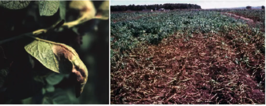
Fig. 1.1.6:
Infecting zoospore cysts from the related species
Phy- tophthora cinnamonii [9].Fig. 1.1.7: Pictures of a leaf, showing symptoms of a late blight infection
(left) and of a field under disease (right) (Source: SCRI).
1.2.Molecular interaction between Phytophthora infestans and Solanum tuberosum
1.2.1.Genetic basis of resistance of
Solanum tuberosum against late blight:Qualitative and quantitative resistance
The classic view of the gene-for-gene model assumes major single locus resistance genes interacting with corresponding avirulence genes [16, 17]. Eleven avirulence (avr) genes in Phytophthora infestans and accordingly eleven resistance (R) genes in Solanum tuberosum are described (fig. 1.2.1.1). Five of these genes, R1, R2, R3, R6 and R7, have been located on the genetic map [18, 19]. Following this categorization system, the race- nomenclature of Phytophthora infestans strains is defined. This nomenclature follows the
virulence (or the ability to overcome the resistance gene) of the oomycete and consequently introduces a virulence (vir) gene nomenclature. The qualitative resistance genes are able to sense the corresponding avirulence gene. This perception of the
Race1
(vir1) Race2
(vir2) Race3
(vir3) Race4
(vir4) Race5
(vir5) Race6
(vir6) Race7
(vir7) Race8
(vir8) Race9
(vir9) Race10
(vir10) Race11 (vir11) R1
C. I. In. I. In. I. In. I. In. I. In. I. In. I. In. I. In. I. In. I. In. I.
R2
In. I. C. I. In. I. In. I. In. I. In. I. In. I. In. I. In. I. In. I. In. I.
R3
In. I. In. I. C. I. In. I. In. I. In. I. In. I. In. I. In. I. In. I. In. I.
R4
In. I. In. I. In. I. C. I. In. I. In. I. In. I. In. I. In. I. In. I. In. I.
R5
In. I. In. I. In. I. In. I. C. I. In. I. In. I. In. I. In. I. In. I. In. I.
R6
In. I. In. I. In. I. In. I. In. I. C. I. In. I. In. I. In. I. In. I. In. I.
R7
In. I. In. I. In. I. In. I. In. I. In. I. C. I. In. I. In. I. In. I. In. I.
R8
In. I. In. I. In. I. In. I. In. I. In. I. In. I. C. I. In. I. In. I. In. I.
R9
In. I. In. I. In. I. In. I. In. I. In. I. In. I. In. I. C. I. In. I. In. I.
R10
In. I. In. I. In. I. In. I. In. I. In. I. In. I. In. I. In. I. C. I. In. I.
R11
In. I. In. I. In. I. In. I. In. I. In. I. In. I. In. I. In. I. In. I. C. I.
Fig. 1.2.1.1: Schematic matrix of the classical potato resistance model and the race
specification of
Phytophthora infestans. R1-R11 stand for the different resistance genes in-trogressed from
Solanum demissum. Race1-11 specifies the nomenclature of Phytophthorastrains, classified after the possible interaction with an
R-gene. C.I means compatibleinteraction (leading to an infection of the host). In. I means incompatible interaction
(leading to an hypersensitive resistance response of the host).
pathogen induces a resistance response in form of a hypersensitive cell death reaction.
This kind of defense response can be seen in analogy to the inflammation in animal systems [20]. Formal pathology calls this type of host/pathogen interaction as
“incompatible interaction“ between the host and the pathogen. The opposite a successful infection of the host, what is termed as “compatible interaction“.
The genetic model of resistance is based on the resistance genes, which have been introgressed before the A
2mating type was introduced to Europe and the complexity of pathogen strains potentiated. The contemporary active populations in Middle Europe are classified after this scheme and usually exist as complex or semi-complex races, containing a mix of the known virulences or even all. At the moment the virulence identification is carried out in an infection assay using a standard set of potato cultivars.
Current resistance breeding aims to introgress new resistance genes from wild species other than Solanum demissum, such as the Rpi-blb genes [21] from Solanum bulbocastanum. This will make a new classification system necessary in the future.
Due to a non-mendelian segregation of R10 and the detection that the R3 locus consists out of the two closely linked genes R3a and R3b, the molecular basis of the R-genes stays in discussion [22, 23].
In addition to the qualitative resistance, also the quantitative or field resistance is known [24, 25]. The underlying physiology is rather a strengthening of the plant. This phenotype is additionally known as horizontal resistance. The genetic basis can be genes like StAOS2 [26], having a role in jasmonate synthesis. Jasmonic acid is involved in unspecific stress signalling [19, 26-28].
Fig. 1.2.1.2:
Comparative scheme of the relationship between quantitative and
qualitative resistance within the phenotype resistance as whole [146].
The distribution of these phenotypes follows the rules of quantitative traits. This differs from R-genes, which follow the distribution of qualitative traits. In plants, both traits are acting together. On one side the horizontal resistance which mediates an unspecific protection against a broad range of pathogens and on the other side the specific resistance genes, which recognize specific stimuli (fig. 1.2.1.2).
1.2.2.The RXLR-Effectors of Phytophthora infestans
Any phase of the infection requires the establishment of an interaction zone between the host and the pathogen. For this, a special
cellular structure, named as „haustorium“ is established (fig. 1.2.2.1). Outgoing from this the further interconnection is directed. During the initial stage, Phytophthora secretes various proteins and molecules, which prepare for the further colonization. This progress, known as cellular reprogramming, is essential to protect the pathogen against unspecific defense responses from the host.
To this belongs the secretion of antimicrobial proteins, like chitinases, bactinecin, serine- and cysteine protease inhibitors and other direct attacking molecules [29, 30]. These
secreted proteins, are currently, together with traditional elicitors, summarized as effectors.
These effectors are released in the apoplastic space as well as in the cytoplasm [5, 29].
Recently, two classes of effector protein could be classified. Based on their characteristic amino acid motif they are called RXLR (harboring the sequence RxLR) and crinkler (CRN motif). These motifs seem to be highly conserved among secreted proteins and play a role for the introduction into the host-cell [15, 30-33]. It is assumed that avirulence and virulence genes are present among this genes, playing the major part of the decision between incompatible and compatible interaction [34, 35].
Fig. 1.2.2.1: General scheme of the
interaction compartment between of
pathogens and the plant [148].
1.2.3.Types of resistance mediating genes
Currently, 5 different classes of gene products are known in plants, which mediate resistance against pathogens.
These classes are built from five different domains. The major c l a s s , t h e N B S - L R R c l a s s , combines a nucleotide binding site (NBS-domain) and leucine rich repeat (LRR domain). Additionally, the N-terminal region contains a coiled-coil domain (CC) or a Toll- interleukin receptor like (TIR) domain. The TIR- and CC-NBS- LRR type genes are summarized as NB-ARC type genes, which
share structural similarities with the NACHT-LRR or CATERPILLER family in animal
systems [20, 36, 37]. This class of genes is widely spread and early estimations
prognosed more than 200 genes of this type in Arabidopsis thaliana [38]. In addition, a
kinase domain can be found among resistance genes [34, 39]. The simplest resistance
gene, detected so far, is the pto gene from Solanum lycopersicum, which only contains a
kinase domain and is classified as serine/threonine kinase. This gene is not able to
mediate resistance on its own and needs prf as a second gene, which belongs to the NBS-
LRR type class [40-42]. One of the major differences within the resistance genes affects
the intracellular localization so far known. On the one hand genes exist, which are
associated to the cellular membrane. To this group belong the cf-genes or fls2. In the case
of fls2, it has been shown, that the gene is responsible for the extracellular recognition of a
certain kind of Flagellin (flg22) and belongs to the class of PAMP-receptors [43]. On the
other hand, the NBS-LRR genes have not been found to be anchored in the membrane
Although an indirect association might be possible [39]. Despite of this fact, it should be
remembered, that a clear differentiation of PAMP-triggered immunity and R-gene triggered
immunity should not be made as both parts belong to the higher order defense system and
Fig. 1.2.3.1: Schematic illustration of the five knownclasses of resistance genes and their localization in relation
to the cellular membrane. Additionally known examples of
plant resistance genes are given below [39].
probably both ways stand in a clear cross-talk [44]. As outcome of both mechanisms, the
hypersensitive response remains the same [45]. The precise molecular mechanism of the
avirulence factor recognition through the NBS-LRR type genes is still unknown. Up to date
it is not clear wether it is a direct molecular interaction or a sensing mechanism via an
indirect „guarding“ molecule as it is proposed by the guard hypothesis [46].
1.3.R1 is a member of the CC-NBS-LRR gene family
1.3.1.R1 and the resistance hotspot on Chromosome V
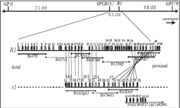
Resistance genes are often organized in clusters. In Solanum tuberosum, several of these hot spots for resistance are known. These often co-localize with QTL regions for resistance as well. One of the most prominent hot spots for resistance is localized on chromosome V between the anchoring markers GP21 and GP179 [47] (for a comparison see Fig. 1.3.2.3).
In addition to the known late blight resistance gene R1, this region harbors several genes with function in resistance to nematodes and viruses as well as QTL-regions being involved in resistance to late blight and nematodes. Additionally, a part of a QTL-region for resistance to bacterial diseases [48, 49] is located in this region. Beside many additional relevant regions, other remarkably hot spots for resistance are located on chromosome III tagged by the markers TG134 and Pt2 [50-52], on chromosome IV tagged by TG62 and GP180-a [48, 49], on chromosome V close to GP78 and GP22 [48, 49, 53-56] and on chromosome XI near STM2005 and STM0025 [50,
57].
R1 by itself has been shown to mediate qualitative resistance [54] and the predicted gene product encodes a protein of 1313 amino acids. Based on the Prosite database, it contains a leucine zipper domain and a nucleotide binding site. Additionally four glycosylation sites, a cAMP-phosphorylations site, various CKcasein kinase II- and phoshokinase c- phosphorylation sites, as well as 3 sites for myristilation and one amidation site can be found (Source: Prosite database [58]). Summing up the predicted motif information, this gene is formally classified as CC-NBS-LLR class gene and belong to the group of classical single locus resistance genes.
Fig. 1.3.1.1: Picture of the R1-
specific phenotype. Seen is a leaf of
the potato cultivar Desirée (A) and
from a
R1-transgenic Desirée plant9 days after infection with a P. infes-
tans race 4 isolate[54].
1.3.2.Organization of the contigs R1 and r1
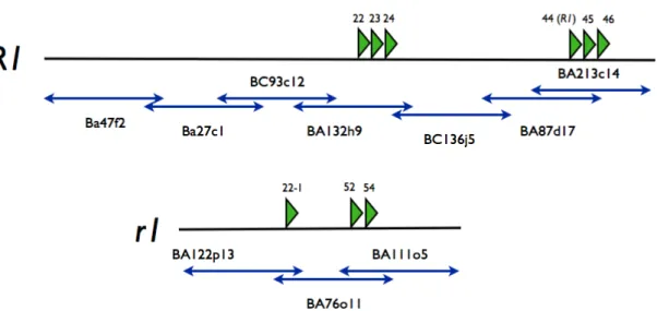
Sequencing and annotation of 10 BAC clones located in the region of the resistance hot spot on chromosome V of Solanum tuberosum was able to unravel various open reading frames (ORFs) (see Fig. 1.3.2.1). Among these were eight ORFs, whose predicted products share a high degree of sequence homology to the known resistance gene R1.
These open reading frames are located on the two contigs R1 and r1, which are likely of different origin. Differing to the r1-contig, the R1-contig originated from an introgressed region from Solanum demissum. The six open reading frames 22, 23, 24, 44 (R1), 45, and 46 are located on the introgressed region. In the r1-contig, with number 22-1, 52 and 54, three additional ORFs have been detected. Together, these nine open reading frames form the R1-family [55].
The relationship between the members of the R1 gene family seems to form two homology groups with three members each and a third group with distantly related members. The
Fig. 1.3.2.1: Map of the sequenced contig of Solanum tuberosum cv. P6/210. Two
differing orthologues regions named as R1 and
r1 were identified. The R1 contigharbors the R1 gene. On both contigs, in total 8 open reading frames have been
identified, whose putative products share a high similarity with R1 [55].
first group contains the R1 gene, ORF54 and ORF 22-1. Remarkably, both open reading frames being most similar to R1, are located on the r1-contig. A different observation is made in the second group containing ORF24, ORF45 and ORF23. All of these open reading frames are localized on the R1-contig. The greatest similarity
occurs between ORF24 and ORF45. The genomic sequence of these loci share a homology of almost 100% several kilobases up- and downstream of the putative coding region. It is most likely that the members of this family evolved from duplication events and these two open reading frames reflect the youngest. The most diverse member of this group of predicted genes is ORF 46. (see Fig. 1.3.2.2)
Fig. 1.3.2.3: Function map of chromo-
some V of
S. tuberosum. The green bars in-dicate QTL regions for resistance to fungal p a t h o g e n s a n d o o m y c e t e s (source:PoMaMo (modified) [18]).
Fig. 1.3.2.2: Phenetic tree of nine open reading
frames, sharing a high sequence homology with
the
R1 gene. The tree is based on the predictedamino acid sequences.
1.4.Solanum nigrum and its role as source for resistance
Solanum nigrum , black nightshade with common n a m e , b e l o n g s t o t h e Solanaceae family (see Fig.
1.4.1). It has its natural habitat all around the world among moderate climate and humidity conditions. As an endemic Eurasian species [59] it grows under the same c o n d i t i o n s a s c u l t i v a t e d potato. In middle Europe it
often can be seen as weed near the potato cultivation areas. In recent times it has been the object of health related studies [60-62]. It is examined for an anti carcinogenic effect of polyphenols, which are present in this plant. In contrast to traditional prejudice, most parts of this plant like the berries, seem not to be poisonous [59].
Additionally, mainly due to the work of Ian Baldwin and coworkers (at the MPI for Chemical Ecology), since a few years, this plant is used as a model organism to study plant-herbivore interaction in molecular ecological studies [63, 64]. Although, it has been reported that Solanum nigrum can be infected by certain strains of Phytophthora infestans [65], the general situation in the field is an incompatible interaction with the pathogen. For potato resistance breeding, Solanum nigrum is with its ability to resist late blight infections to the level of immunity, a very promising plant as genetic source for
Fig. 1.4.1: Taxonomic overview of some members of theSolanaceae family. The taxon Solanum tuberosum subsp. andigena includes both subspecies tuberosa and andigena (source: NCBI).
Fig. 1.4.2: Picture of a flowering Solanum nigrum
plant (Picture taken by Dr. Heibges, this labora-
tory).
made and showed that it is possible to transfer the resistance capacities of this plants.
Nevertheless, this plant also transfers unfavorable characteristics, like the lack of tuber formation. Additionally, somatic hybrids showed a high level of sterility and therefore were useless as pre-breeding material [67, 68].
One important question concerns the underlying mechanism of resistance and at the and the question wether it is a host with a powerful resistance-gene-like recognition system, or wether a PAMP-triggered immune response is the reason for the avoidance of infection.
This is not clear yet although genomic sequences with high homology to the R1 resistance gene of Solanum tuberosum have been identified in S. nigrum (fig. 1.4.3-1.4.5).
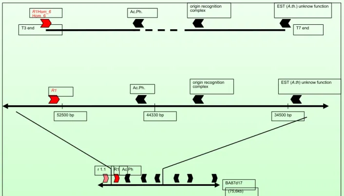
EST (A.th) unknow function origin recognition
complex
52500 bp 44330 bp 34500 bp
Ac.Ph.
R1
BA87d17 (75,6kb) R1
r 1.1 Ac.Ph
T3 end R1Hom_6 Hom_6
T7 end Ac.Ph.
origin recognition
complex EST (A.th.) unknow function
S. nigrum Clone 6 is orthologous to the R1 region in S. tuberosum but truncated at the 5‘ end
Fig. 1.4.3: Identified region of syntheny between Solanum tuberosum
and Solanum nigrum. Shown
is the organization of a genomic fragment from Solanum nigrum (upper) and a corresponding region
on BAC BA87d17 from Solanum tuberosum /middle and lower)(data from Tatjana von Frey-Jost, this
laboratory)
Fig. 1.4.5: Results of a Southern Blot
after hybridization with a
R1 specificprobe. Several cultivars of
Solanum tuberosum (P3-Desirée) have been used aswell as the related species
Solanum dulcamara, Solanum nigrum and Solanum melongana. The BAC BA87d17 has beenused as control (data from Dr. Heibges, this laboratory).
Fig. 1.4.4: Results of a Southern Blot after
hybridization with a
R1 specific probe. Seven cosmidclones from a genomic library of
Solanum nigrum havebeen used (1-7)and the BAC BA87d17 from
Solanum tuberosum as control (B) (data from Tatjana von Frey-Jost, this laboratory).
1.5.Methods for expression analysis
1.5.1. Single target driven methods
The first described method for the analysis of ribonucleic acids has been published in 1977. In this approach, the 1975 published technique of the Southern Blot for the detection of DNA fragments in a complex mixture has been modified to blot RNA onto a nitrocellulose membrane instead of genomic DNA [69, 70]. This new method, known as Northern Blot, was the beginning of expression analysis [70]. The next important step was the ability to synthesize complementary DNA (cDNA) of the RNA [71] in combination with the polymerase chain reaction (PCR) [72].
At present three different methods for the analysis of specific transcripts are known. The method with the broadest use is the reverse transcriptase (RT-)PCR. cDNA is generated and a specific fragment is amplified using a PCR reaction. The signal detection and further analysis is performed using agarose gel electrophoresis. This method has been improved by the quantitative reverse transcriptase (qRT-)PCR, where the amplified fragment detected by a fluorometric measurement during the PCR reaction.
Another method, with broader use in medical applications for detection of retroviruses is the branched DNA or bDNA-assay. This method is
mainly used to detect very low amounts of transcripts in blood or tissues but generally can be applied for any kind of nucleic acids. The specific nucleic acid is bound by an immobilized capture probe. Afterwards the signal of the bound molecules is amplified using of bDNA label probes.
And since a few years the Northern Blot has found a new application area. This was initiated by the discovery of small RNAs for which the production of cDNA is laborious and far away from being a
standard technique. For the analysis of this small molecule class the Northern blot has become the standard technique.
Fig. 1.5.1.1: Schematic picture of the
principle of a bDNA-assay. AP stand for
alkaline phosphatase which is used for
the production of a chemoluminiscence
signal [149].
1.5.2. Methods for transcriptome analysis
Besides expression analysis for specific target genes, methods have been developed to generate whole transcriptome data. One of the first approaches in this direction the generation of cDNA-libraries and random sequencing of clones to generate EST (expressed sequence tags) information. A first approach in a large scale has been performed in the within the Human Genome Project [73]. In addition, using differential display, a way has been found to compare two libraries [74]. Although being far away from generating quantitative data, this EST-information was the basis for the first whole transcriptome analysis method called cDNA microarray. In this method, the principle of the Northern Blot has been turned around by immobilizing multiple probes on an array and hybridizing this array with first a radioactive and later flourophoric labeled cDNAs. The first generation was able to detect the presence or absence of a given transcript. The second generation found a way to quantify the fluorometric signal [75-77]. Nevertheless, these array based technologies were limited by the availability of sequence information.
This problem wanted to be solved by the application of an expectation independent method, the first attempt in this direction was to raise the quantitative efficiency of EST- sequencing. This has be done, by the generation of small 3´-sequence tags, which were ligated into concatamers. These concatamers were subsequently cloned and sequenced.
This method called serial analysis of gene expression (SAGE) [78], was still limited by the cloning step and the costs of traditional Sanger sequencing [79] and was not able to generate deep transcriptome information in a cost effective manner.
The introduction of „Next Generation Sequencing“ methods (summarized as massive
parallel sequencing) is able to multiply the number of sequenced tags per reaction and has
opened new possibilities de-novo transcriptome analysis. The major applications of these
high throughput sequencing technologies are the sequencing of whole transcriptomes
combined with an assembly and an annotation to a given reference genome. The
quantification of the amount of a certain transcript is defined by the coverage rate. This
method is known as RNAseq or transcriptome sequencing. On the other hand SAGE can
be applied in an improved manner. In this method the quantification is done by direct
counting of the number of sequenced tags , which are localized on a specific region of the
transcript. These methods are known as Digital gene expression, DeepSAGE, LongSAGE or SuperSAGE [80-86].
DeepSAGE, in particular, works by creating small ditag-libraries of cDNA fragments, which are located close to the 3´ end of a transcript. To achieve this, two restriction enzymes are required, which recognize the same restriction site, but cleave different positions. One of the most commonly used enzyme system consists of NlaIII (or DpnII) and MmeI, which both identify a CATG motif. Outgoing from this site, a 18-20 bases large fragment is extracted. Using this
principle, it is possible to extract a specific region of a transcript. The advantage of this method compared to whole transcriptome sequencing is that less sequenced molecules are needed to achieve a comparable quantitative information to RNAseq based experiments. The disadvantage is the short length of sequence information which is associated with ambiguities.
1.6.Massive parallel sequencing
Fig. 1.5.2.1: Scheme of the generation of
SAGE-libraries. Immobilized double strand cDNA is digested with two restric- tion enzymes using the same CATG re- striction site. The final cDNA fragment (red) is flanked by two linkers (blue and green).
Table 1.6.2: Summary of the performance features of presently available
„Next Generation Sequencing“ platforms.*
System Provider Read length No of sequences per run
Sequencing principle
Template amplification 454 flx
Roche app. 400 bp up to 1 x 10
6PyrosequencingEmulsion
PCR
GenomeAnalyzer
Illumina up to 100 bp up to 200 x 10
6Sanger Cluster generation
HeliscopeHelicos 25 – 55 bp 8 – 400 x 10
6Sanger Not necessary
SolidApplied
Biosystems
50 bp up to 2 x 10
9Probe ligation Emulsion PCR
*The data is based on the specifications of the providers [88-90, 150].
At the moment four different platforms for massive parallel sequencing are available. The most prominent ones are the „Genome Analyzer“ from Illumina, San Diego (USA) (former Solexa) and the 454 FLX system from Roche, Penzberg (Germany) [87]. Quite new is the Solid system from Applied Biosystems, Foster City (USA) and the most recent invented platform is the „Heliscope Sequencer“ from Helios, Cambridge (USA). Interestingly, three of the four different systems follow a different sequencing method. The 454 sequencing technology delivers the longest reads and uses pyrosequencing to generate sequences [88]. The Helicos and Illumina systems use a modification of Sanger sequencing [89]. The Solid system uses a competitive ligation system of fluorescent labeled dibaseprobes as sequencing system. These dibaseprobes are small single strand nucleic acid fragments which consist of two bases at the 3´-end and additional placeholder bases. They are labeled with a flourophoric dye which is specific for the first two bases. The speciality of this system is a two-fold sequence detection of each base, achieving an enhanced sequencing accuracy compared to other methods [90]. The main characteristics are summarized in Table 1.6.2.
1.7.Small introduction to the relevant statistical tests for this study
Whenever large amounts of data have to be analysed like it is the case in transcriptomic experiments, it is necessary to find a way to distinguish between interesting and uninteresting observation. Statistical methods can help to characterize the data mathematically and try to identify the most interesting values in a large amount of data.
Especial tests for significance of observation can be a useful tool for the identification of interesting values and a help for the interpretation of results.
One of the most crucial prerequisites for statistical tests is the assessment of the proper probability distribution. One of the most special features of data, generated by Massive parallel sequencing methods is the discrete distribution. Unlike, most empiric data , it does not follow the normal distribution.
During this thesis, five statistical tests, which do not require a normal distribution of the data, have been performed and validated. Additionally some characteristics of Student´s t- test are shown.
The G-test statistics [91] uses in principle a series of
χ2-distribution tests. In this analysis
are within a group or between two groups. By the application of seperate tests for each hypothesis they have to be rejected or accepted individually to pass the overall test.
Usually it is desired that differences between two grous are occurr and that no differences within a group are detected.
Another implemented test is included in the R-package Sagenhaft and is called sage.test [92]. This test is a variation of Fisher´s Exact test and compares the proportion of each sequence tag in a library to the proportion of the same tag in another library (contingency table statistic). In this test, the original Fisher´s exact test has been modified in the way that a negative binomial approach has been used. This was necessary far an application in large data-matrixes.
In contrast to the two established tests mentioned above, which were already used for the
analysis of traditional SAGE-libraries, edgeR, BaySeq [93, 94] and a permutation test
procedure have been used as well. The first two tests were developed specifically for the
analysis of transcriptome data resulting from massive parallel sequencing methods. At the
moment, these tests are still in an experimental stage and the permutation test had to be
developed specifically.
1.8.Motivation for this study
The beginning of the project was the R1 resistance gene and the genomic region surrounding it that were classified by the ORF-prediction and annotation algorithms as Disease resistance genes. These findings implied the existence of a gene family and showed that the R1 gene is not unique in Solanum tuberosum. This hypothesis was supported by the identification
of putative homologues genes in Solanum nigrum.
The idea arose that some of these genes have similar f u n c t i o n s i n p a t h o g e n recognition and that among them may be a member, which is involved in the quantitative effect, observed in from the h o t s p o t r e g i o n o n Chromosome V of Solanum t u b e r o s u m . I n S o l a n u m
nigrum, one of the homologous genes could involved in the broad resistance of this plant.
Additionally, it was necessary to face the truth, that besides the molecular structure, which is originating from the presence of the R1-gene, little was known, and just a rough idea about its acting mechanism existed (see fig. 1.8.1).
The prerequisite for further studies on the function of the R1 family was a further description of the consequences, which result of the presence of this gene. To achieve this, a comparative transcriptome analysis of R1-transgenic plants was performed. In this approach, plants were included, which do not harbor the R1-gene. This selection has been expanded by including plants which are transformed with the ORF45. The underlying sequence is highly similar but the presence of this open reading frame does not mediate resistance as R1.
Another question arose from the predicted open reading frames. Experimental evidence
Fig. 1.8.1: Model of the mechanism of the function of R1 atthe beginning of this study.
for expresion was lacking. To test the correctness of the in silico data RT-PCR experiments have been performed.
In Solanum nigrum an additional question was urgent: Is the presence of a R-gene
mediated resistance mechanism the reason for the sensing of Phytophthora infestans or is
it due to a PAMP-mechanism? To get a hint on this, transient expression of putative
effectors of Phytophthora infestans has been performed. The aim was the localization of
the pathogen recognition event by expressing the effectors directly in the cytosol, using a
PVX-based expression system. The identification of necrosis-inducing effectors would be a
good argument for the presence of R-genes of the NBS-LRR type class in Solanum
nigrum, which detects Phytophthora infestans. The major aim of this study, was a
description of the function of R1 and the related sequences. A broad data basis was to be
generated, which can be used to generate new working hypothesis on the function of R1
and other members of the R1 family.
2.Material and Methods
2.1.Standard conditions for plant and oomycete cultivation
Plants, originating from in vitro culture were grown under longday conditions (16h light (80 mmol photons m
-2s
-1), 8h dark) and 21 °C temperature in climate chambers from Brown- Boveri Cie (now: York International), Mannheim (Germany). For race specifications of Phytophthora infestans using the standars differential set of Solanum tuberosum cultivars from the Scottish Crop Research Institute, the plant material was grown under comparable conditions in the green house.
Phytophthora infestans was cultivated on rye agar plates [95], containing small leaves from in vitro culture plants or was propagated on leaflets from susceptible potato cultivars.
The climate conditions were 16h light and 8h of darkness and 18°C for short term cultivation (within ongoing infection experiments) or 12°C for long term storage.
2.2.Chemicals
Enzymes for RNA-extraction and modification were supplied by Ambion, Austin (USA).
Reagents for qPCR-techniques were supplied by Applies Biosystems, Foster City (USA).
Reagents for cDNA synthesis were suppllied primary by Fermentas, St. Leon-Roth (Ger- many) and Invitrogen, Carlsbad (USA). Standard chemicals were supplied by Roth, Karls- ruhe (Germany), Sigma Aldrich, St. Louis (USA) and BioBudget, Krefeld (Germany). Gen- eral enzymes were primarily supplied by Fermentas, St. Leon-Rot (Germany), Invitrogen, Carlsbad (USA), New England Biolabs, Ipswich (USA) and Roche, Penzberg (Germany).
All enzymes were used with buffers of the supplier according to the supplier´s instructions
(if not described differently).
2.3.Infection assays and plant cultivation
For the analysis of infected plant material, plants were grown of 6-8 weeks in a climate chamber under standard conditions. At least one week before the beginning of infection experiments, the plants have been transferred to a special chamber. For infections, the Phytophthora infestans strain R208m
2(originating from the laboratory of Felix Mauch, University of Fribourg (Switzerland) and described in Si-Ammour 2003 [96]) was used.
During the studies the race specificity of the strain was monitored using a detached leaflet assay and the differential set of potato lines originating from the Scottish crop research institute (SCRI).
The detached leaflet assay was made in 12well-plates, using leaf discs of 2 cm in diameter. The disc were laid on water-soaked Whatman paper-discs (using 300µl of water). Inoculum was produced by washing the sporangia from infected potato leaves (between 8-11 days after infection) using approximately 1-2 ml deionized water for 2-4 infected leaflets (depending of the degree of infection of the used source). As leaf source 6-8 week old plants and the third to fifth leaf were used. The sporangia concentration was quantified using a Neubauer chamber and sporulation of the zoospores was induced by incubating the sporangia suspension at 4-8°C for 3-6 hours. The climate conditions during infections were 16h day with a temperature of 18 °C and 8 h night with 14 °C in a climate chamber from Ehret, Emmendingen (Germany). The successful sporulation was confirmed optically using a light microscope. Directly before infection experiments, the concentration was adjusted to 30000-60000 sporangia/ml. Infections were done by placing a droplet of 20 µl inoculum on the leaf disc. The infection was observed optically with a binocular microscope from the fifth to eighth day post infection. The occurrence of sporangia harboring mycelia on the leaf was the criterion for a positive infection.
For the harvest of material used in chapter 3.1 3.2, 25 plants were grown under conditions described in chapter 2.1 to an age of 10 weeks in case of Solanum tuberosum P6/210 and 6-10 in case of Solanum nigrum P4. Material was harvested and pooled from 5-15 plants.
Harvested material was frozen immediately in liquid nitrogen and stored at -80 °C.
2.4.Confocal microscopy
For confocal microscopy, leaves from Solanum tuberosum v. Desirée which have been grown for 6-8 weeks under standard conditions as described in chapter 2.1 were used.
The infections were made similar to the detached leaflet assay described in chapter 2.3 with following exceptions. Instead of leaf discs, whole leaves were used, which were placed on moist blotting paper on a metal grid within a clear, with parafilm closed plastic box. Infections were made by placing 25-35 droplets of inoculum on each leaflet.
For microscopy small pieces of the infected leaves at three days after infection were stained with a solution of 0,02% diethanol for approximately 10 seconds. Pictures were taken using a confocal laser scanning microscopy (Leica SP2 AOBS, Leica Microsystems, Wetzlar (Germany)). For excitation of the flourescence laser light with 405 nm wavelength was used. The detection of the flourescence emission was made using a filter for wavelength of 420-520 nm.
Confocal microscopy pictures were taken three days after infection under aid of Dr. Elmon Schmelzer and the Cemic group at the Max-Planck-Institute for Plant Breeding Research.
2.5.Molecular biological methods
2.5.1.Polymerase chain reaction (PCR)
The standard reaction mixture included following components:
1 µl Template-DNA (50 ng genomic DNA, 1 ng BAC-DNA) PCR-buffer Amplikon III
2,5 mM MgCl
2200 µM dNTP-Mix 0,25 µM Forward-Primer 0,25 µM Reverse-Primer
1 u recombinant Taq DNA-Polymerase +H
2O to final volume of 20µl
Standard PCR-program included following steps:
2 minutes 94 °C 30 seconds 93 °C
30 seconds T
Ax35 30 second 72 °C
5 minutes 72°C
Linear-PCR for the amplification of ORF23
The polymerase chain reaction in case of ORF23 was done under standard conditions with the exception that the reverse primer was added to the reaction mixture after 15 cycles of amplification.
Table 2.5.1.1: Overview of used oligonucleotides for polymerase chain reaction
Name Sequence (5´-3´) Annealing
temperature [°C]
Product size (bp)
Application
TC173455_f
TGCTGGTGAACCCACAAAGCCC55 306 qRT-PCR
TC173455_r
TCCGCCTTGGTCAACCCTGC55 306 qRT-PCR
TC180208_f
TGCGCAAGGGTTTGGCCTGT55 337 qRT-PCR
TC180208_r
ACCACCGCCTCCTCCGGTTT55 337 qRT-PCR
TC173049_f
GCTCATGGCGGGGAAGGAGG55 140 qRT-PCR
TC173049_r
CGCGCCGATGGCGAGTAAGT55 140 qRT-PCR
TC163043_f
TGTGGAGGCGAGCTCTGGTGT55 160 qRT-PCR
TC163043_r
GACCTCCAGTTCCGCCGCTG55 160 qRT-PCR
TC173953_f
GGCATTGGGGTTACATGGATGGTCC55 200 qRT-PCR
TC173953_r
TGCCATCTCACTTGTGGATTCGCC55 200 qRT-PCR
TC165331_f
TGGCTGCAGCAGTACGGAACA55 240 qRT-PCR
TC165331_r
GCTCCACCGATGCAGGACCC55 240 qRT-PCR
TC172861_f
TGATTGGCGTGCCAACCCCT55 144 qRT-PCR
TC172861_r
CCCCCACCTGCAGACCGAGT55 144 qRT-PCR
TC168403_f
TGGGTTGGCGAGGAAAGCGG55 173 qRT-PCR
TC168403_r
AGCAGGGTAAGAGAGTGGGGGT55 173 qRT-PCR
TC182394_f
TGGTCCTCCGTCTCCGTGGTG55 378 qRT-PCR
TC182394_r
CCTCTGCTGGTCCGGTGGGA55 378 qRT-PCR
TC170569_f
ACCGCAGCAGGTCCATGCAA55 166 qRT-PCR
TC170569_r
CCCTTTCGTCTTTCCGCTCACCG55 166 qRT-PCR
TC190958_f
CAGCTGAGCAGGGACGGCAG55 146 qRT-PCR
TC190958_r
GGAGCAGCATCAACGGGTGC55 146 qRT-PCR TC174692_f
GGAGCGACAGCGATTTGTACGTG55 200 qRT-PCR
TC174692_r
GCTTGAACCTTCCCCTCGCGT55 200 qRT-PCR
TC173012_f
GTACGGCCTTCTGCACCGCT55 249 qRT-PCR
TC173012_r
GTGACCTTTGCGCCCGCTCT55 249 qRT-PCR
TC176096_f
CTGTAGTGGCTGCCCGTGCC55 169 qRT-PCR
TC176096_r
ACGCGTGCTGCTTTTCCCAT55 169 qRT-PCR
TC190958_f
CAGCTGAGCAGGGACGGCAG55 146 qRT-PCR
TC190958_r
GGAGCAGCATCAACGGGTGC55 146 qRT-PCR
TC184597_f
GCTGGACCGCTCAAATGCTGC55 212 qRT-PCR
TC184597_r
CGGCAAAGAGGCGCAGAAGC55 212 qRT-PCR
TC183138_f
AGTGTCGATGCTGAACTGGTGGA55 191 qRT-PCR
TC183138_r
TGCCTTCCCGCTGTCAAATCCT55 191 qRT-PCR
st22-1_f
TAGGGATCAGATCAGTACC52 162 RT-PCR
st22-1_r
GAAATTGAAAAAGCAGGAGAG52 162 RT-PCR
st23_f
GTTCTAAAACCATAAATGGTACG55 348 RT-PCR
st23_r
GCTACATTTGTTTGAAACAAAGC55 348 RT-PCR
st24/45_f
CTCAAGAATCAACTTCAAGTTG55 314 RT-PCR
st24/45_r
CATCTTCAAACCCAACAATTTC55 314 RT-PCR
st46_f
CGAATGGGGTAGTTACATGC52 119 RT-PCR
st46_r
GCATGTTTGGTTCATCAGTG52 119 RT-PCR
st52_f
CCAAGCATGGCCCCCTATG55 278 RT-PCR
st52_r
CCCTGTTTCCTGGAACAAAAAG55 278 RT-PCR
st54_f
CTGTATCTTGAAAAAGTTTGGG55 586 RT-PCR
st54_r
ATGCTTCTTTTGAGCTTG55 586 RT-PCR
snR1.4_f
ACTGCATCGATGTCAAGATCT57 320
RT-PCR (developed by Tatjana von Frey- Jost, this
laboratory) snR1.4_r
CCCTTGAGCGTTAAAGATAAC57 320
RT-PCR (developed by Tatjana von Frey- Jost, this
laboratory)
snR1.5_f
AGACCTAGATTCTCTACTGAAGC
55 410
RT-PCR (developed by Tatjana von Frey- Jost, this
laboratory) SnR1.5_r
CTTTTTTCTCCGTCTTTGCT

![Fig. 1.1.3: Schematic alignment of genomic areas from the three Phytophthora species Phytophthora ramorum (top), sojae (middle) and infestans (bottom) [5].](https://thumb-eu.123doks.com/thumbv2/1library_info/3622148.1501858/15.892.515.809.107.434/schematic-alignment-genomic-phytophthora-species-phytophthora-ramorum-infestans.webp)
![Fig. 1.2.1.2: Comparative scheme of the relationship between quantitative and qualitative resistance within the phenotype resistance as whole [146].](https://thumb-eu.123doks.com/thumbv2/1library_info/3622148.1501858/19.892.154.745.819.1065/comparative-scheme-relationship-quantitative-qualitative-resistance-phenotype-resistance.webp)
![Fig. 1.2.2.1: General scheme of the interaction compartment between of pathogens and the plant [148].](https://thumb-eu.123doks.com/thumbv2/1library_info/3622148.1501858/20.892.464.804.429.720/fig-general-scheme-interaction-compartment-pathogens-plant.webp)
![Fig. 1.3.2.3: Function map of chromo- chromo-some V of S. tuberosum. The green bars in-dicate QTL regions for resistance to fungal p a t h o g e n s a n d o o m y c e t e s (source:PoMaMo (modified) [18]).](https://thumb-eu.123doks.com/thumbv2/1library_info/3622148.1501858/25.892.497.803.101.983/function-chromo-chromo-tuberosum-regions-resistance-pomamo-modified.webp)