2013 ESH/ESC Guidelines for the management of arterial hypertension
The Task Force for the management of arterial hypertension of the European Society of Hypertension (ESH) and of the European Society of Cardiology (ESC)
Authors/Task Force Members: Giuseppe Mancia (Chairperson) (Italy) * , Robert Fagard (Chairperson) (Belgium) * , Krzysztof Narkiewicz (Section co-ordinator) (Poland), Josep Redon (Section co-ordinator) (Spain), Alberto Zanchetti (Section co-ordinator) (Italy), Michael Bo¨hm (Germany), Thierry Christiaens (Belgium), Renata Cifkova (Czech Republic), Guy De Backer (Belgium), Anna Dominiczak (UK),
Maurizio Galderisi (Italy), Diederick E. Grobbee (Netherlands), Tiny Jaarsma (Sweden), Paulus Kirchhof (Germany/UK), Sverre E. Kjeldsen (Norway), Ste´phane Laurent (France), Athanasios J. Manolis (Greece), Peter M. Nilsson (Sweden), Luis Miguel Ruilope (Spain), Roland E. Schmieder (Germany), Per Anton Sirnes (Norway), Peter Sleight (UK), Margus Viigimaa (Estonia), Bernard Waeber (Switzerland), Faiez Zannad (France)
ESH Scientific Council: Josep Redon (President) (Spain), Anna Dominiczak (UK), Krzysztof Narkiewicz (Poland), Peter M. Nilsson (Sweden), Michel Burnier (Switzerland), Margus Viigimaa (Estonia), Ettore Ambrosioni (Italy), Mark Caufield (UK), Antonio Coca (Spain), Michael Hecht Olsen (Denmark), Roland E. Schmieder (Germany), Costas Tsioufis (Greece), Philippe van de Borne (Belgium).
ESC Committee for Practice Guidelines (CPG): Jose Luis Zamorano (Chairperson) (Spain), Stephan Achenbach (Germany), Helmut Baumgartner (Germany), Jeroen J. Bax (Netherlands), He´ctor Bueno (Spain), Veronica Dean (France), Christi Deaton (UK), Cetin Erol (Turkey), Robert Fagard (Belgium), Roberto Ferrari (Italy), David Hasdai (Israel), Arno W. Hoes (Netherlands), Paulus Kirchhof (Germany/UK), Juhani Knuuti (Finland), Philippe Kolh (Belgium), Patrizio Lancellotti (Belgium), Ales Linhart (Czech Republic), Petros Nihoyannopoulos (UK), Massimo F. Piepoli (Italy), Piotr Ponikowski (Poland), Per Anton Sirnes (Norway), Juan Luis Tamargo (Spain), Michal Tendera (Poland), Adam Torbicki (Poland), William Wijns (Belgium), Stephan Windecker (Switzerland).
*Corresponding authors: The two chairmen equally contributed to the document. Chairperson ESH: Professor Giuseppe Mancia, Centro di Fisiologia Clinica e Ipertensione, Via F. Sforza, 35, 20121 Milano, Italy. Tel:+39 039 233 3357, Fax:+39 039 322 274. Email:giuseppe.mancia@unimib.it. Chairperson ESC: Professor Robert Fagard, Hypertension & Cardiovascular Rehab. Unit, KU Leuven University, Herestraat 49, 3000 Leuven, Belgium. Tel:+32 16 348 707, Fax:+32 16 343 766, Email:robert.fagard@uzleuven.be
These guidelines also appear in theJournal of Hypertension, doi: 10.1097/01.hjh.0000431740.32696.cc and inBlood Pressure, doi: 10.3109/08037051.2013.812549.
With special thanks to Mrs Clara Sincich and Mrs Donatella Mihalich for their contribution.
Other ESC entities having participated in the development of this document:
ESC Associations: Heart Failure Association (HFA), European Association of Cardiovascular Imaging (EACVI), European Association for Cardiovascular Prevention & Rehabilitation (EACPR), European Heart Rhythm Association (EHRA)
ESC Working Groups: Hypertension and the Heart, Cardiovascular Pharmacology and Drug Therapy ESC Councils: Cardiovascular Primary Care, Cardiovascular Nursing and Allied Professions, Cardiology Practice
The content of these European Society of Cardiology (ESC) and European Society of Hypertension (ESH) Guidelines has been published for personal and educational use only. No com- mercial use is authorized. No part of the ESC Guidelines may be translated or reproduced in any form without written permission from the ESC. Permission can be obtained upon sub- mission of a written request to Oxford University Press, the publisher of the European Heart Journal and the party authorized to handle such permissions on behalf of the ESC.
Disclaimer.The ESH/ESC Guidelines represent the views of the ESH and ESC and were arrived at after careful consideration of the available evidence at the time they were written.
Health professionals are encouraged to take them fully into account when exercising their clinical judgement. The guidelines do not, however, override the individual responsibility of health professionals to make appropriate decisions in the circumstances of the individual patients, in consultation with that patient, and where appropriate and necessary the patient’s guardian or carer. It is also the health professional’s responsibility to verify the rules and regulations applicable to drugs and devices at the time of prescription.
&The European Society of Hypertension (ESH) and European Society of Cardiology (ESC) 2013. All rights reserved. For permissions please email: journals.permissions@oup.com.
by guest on February 8, 2014http://eurheartj.oxfordjournals.org/Downloaded from
Document Reviewers: Denis L. Clement (ESH Review Co-ordinator) (Belgium), Antonio Coca (ESH Review Co-ordinator) (Spain), Thierry C. Gillebert (ESC Review Co-ordinator) (Belgium), Michal Tendera (ESC Review Co-ordinator) (Poland), Enrico Agabiti Rosei (Italy), Ettore Ambrosioni (Italy), Stefan D. Anker (Germany), Johann Bauersachs (Germany), Jana Brguljan Hitij (Slovenia), Mark Caulfield (UK), Marc De Buyzere (Belgium), Sabina De Geest (Switzerland), Genevie`ve Anne Derumeaux (France), Serap Erdine (Turkey), Csaba Farsang (Hungary), Christian Funck-Brentano (France), Vjekoslav Gerc (Bosnia & Herzegovina), Giuseppe Germano (Italy), Stephan Gielen (Germany), Herman Haller (Germany), Arno W. Hoes (Netherlands), Jens Jordan (Germany), Thomas Kahan (Sweden), Michel Komajda (France), Dragan Lovic (Serbia), Heiko Mahrholdt (Germany),
Michael Hecht Olsen (Denmark), Jan Ostergren (Sweden), Gianfranco Parati (Italy), Joep Perk (Sweden), Jorge Polonia (Portugal), Bogdan A. Popescu (Romania), Zˇ eljko Reiner (Croatia), Lars Ryde´n (Sweden), Yuriy Sirenko (Ukraine), Alice Stanton (Ireland), Harry Struijker-Boudier (Netherlands), Costas Tsioufis (Greece), Philippe van de Borne (Belgium), Charalambos Vlachopoulos (Greece), Massimo Volpe (Italy), David A. Wood (UK).
The affiliations of the Task Force Members are listed in the Appendix. The disclosure forms of the authors and reviewers are available on the respective society websites http://www.eshonline.org and www.escardio.org/guidelines
Online publish-ahead-of-print 14 June 2013
- - - -- - - -
Keywords Hypertension † Guidelines † Antihypertensive treatment † Blood pressure † Blood pressure measurement † Cardiovascular risk † Cardiovascular complications † Device therapy † Follow-up
† Lifestyle † Organ damage
Table of Contents
Abbreviations and acronyms . . . .2162
1 Introduction . . . .2163
1.1 Principles . . . .2163
1.2 New aspects . . . .2163
2 Epidemiological aspects . . . .2164
2.1 Relationship of blood pressure to cardiovascular and renal damage . . . .2164
2.2 Definition and classification of hypertension . . . .2165
2.3 Prevalence of hypertension . . . .2165
2.4 Hypertension and total cardiovascular risk . . . .2165
2.4.1 Assessment of total cardiovascular risk . . . .2165
2.4.2 Limitations . . . .2166
2.4.3 Summary of recommendations on total cardiovascular risk assessment . . . .2167
3 Diagnostic evaluation . . . .2167
3.1 Bood pressure measurement . . . .2168
3.1.1 Office or clinic blood pressure . . . .2168
3.1.2 Out-of-office blood pressure . . . .2168
3.1.3 White-coat (or isolated office) hypertension and masked (or isolated ambulatory) hypertension . . . .2170
3.1.4 Clinical indications for out-of-office blood pressure . .2170 3.1.5 Blood pressure during exercise and laboratory stress .2171 3.1.6 Central blood pressure . . . .2172
3.2 Medical history . . . .2172
3.3 Physical examination . . . .2173
3.4 Summary of recommendations on blood pressure measurement, history, and physical examination . . . .2173
3.5 Laboratory investigations . . . .2173
3.6 Genetics . . . .2173
3.7 Searching for asymptomatic organ damage . . . .2174
3.7.1 Heart . . . .2174
3.7.2 Blood vessels . . . .2176
3.7.3 Kidney . . . .2176
3.7.4 Fundoscopy . . . .2177
3.7.5 Brain . . . .2177
3.7.6 Clinical value and limitations . . . .2177
3.7.7 Summary of recommendations on the search for asymptomatic organ damage, cardiovascular disease, and chronic kidney disease . . . .2178
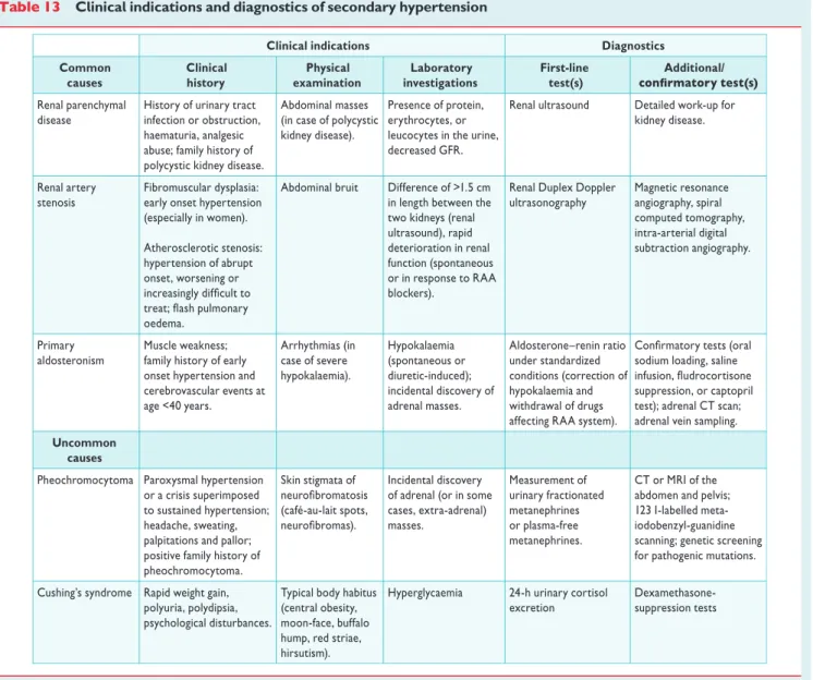
3.8 Searching for secondary forms of hypertension . . . .2178
4 Treatment approach . . . .2178
4.1 Evidence favouring therapeutic reduction of high blood pressure . . . .2178
4.2 When to initiate antihypertensive drug treatment . . . . .2178
4.2.1 Recommendations of previous Guidelines . . . .2178
4.2.2 Grade 2 and 3 hypertension and high-risk grade 1 hypertension . . . .2179
4.2.3 Low-to-moderate risk, grade 1 hypertension . . . .2179
4.2.4 Isolated systolic hypertension in youth . . . .2181
4.2.5 Grade 1 hypertension in the elderly . . . .2181
4.2.6 High normal blood pressure . . . .2181
4.2.7 Summary of recommendations on initiation of antihypertensive drug treatment . . . .2181
4.3 Blood pressure treatment targets . . . .2182
4.3.1 Recommendations of previous Guidelines . . . .2182
4.3.2 Low-to-moderate risk hypertensive patients . . . .2182
4.3.3 Hypertension in the elderly . . . .2182
4.3.4 High-risk patients . . . .2182
by guest on February 8, 2014http://eurheartj.oxfordjournals.org/Downloaded from
4.3.5 The ‘lower the better’ vs. the J-shaped curve
hypothesis . . . .2183
4.3.6 Evidence on target blood pressure from organ damage studies . . . .2184
4.3.7 Clinic vs. home and ambulatory blood pressure targets . . . .2184
4.3.8 Summary of recommendations on blood pressure targets in hypertensive patients . . . .2184
5 Treatment strategies . . . .2185
5.1 Lifestyle changes . . . .2185
5.1.1 Salt restriction . . . .2185
5.1.2 Moderation of alcohol consumption . . . .2185
5.1.3 Other dietary changes . . . .2185
5.1.4 Weight reduction . . . .2185
5.1.5 Regular physical exercise . . . .2186
5.1.6 Smoking cessation . . . .2186
5.1.7 Summary of recommendations on adoption of lifestyle changes . . . .2186
5.2 Pharmacological therapy . . . .2187
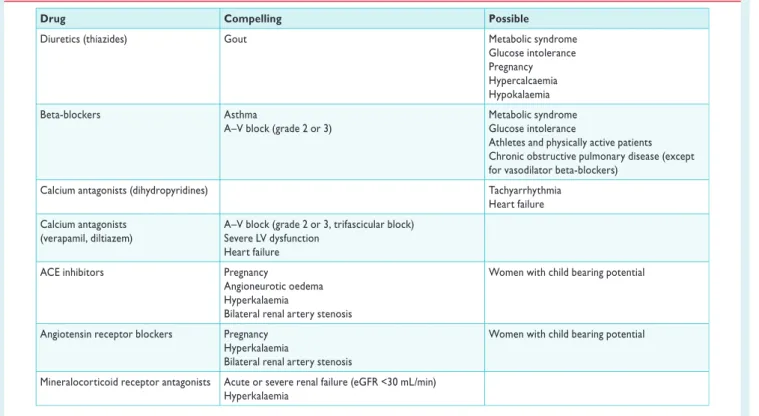
5.2.1 Choice of antihypertensive drugs . . . .2187
5.2.2 Monotherapy and combination therapy . . . .2189
5.2.3 Summary of recommendations on treatment strategies and choice of drugs . . . .2193
6 Treatment strategies in special conditions . . . .2194
6.1 White-coat hypertension . . . .2194
6.2 Masked hypertension . . . .2194
6.2.1 Summary of recommendations on treatment strategies in white-coat and masked hypertension . . . .2194
6.3 Elderly . . . .2194
6.3.1 Summary of recommendations on antihypertensive treatment strategies in the elderly . . . .2195
6.4 Young adults . . . .2195
6.5 Women . . . .2195
6.5.1 Oral contraceptives . . . .2195
6.5.2 Hormone replacement therapy . . . .2196
6.5.3 Pregnancy . . . .2196
6.5.4 Long-term cardiovascular consequences in gestational hypertension . . . .2196
6.5.5 Summary of recommendations on treatment strategies in hypertensive women . . . .2197
6.6 Diabetes mellitus . . . .2197
6.6.1 Summary of recommendations on treatment strategies in patients with diabetes . . . .2198
6.7 Metabolic syndrome . . . .2198
6.7.1 Summary of recommendations on treatment strategies in hypertensive patients with metabolic syndrome 2198 6.8 Obstructive sleep apnoea . . . .2199
6.9 Diabetic and non-diabetic nephropathy . . . .2199
6.9.1 Summary of recommendations on therapeutic strategies in hypertensive patients with nephropathy . . . . .2200
6.9.2 Chronic kidney disease stage 5D . . . .2200
6.10 Cerebrovascular disease . . . .2200
6.10.1 Acute stroke . . . .2200
6.10.2 Previous stroke or transient ischaemic attack . . . . .2200
6.10.3 Cognitive dysfunction and white matter lesions . . .2200
6.10.4 Summary of recommendations on therapeutic strategies in hypertensive patients with cerebrovascular disease . . . .2201
6.11 Heart disease . . . .2201
6.11.1 Coronary heart disease . . . .2201
6.11.2 Heart failure . . . .2201
6.11.3 Atrial fibrillation . . . .2201
6.11.4 Left ventricular hypertrophy . . . .2202
6.11.5 Summary of recommendations on therapeutic strategies in hypertensive patients with heart disease . . . .2202
6.12 Atherosclerosis, arteriosclerosis, and peripheral artery disease . . . .2203
6.12.1 Carotid atherosclerosis . . . .2203
6.12.2 Increased arterial stiffness . . . .2203
6.12.3 Peripheral artery disease . . . .2203
6.12.4 Summary of recommendations on therapeutic strategies in hypertensive patients with atherosclerosis, arteriosclerosis, and peripheral artery disease . . . .2203
6.13 Sexual dysfunction . . . .2203
6.14 Resistant hypertension . . . .2204
6.14.1 Carotid baroreceptor stimulation . . . .2204
6.14.2 Renal denervation . . . .2205
6.14.3 Other invasive approaches . . . .2205
6.14.4 Follow-up in resistant hypertension . . . .2205
6.14.5 Summary of recommendations on therapeutic strategies in patients with resistant hypertension . . . .2205
6.15 Malignant hypertension . . . .2206
6.16 Hypertensive emergencies and urgencies . . . .2206
6.17 Perioperative management of hypertension . . . .2206
6.18 Renovascular hypertension . . . .2206
6.19 Primary aldosteronism . . . .2206
7 Treatment of associated risk factors . . . .2207
7.1 Lipid-lowering agents . . . .2207
7.2 Antiplatelet therapy . . . .2207
7.3 Treatment of hyperglycaemia . . . .2207
7.4 Summary of recommendations on treatment of risk factors associated with hypertension . . . .2208
8 Follow-up . . . .2208
8.1 Follow-up of hypertensive patients . . . .2208
8.2 Follow-up of subjects with high normal blood pressure and white-coat hypertension . . . .2208
8.3 Elevated blood pressure at control visits . . . .2208
8.4 Continued search for asymptomatic organ damage . . . . .2209
8.5 Can antihypertensive medications be reduced or stopped?2209 9 Improvement of blood pressure control in hypertension . . . . .2209
10 Hypertension disease management . . . .2210
10.1 Team approach in disease management . . . .2211
10.2 Mode of care delivery . . . .2211
10.3 The role of information and communication technologies2211 11 Gaps in evidence and need for future trials . . . .2212
APPENDIX: Task Force members affiliations . . . .2212
References . . . .2213
by guest on February 8, 2014http://eurheartj.oxfordjournals.org/Downloaded from
Abbreviations and acronyms
ABCD Appropriate Blood pressure Control in Diabetes ABI ankle – brachial index
ABPM ambulatory blood pressure monitoring
ACCESS Acute Candesartan Cilexetil Therapy in Stroke Sur- vival
ACCOMPLISH Avoiding Cardiovascular Events in Combination Therapy in Patients Living with Systolic Hyperten- sion
ACCORD Action to Control Cardiovascular Risk in Diabetes ACE angiotensin-converting enzyme
ACTIVE I Atrial Fibrillation Clopidogrel Trial with Irbesartan for Prevention of Vascular Events
ADVANCE Action in Diabetes and Vascular Disease: Preterax and Diamicron-MR Controlled Evaluation AHEAD Action for HEAlth in Diabetes
ALLHAT Antihypertensive and Lipid-Lowering Treatment to Prevent Heart ATtack
ALTITUDE ALiskiren Trial In Type 2 Diabetes Using Cardio-renal Endpoints
ANTIPAF ANgioTensin II Antagonist In Paroxysmal Atrial Fib- rillation
APOLLO A Randomized Controlled Trial of Aliskiren in the Prevention of Major Cardiovascular Events in Elderly People
ARB angiotensin receptor blocker ARIC Atherosclerosis Risk In Communities ARR aldosterone renin ratio
ASCOT Anglo-Scandinavian Cardiac Outcomes Trial ASCOT-LLA Anglo-Scandinavian Cardiac Outcomes Trial—
Lipid Lowering Arm
ASTRAL Angioplasty and STenting for Renal Artery Lesions
A-V atrioventricular
BB beta-blocker
BMI body mass index
BP blood pressure
BSA body surface area
CA calcium antagonist
CABG coronary artery bypass graft CAPPP CAPtopril Prevention Project
CAPRAF CAndesartan in the Prevention of Relapsing Atrial Fibrillation
CHD coronary heart disease
CHHIPS Controlling Hypertension and Hypertension Im- mediately Post-Stroke
CKD chronic kidney disease
CKD-EPI Chronic Kidney Disease—EPIdemiology collabor- ation
CONVINCE Controlled ONset Verapamil INvestigation of CV Endpoints
CT computed tomography
CV cardiovascular
CVD cardiovascular disease
D diuretic
DASH Dietary Approaches to Stop Hypertension DBP diastolic blood pressure
DCCT Diabetes Control and Complications Study DIRECT DIabetic REtinopathy Candesartan Trials
DM diabetes mellitus
DPP-4 dipeptidyl peptidase 4
EAS European Atherosclerosis Society
EASD European Association for the Study of Diabetes
ECG electrocardiogram
EF ejection fraction
eGFR estimated glomerular filtration rate
ELSA European Lacidipine Study on Atherosclerosis ESC European Society of Cardiology
ESH European Society of Hypertension ESRD end-stage renal disease
EXPLOR Amlodipine – Valsartan Combination Decreases Central Systolic Blood Pressure more Effectively than the Amlodipine – Atenolol Combination FDA U.S. Food and Drug Administration
FEVER Felodipine EVent Reduction study
GISSI-AF Gruppo Italiano per lo Studio della Sopravvivenza nell’Infarto Miocardico-Atrial Fibrillation
HbA1c glycated haemoglobin
HBPM home blood pressure monitoring HOPE Heart Outcomes Prevention Evaluation HOT Hypertension Optimal Treatment HRT hormone replacement therapy
HT hypertension
HYVET HYpertension in the Very Elderly Trial IMT intima-media thickness
I-PRESERVE Irbesartan in Heart Failure with Preserved Systolic Function
INTERHEART Effect of Potentially Modifiable Risk Factors asso- ciated with Myocardial Infarction in 52 Countries INVEST INternational VErapamil SR/T Trandolapril ISH Isolated systolic hypertension
JNC Joint National Committee
JUPITER Justification for the Use of Statins in Primary Preven- tion: an Intervention Trial Evaluating Rosuvastatin LAVi left atrial volume index
LIFE Losartan Intervention For Endpoint Reduction in Hypertensives
LV left ventricle/left ventricular LVH left ventricular hypertrophy LVM left ventricular mass
MDRD Modification of Diet in Renal Disease MRFIT Multiple Risk Factor Intervention Trial MRI magnetic resonance imaging
NORDIL The Nordic Diltiazem Intervention study
OC oral contraceptive
OD organ damage
ONTARGET ONgoing Telmisartan Alone and in Combination with Ramipril Global Endpoint Trial
PAD peripheral artery disease
PATHS Prevention And Treatment of Hypertension Study PCI percutaneous coronary intervention
by guest on February 8, 2014http://eurheartj.oxfordjournals.org/Downloaded from
PPAR peroxisome proliferator-activated receptor PREVEND Prevention of REnal and Vascular ENdstage Disease PROFESS Prevention Regimen for Effectively Avoiding Sec-
ondary Strokes
PROGRESS Perindopril Protection Against Recurrent Stroke Study
PWV pulse wave velocity QALY Quality adjusted life years RAA renin-angiotensin-aldosterone RAS renin-angiotensin system RCT randomized controlled trials
RF risk factor
ROADMAP Randomized Olmesartan And Diabetes MicroAl- buminuria Prevention
SBP systolic blood pressure
SCAST Angiotensin-Receptor Blocker Candesartan for Treatment of Acute STroke
SCOPE Study on COgnition and Prognosis in the Elderly SCORE Systematic COronary Risk Evaluation
SHEP Systolic Hypertension in the Elderly Program STOP Swedish Trials in Old Patients with Hypertension STOP-2 The second Swedish Trial in Old Patients with
Hypertension
SYSTCHINA SYSTolic Hypertension in the Elderly: Chinese trial SYSTEUR SYSTolic Hypertension in Europe
TIA transient ischaemic attack TOHP Trials Of Hypertension Prevention
TRANSCEND Telmisartan Randomised AssessmeNt Study in ACE iNtolerant subjects with cardiovascular Disease
UKPDS United Kingdom Prospective Diabetes Study VADT Veterans’ Affairs Diabetes Trial
VALUE Valsartan Antihypertensive Long-term Use Evaluation
WHO World Health Organization
1 Introduction
1.1 Principles
The 2013 guidelines on hypertension of the European Society of Hypertension (ESH) and the European Society of Cardiology (ESC) follow the guidelines jointly issued by the two societies in 2003 and 2007.1,2Publication of a new document 6 years after the previous one was felt to be timely because, over this period, important studies have been conducted and many new results have been pub- lished on both the diagnosis and treatment of individuals with an ele- vated blood pressure (BP), making refinements, modifications and expansion of the previous recommendations necessary.
The 2013 ESH/ESC guidelines continue to adhere to some funda- mental principles that inspired the 2003 and 2007 guidelines, namely (i) to base recommendations on properly conducted studies identi- fied from an extensive review of the literature, (ii) to consider, as the highest priority, data from randomized, controlled trials (RCTs) and their meta-analyses, but not to disregard—particularly when dealing with diagnostic aspects—the results of observational
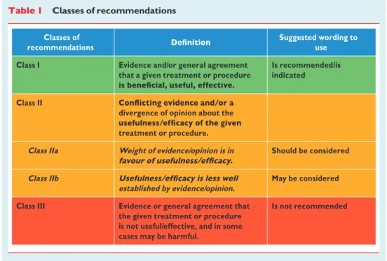
and other studies of appropriate scientific calibre, and (iii) to grade the level of scientific evidence and the strength of recommendations on major diagnostic and treatment issues as in European guidelines on other diseases, according to ESC recommendations (Tables1and2).
While it was not done in the 2003 and 2007 guidelines, providing the recommendation class and the level of evidence is now regarded as important for providing interested readers with a standard approach, by which to compare the state of knowledge across different fields of medicine. It was also thought that this could more effectively alert physicians on recommendations that are based on the opinions of the experts rather than on evidence. This is not uncommon in medi- cine because, for a great part of daily medical practice, no good science is available and recommendations must therefore stem from common sense and personal clinical experience, both of which can be fallible. When appropriately recognized, this can avoid guidelines being perceived as prescriptive and favour the per- formance of studies where opinion prevails and evidence is lacking.
A fourth principle, in line with its educational purpose, is to provide a large number of tables and a set of concise recommendations that could be easily and rapidly consulted by physicians in their routine practice.
The European members of the Task Force in charge of the 2013 guidelines on hypertension have been appointed by the ESH and ESC, based on their recognized expertise and absence of major con- flicts of interest [their declaration of interest forms can be found on the ESC website (www.escardio.org/guidelines) and ESH website (www.eshonline.org)]. Each member was assigned a specific writing task, which was reviewed by three co-ordinators and then by two chairmen, one appointed by ESH and another by ESC. The text was finalized over approximately 18 months, during which the Task Force members met collectively several times and corre- sponded intensively with one another between meetings. Before publication, the document was also assessed twice by 42 European reviewers, half selected by ESH and half by ESC. It can thus be confi- dently stated that the recommendations issued by the 2013 ESH/ESC guidelines on hypertension largely reflect the state of the art on hypertension, as viewed by scientists and physicians in Europe.
Expenses for meetings and the remaining work have been shared by ESH and ESC.
1.2 New aspects
Because of new evidence on several diagnostic and therapeutic aspects of hypertension, the present guidelines differ in many respects from the previous ones.2Some of the most important differ- ences are listed below:
(1) Epidemiological data on hypertension and BP control in Europe.
(2) Strengthening of the prognostic value of home blood pressure monitoring (HBPM) and of its role for diagnosis and manage- ment of hypertension, next to ambulatory blood pressure mon- itoring (ABPM).
(3) Update of the prognostic significance of night-time BP, white- coat hypertension and masked hypertension.
(4) Re-emphasis on integration of BP, cardiovascular (CV) risk factors, asymptomatic organ damage (OD) and clinical compli- cations for total CV risk assessment.
by guest on February 8, 2014http://eurheartj.oxfordjournals.org/Downloaded from
(5) Update of the prognostic significance of asymptomatic OD, including heart, blood vessels, kidney, eye and brain.
(6) Reconsideration of the risk of overweight and target body mass index (BMI) in hypertension.
(7) Hypertension in young people.
(8) Initiation of antihypertensive treatment. More evidence-based criteria and no drug treatment of high normal BP.
(9) Target BP for treatment. More evidence-based criteria and unified target systolic blood pressure (SBP) (,140 mmHg) in both higher and lower CV risk patients.
(10) Liberal approach to initial monotherapy, without any all-ranking purpose.
(11) Revised schema for priorital two-drug combinations.
(12) New therapeutic algorithms for achieving target BP.
(13) Extended section on therapeutic strategies in special conditions.
(14) Revised recommendations on treatment of hypertension in the elderly.
(15) Drug treatment of octogenarians.
(16) Special attention to resistant hypertension and new treatment approaches.
(17) Increased attention to OD-guided therapy.
(18) New approaches to chronic management of hypertensive disease.
2 Epidemiological aspects
2.1 Relationship of blood pressure to cardiovascular and renal damage
The relationship between BP values and CV and renal morbid- and fatal events has been addressed in a large number of observational studies.3The results, reported in detail in the 2003 and 2007 ESH/
ESC guidelines,1,2can be summarized as follows:
(1) Office BP bears an independent continuous relationship with the incidence of several CV events [stroke, myocardial infarction, sudden death, heart failure and peripheral artery disease (PAD)] as well as of end-stage renal disease (ESRD).3–5This is true at all ages and in all ethnic groups.6,7
(2) The relationship with BP extends from high BP levels to rela- tively low values of 110 – 115 mmHg for SBP and 70 – 75 mmHg for diastolic BP (DBP). SBP appears to be a better predictor of events than DBP after the age of 50 years,8,9and in elderly individuals pulse pressure (the difference between SBP and DBP values) has been reported to have a possible additional prognostic role.10This is indicated also by the par- ticularly high CV risk exhibited by patients with an elevated SBP and a normal or low DBP [isolated systolic hypertension (ISH)].11
(3) A continuous relationship with events is also exhibited by out-of-office BP values, such as those obtained by ABPM and HBPM (see Section 3.1.2).
Table 1 Classes of recommendations
Classes of recommendations
Suggested wording to use Class I Evidence and/or general agreement
that a given treatment or procedure
Is recommended/is indicated
Class II
divergence of opinion about the treatment or procedure.
Class IIa Weight of evidence/opinion is in Should be considered
Class IIb
established by evidence/opinion.
May be considered
Class III Evidence or general agreement that the given treatment or procedure is not useful/effective, and in some cases may be harmful.
Is not recommended
Table 2 Levels of Evidence Level of
evidence A
Data derived from multiple randomized clinical trials or meta-analyses.
Level of evidence B
Data derived from a single randomized clinical trial or large non-randomized studies.
Level of evidence C
Consensus of opinion of the experts and/or small studies, retrospective studies, registries.
by guest on February 8, 2014http://eurheartj.oxfordjournals.org/Downloaded from
(4) The relationship between BP and CV morbidity and mortality is modified by the concomitance of other CV risk factors.
Metabolic risk factors are more common when BP is high than when it is low.12,13
2.2 Definition and classification of hypertension
The continuous relationship between BP and CV and renal events makes the distinction between normotension and hypertension dif- ficult when based on cut-off BP values. This is even more so because, in the general population, SBP and DBP values have a uni- modal distribution.14In practice, however, cut-off BP values are uni- versally used, both to simplify the diagnostic approach and to facilitate the decision about treatment. The recommended classification is un- changed from the 2003 and 2007 ESH/ESC guidelines (Table3).
Hypertension is defined as values ≥140 mmHg SBP and/or
≥90 mmHg DBP, based on the evidence from RCTs that in patients with these BP values treatment-induced BP reductions are beneficial (see Sections 4.1 and 4.2). The same classification is used in young, middle-aged and elderly subjects, whereas different criteria, based on percentiles, are adopted in children and teenagers for whom data from interventional trials are not available. Details on BP classi- fication in boys and girls according to their age and height can be found in the ESH’s report on the diagnosis, evaluation and treatment of high BP in children and adolescents.15
2.3 Prevalence of hypertension
Limited comparable data are available on the prevalence of hyperten- sion and the temporal trends of BP values in different European coun- tries.16Overall the prevalence of hypertension appears to be around 30 – 45% of the general population, with a steep increase with ageing.
There also appear to be noticeable differences in the average BP levels across countries, with no systematic trends towards BP changes in the past decade.17–37
Owing to the difficulty of obtaining comparable results among countries and over time, the use of a surrogate of hypertension status has been suggested.38Stroke mortality is a good candidate, because hypertension is by far the most important cause of this
event. A close relationship between prevalence of hypertension and mortality for stroke has been reported.39 The incidence and trends of stroke mortality in Europe have been analysed by use of World Health Organization (WHO) statistics. Western Euro- pean countries exhibit a downward trend, in contrast to eastern European countries, which show a clear-cut increase in death rates from stroke.40
2.4 Hypertension and total cardiovascular risk
For a long time, hypertension guidelines focused on BP values as the only- or main variables determining the need for—and the type of—
treatment. In 1994, the ESC, ESH and European Atherosclerosis Society (EAS) developed joint recommendations on prevention of coronary heart disease (CHD) in clinical practice,41and emphasized that prevention of CHD should be related to quantification of total (or global) CV risk. This approach is now generally accepted and had already been integrated into the 2003 and 2007 ESH/ESC guide- lines for the management of arterial hypertension.1,2The concept is based on the fact that only a small fraction of the hypertensive popu- lation has an elevation of BP alone, with the majority exhibiting add- itional CV risk factors. Furthermore, when concomitantly present, BP and other CV risk factors may potentiate each other, leading to a total CV risk that is greater than the sum of its individual components.
Finally, in high-risk individuals, antihypertensive treatment strategies (initiation and intensity of treatment, use of drug combinations, etc.:
see Sections 4, 5, 6 and 7), as well as other treatments, may be differ- ent from those to be implemented in lower-risk individuals. There is evidence that, in high-risk individuals, BP control is more difficult and more frequently requires the combination of antihypertensive drugs with other therapies, such as aggressive lipid-lowering treatments.
The therapeutic approach should consider total CV risk in addition to BP levels in order to maximize cost-effectiveness of the manage- ment of hypertension.
2.4.1 Assessment of total cardiovascular risk
Estimation of total CV risk is easy in particular subgroups of patients, such as those with antecedents of established cardiovascular disease (CVD), diabetes, CHD or with severely elevated single risk factors. In all of these conditions, the total CV risk is high or very high, calling for intensive CV risk-reducing measures. However, a large number of patients with hypertension do not belong to any of the above cat- egories and the identification of those at low, moderate, high or very high risk requires the use of models to estimate total CV risk, so as to be able to adjust the therapeutic approach accordingly.
Several computerized methods have been developed for estimat- ing total CV risk.41–48 Their values and limitations have been reviewed recently.49 The Systematic COronary Risk Evaluation (SCORE) model has been developed based on large European cohort studies. The model estimates the risk of dying from CV (not just coronary) disease over 10 years based on age, gender, smoking habits, total cholesterol and SBP.43The SCORE model allows calibra- tion of the charts for individual countries, which has been done for numerous European countries. At the international level, two sets of charts are provided: one for high-risk and one for low-risk coun- tries. The electronic, interactive version of SCORE, known as Heart- Score (available throughwww.heartscore.org), is adapted to also Table 3 Definitions and classification of office blood
pressure levels (mmHg)a
Category Systolic Diastolic
Optimal <120 and <80
Normal 120–129 and/or 80–84
High normal 130–139 and/or 85–89
Grade 1 hypertension 140–159 and/or 90–99 Grade 2 hypertension 160–179 and/or 100–109
Grade 3 hypertension ≥180 and/or ≥110
Isolated systolic hypertension ≥140 and <90
aThe blood pressure (BP) category is defined by the highest level of BP, whether systolic or diastolic. Isolated systolic hypertension should be graded 1, 2, or 3 according to systolic BP values in the ranges indicated.
by guest on February 8, 2014http://eurheartj.oxfordjournals.org/Downloaded from
allow adjustment for the impact of high-density lipoprotein choles- terol on total CV risk.
The charts and their electronic versions can assist in risk assess- ment and management but must be interpreted in the light of the phy- sician’s knowledge and experience, especially with regard to local conditions. Furthermore, the implication that total CV risk estimation is associated with improved clinical outcomes when compared with other strategies has not been adequately tested.
Risk may be higher than indicated in the charts in:
† Sedentary subjects and those with central obesity; the increased relative risk associated with overweight is greater in younger sub- jects than in older subjects.
† Socially deprived individuals and those from ethnic minorities.
† Subjects with elevated fasting glucose and/or an abnormal glucose tolerance test, who do not meet the diagnostic criteria for dia- betes.
† Individuals with increased triglycerides, fibrinogen, apolipoprotein B, lipoprotein(a) levels and high-sensitivity C-reactive protein.
† Individuals with a family history of premature CVD (before the age of 55 years in men and 65 years in women).
In SCORE, total CV risk is expressed as the absolute risk of dying from CVD within 10 years. Because of its heavy dependence on age, in young patients, absolute total CV risk can be low even in the presence of high BP with additional risk factors. If insufficiently treated, however, this condition may lead to a partly irreversible high-risk condition years later. In younger subjects, treatment decisions should better be guided by quantification of relative risk or by esti- mating heart and vascular age. A relative-risk chart is available in the Joint European Societies’ Guidelines on CVD Prevention in Clinical Practice,50which is helpful when advising young persons.
Further emphasis has been given to identification of asymptomatic OD, since hypertension-related asymptomatic alterations in several organs indicate progression in the CVD continuum, which markedly increases the risk beyond that caused by the simple presence of risk factors. A separate section (Section 3.7) is devoted to searching for asymptomatic OD,51–53where evidence for the additional risk of each subclinical alteration is discussed.
For more than a decade, international guidelines for the manage- ment of hypertension (the 1999 and 2003 WHO/ International Society of Hypertension Guidelines and the 2003 and 2007 ESH/
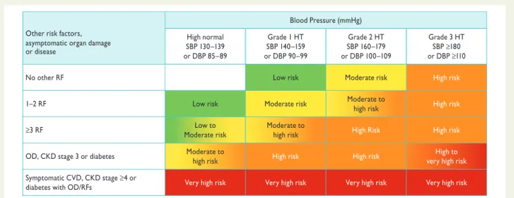
ESC Guidelines)1,2,54,55have stratified CV risk in different categor- ies, based on BP category, CV risk factors, asymptomatic OD and presence of diabetes, symptomatic CVD or chronic kidney disease (CKD), as also done by the 2012 ESC prevention guidelines.50 The classification in low, moderate, high and very high risk is retained in the current guidelines and refers to the 10-year risk of CV mortality as defined by the 2012 ESC prevention guidelines (Figure 1).50 The factors on which the stratification is based are summarized inTable4.
2.4.2 Limitations
All currently available models for CV risk assessment have limitations that must be appreciated. The significance of OD in determining calculation of overall risk is dependent on how carefully the damage is assessed, based on available facilities. Conceptual limita- tions should also be mentioned. One should never forget that the ra- tionale of estimating total CV risk is to govern the best use of limited resources to prevent CVD; that is, to grade preventive measures in relation to the increased risk. Yet, stratification of absolute risk is often used by private or public healthcare providers to establish a barrier, below which treatment is discouraged. It should be kept in
BP = blood pressure; CKD = chronic kidney disease; CV = cardiovascular; CVD = cardiovascular disease; DBP = diastolic blood pressure; HT = hypertension;
OD = organ damage; RF = risk factor; SBP = systolic blood pressure.
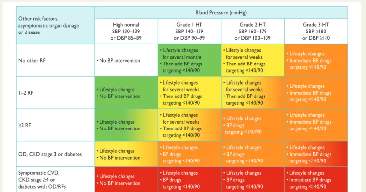
Other risk factors, asymptomatic organ damage or disease
Blood Pressure (mmHg) High normal
SBP 130–139 or DBP 85–89
Grade 1 HT SBP 140–159 or DBP 90–99
Grade 2 HT SBP 160–179 or DBP 100–109
Grade 3 HT SBP ≥180 or DBP ≥110
No other RF Low risk Moderate risk High risk
1–2 RF Low risk Moderate risk Moderate to
high risk High risk
≥3 RF Low to
Moderate risk
Moderate to
high risk High Risk High risk
OD, CKD stage 3 or diabetes Moderate to
high risk High risk High risk High to
very high risk Symptomatic CVD, CKD stage ≥4 or
diabetes with OD/RFs Very high risk Very high risk Very high risk Very high risk
Figure 1 Stratification of total CV risk in categories of low, moderate, high and very high risk according to SBP and DBP and prevalence of RFs, asymptomatic OD, diabetes, CKD stage or symptomatic CVD. Subjects with a high normal office but a raised out-of-office BP (masked hypertension) have a CV risk in the hypertension range. Subjects with a high office BP but normal out-of-office BP (white-coat hypertension), particularly if there is no diabetes, OD, CVD or CKD, have lower risk than sustained hypertension for the same office BP.
by guest on February 8, 2014http://eurheartj.oxfordjournals.org/Downloaded from
mind that any threshold used to define high total CV risk is arbitrary, as well as the use of a cut-off value leading to intensive interventions above this threshold and no action at all below. Finally, there is a strong effect of age on total CV risk models. It is so strong that younger adults (particularly women) are unlikely to reach high-risk levels even when they have more than one major risk factor and a clear increase in relative risk. By contrast, many elderly men (e.g.
.70 years) reach a high total risk level whilst being at very little increased risk relative to their peers. The consequences are that most resources are concentrated in older subjects, whose potential lifespan is relatively short despite intervention, and little attention is given to young subjects at high relative risk despite the fact that, in the absence of intervention, their long-term exposure to an increased risk may lead to a high and partly irreversible risk situation in middle age, with potential shortening of their otherwise longer life expectancy.
2.4.3 Summary of recommendations on total cardiovascular risk assessment
3 Diagnostic evaluation
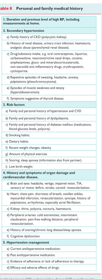
The initial evaluation of a patient with hypertension should (i) confirm the diagnosis of hypertension, (ii) detect causes of secondary hyper- tension, and (iii) assess CV risk, OD and concomitant clinical condi- tions. This calls for BP measurement, medical history including family history, physical examination, laboratory investigations and further diagnostic tests. Some of the investigations are needed in all patients;
others only in specific patient groups.
Table 4 Factors—other than office BP—influencing prognosis; used for stratification of total CV risk inFigure1
Risk factors Male sex
Age (men ≥55 years; women ≥65 years) Smoking
Dyslipidaemia
Total cholesterol >4.9 mmol/L (190 mg/dL), and/or
Low-density lipoprotein cholesterol >3.0 mmol/L (115 mg/dL), and/or
High-density lipoprotein cholesterol: men <1.0 mmol/L (40 mg/dL), women <1.2 mmol/L (46 mg/dL), and/or Triglycerides >1.7 mmol/L (150 mg/dL)
Fasting plasma glucose 5.6–6.9 mmol/L (102–125 mg/dL) Abnormal glucose tolerance test
Obesity [BMI ≥30 kg/m2 (height2)]
Abdominal obesity (waist circumference: men ≥102 cm;
women ≥88 cm) (in Caucasians)
Family history of premature CVD (men aged <55 years;
women aged <65 years) Asymptomatic organ damage
Pulse pressure (in the elderly) ≥60 mmHg
Electrocardiographic LVH (Sokolow–Lyon index >3.5 mV;
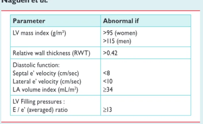
RaVL >1.1 mV; Cornell voltage duration product >244 mV*ms), or Echocardiographic LVH [LVM index: men >115 g/m2;
women >95 g/m2 (BSA)]a
Carotid wall thickening (IMT >0.9 mm) or plaque Carotid–femoral PWV >10 m/s
Ankle-brachial index <0.9
Microalbuminuria (30–300 mg/24 h), or albumin–creatinine ratio (30–300 mg/g; 3.4–34 mg/mmol) (preferentially on morning spot urine)
Diabetes mellitus
Fasting plasma glucose ≥7.0 mmol/L (126 mg/dL) on two repeated measurements, and/or
HbA1c >7% (53 mmol/mol), and/or
Post-load plasma glucose >11.0 mmol/L (198 mg/dL) Established CV or renal disease
Cerebrovascular disease: ischaemic stroke; cerebral haemorrhage;
transient ischaemic attack
CHD: myocardial infarction; angina; myocardial revascularization with PCI or CABG
Heart failure, including heart failure with preserved EF Symptomatic lower extremities peripheral artery disease CKD with eGFR <30 mL/min/1.73m2 (BSA); proteinuria (>300 mg/24 h).
Advanced retinopathy: haemorrhages or exudates, papilloedema CKD with eGFR 30–60 mL/min/1.73 m2 (BSA)
BMI¼body mass index; BP¼blood pressure; BSA¼body surface area; CABG¼ coronary artery bypass graft; CHD¼coronary heart disease; CKD¼chronic kidney disease; CV¼cardiovascular; CVD¼cardiovascular disease; EF¼ejection fraction; eGFR¼estimated glomerular filtration rate; HbA1c¼glycated haemoglobin; IMT¼intima-media thickness; LVH¼left ventricular hypertrophy;
LVM¼left ventricular mass; PCI¼percutaneous coronary intervention; PWV¼ pulse wave velocity.
aRisk maximal for concentric LVH: increased LVM index with a wall thickness/radius ratio of.0.42.
Total cardiovascular risk assessment
Recommendations Classa Levelb Ref.C In asymptomatic subjects
with hypertension but free of CVD, CKD, and diabetes, using the SCORE model is recommended as a minimal requirement.
I B 43
As there is evidence that OD predicts CV death independently of SCORE, a search for OD should be considered, particularly in individuals at moderate risk.
IIa B 51, 53
It is recommended that decisions on treatment strategies depend on the initial
level of total CV risk. I B 41, 42, 50
CKD¼chronic kidney disease; CV¼cardiovascular; CVD¼cardiovascular disease; OD¼organ damage; SCORE¼Systematic COronary Risk Evaluation
aClass of recommendation.
bLevel of evidence.
cReference(s) supporting recommendation(s).
by guest on February 8, 2014http://eurheartj.oxfordjournals.org/Downloaded from
3.1 Bood pressure measurement
3.1.1 Office or clinic blood pressure
At present, BP can no longer be estimated using a mercury sphygmo- manometer in many—although not all—European countries. Aus- cultatory or oscillometric semiautomatic sphygmomanometers are used instead. These devices should be validated according to standar- dized protocols and their accuracy should be checked periodically through calibration in a technical laboratory.56Measurement of BP at the upper arm is preferred and cuff and bladder dimensions should be adapted to the arm circumference. In the event of a signifi- cant (.10 mmHg) and consistent SBP difference between arms, which has been shown to carry an increased CV risk,57the arm with the higher BP values should be used. A between-arms difference is meaningful if demonstrated by simultaneous arm measurement; if one gets a difference between arms with sequential measurement, it could be due to BP variability. In elderly subjects, diabetic patients and in other conditions in which orthostatic hypotension may be fre- quent or suspected, it is recommended that BP be measured 1 min and 3 min after assumption of the standing position. Orthostatic hypotension—defined as a reduction in SBP of≥20 mmHg or in DBP of≥10 mmHg within 3 min of standing—has been shown to carry a worse prognosis for mortality and CV events.58,59If feasible, automated recording of multiple BP readings in the office with the patient seated in an isolated room, though providing less information overall, might be considered as a means to improve reproducibility and make office BP values closer to those provided by daytime ABPM or HBPM,60,61. BP measurements should always be associated with measurement of heart rate, because resting heart rate values in- dependently predict CV morbid or fatal events in several conditions, including hypertension.62,63Instructions for correct office BP mea- surements are summarized inTable5.
3.1.2 Out-of-office blood pressure
The major advantage of out-of-office BP monitoring is that it provides a large number of BP measurements away from the medical environ- ment, which represents a more reliable assessment of actual BP than office BP. Out-of-office BP is commonly assessed by ABPM or HBPM, usually by self-measurement. A few general principles and remarks hold for the two types of monitoring, in addition to recommenda- tions for office BP measurement:64–67
† The procedure should be adequately explained to the patient, with verbal and written instructions; in addition, self-measurement of BP requires appropriate training under medical supervision.
† Interpretation of the results should take into account that the re- producibility of out-of-office BP measurements is reasonably good for 24-h, day and night BP averages but less for shorter periods within the 24 hs and for more complex and derived indices.68
† ABPM and HBPM provide somewhat different information on the subject’s BP status and risk and the two methods should thus be regarded as complementary, rather than competitive or alterna- tive. The correspondence between measurements with ABPM and HBPM is fair to moderate.
† Office BP is usually higher than ambulatory and home BP and the difference increases as office BP increases. Cut-off values for the definition of hypertension for home and ambulatory BP, according
to the ESH Working Group on BP Monitoring, are reported in Table6.64–67
† Devices should have been evaluated and validated according to international standardized protocols and should be properly maintained and regularly calibrated; at least every 6 months. The validation status can be obtained on dedicated websites.
Table 5 Office blood pressure measurement
• To allow the patients to sit for 3–5 minutes before beginning BP measurements.
• To take at least two BP measurements, in the sitting position, spaced 1–2 min apart, and additional measurements if the
rst two are quite different. Consider the average BP if deemed appropriate.
• To take repeated measurements of BP to improve accuracy in p
• To use a standard bladder (12–13 cm wide and 35 cm long), but have a larger and a smaller bladder available for large (arm circumference >32 cm) and thin arms, respectively.
• To have the cuff at the heart level, whatever the position of the patient.
• When adopting the auscultatory method, use phase I and V (disappearance) Korotkoff sounds to identify systolic and diastolic BP, respectively.
• T
differences. In this instance, take the arm with the higher value as the reference.
• T
the standing position in elderly subjects, diabetic patients, and in other conditions in which orthostatic hypotension may be frequent or suspected.
• To measure, in case of conventional BP measurement, heart rate by pulse palpation (at least 30 s) after the second measurement in the sitting position.
BP¼blood pressure.
Table 6 Definitions of hypertension by office and out-of-office blood pressure levels
Category Systolic BP
(mmHg)
Diastolic BP (mmHg)
Daytime (or awake) ≥135 and/or ≥85
Nighttime (or asleep) ≥120 and/or ≥70
24-h ≥130 and/or ≥80
Home BP ≥135 and/or ≥85
140 and/or 90
Office BP Ambulatory BP
≥ ≥
BP¼blood pressure.
by guest on February 8, 2014http://eurheartj.oxfordjournals.org/Downloaded from
3.1.2.1 Ambulatory blood pressure monitoring
3.1.2.1.1 Methodological aspects A number of methodological aspects have been addressed by the ESH Working Group on Blood Pressure Monitoring.64,65 ABPM is performed with the patient wearing a portable BP measuring device, usually on the non-dominant arm, for a 24 – 25 h period, so that it gives information on BP during daily activities and at night during sleep. At the time of fitting of the portable device, the difference between the initial values and those from BP measurement by the operator should not be greater than 5 mmHg. In the event of a larger difference, the ABPM cuff should be removed and fitted again. The patient is instructed to engage in normal activities but to refrain from strenuous exercise and, at the time of cuff inflation, to stop moving and talking and keep the arm still with the cuff at heart level. The patient is asked to provide infor- mation in a diary on symptoms and events that may influence BP, in addition to the times of drug ingestion, meals and going to- and rising from bed. In clinical practice, measurements are often made at 15 min intervals during the day and every 30 min overnight; exces- sive intervals between BP readings should be avoided because they reduce the accuracy of 24-h BP estimates.69It may be recommended that measurements be made at the same frequency during the day and night—for example every 20 min throughout. The measurements are downloaded to a computer and a range of analyses can be performed. At least 70% of BPs during daytime and night-time periods should be satisfactory, or else the monitoring should be repeated. The detection of artifactual readings and the handling of outlying values have been subject to debate but, if there are suf- ficient measurements, editing is not considered necessary and only grossly incorrect readings should be deleted. It is noteworthy that readings may not be accurate when the cardiac rhythm is marked- ly irregular.70
3.1.2.1.2 Daytime, night-time and 24-hour blood pressure In addition to the visual plot, average daytime, night-time and 24-h BP are the most commonly used variables in clinical practice. Average daytime and night-time BP can be calculated from the diary on the basis of the times of getting up and going to bed. An alternative method is to use short, fixed time periods, in which the rising and retiring periods—which differ from patient to patient—are eliminated. It has, for example, been shown that average BPs from 10 am to 8 pm and from midnight to 6 am correspond well with the actual waking and sleeping BPs,71but other short, fixed time periods have been pro- posed, such as from 9 am to 9 pm and from 1 am to 6 am. In the event of different measurement intervals during the day and the night, and to account for missing values, it is recommended that average 24-h BP be weighted for the intervals between successive readings or to cal- culate the mean of the 24 hourly averages to avoid overestimation of average 24-h BP.72
The night-to-day BP ratio represents the ratio between average night-time and daytime BP. BP normally decreases during the night—defined as ‘dipping’. Although the degree of night-time dipping has a normal distribution in a population setting, it is generally agreed that the finding of a nocturnal BP fall of.10% of daytime values (night – day BP ratio,0.9) will be accepted as an arbitrary cut-off to define subjects as ‘dippers’. Recently, more dipping categories have been proposed: absence of dipping, i.e. nocturnal BP increase (ratio .1.0); mild dipping (0.9,ratio≤1.0); dipping (0.8,ratio≤0.9); and extreme dipping (ratio≤0.8). One should bear in mind that the reproducibility of the dipping pattern is limited.73,74 Possible reasons for absence of dipping are sleep
disturbance, obstructive sleep apnoea, obesity, high salt intake in salt- sensitive subjects, orthostatic hypotension, autonomic dysfunction, chronic kidney disease (CKD), diabetic neuropathy and old age.
3.1.2.1.3 Additional analyses A number of additional indices may be derived from ABPM recordings.75–81They include: BP variability,75 morning BP surge,76,77,81blood pressure load,78and the ambulatory arterial stiffness index.79,80However, their added predictive value is not yet clear and they should thus be regarded as experimental, with no routine clinical use. Several of these indices are discussed in detail in ESH position papers and guidelines,64,65including informa- tion on facilities recommended for ABPM software in clinical prac- tice, which include the need for a standardized clinical report, an interpretative report, a trend report to compare recordings obtained over time and a research report, offering a series of additional para- meters such as those listed above.
3.1.2.1.4 Prognostic significance of ambulatory blood pressure Several studies have shown that hypertensive patients’ left ventricular hyper- trophy (LVH), increased carotid intima-media thickness (IMT) and other markers of OD correlate with ambulatory BP more closely than with office BP.82,83Furthermore, 24-h average BP has been con- sistently shown to have a stronger relationship with morbid or fatal events than office BP.84–87 There are studies in which accurately measured office BP had a predictive value similar to ambulatory BP.87 Evidence from meta-analyses of published observational studies and pooled individual data,88–90however, has shown that ambulatory BP in general is a more sensitive risk predictor of clinical CV outcomes, such as coronary morbid or fatal events and stroke, than office BP. The superiority of ambulatory BP has been shown in the general population, in young and old, in men and women, in untreated and treated hypertensive patients, in patients at high risk and in patients with CV or renal disease.89–93Studies that accounted for daytime and night-time BP in the same statistical model found that night-time BP is a stronger predictor than daytime BP.90,94 The night – day ratio is a significant predictor of clinical CV outcomes but adds little prognostic information over and above 24-h BP.94,95 With regard to the dipping pattern, the most consistent finding is that the incidence of CV events is higher in patients with a lesser or no drop in nocturnal BP than in those with greater drop,89,91,92,95,96
although the limited reproducibility of this phe- nomenon limits the reliability of the results for small between- group differences.89,91,92,95
Extreme dippers may have an increased risk for stroke.97However, data on the increased CV risk in extreme dippers are inconsistent and thus the clinical significance of this phe- nomenon is uncertain.89,95
3.1.2.2 Home blood pressure monitoring
3.1.2.2.1 Methodological aspects The ESH Working Group on Blood Pressure Monitoring has proposed a number of recommendations for HBPM.66,67The technique usually involves self-measurement of BP but, in some patients, the support of a trained health provider or family member may be needed. Devices worn on the wrist are cur- rently not recommended but their use might be justified in obese sub- jects with extremely large arm circumference. For diagnostic evaluation, BP should be measured daily on at least 3 – 4 days and pref- erably on 7 consecutive days; in the mornings as well as in the eve- nings. BP is measured in a quiet room, with the patient in the seated position, back and arm supported, after 5 min of rest and with two measurements per occasion taken 1 – 2 min apart: the results are reported in a standardized logbook immediately after
by guest on February 8, 2014http://eurheartj.oxfordjournals.org/Downloaded from
each measurement. However, BP values reported by the patient may not always be reliable, which can be overcome by storage in a memory-equipped device. Home BP is the average of these readings, with exclusion of the first monitoring day. Use of telemonitoring and smartphone applications for HBPM may be of further advantage.98,99 Interpretation of the results should always be under the close guid- ance of the physician.
When compared with office BP, HBPM yields multiple measure- ments over several days, or even longer periods, taken in the indivi- dual’s usual environment. Compared with ambulatory BP, it provides measurements over extended periods and day-to-day BP variability, is cheaper,100more widely available and more easily re- peatable. However, unlike ABPM, it does not provide BP data during routine, day-to-day activities and during sleep, or the quantifi- cation of short-term BP variability.101
3.1.2.2.2 Prognostic significance of home BP Home BP is more closely related to hypertension-induced OD than office BP, particularly LVH,82,83and recent meta-analyses of the few prospective studies in the general population, in primary care and in hypertensive patients, indicate that the prediction of CV morbidity and mortality is significantly better with home BP than with office BP.102,103 Studies in which both ABPM and HBPM were performed show that home BP is at least as well correlated with OD as is the ambulatory BP,82,83and that the prognostic significance of home BP is similar to that of ambulatory BP after adjustment for age and gender.104,105
3.1.3 White-coat (or isolated office) hypertension and masked (or isolated ambulatory) hypertension Office BP is usually higher than BP measured out of the office, which has been ascribed to the alerting response, anxiety and/or a condi- tional response to the unusual situation,106and in which regression to the mean may play a role. Although several factors involved in office or out-of-office BP modulation may be involved,107the differ- ence between the two is usually referred to—although somewhat improperly—as the ‘white-coat effect’,107,108whereas ‘white-coat-’
or ‘isolated office-’ or ‘isolated clinic hypertension’ refers to the con- dition in which BP is elevated in the office at repeated visits and normal out of the office, either on ABPM or HBPM. Conversely, BP may be normal in the office and abnormally high out of the medical environment, which is termed ‘masked-’ or ‘isolated ambulatory hypertension’. The terms ‘true-’ or ‘consistent normotension’ and
‘sustained hypertension’ are used when both types of BP measure- ment are, respectively, normal or abnormal. Whereas the cut-off value for office BP is the conventional 140/90 mmHg, most studies in white-coat or masked hypertension have used a cut-off value of 135/85 mmHg for out-of-office daytime or home BP and 130/
80 mmHg for 24-h BP. Notably, there is only moderate agreement between the definition of white-coat or masked hypertension diag- nosed by ABPM or HBPM.101It is recommended that the terms
‘white-coat hypertension’ and ‘masked hypertension’ be reserved to define untreated individuals.
3.1.3.1 White-coat hypertension
Based on four population studies, the overall prevalence of white- coat hypertension averaged 13% (range 9 – 16%) and it amounted to about 32% (range 25 – 46%) among hypertensive subjects in these surveys.109Factors related to increased prevalence of white-
coat hypertension are: age, female sex and non-smoking. Prevalence is lower in the case of target OD or when office BP is based on repeated measurements or when measured by a nurse or another healthcare provider.110,111 The prevalence is also related to the level of office BP: for example, the percentage of white-coat hyper- tension amounts to about 55% in grade 1 hypertension and to only about 10% in grade 3 hypertension.110OD is less prevalent in white- coat hypertension than in sustained hypertension and prospective studies have consistently shown this to be the case also for CV events.105,109,112,113
Whether subjects with white-coat hypertension can be equalled to true normotensive individuals is an issue still under debate because, in some studies, the long-term CV risk of this condi- tion was found to be intermediate between sustained hypertension and true normotension,105whereas in meta-analyses it was not sig- nificantly different from true normotension when adjusted for age, gender and other covariates.109,112,113
The possibility exists that, because white-coat hypertensive patients are frequently treated, the reduction of clinic BP leads to a reduced incidence of CV events.112 Other factors to consider are that, compared with true normotensive subjects, in white-coat hypertensive patients, (i) out-of-office BP is higher,105,109(ii) asymptomatic OD such as LVH may be more frequent,114and (iii) this is the case also for metabolic risk factors and long-term risk of new-onset diabetes and progression to sustained hypertension.115,116It is recommended that the diagnosis of white-coat hypertension be confirmed within 3 – 6 months and these patients be investigated and followed-up closely, including repeated out-of-office BP measurements (see Section 6.1).
3.1.3.2 Masked hypertension
The prevalence of masked hypertension averages about 13%
(range 10 – 17%) in population-based studies.109 Several factors may raise out-of-office BP relative to office BP, such as younger age, male gender, smoking, alcohol consumption, physical activity, exercise-induced hypertension, anxiety, job stress, obesity, diabetes, CKD and family history of hypertension and the prevalence is higher when office BP is in the high normal range.117Masked hypertension is frequently associated with other risk factors, asymptomatic OD and increased risk of diabetes and sustained hypertension.114–119 Meta-analyses of prospective studies indicate that the incidence of CV events is about two times higher than in true normotension and is similar to the incidence in sustained hypertension.109,112,117
The fact that masked hypertension is largely undetected and untreated may have contributed to this finding. In diabetic patients masked hypertension is associated with an increased risk of nephro- pathy, especially when the BP elevation occurs mainly during the night.120,121
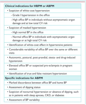
3.1.4 Clinical indications for out-of-office blood pressure It is now generally accepted that out-of-office BP is an important adjunct to conventional office BP measurement, but the latter cur- rently remains the ‘gold standard’ for screening, diagnosis and man- agement of hypertension. The time-honoured value of office BP, however, has to be balanced against its important limitations, which have led to the increasingly frequent suggestion that out-of-office BP measurements play an important role in hypertension manage- ment. Although there are important differences between ABPM
by guest on February 8, 2014http://eurheartj.oxfordjournals.org/Downloaded from