Management of progressive genu varum in a patient with Dyggve-Melchior-Clausen syndrome
Management einer ausgeprägten Genu varum-Fehlstellung bei einem Patienten mit Dyggve-Melchior-Clausen Syndrom
Abstract
We describe the orthopaedic management of progressive genu varum in a child who manifested the full phenotypic characterization of Dyggve- Melchior-Clausen syndrome.
Vladimir Kenis
1Alexey Baindurashvili
1Evgeniy Melchenko
1Keywords:Dyggve-Melchior-Clausen syndrome, genu varum, radiographs
Franz Grill
2Ali Al Kaissi
2,3Zusammenfassung
Wir beschreiben das orthopädische Management einer schweren Genu varum-Fehlstellung am Fall eines Kindes mit voller phänotypischer Ausprägung eines Dyggve-Melchior-Clausen Syndroms.
1 Pediatric Orthopedic Institute n.a. H. Turner, Department of Foot and Ankle Surgery, Schlüsselwörter:Dyggve-Melchior-Clausen Syndrom, Genu varum,
Röntgenbilder
Neuroorthopaedics and Systemic Disorders, Saint- Petersburg, Russia 2 Orthopaedic Hospital of
Speising, Paediatric Department, Vienna, Austria 3 Ludwig Boltzmann Institute
of Osteology at the Hanusch Hospital of WGKK and AUVA Trauma Centre Meidling, First Medical Department, Hanusch Hospital, Vienna, Austria
Background
Dyggve-Melchior-Clausen syndrome (DMC) and Smith- McCort dysplasia (SMC) are rare autosomal recessive osteochondrodysplasias. DMC was first described by Dyggve et al. in 1962 and SMC was originally described by Smith and McCort in 1958 as skeletal dysplasias. Be- cause both of the genes responsible for these disorders are localized on the same chromosome, they are thought of as allelic disorders. DMC and SMC are both disorders of bone and cartilage that are characterized by short trunk and extremities and a barrel shaped chest, in addition to mental retardation and microcephaly [1], [2], [3], [4]. The radiographic appearances of both disorders are similar.
Because of vertebral changes, differential diagnoses must include achondroplasia, brachyolmia (spondylodysplasia), spondylometaphyseal dysplasia, spondyloepimetaphyseal dysplasia, Morquio syndrome and metatropic dysplasia,
which all cause irregularities on the end-plates of verte- brae and platyspondyly [5]. Several studies have been published regarding DMC syndrome, very little included the orthopaedic management.
Case presentation
A 10-year-old boy was referred to our department because of short stature, waddling gait and skeletal maldevelop- ment. He was born in term after uneventful gestation, with birth length of 47 cm, and weight of 3100 g. He is the second son of the mother, but from the second mar- riage of non-consanguineous parents. Parents and first child are healthy without any family history of short stature, pathological gait, mental retardation and skeletal maldevelopment. His subsequent course of development has been of relative retardation in acquiring the skills of motor development. Walking has been achieved at the
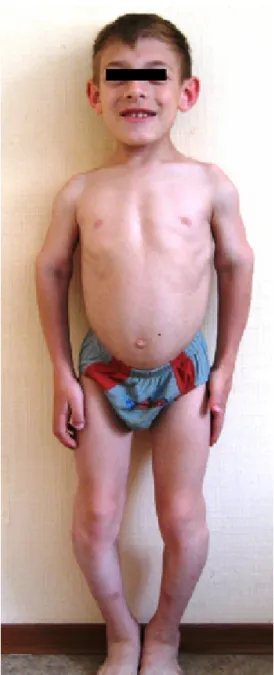
age of 18 months albeit with difficulty. A crouching stance was a prominent feature. Clinical examination at the age of 10 years showed: short stature (–3SD). Occipito- frontal circumference was around (–1SD). The phenotypic features were characteristic for DMC syndrome (Figure 1).
Skeletal survey and imagings were specific for this dys- plasia, especially on pelvis radiographs – lacy appearance of the iliac crests were evident (Figure 2, Figure 3, Figure 4). Hearing and vision were normal. Neurological examination was normal but intelligence was subnormal.
Craniocervical 3D reconstruction CT scan showed normal atlanto-dens distance with no odontoid hypoplasia. Note the calvarial thickening of the occipital region (Figure 5).
Figure 1: A boy manifesting coarse facies, sloping forehead with V shaped frontal hair, thick eye-brows, prominent maxilla,
prognathism and large/protruding ears, short neck, and barrel-shaped thorax. The upper limbs (rhizomelic) showed bilateral contractures of the elbows and the lower limbs showed
extensive genu varum.
Laboratory examinations showed normal phosphorus, alkaline phosphatase and calcium levels. Urine amino- acids and urinary excretion of mucopolysaccharides have been investigated (two dimensional electrophoresis of glycosaminoglycans in the urine and measurement of alpha-L-iduronidase and galactose-6-sulphatase enzyme activities were performed to exclude MPS type I and MPS type IVA respectively.
Treatment
The patient was firstly observed in our clinic at the age of 10 years after several years of observation by geneti- cists with the diagnosis of “spondylo-epi-metaphyseal dysplasia, non-verified type”. He underwent regular physical therapy aimed to enhance the mobility of the spine and the articulations.
In the last 2 years, we observed significant progression of his genu vara. On the antero-posterior standing radio- graphs the amount of varus deformity was 25° on the right side and 22° on the left side. Thereby, we used a guided growth technique, aiming to prevent further deteri- oration (Figure 6). The progressive nature of lower limb deformities and the poor quality of the osseous tissue are adverse factors against the performance of extensive bony surgery such as (osteotomies with internal or exter- nal fixation). Thus, minimal-invasive surgery without long- lasting immobilisation and full weight bearing immediately after procedure seems to be more advantageous. The post-operative follow-up for the period of six-months, no further progression of the deformity was notable.
The antero-posterior radiographs of the hips showed in- stability secondary to defective ossification of the capital femoral epiphyses and the acetabulae (micro-architectural deterioration of bone tissue). Migration percentage was 0.8 on the right side and 0.7 on the left side by the bony landmarks. The central-edge angle (Wiberg angle) was –20° on the both sides and Shenton line was somehow distorted. MRI-imagings confirmed the defective ossifica- tion of the femoral heads and the acetabular edges with subsequent lateral displacement of the ossification centres. Cartilaginous coverage of the femoral heads was more than 60% (Wiberg angle of +10° by cartilaginous landmarks). At this stage, however, surgical realignment was thought to be non-mandatory. But, nevertheless rapid and severe hip deterioration has to be expected.
Genetics
Neumann et al. [4] looked at 2 families with DMC and 1 with Smith-McCort, and found mutations in the DMC families but not in the Smith-McCort family. El Ghouzzi et al. [6] demonstrated mutations in a novel gene – dymeclin in families mapping to 18q21.1. The gene transcript is expressed in chondrocytes and brain but is of unknown function. Cohn et al. [7] also demonstrated mutations in Dygvve-Melchior-Clausen and Smith-McCort cases. In the
Kenis et al.: Management of progressive genu varum in a patient ...
Figure 2: Anteroposterior pelvis radiograph showed a dysplastic pelvis. The iliac crest showed a pathognomonic, semi lunar, irregular lacy margin with a wide irregular sacro-iliac joint. Note the supero-lateral collapse of the femoral heads bilaterally causing lateral displacement of the femoral heads (associated with epi-metaphyseal irregularities). Defective ossification of
the acetabuli was evident.
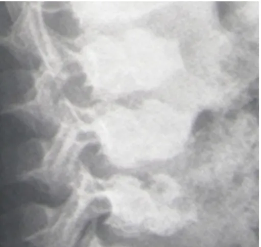
Figure 3: Lateral spine radiograph showed platyspondyly, double vertebral hump (beaked/wedged) vertebrae associated with irregular end-plates.
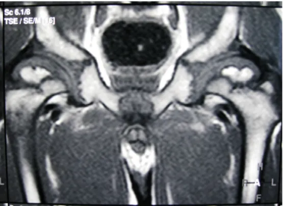
Figure 4: MRI image showed defective ossification of the femoral heads confirmed the superolateral femoral head collapse (a feature of progressive dysplasia and severe joint degeneration) associated with femoral head osteonecrosis (note the medial
beaking of the femoral heads). The latter is the reason of femoral head collapse.
Figure 5: 3D reconstruction CT scan showed normal atlanto-dens distance with no odontoid hypoplasia. Note the calvarial thickening of the occipital region.
Kenis et al.: Management of progressive genu varum in a patient ...
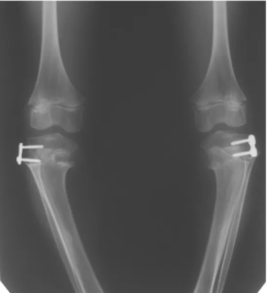
Figure 6: Anteroposterior standing radiograph of the knee joints, 3 months after temporary lateral hemiepyphyseodesis of both tibiae
former there were null mutations. In a review by Paupe et al. [8] it is stated that to date, there have been 16 dif- ferent mutations found in 21 families.
Discussion
Dyggve, Melchior, and Clausen reported three siblings from a consanguineous mating in Greenland with “Mor- quio-Ullrich disease” [1], [2], although earlier examples appear to have been published [9]. Patients with DMC syndrome are different than patients with Morquio syn- drome.
DMC patients had no corneal clouding, no urinary muco- polysaccharides and are mentally retarded. The main features are mental retardation, short stature, short-trunk dwarfism, progressive sternal bulging, and delayed bone age.
There is genetic heterogeneity, both autosomal recessive and X-linked recessive (Smith-McCort syndrome) forms have been recognized [3]. In the autosomal recessive form, atlantoaxial instability predisposes to spinal cord
compression [10], [11]. Adult height ranges between 115–127 cm and in the X-linked form between 130–150 cm. By the age of 5 years, disproportionate short stature becomes evident. Joint mobility is somehow restricted.
The skeletal changes are present by the age of 4–5 years when flattening of the vertebral bodies associated with radiolucent depression in their mid-portion, can be detect- ed. Within a few years the vertebral bodies become flat and there is a lacy appearance of the iliac crest on pelvic radiographs. The femoral heads are delayed in their ossi- fication and they might be fragmented. Progressive hip dislocation might occur. The skull commonly shows doli- chocephaly or microcephaly. The chest is barrel-shape with protruding sternum. The knees have genu valgum or genu varum. The cervical spine may have a narrow canal [1], [2], [5].
Naffah and Taleb [11] reported odontoid hypoplasia and C1/2 instability requiring spinal fusion in two patients with DMC syndrome. Cervical spine instability has been considered as a serious danger of severe neurological deficit. Shorr [12] described odontoid hypoplasia and coxa magna
Yunis et al. [13] reported a large X-linked pedigree where mental retardation was not a feature and suggested that the cases reported by Smith and McCort [14] and Burns et al. [3], could be examples of further X-linked cases where mental retardation may not be a feature. Nakamura et al. [15] reported two further cases of the Smith-McCort phenotype. They also showed from iliac crest biopsies that the rough endoplasmic reticulum contains fine granular or amorphous material. This is also seen in cases with mental retardation.
Conclusion
Preventive and corrective orthopaedic surgery is an option in complex management of lower limb deformities. Min- imal-invasive surgery (guided growth) may be preferable and further investigations with long-term follow-up are the baseline procedures in the management of such pa- tients.
Learning points
1. Mucopolysaccharidosis Type IVA is one of the most important diseases that must be distinguished from DMC and SMD with radiographic findings.
2. In contrast to Morquio syndrome, our patient has no mucopolysacchariduria and no corneal clouding.
3. Noteworthy features of normal hearing, normal teeth and a normal odontoid with no subsequent atlanto- axial instability, but microcephaly and mental sub- normality were notable in our present patient.
Notes
Competing interests
The authors declare that they have no competing in- terests.
Informed consent
Our research project has been approved by the Medical University of Vienna (Ethics committee, EK Nr: 921/2009) and informed consent was obtained from the patients guardians.
References
1. Dyggve HV, Melchior JC, Clausen J. Morquio-Ullrich's disease: an inborn error of metabolism? Arch Dis Child. 1962;37(195):525- 34. Available from: http://www.ncbi.nlm.nih.gov/pmc/articles/
PMC2012915/
2. Dyggve HV, Melchior JC, Clausen J, Rastogi SC. The Dyggve- Melchior-Clausen (DMC) syndrome. A 15 year follow-up and a survey of the present clinical and chemical findings.
Neuropadiatrie. 1977;8(4):429-42. DOI: 10.1055/s-0028- 1091538
3. Burns C, Powell BR, Hsia YE, Reinker K. Dyggve-Melchior-Clausen syndrome: report of seven patients with the Smith-McCort variant and review of the literature. J Pediatr Orthop. 2003;23(1):88-93.
DOI: 10.1097/01241398-200301000-00018
4. Neumann LM, El Ghouzzi V, Paupe V, Weber HP, Fastnacht E, Leenen A, Lyding S, Klusmann A, Mayatepek E, Pelz J, Cormier- Daire V. Dyggve-Melchior-Clausen syndrome and Smith-McCort dysplasia: clinical and molecular findings in three families supporting genetic heterogeneity in Smith-McCort dysplasia. Am J Med Genet A. 2006;140(5):421-6. DOI: 10.1002/ajmg.a.31090 5. Spranger JW, Brill PW, Poznanski AK. Bone dysplasias: An atlas of genetic disorders of skeletal development. 2nd ed. New York:
Oxford University Press; 2002.
6. El Ghouzzi V, Dagoneau N, Kinning E, Thauvin-Robinet C, Chemaitilly W, Prost-Squarcioni C, Al-Gazali LI, Verloes A, Le Merrer M, Munnich A, Trembath RC, Cormier-Daire V. Mutations in a novel gene Dymeclin (FLJ20071) are responsible for Dyggve- Melchior-Clausen syndrome. Hum Mol Genet. 2003;12(3):357- 64. DOI: 10.1093/hmg/ddg029
7. Cohn DH, Ehtesham N, Krakow D, Unger S, Shanske A, Reinker K, Powell BR, Rimoin DL. Mental retardation and abnormal skeletal development (Dyggve-Melchior-Clausen dysplasia) due to mutations in a novel, evolutionarily conserved gene. Am J Hum Genet. 2003;72(2):419-28. DOI: 10.1086/346176
8. Paupe V, Gilbert T, Le Merrer M, Munnich A, Cormier-Daire V, El Ghouzzi V. Recent advances in Dyggve-Melchior-Clausen syndrome. Mol Genet Metab. 2004;83(1-2):51-9. DOI:
10.1016/j.ymgme.2004.08.012
9. Farrell MJ, Maloney JD, Yakovlev PI. Morquio's disease associated with mental defect. Arch Neurol Psychiatry. 1942;48(3):456-64.
10. Naffah J. The Dyggve-Melchior-Clausen syndrome. Am J Hum Genet. 1976;28(6):607-14.
11. Naffah J, Taleb N. Deux nouveaux cas de syndrome de Dyggve- Melchior-Clausen avec hypoplasie de l'apophyse odontoide et compression spinale [2 further cases of Dyggve-Melchior-Clausen syndrome with hypoplasia of the odontoid apophysis and spinal compression]. Arch Fr Pediatr.1974;31(10):985-92.
12. Schorr S, Legum C, Ochshorn M, Hirsch M, Moses S, Lasch EE, El-Masri M. The Dyggve-Melchio-Clausen syndrome. AJR Am J Roentgenol. 1977;128(1):107-13.
13. Yunis E, Fontalvo J, Quintero L. X-linked Dyggve-Melchior-Clausen syndrome. Clin Genet. 1980;18(4):284-90. DOI: 10.1111/j.1399- 0004.1980.tb00887.x
14. Smith R, McCort JJ. Osteochondrodystrophy (Morquio-Brailsford type); occurrence in three siblings. Calif Med. 1958;88(1):55-9.
15. Nakamura K, Kurokawa T, Nagano A, Nakamura S, Taniguchi K, Hamazaki M. Dyggve-Melchior-Clausen syndrome without mental retardation (Smith-McCort dysplasia): morphological findings in the growth plate of the iliac crest. Am J Med Genet.
1997;72(1):11-7. DOI: 10.1002/(SICI)1096- 8628(19971003)72:1<11::AID-AJMG3>3.0.CO;2-Y
Corresponding author:
Ali Al Kaissi
Ludwig Boltzmann Institute of Osteology at the Hanusch Hospital of WGKK and AUVA Trauma Centre Meidling, First Medical Department, Hanusch Hospital, Heinrich Collin Str. 30, A-1140, Vienna, Austria
ali.alkaissi@osteologie.at
Kenis et al.: Management of progressive genu varum in a patient ...
Please cite as
Kenis V, Baindurashvili A, Melchenko E, Grill F, Al Kaissi A. Management of progressive genu varum in a patient with Dyggve-Melchior-Clausen syndrome. GMS Ger Med Sci. 2011;9:Doc25.
DOI: 10.3205/000148, URN: urn:nbn:de:0183-0001489
This article is freely available from
http://www.egms.de/en/journals/gms/2011-9/000148.shtml
Received:2011-06-03 Revised:2011-08-25 Published:2011-09-20
Copyright
©2011 Kenis et al. This is an Open Access article distributed under the terms of the Creative Commons Attribution License
(http://creativecommons.org/licenses/by-nc-nd/3.0/deed.en). You are free: to Share — to copy, distribute and transmit the work, provided the original author and source are credited.