https://doi.org/10.5852/ejt.2019.517 www.europeanjournaloftaxonomy.eu 2019 · Fernandes M.R. & Pimenta A.D.
This work is licensed under a Creative Commons Attribution License (CC BY 4.0).
M o n o g r a p h
urn:lsid:zoobank.org:pub:CAC6F8AF-ED37-4989-9672-68316920750B
Basic anatomy of species of Triphoridae (Gastropoda, Triphoroidea) from Brazil
Maurício Romulo FERNANDES
1,2,*& Alexandre Dias PIMENTA
21
Departamento de Zoologia, Instituto de Biociências, Universidade Federal do Estado do Rio de Janeiro, Av. Pasteur, 458, Urca, 22290-240, Rio de Janeiro, Brazil.
2
Departamento de Invertebrados, Museu Nacional, Universidade Federal do Rio de Janeiro, Quinta da Boa Vista, São Cristóvão, 20940-040, Rio de Janeiro, Brazil.
*
corresponding author: mauriciofernandes14@hotmail.com
2
Email: alexpim@mn.ufrj.br
1
urn:lsid:zoobank.org:author:6D9B33AF-4E10-4C8C-A2F4-EF21BBF829AC
2
urn:lsid:zoobank.org:author:F25DDF06-C6B5-42BA-9EC0-27E86FAD16A5
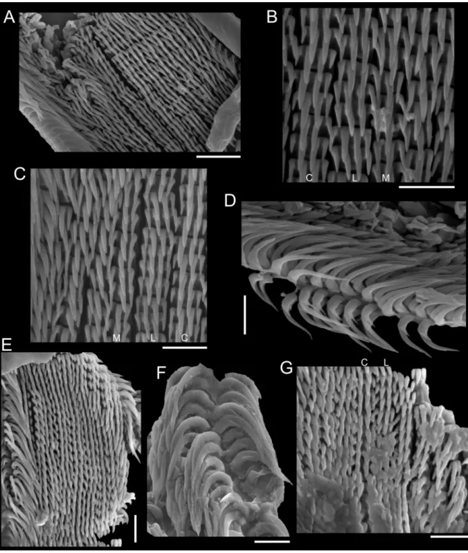
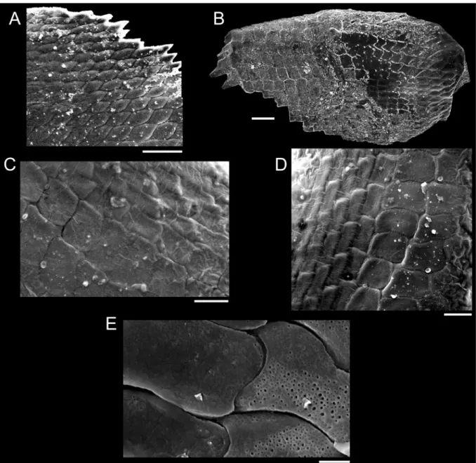
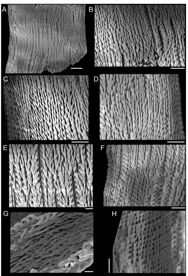
Abstract. The alpha-taxonomy of triphorids is still largely based on the study of the shell, and the scarcity of studies dealing with their anatomy is a result of the diffi culty of sampling live animals and their very small size. Whereas radula and operculum are important structures in the taxonomy at the generic level, the jaw of triphorids has never been properly studied, being regarded as presenting a morphological homogeneity. The present research explored the basic anatomy (especially internal hard structures: operculum, jaw and radula) of 12 species from Brazil, distributed in 11 genera: Cheirodonta Marshall, 1983 (with a new generic allocation, Cheirodonta dupliniana (Olsson, 1916) comb. nov.), Cosmotriphora Olsson & Harbison, 1953, Iniforis Jousseaume, 1884, Latitriphora Marshall, 1983, Metaxia Monterosato, 1884, Monophorus Grillo, 1877, Nanaphora Laseron, 1958, Nototriphora Marshall, 1983, Sagenotriphora Marshall, 1983, Similiphora Bouchet, 1985 and Strobiligera Dall, 1924; in addition, the basic anatomy of the Caribbean species “Inella” harryleei Rolán & Fernández- Garcés, 2008 was analysed. Radular examination showed that the majority of species studied is properly allocated in their genera after comparisons in the literature with respective type species, albeit a few species are clearly in need of a new generic allocation. The jaw of triphorids is remarkably heterogeneous, displaying different patterns of scales and micro-pores between outer and inner sides.
Keywords. Taxonomy, morphology, radula, jaw, operculum.
Fernandes M.R. & Pimenta A.D. 2019. Basic anatomy of species of Triphoridae (Gastropoda, Triphoroidea) from Brazil. European Journal of Taxonomy 517: 1–60. https://doi.org/10.5852/ejt.2019.517
Introduction
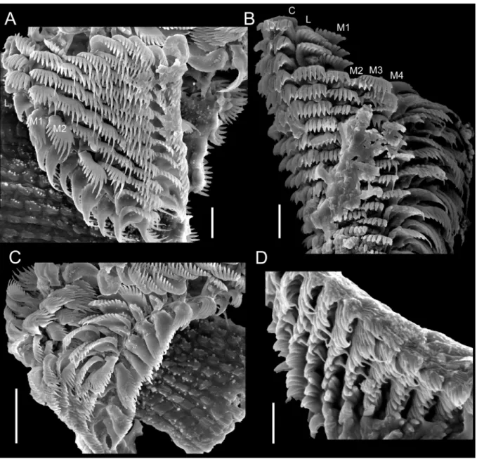
Members of the family Triphoridae are sponge feeders, and their enormous radular diversifi cation is believed to constitute an adaptation to the great morphological plasticity in Porifera (Marshall 1983;
Wells 1998). Despite being one of the most speciose families of marine molluscs in the world (Bouchet
et al. 2002; Albano et al. 2011), with 642 Recent valid species (WoRMS 2018) and hundreds of undescribed ones, most of them are known only by their shells. The scarcity of studies dealing with the anatomy of triphorids is a result of the diffi culty of sampling live animals, as dredging usually furnishes only empty shells, as well as of their small size; adults of most species reach less than 10 mm in shell length (Wells 1998).
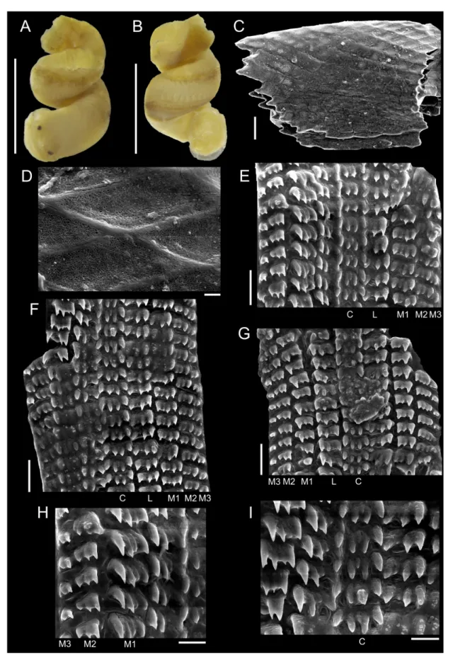
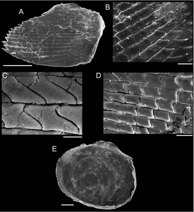
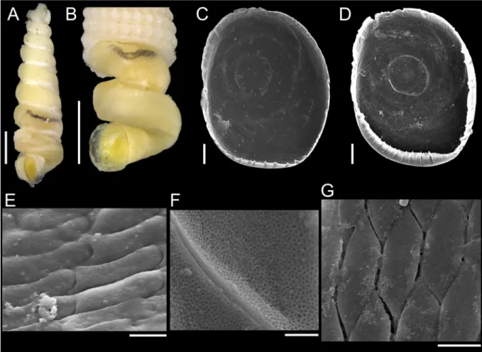
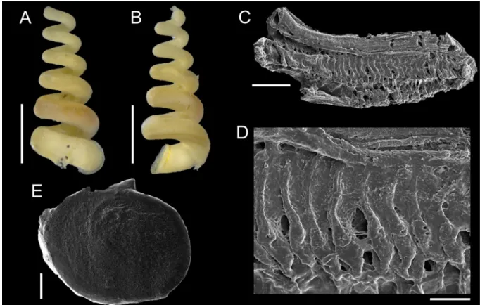
The sinistral coiling of triphorids (not considering the subfamily Metaxiinae, which is composed of species with dextral coiling) makes them unusual in the common sense of gastropods’ anatomy: osphradium and ctenidium lie in the right side of the body, whereas rectum and genital duct lie in the left side (Fretter 1951). They have an usually thin and corneous operculum, circular to elliptical, with two to seven whorls (Marshall 1983). The great variation in number, shape and size of radular teeth makes Triphoridae probably the gastropod family with the highest diversity of radula, well aware that this structure is a very important attribute to generic delimitations (Marshall 1983; Wells 1998). The radular ribbon is long and thin, bearing hundreds of teeth rows, with formula (1-30)-1-1-1-(1-30) (Marshall 1983).
Triphorids have elongated cephalic tentacles, eyes positioned at their bases, and an acrembolic proboscis. They have a narrow foot and a well-developed posterior pedal gland, without pedal tentacles (Wells 1998). Distinct mantle tentacles extend into the posterior canal, and their length may serve to taxonomic purposes (Nützel 1998), although these structures were barely noticed in previous works.
Triphorids also show a typical glandular pouch that lies dorsally to the esophagus, opening at the end of the posterior esophagus (Kosuge 1966). They are dioecious but males lack a penis, transferring mobile spermatozeugmata into the pallial cavity of the female, which has an elaborated reproductive system (Kosuge 1966).
The majority of studies comprizing the anatomy of triphorids is related only to their internal hard structures, especially opercula and radula (e.g., Kosuge 1966; Marshall 1983; Bandel 1984; Bouchet 1985; Rolán & Fernández-Garcés 1994, 1995, 2008; Nützel 1998), with very few studies illustrating or describing jaws (Risbec 1943; Fretter 1951; Kosuge 1966; Bouchet 1985; Nützel 1998). Colored photographs of the external morphology of soft parts of triphorids are scarce (e.g., Bouchet & Guillemot 1978; Redfern 2013; Stephens & Vafi adis 2015). There are even rarer studies illustrating particular aspects of the reproductive, digestive or nervous systems (e.g., Risbec 1943; Fretter 1951; Johansson 1953;
Marcus & Marcus 1963; Kosuge 1966; Houston 1985), owing to the limitation of drawings on camera lucida of these small animals. Haszprunar (1985) and Healy (1990) conducted ultrastructural histologic sections on a few triphorids (the former on the osphradium, the latter on the spermatozeugmata), and Golding et al. (2009) provided histological sections of the proboscis of one species.
Recent studies increased the total number of triphorids known from Brazil to 66 (Fernandes & Pimenta in prep.). All of them were studied only by their shells, precluding comparisons with Caribbean specimens illustrated by the external morphology of soft parts, operculum or radula (Bandel 1984; Rolán &
Fernández-Garcés 1994, 1995, 2008; Redfern 2013). An exception to this is Marshallora cf. nigrocincta (C.B. Adams, 1839), which was studied by Marcus & Marcus (1963) in southeastern Brazil and was the target of an integrative taxonomic approach (Fernandes et al. in prep.), thus not being evaluated in the present study. A summary of studies dealing with anatomical aspects of the triphorid genera addressed in this work is furnished below (Table 1).
The objective of this research is to examine basic aspects of the anatomy of triphorids occurring in
Brazil and to provide comparisons at generic and specifi c levels. For this purpose, colored photographs
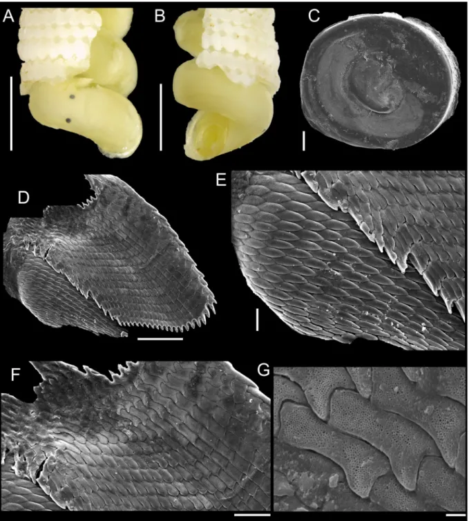
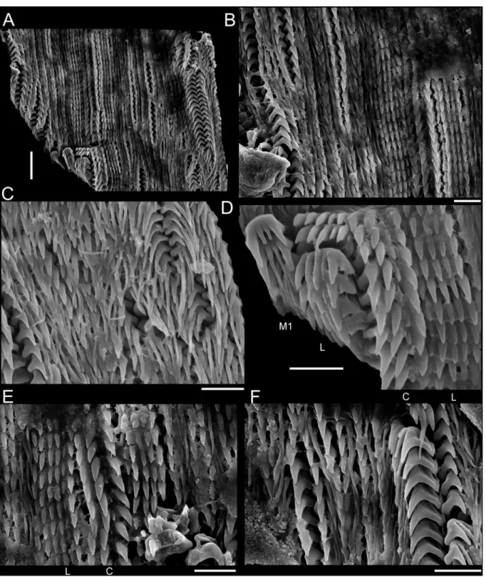
of the external morphology of soft parts and scanning electron microscope (SEM) images of internal
hard structures, i.e., opercula, radulae and jaws, were conducted. The importance of these structures in
the phylogenetic context of Triphoridae is also discussed.
Table 1 (continued on 2 next pages). Previous morphological studies on Triphoridae, regarded only genera addressed in this work. Some studies illustrated but did not describe the respective morphological feature(s), or the inverse. Type species are indicated by an asterisk (*). Abbreviations: EA = Eastern Atlantic (comprising the Mediterranean); WA = Western Atlantic; WP = Western Pacifi c.
Species Geographical
range Morphological features References
Metaxia Monterosato, 1884 Metaxia rugulosa
(C.B. Adams, 1850) WA External morphology Redfern (2013)
*Metaxia metaxa
(Delle Chiaje, 1828) EA External morphology; radula Bouchet (1985)
Metaxia exaltata (Powell, 1930) WP External morphology; operculum;
jaw; radula Marshall (1977)
Metaxia spp. WP Operculum; jaw; radula Nützel (1998)
Cheirodonta Marshall, 1983
*Cheirodonta pallescens
(Jeffreys, 1867) EA External morphology; operculum;
radula
Fretter (1951), described as Triphora perversa;
Bouchet & Guillemot (1978); Bouchet (1985);
Cheirodonta labiata
(A. Adams, 1854) WP Operculum; radula Marshall (1983)
Cosmotriphora Olsson & Harbison, 1953
*Cosmotriphora melanura
(C.B. Adams, 1850) WA/EA External morphology; operculum;
radula Bouchet (1985); Rolán &
Fernández-Garcés (1994) Iniforis Jousseaume, 1884
Iniforis turristhomae (Holten, 1802) WA External morphology; operculum;
radula Bandel (1984); Rolán &
Fernández-Garcés (1993)
*Iniforis malvacea Jousseaume,
1884 WP Radula Marshall (1983)
Iniforis violacea
(Quoy & Gaimard, 1834) WP Radula Marshall (1983)
Latitriphora Marshall, 1983 Latitriphora albida
(A. Adams, 1854) WA External morphology; operculum;
radula
Rolán & Fernández-Garcés (1995); Redfern (2013) Monophorus Grillo, 1877
Monophorus olivaceus (Dall, 1889) WA External morphology; radula Rolán & Fernández-Garcés (1994)
Monophorus ateralbus
Rolán & Fernández-Garcés, 1994 WA External morphology; radula Rolán & Fernández-Garcés (1994)
*Monophorus perversus
(Linnaeus, 1758) EA External morphology; radula Bouchet (1985)
Monophorus thiriotae
Bouchet, 1985 EA External morphology; radula Bouchet (1985)
Monophorus erythrosoma
(Bouchet & Guillemot, 1978) EA External morphology; radula Bouchet & Guillemot (1978); Bouchet (1985);
Fernandes & Rolán (1988) Monophorus verdensis
Fernandes & Rolán, 1988 EA External morphology; operculum;
radula Fernandes & Rolán (1988)
Species Geographical
range Morphological features References
Monophorus pantherinus
Rolán & Peñas, 2001 EA External morphology; operculum;
radula Rolán & Peñas (2001) Monophorus alboranensis
Rolán & Peñas, 2001 EA Operculum Rolán & Peñas (2001)
Monophorus amicitiae
Romani, 2015 EA Operculum Romani (2015)
Monophorus sp. EA External morphology; radula Rolán & Peñas (2001) Monophorus angasi
(Crosse & Fischer, 1865) WP Operculum; radula Marshall (1983)
Monophorus nigrofuscus
(A. Adams, 1854) WP Operculum; radula Marshall (1983)
Monophorus fascelinus
(Suter, 1908) WP External morphology Nützel (1998)
Nanaphora Laseron, 1958 Nanaphora verbernei
(Moolenbeek & Faber, 1989) WA External morphology; operculum;
radula
Rolán & Fernández-Garcés (1994)
Nanaphora decollata
(Rolán & Fernández-Garcés, 1994) WA External morphology; operculum;
radula
Rolán & Fernández-Garcés (1994)
Nanaphora albogemmata
(Laseron, 1958) WP Operculum; radula Marshall (1983)
Nanaphora aff. albogemmata
(Laseron, 1958) WP Radula Nützel (1998)
Nototriphora Marshall, 1983 Nototriphora decorata
(C.B. Adams, 1850) WA External morphology; operculum;
radula
Garcia & Luque (1986);
Rolán & Fernández-Garcés (1994)
Nototriphora canarica
(Nordsieck & Talavera, 1979) EA External morphology; operculum;
radula
Fernandes & Rolán (1988);
Bouchet (1997)
Nototriphora vestita Marshall, 1983 WP Operculum Marshall (1983)
*Nototriphora aupouria
(Powell, 1937) WP Operculum; radula Marshall (1983)
Sagenotriphora Marshall, 1983 Sagenotriphora osclausum
(Rolán & Fernández-Garcés, 1995) WA Operculum; radula Rolán & Fernández-Garcés (2008)
Sagenotriphora candidula
Rolán & Lee, 2008 WA Operculum; radula Rolán & Fernández-Garcés (2008)
*Sagenotriphora ampulla
(Hedley, 1903) WP Operculum; radula Marshall (1983)
Similiphora Bouchet, 1985 Similiphora intermedia
(C.B. Adams, 1850) WA External morphology Rolán & Fernández-Garcés (1995)
*Similiphora similior
(Bouchet & Guillemot, 1978) EA External morphology; radula
Bouchet & Guillemot (1978); Bouchet (1985,
1997)
Table 1 (continued). Previous morphological studies on Triphoridae.
Material and methods
Some lots herein examined were obtained from malacological collections in Brazil and USA. Other specimens were sampled by the authors after 2014, with fi eld works specifi cally devoted to obtain triphorids along the Brazilian coast. A few illustrated specimens are from Caribbean or adjacencies, but always related to species that also occur in Brazil.
Acronyms of institutions
BMSM = The Bailey-Matthews National Shell Museum, Sanibel, USA FLMNH = Florida Museum of Natural History, Gainesville, USA
MNRJ = Museu Nacional, Universidade Federal do Rio de Janeiro, Rio de Janeiro, Brazil MZUSP/MZSP = Museu de Zoologia da Universidade de São Paulo, São Paulo, Brazil
NMNH/USNM = National Museum of Natural History, Washington DC, USA
ZUEC-GAS = collection of Gastropoda of the Museu de Zoologia of Universidade Estadual de Campinas – UNICAMP, Campinas, Brazil
Sampling was conducted by snorkeling or SCUBA. Triphorids are too small to be seen and hand- collected in great amounts underwater, thus techniques adopted were: brushing of sponges, considered the most suitable method to sample triphorids (Albano et al. 2011); brushing of the underside of stones;
and sieving of algae. The mesh size was 0.7 mm, with shallow sieves employed during snorkeling and a basket composed of PVC used during SCUBA dives. The material was anesthetized with 3.5% MgCl
2. Specimens were fi xed in ethanol 80% to 100% and stored at MNRJ. Unfortunately, this material was lost after the fi re at MNRJ on September 2018 (Zamudio et al. 2018).
Shells were photographed prior to anatomical studies. They were initially cracked to allow the image record of the external morphology of soft parts (hereafter, the term ‘external morphology’ will be applied exclusively to soft parts). Some shells were dissolved using Railliet-Henry’s fl uid (1L: 930 mL of distilled water + 6 g of sodium chloride + 50 mL of formaldehyde 37% + 20 mL of glacial acetic acid).
The external morphology was photographed in a camera Zeiss AxioCam ICc5 coupled to the stereo microscope Zeiss Discovery.V20.
To illustrate the operculum, it was removed from the foot of the animal with a pair of fi ne forceps/
needles and mechanically cleaned. The operculum was positioned on the carbon tape by partly (not completely) touching its surface, in order to facilitate the vizualization of the whorls (Geiger et al.
2007), and was subsequently analyzed in a SEM model JEOL JSM-6390LV. To illustrate radulae and jaws, diluted bleach was applied in the anterior body; after visualizing them, they were mechanically
Species Geographical
range Morphological features References
Similiphora triclotae Bouchet, 1997 EA External morphology; operculum;
radula Bouchet (1997)
Strobiligera Dall, 1924 Strobiligera lubrica
Bouchet & Warén, 1993 EA Operculum; radula Bouchet & Warén (1993) Strobiligera brychia
(Bouchet & Guillemot, 1978) EA Radula Bouchet (1985)