Research Collection
Doctoral Thesis
Investigation of Folate-Based Radiopharmaceuticals for Theragnostic Application and Combination Therapy
Author(s):
Guzik, Patrycja Publication Date:
2021
Permanent Link:
https://doi.org/10.3929/ethz-b-000477435
Rights / License:
In Copyright - Non-Commercial Use Permitted
This page was generated automatically upon download from the ETH Zurich Research Collection. For more information please consult the Terms of use.
DISS. ETH NO. 27348
Investigation of Folate-Based Radiopharmaceuticals for Theragnostic Application and Combination Therapy
A thesis submitted to attain the degree of DOCTOR OF SCIENCES of ETH ZURICH
(Dr. sc. ETH Zurich)
presented by
PATRYCJA SYLWIA GUZIK
MSc Eng in Biotechnology, Warsaw University of Technology
born on 17.08.1991 citizen of Poland
accepted on the recommendation of Prof. Dr. Roger Schibli, examiner Prof. Dr. Simon M. Ametamey, co-examiner
PD Dr. Cristina Müller, co-examiner
2021
Acknowledgment
I would like to express my profound appreciation to everyone who has contributed to this work. My sincere gratitude goes to Prof. Dr. Roger Schibli and PD Dr. Cristina Müller for giving me the opportunity to be a part of these outstanding research projects and for continuous guidance and support during my PhD. I would like to extend my gratitude to Prof. Dr. Simon Ametamey for being a member of the examination committee and reviewing my thesis. I would like to thank the members of my doctoral committee, Prof. Dr. med. Alfred Zipellius and Prof. Dr. Gisbert Schneider, for their kindness, fruitful discussions and valuable advice.
I would like to thank Merck & Cie Schaffhausen, particularly Dr. Viola Groehn and Dr. Rudolf Moser, as well as ITM GmbH and Dr. Konstantin Zhernosekov, for the financial support of the projects. I am also grateful to the Swiss Excellence Scholarship for the provided funding.
My special thanks go to Dr. Klaudia Siwowska who introduced me to the projects and was the greatest support during my first year. Moreover, I would like to thank everyone who contributed to the Folate projects: Dr. Silvan Boss and Annette Krämer for the production of 18F-tracers; Dr. Martina Benešová and Luisa Deberle for their support and efforts invested in producing 18F-folates and the synthesis of reduced folate conjugates; Raffaella Schmid, Susan Cohrs, and Fan Sozzi for providing excellent support for my experiments; Tanja Landolt and Magdalena Ratz for the synthesis and in vitro evaluation of the radiofolates in the scope of their Master theses. My deep appreciation goes to the former and present Postdocs of the Nuclide Chemistry group, Dr. Hsin-Yu Fang and Dr. Francesca Borgna, for valuable discussions and sharing their knowledge and experience. I would like to thank Dr. Josep Monné Rodríguez for the support and expertise in histopathological evaluation of animal tissues. My sincere thanks go also to Dr. Peter Bernhardt for his help with dose estimation studies.
I wish to thank my PhD fellows from the Nuclide Chemistry group, Dr. Christoph Umbricht, Viviane Tschan, Anna Becker and Sarah Busslinger, for their support in the lab and a great working atmosphere.
I would also like to thank all the former and present PhD students of the Schibli Group and CRS members at PSI and ETHZ for a good time and funny memories.
Special thanks and words of appreciation go to my cherished friends, Wiola, Mario and Filip.
Last but not least, I wish to thank my wonderful family. I could not have completed this challenging journey without them.
Thank you!
Contents
Summary ... 1
Zusammenfassung ... 5
Abbreviations ... 9
I. General Introduction ... 11
1.1. Clinical Potential of Tumor-Targeting Radiopharmaceuticals ... 12
1.1.1. General Features of Diagnostic and Therapeutic Radiopharmaceuticals ... 12
1.1.2. Clinically Used Receptor-Targeted Radiopharmaceuticals for Cancer Diagnosis and Therapy ... 13
1.2. Properties of Folates and Mechanisms of Their Cellular Uptake ... 14
1.2.1. Folate Transporters and Receptors ... 15
1.3. Concepts of FR-Targeting Pharmaceuticals ... 17
1.4. FR-Targeted Radioconjugates for Imaging of Cancer and Inflammatory Diseases ... 19
1.4.1. Folate-Conjugates Labeled with Radiometals ... 19
1.4.2. Folate-Based 18F-Radiotracers ... 21
1.5. Low Tumor-to-Kidney Ratio as a Major Challenge of FR-Targeted Radionuclide Therapy 22 1.5.1. Pharmacological Approach to Reduce Kidney Uptake of Radiofolates ... 22
1.5.2. Chemical Modification of Radiofolates to Improve the Pharmacokinetic Profile ... 23
1.6. Cancer Therapy with Immune Checkpoint Inhibitors (ICIs) ... 24
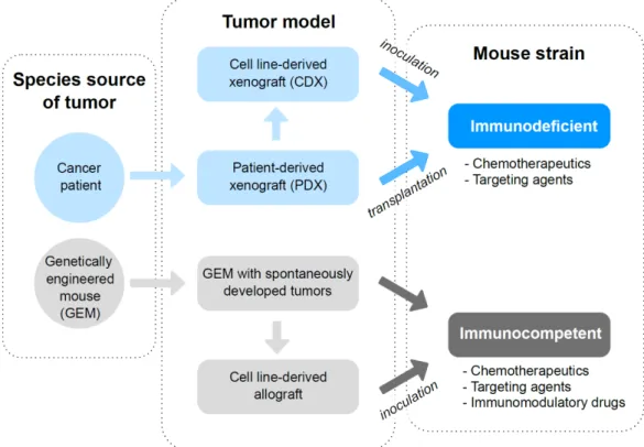
1.7. Characteristics and Applications of Tumor Mouse Models for Investigation of FR-Targeting Agents ... 26
1.8. Aim of the Thesis ... 29
II. Identification of a PET Radiotracer for Imaging of the Folate Receptor-α – a Potential Tool to Select Patients for Targeted Tumor Therapy ... 31
2.1. Introduction ... 32
2.2. Materials & Methods ... 34
2.2.1. Folate Derivatives ... 34
2.2.2. Cell Culture ... 34
2.2.3. Western Blot ... 34
2.2.4. Cell Internalization of 6R/6S-[18F]Aza-5-MTHF and [18F]AzaFol ... 35
2.2.5. FRα- and FRβ-Binding Affinity (IC50 Values) ... 35
2.2.6. Autoradiography Studies ... 36
2.2.7. Immunohistochemistry ... 36
2.2.8. In Vivo Studies ... 37
2.2.9. Biodistribution Studies ... 37
2.2.10. PET/CT Imaging Studies ... 37
2.3. Results ... 38
2.3.2. Uptake of 18F-Radiotracers in RT16 and D4 Cells ... 39
2.3.3. Receptor-Binding Affinity of Fluorinated Folates (IC50 Values) ... 39
2.3.4. Autoradiography Studies Using 18F-labeled Folate Radiotracers ... 41
2.3.5. Biodistribution of 18F-labeled Folate Radiotracers ... 42
2.3.6. PET/CT Imaging Studies Using 18F-lableled Folate Radiotracers ... 45
2.4. Discussion ... 47
2.5. Conclusion ... 48
III. Combining Albumin-Binding Properties and Interaction with Pemetrexed to Improve the Tissue Distribution of Radiofolates ... 49
3.1. Introduction ... 50
3.2. Materials & Methods ... 52
3.2.1. Preparation of [177Lu]Lu-Folate ... 52
3.2.2. Cell Culture ... 52
3.2.3. In Vivo Studies ... 52
3.2.4. Biodistribution Studies ... 52
3.2.5. Statistics ... 53
3.2.6. SPECT/CT Studies ... 53
3.3. Results ... 54
3.3.1. Biodistribution Studies ... 54
3.3.2. In Vivo SPECT/CT Experiments ... 58
3.4. Discussion ... 60
3.5. Conclusion ... 61
IV. Preclinical Evaluation of 5-Methyltetrahydro-folate-Based Radioconjugates – New Perspectives for Folate Receptor-Targeted Radionuclide Therapy ... 63
4.1. Introduction ... 64
4.2. Materials & Methods ... 66
4.2.1. Radiolabeling and Quality Control of the Folate Radioconjugates ... 66
4.2.2. In Vitro Stability of Folate Radioconjugates in PBS ... 66
4.2.3. In Vitro Stability of Folate Radioconjugates in Human Plasma ... 66
4.2.4. Determination of LogD Values ... 67
4.2.5. Binding Affinity to Mouse and Human Plasma Proteins ... 67
4.2.6. Tumor Cell Culture and Cell Uptake Studies ... 68
4.2.7. In Vivo Studies ... 69
4.2.8. In Vivo Stability of Folate Radioconjugates in Blood Plasma ... 69
4.2.9. Biodistribution Studies ... 69
4.2.10. Determination of Areas Under the Curve ... 70
4.2.11. SPECT/CT Imaging Studies ... 70
4.2.12. Therapy Study ... 70
4.2.13. Assessment of the Therapy Study ... 71
4.2.14. Statistical Analysis ... 72
4.3. Results ... 73
4.3.1. Radiolabeling and Quality Control of Folate Radioconjugates ... 73
4.3.2. In Vitro Stability and LogD Values of [177Lu]Lu-Folate Conjugates ... 73
4.3.3. Binding Affinity to Mouse and Human Plasma Proteins ... 73
4.3.4. Cell Uptake and Internalization Studies ... 74
4.3.5. In Vivo Stability of Folate Radioconjugates ... 75
4.3.6. Biodistribution Studies ... 75
4.3.7. Determination of Areas Under the Curve (AUC) ... 79
4.3.8. SPECT/CT Imaging Studies ... 81
4.3.9. Therapy Study ... 85
4.3.10. Assessment of the Therapy Study ... 85
4.4. Discussion ... 89
4.5. Conclusion ... 90
V. Promising Potential of [177Lu]Lu-DOTA-Folate to Enhance Tumor Response to Immunotherapy – a Preclinical Study Using a Syngeneic Breast Cancer Model ... 91
5.1. Introduction ... 92
5.2. Materials & Methods ... 94
5.2.1. Radiosynthesis of the [177Lu]Lu-DOTA-Folate ... 94
5.2.2. Tumor Cell Culture ... 94
5.2.3. Determination of FR Expression in NF9006 Cells Relative to KB Cells ... 94
5.2.4. Cell Uptake and Internalization ... 95
5.2.5. NF9006 and KB Cell Uptake of Variable Molar Amounts of [177Lu]Lu-DOTA-Folate .... 96
5.2.6. In Vitro Autoradiography ... 96
5.2.7. In Vivo Experiments ... 96
5.2.8. Biodistribution and Dosimetry of [177Lu]Lu-DOTA-Folate ... 97
5.2.9. SPECT/CT Imaging Studies ... 97
5.2.10. NF9006 Tumor Response to [177Lu]Lu-DOTA-Folate Administration ... 98
5.2.11. Therapy Study ... 98
5.2.12. Assessment of the Therapy ... 99
5.2.13. Assessment of PET Radiotracers for Monitoring NF9006 Tumors ... 100
5.2.14. Statistical Analysis ... 100
5.3. Results ... 102
5.3.1. Radiolabeling of the Folate Radioconjugate ... 102
5.3.2. Determination of FR-Expression in NF9006 Cells Relative to KB Cells ... 102
5.3.3. Cell Uptake and Internalization ... 103
5.3.4. NF9006 and KB Cell Uptake of Variable Molar Amounts of [177Lu]Lu-DOTA-Folate .. 103
5.3.5. In Vitro Autoradiography ... 104
5.3.7. SPECT/CT Imaging Studies ... 106
5.3.8. NF9006 Tumor Response to Variable Quantities of [177Lu]Lu-DOTA-Folate ... 106
5.3.9. Therapy Study Using [177Lu]Lu-DOTA-Folate and an Anti-CTLA-4 Antibody ... 107
5.3.10. Assessment of Potential Early Side Effects After Therapy ... 111
5.3.11. Assessment of PET Radiotracers for Monitoring NF9006 Tumors ... 112
5.4. Discussion ... 114
5.5. Conclusion ... 115
VI. Imaging Studies of FR-positive Tumors Using Transgenic Mouse Model of Mammary Gland Carcinoma ... 117
6.1. Introduction ... 118
6.2. Materials & Methods ... 120
6.2.1. Radiosynthesis of [177Lu]Lu-DOTA-Folate ... 120
6.2.2. In Vivo Studies ... 120
6.2.3. SPECT/CT Imaging Studies ... 121
6.2.4. Biodistribution Study ... 121
6.2.5. In Vitro Autoradiography ... 122
6.3. Results & Discussion ... 123
6.3.1. [177Lu]Lu-DOTA-Folate Binding to Spontaneous Tumors Is FR-Specific ... 123
6.3.2. Spontaneous Tumors of MMTV-neu Mice Can Be Imaged with [177Lu]Lu-DOTA-Folate . ... 124
6.3.3. Injection of [177Lu]Lu-DOTA-Folate Caused Shrinkage of Spontaneous Tumors Over Time ... 125
6.3.4. Follow-up Injections of [177Lu]Lu-DOTA-Folate Showed Reduced Tumor Uptake ... 128
6.3.5. The Tissue Uptake of [177Lu]Lu-DOTA-Folate Might Be Influenced by the Diet ... 128
6.4. Conclusion ... 130
VII. Conclusions & Outlook ... 131
References ... 137
Summary
The theragnostic concept in nuclear medicine makes use of radiolabeled ligands for targeting tumor- associated markers suitable for imaging and therapy of cancer diseases. The folate receptor-α (FRα) is a potential target to address numerous tumors of epithelial origin, including gynecological cancers, breast and lung cancers. Importantly, the FR presents also as isoform β that is a marker of activated macrophages involved in inflammatory diseases. Commonly, FR-specific binding of radioconjugates was achieved by employment of folic acid as a targeting agent that binds to both FR isoforms with nanomolar affinity. Although a distinct uptake of radiofolates was observed in tumors, the concomitant binding to the FRs expressed in the kidneys remained of concern due to the risk of damage to the kidneys caused by particle-emitting radiation of therapeutic radioconjugates. Modification of a folic acid conjugate with an albumin binder enhanced blood retention and resulted in tremendous increase of the tumor-to-kidney ratio. This in turn allowed for using radiofolates in preclinical therapy setting for the first time. The unselective binding of folic acid to the FRα and FRβ may be unfavorable for selection of cancer patients who could be inadequately treated based on false-positive (FRβ-based) imaging results.
The focus of this thesis was, therefore, to improve radiofolates in the aspect of their diagnostic value for FRα-expressing tumors and therapeutic potential.
Wang et al. reported in 1992 that the 6S isomer of 5-methyltetrahydrofolate (MTHF) binds with higher affinity to the FRα than to the FRβ. Thus, for the purpose of developing a tumor-selective (= FRα- selective) imaging agent, 5-MTHF was proposed as an alternative targeting molecule instead of folic acid. The design and syntheses of a series of 18F-labeled 5-MTHF derivatives were performed by Boss et al. In the present thesis (chapter 2), the selectivity of the PET radiofolates, 6R-[18F]Aza-5-MTHF*
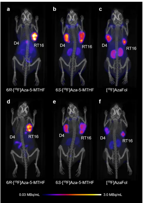
and 6S-[18F]Aza-5-MTHF*, was investigated in comparison to the folic acid-based analogue, [18F]AzaFol. Comparative studies with FRα- or FRβ-expressing cells performed in vitro revealed ~12- fold higher accumulation of 6R-[18F]Aza-5-MTHF in FRα-positive cells than in FRβ-expressing cells (~62% vs. ~5% of total added activity) and ~43-fold higher binding affinity to the FRα (IC50=1.8 nM) than to the FRβ (IC50=77 nM). Neither 6S-[18F]Aza-5-MTHF nor [18F]AzaFol demonstrated differentiated binding to FR isoforms. In vivo studies with a mouse model bearing a FRα- and a FRβ- positive xenograft confirmed the in vitro findings and clearly indicated FRα selectivity of 6R-[18F]Aza- 5-MTHF. As a next step, experiments with a mouse model that combines a FRα-positive tumor and FRβ-positive activated macrophages in inflammation will be necessary to confirm the
__________________________
* The R/S nomenclature changes with the introduction of the heteroaromate in 18F-labeled aza-5-MTHFs
6S/6R-5-MTHF = 6-[[4-[(2-Amino-5-methyl-4-oxo-1,6,7,8-tetrahydropteridin-6-yl)methylamino]benzamido]4-carboxy- butanoate
6R/6S-[18F]Aza-5-MTHF = 6-[[4-[(2-Amino-5-methyl-4-oxo-1,6,7,8-tetrahydropteridin-6-yl)methylamino]2-[18F]fluoro- nicotinamido]4-carboxybutanoate acid
feasibility to unambiguously image tumors in the presence of inflammatory sites.
It was previously shown in our group that pre-injection of the antifolate, pemetrexed (PMX), reduced renal accumulation of conventional radiofolates without simultaneous blockade of the uptake in the tumor. In this thesis (chapter 3) we combined this strategy with the albumin-binding folic acid radioconjugate ([177Lu]Lu-cm13) to further improve the tissue distribution profile which is particularly important in view of therapeutic application. Biodistribution studies with mice that were injected with PMX prior to the [177Lu]Lu-cm13, demonstrated increased tumor-to-kidney ratios by 33–53%, depending on the type of FR-positive tumor. It was found that additional injections of PMX, at 3 h or 7 h after the administration of the radioconjugate resulted in 46–72% higher ratios as compared to the values obtained with radiofolate alone. These results were confirmed by SPECT/CT scans which readily visualized the tumor xenografts, whereas accumulation of [177Lu]Lu-folate in the kidneys was significantly reduced in mice injected with PMX. Although PMX had a positive impact in terms of reducing the kidney uptake of albumin-binding radiofolates, the effect was only moderate. Moreover, the repeated injections and potential toxicity of PMX make this concept challenging with regard to clinical application.
The concept of exchanging the FR-targeting molecule and replacing folic acid with 6R- or 6S-isomer of 5-MTHF was employed for albumin-binding folate conjugates as well (chapter 4). 6R-RedFol-1 and 6S- RedFol-1 were radiolabeled with 177Lu and compared in vitro and in vivo to [177Lu]Lu-OxFol-1, a previously developed and characterized folic acid-based FR-targeting agent. Both [177Lu]Lu-6R- RedFol-1 and [177Lu]Lu-6S-RedFol-1 demonstrated similar in vitro properties as [177Lu]Lu-OxFol-1, however, the biodistribution studies revealed 4–5-fold enhanced blood retention in the case of the 5- MTHF radioconjugates. Notably, the tumor uptake expressed as integrated area under the time-activity curve (AUC0→120h) of [177Lu]Lu-6R-RedFol-1 and [177Lu]Lu-6S-RedFol-1 was 3.2-fold and 3.6-fold increased in comparison to [177Lu]Lu-OxFol-1, respectively. In the case of [177Lu]Lu-6S-RedFol-1 the kidney uptake was ~3-fold increased, whereas the renal retention of [177Lu]Lu-6R-RedFol-1 was similar to that of [177Lu]Lu-OxFol-1. This led to an almost 4-fold increased tumor-to-kidney AUC0→120h ratio for [177Lu]Lu-6R-RedFol-1 than for [177Lu]Lu-6S-RedFol-1 and [177Lu]Lu-OxFol-1. In a comparative therapy study, it was demonstrated that at equal activity (10 MBq), the therapeutic effect of [177Lu]Lu- 6R-RedFol-1 was better than that of [177Lu]Lu-OxFol-1, reflected by a slower tumor growth and, consequently, an increased median survival time (49 d and 34 d, respectively) as compared to the control (22 d). If radioconjugates were applied at 15 MBq activity, mice injected with [177Lu]Lu-OxFol-1 showed median survival of 44 d, whereas all mice treated with [177Lu]Lu-6R-RedFol-1 survived until the end of the study (>56 d).
It was previously shown by other research groups that a radiation stimulus can improve the tumor response to therapy with immune checkpoint inhibitors in preclinical and clinical studies. This effect is caused by increased infiltration of immune cells upon tumor irradiation and is described as a switch from an immunologically “cold” tumor to a “hot” tumor. We envisioned that such sensitizing effect
could be achieved using FR-targeting radioconjugate (chapter 5). The concept was tested in a syngeneic mouse model established from NF9006 mammary gland tumor cells derived from MMTV-neu transgenic mice. The model was characterized in our group with regard to FR expression and in vitro and in vivo targeting. The 177Lu-DOTA-folate conjugate (folic acid-based) applied at a low activity (5 MBq), resulting in an absorbed tumor dose of 3.5 Gy, sensitized the FR-positive NF9006 tumors to anti-CTLA-4 immunotherapy which was manifested by reduced tumor growth. This resulted in a significantly improved median survival of mice (>70 days) as compared to mice that received the
177Lu-folate conjugate or the anti-CTLA-4 antibody alone. Each modality had only a minor effect on tumor growth and did not substantially increase the median survival (23 d and 19 d, respectively) as compared to untreated controls (12 d). Future studies may be directed towards the investigation of the immune-profile to characterize the changes in tumor microenvironment upon the treatment with 177Lu- folate conjugate and the anti-CTLA-4 antibody.
Transgenic mice with spontaneously developed tumors present a more representative preclinical cancer model than mice bearing subcutaneous human tumors. This refers primarily to the higher complexity of spontaneous models, and the interplay between tumor microenvironment and the immune system. In chapter 6 of this thesis, we set out to investigate the uptake of 177Lu-DOTA-folate conjugate in spontaneous breast tumors of MMTV-neu mouse strain, and compare it with NF9006 syngeneic model.
SPECT/CT images demonstrated significant uptake of the radiofolate in the tumors and kidneys. The quantity of injected activity for imaging (25 MBq) was revealed to have a therapeutic effect as tumors started to shrink shortly after. Subsequent follow-up scans with [177Lu]Lu-DOTA-folate allowed visualizing the changes in sub-tissue distribution of radiofolate over time. The MMTV-neu transgenic mice could serve as a more sophisticated model for investigation of FR-targeted therapies in combination with immune checkpoint inhibitors and other immune-modulatory agents.
The results of this thesis represent a major step towards the theragnostic application of radiofolates. The new generation of 5-MTHF-based 18F-radiotracers were designed for unambiguous cancer imaging using PET. On the other hand, the 5-MTHF-based 177Lu-folate conjugates will enable a safer therapeutic application by preventing damage to radiosensitive kidneys. Finally, it was demonstrated that the application of folate radioconjugates can sensitize tumors to immunotherapeutic effects, which may open new perspectives for future management of FR-positive cancer diseases.
Zusammenfassung
Das theragnostische Konzept in der Nuklearmedizin beruht auf der Verwendung von Radioliganden, welche an tumorassoziierte Zielstrukturen binden, die sich für die Bildgebung und Therapie von Krebserkrankungen eignen. Der Folatrezeptor-α (FRα) stellt eine solche Zielstruktur dar, weil er auf zahlreichen Tumoren epithelialen Ursprungs wie gynäkologische Krebsarten, Brust- und Lungenkrebs vorkommt. Darüber hinaus wird die β-Isoform des FRs von aktivierten Makrophagen, welche in Entzündungskrankheiten involviert sind, exprimiert. Eine FR-spezifische Bindung von Radiokonjugaten wurde durch den Einsatz von Folsäure als Ligand erreicht. Folsäure bindet mit einer Affinität im nanomolaren Bereich an beide FR Isoformen. Obwohl eine deutliche Aufnahme der Radiofolate in Tumoren beobachtet wurde, erwies sich deren gleichzeitige Bindung an die in den Nieren exprimierten FR als ungünstig, da die partikelemittierende Strahlung therapeutischer Radiokonjugate das Risiko einer Nierenschädigung birgt. Die Modifikation eines Folatkonjugats mit einer albuminbindenden Einheit führte zu einer Verlängerung dessen Blutzirkulation und dadurch zu einer enormen Verbesserung des Tumor-zu-Nieren-Verhältnisses der angereicherten Aktivität. Dies wiederum ermöglichte erstmals den Einsatz von Radiofolaten in präklinischen Therapiestudien. Die nicht-selektive Bindung der Folsäure an den FRα könnte sich jedoch in Bezug auf die Selektion von Krebspatienten als ungünstig erweisen, da aufgrund der potentiell falsch-positiven (FRβ-basierten) Bildgebungsresultate, Patienten einer nicht angemessenen Behandlung unterzogen werden könnten.
Daher lag der Schwerpunkt dieser Arbeit auf der Untersuchung von Radiofolaten hinsichtlich deren diagnostischer Bedeutung für FRα exprimierende Tumoren und deren therapeutischen Potenzials.
Wang et al. zeigte im Jahr 1992, dass die Bindungsaffinität des 6S-Isomer des 5-Methyltetrahydrofolats (MTHF) zum FRα höher ist als zum FRβ. Aus diesem Grund wurde für die Entwicklung eines tumorselektiven (d.h. FRα-selektiven) Folatkonjugates das 5-MTHF anstelle von Folsäure als alternativer Ligand vorgeschlagen. Das Design und die Synthesen einer Reihe von 18F-markierten 5- MTHF-Derivaten wurden von Boss et al. etabliert. In der vorliegenden Arbeit wurde die Selektivität der PET Radiofolate, 6R-[18F]Aza-5-MTHF* und 6S-[18F]Aza-5-MTHF*, im Vergleich zu dem auf Folsäure basierenden Analogon [18F]AzaFol untersucht (Kapitel 2). In vitro durchgeführte Vergleichsstudien mit FRα- oder FRβ-exprimierenden Zellen zeigten für 6R-[18F]Aza-5-MTHF eine
~12-fach höhere Akkumulation in FRα-positiven Zellen gegenüber FRβ-exprimierenden Zellen (~62%
vs. ~5% der gesamten hinzugefügten Aktivität) und eine ~43-fach höhere Bindungsaffinität zum FRα
___________________________
*Die R/S-Nomenklatur ändert sich durch die Einführung des Heteroaromaten in die Struktur des 18F-markierten aza-5-MTHF 6S/6R-5-MTHF = 6-[[4-[(2-Amino-5-methyl-4-oxo-1,6,7,8-tetrahydropteridin-6-yl)methylamino]benzamido]4-carboxy- butansäure
6R/6S-[18F]Aza-5-MTHF = 6-[[4-[(2-Amino-5-methyl-4-oxo-1,6,7,8-tetrahydropteridin-6-yl)methylamino]2-[18F]fluoro- nicotinamido]4-carboxybutansäure
(IC50=1.8 nM) als zum FRβ (IC50=77 nM). Weder 6S-[18F]Aza-5-MTHF noch [18F]AzaFol zeigten jedoch eine selektive Bindung an den FRα. In vivo Studien, die auf einem Mausmodell mit FRα- und FRβ-positiven Xenograften beruhten, bestätigten die in vitro Resultate und zeigten die selektive Bindung von 6R-[18F]Aza-5-MTHF an den FRα. Um die Möglichkeit einer eindeutigen Darstellung von Tumoren in Gegenwart von Entzündungsherden zu verifizieren, sollen in einem nächsten Schritt Experimente mit einem Mausmodell durchgeführt werden, welches einen FRα-positiven Tumor und gleichzeitig eine Entzündung, welche FRβ-positive aktivierte Makrophagen involviert, kombiniert.
In unserer Gruppe wurde bereits gezeigt, dass die Injektion des Antifolats Pemetrexed (PMX) vor dem Radiofolat die renalen Aktivitätsakkumulation reduziert, ohne dabei die Aufnahme in den Tumor zu blockieren. In dieser Arbeit kombinierten wir diese Strategie mit dem albuminbindenden Folatkonjugat [177Lu]Lu-cm13, um das Gewebeverteilungsprofil weiter zu verbessern (Kapitel 3). Dies ist im Hinblick auf eine therapeutische Anwendung besonders wichtig. Biodistributionsstudien mit Mäusen, denen PMX vor der Injektion des [177Lu]Lu-cm13 appliziert wurde, zeigten je nach Xenograft ein um 33–53%
erhöhtes Tumor-zu-Nieren-Verhältnis. Eine zusätzliche PMX Injektion, drei oder sieben Stunden nach Verabreichung des Radiokonjugats, führte zu einem 46–72% höheren Tumor-zu-Nieren Verhältnis verglichen mit den Werten, welche mit dem Radiofolat alleine erzielt wurden. Diese Ergebnisse entsprachen den SPECT/CT Bildern, die eine den Tumor gut visualisierten, während die Nierenakkumulation von [177Lu]Lu-Folat in Mäusen, denen PMX appliziert wurde, signifikant reduziert wurde. Obwohl PMX bezüglich der Verringerung der Nierenaufnahme von albuminbindenden Radiofolaten einen positiven Einfluss hatte, war der Effekt nur mässig. Wiederholte Injektionen des potenziell toxischen PMX wäre für eine klinische Anwendung jedoch eher schwierig realisierbar.
Die Strategie, Folsäure als FR-bindendes Molekül mit den 6R- und 6S-Isomeren von 5-MTHF zu ersetzen, wurde auch auf die albuminbindenden Folatkonjugate übertragen (Kapitel 4). 6R-RedFol-1 und 6S-RedFol-1 wurden mit 177Lu radioaktiv markiert und die in vitro und in vivo Eigenschaften der beiden Konjugate mit jenen von [177Lu]Lu-OxFol-1 verglichen. Sowohl [177Lu]Lu-6R-RedFol-1 also auch [177Lu]Lu-6S-RedFol-1 wiesen ähnliche in vitro Eigenschaften wie [177Lu]Lu-OxFol-1 auf, jedoch konnte in Bioverteilungsstudien gezeigt werden, dass die 5-MTHF basierenden Radiokonjugate eine 4–
5-fach höhere Blutretention aufwiesen. Die Aufnahme von [177Lu]Lu-6R-RedFol-1 und [177Lu]Lu-6S- RedFol-1 in den Tumor, ausgedrückt als integrierte Fläche unter der Zeit-Aktivitätskurve (AUC0→120h), war im Vergleich zu [177Lu]Lu-OxFol-1 um einen Faktor 3.2, respektive 3.6 grösser. Während [177Lu]Lu-6S-RedFol-1 eine dem [177Lu]Lu-OxFol-1 vergleichbare Nierenaufnahme zeigte, war diese für [177Lu]Lu-6S-RedFol-1 ~3-fach erhöht. Dies führte zu einem fast 4-fach höheren Tumor-zu-Nieren- Verhältnis (AUC0→120h) für [177Lu]Lu-6R-RedFol-1 im Vergleich zu [177Lu]Lu-6S-RedFol-1 und [177Lu]Lu-OxFol-1. In einer Therapievergleichsstudie wurde gezeigt, dass bei gleicher Aktivität (10 MBq) mit [177Lu]Lu-6R-RedFol-1 ein langsameres Tumorwachstum und eine längere mittlere
Überlebenszeit (49 Tage) erzielt werden konnte als dies bei Mäusen, die mit [177Lu]Lu-OxFol-1 behandelt wurden (mittlere Überlebenszeit 34 Tage) oder gar nicht behandelt wurden (mittlere Überlebenszeit 22 Tage) der Fall war. Bei einer Aktivität von 15 MBq überlebten die Mäuse, die mit [177Lu]Lu-OxFol-1 injiziert wurden, im Schnitt 44 Tage während alle Mäuse, denen [177Lu]Lu-6R- RedFol-1 verabreicht wurde, bis zum Ende der Studie (>56 Tage) überlebten.
Es wurde bereits von anderen Forschungsgruppen in präklinischen und klinischen Studien gezeigt, dass ein Strahlungsstimulus die Sensitivität gegenüber einer Therapie mit Immuncheckpoint Inhibitoren verbessern kann. Dieser Effekt wurde durch eine erhöhte Infiltration von Immunzellen nach der Tumorbestrahlung verursacht und demzufolge als Umwandlung eines immunologisch «kalten» in einen
«heissen» Tumor bezeichnet. Ein solcher Effekt zur Sensibilisierung des Tumors könnte mit einem folatrezeptorbindenden Radiokonjugat erzielt werden (Kapitel 5). Dieses Prinzip wurde in einem syngenen Mausmodell getestet, welches mit NF9006 Milchdrüsentumorzellen von MMTV-neu transgenen Mäusen etabliert wurde. Das Modell wurde hinsichtlich der Folatrezeptorexpression und der in vitro und in vivo Bindung von unserer Gruppe charakterisiert. Das 177Lu-DOTA-Folatkonjugat, welches auf Folsäure basiert, zeigte bei einer tiefen injizierten Aktivität (5 MBq) und einer absorbierten Tumordosis von 3.5 Gy einen sensibilisierenden Effekt auf die FR-positiven NF9006 Tumoren gegenüber einer anti-CTLA-4 Immuntherapie. Dies führte zu einem verringerten Tumorwachstum und einer längeren mittleren Überlebenszeit der Mäuse (>70 Tage) im Vergleich zu Mäusen, die entweder nur das 177Lu-Folatkonjugat oder nur den anti-CTLA-4 Antikörper erhalten hatten. Beide Monotherapien zeigten einen minimalen Einfluss auf das Tumorwachstum und konnten die mittlere Überlebenszeit gegenüber der Kontrollgruppe nicht massgeblich verlängern (177Lu-Folatkonjugat:
23 Tage; anti-CTLA-4: 19 Tage; Kontrollgruppe: 12 Tage). Weiterführende Studien könnten sich dem Erstellen von Immunprofilen widmen, um die Veränderungen der Tumormikroumgebung nach einer Therapie mit 177Lu-Folatkonjugat und anti-CTLA-4 Antikörper zu charakterisieren.
Transgene Mäuse mit sich spontan entwickelnden Tumoren sind für die klinische Situation repräsentativer als Mäuse mit subkutanen humanen Tumoren. Dies bezieht sich vor allem auf die höhere Komplexität der spontan entwickelten Tumoren und das Zusammenspiel zwischen der Tumorumgebung und des Immunsystems. In Kapitel 6 dieser Arbeit untersuchten wir die Aufnahme von 177Lu-DOTA- Folatkonjugat in die spontanen Brusttumore von MMTV-neu Mäusen und verglichen diese mit dem NF9006 syngenen Modell. SPECT/CT Bilder zeigten eine signifikante Aufnahme des Radiofolats in den Tumoren und Nieren. Die Menge injizierter Aktivität für die Bildgebung (25 MBq) hatte einen therapeutischen Effekt und die Tumoren begannen bereits kurze Zeit nach der Injektion zu schrumpfen.
Die darauffolgende Bildgebung mit [177Lu]Lu-DOTA-Folat erlaubten Veränderungen der Verteilung der Aktivität in den Geweben über die Zeit aufzuzeigen. Das MMTV-neu transgene Mausmodell könnte als ein weiterentwickeltes Modell für die Untersuchung von FR-gezielten Therapien in Kombination mit Immuncheckpoint Inhibitoren oder anderen immunregulierenden Wirkstoffen dienen.
Die Resultate dieser Arbeit stellen einen weiteren Schritt in der Entwicklung von Radiofolaten in Richtung theragnostischer Anwendung dar. Die neue Generation von 5-MTHF-basierenden 18F- Radiofolaten wurden für eine eindeutige Krebsbildgebung mittels PET entworfen. Die 5-MTHF- basierenden 177Lu-Folatkonjugate scheinen eine sichere therapeutische Anwendung zu ermöglichen, um Gewebsschäden in den radiosensitiven Nieren zu vermeiden. Ausserdem wurde gezeigt, dass Radiofolatkonjugate die Tumoren für eine Immuntherapie sensibilisieren können. Dies könnte neue Perspektiven für die Behandlung von Krebserkrankungen mit FR-positiven Tumoren eröffnen.
Abbreviations
ALP Alkaline phosphatase
ALB Albumin
AML Acute myeloid leukemia
ANOVA Analysis of variance
AUC Area under the curve
Bl Blood/ Urinary bladder
BUN Blood urea nitrogen
B50 Half maximum binding
CDX Cell line-derived xenograft
CT Computed tomography
CML Chronic myeloid leukemia
CRE Creatinine
Da Dalton (= 1 g/mol)
DMSO Dimethyl sulfoxide
DOTA 1,4,7,10-tetraazacyclododecane-1,4,7,10-tetraacetic acid
DTPA Diethylenetriaminepentaacetic acid
EDTA Ethylenediaminetetraacetic acid
FA Folic acid
FCS Fetal calf serum
FFRPMI Folate-free RPMI cell culture medium
FR Folate receptor
FWHM Full width at half maximum
Gy Gray (J/kg)
HCl Hydrochloric acid
HE Hematoxylin-eosin
HER2 Human epidermal growth factor receptor 2
HPLC High performance liquid chromatography
HRP Horseradish peroxidase
I Intensity of radiation in percent
IA/g Injected activity per gram
IC50 Half-maximal inhibitory concentration
ICI Immune checkpoint inhibitors
IgG Immunoglobulin G
i.p. Intraperitoneal
i.v. Intravenous
keV Kiloelectron volt
Ki Kidneys
L Length of tumor
LET Linear energy transfer
Li Liver
mAb Monoclonal antibody
MBq Megabequerel
MIP Maximum intensity projection
MMTV Mouse mammary tumor virus
5-MTHF 5-Methyltetrahydrofolate
NODAGA 1,4,7-triazacyclononane,1-glutaric acid-4,7-acetic acid
PBS Phosphate-buffered saline
PDX Patient derived xenograft
PET Positron Emission Tomography
p.i. Post-injection
PMX Pemetrexed (antifolate drug)
PSMA Prostate Specific Membrane Antigen
RBW Relative Body Weight
RIPA Radioimmunoprecipitation assay buffer
RPMI Roswell Park Memorial Institute medium
RT Room temperature
RTV Relative tumor volume
s.c. Subcutaneous
SD Standard deviation
SDS-PAGE Sodium dodecyl sulfate polyacrylamide gel electrophoresis
SPECT Single Photon Emission Computed Tomography
TBIL Total bilirubin
TGD Tumor growth delay
TGDI Tumor growth delay index
TGI Tumor growth inhibition
tR Retention time
Tu Tumor
T1/2 Physical half-life
W Width of tumor
I. General Introduction
1.1. Clinical Potential of Tumor-Targeting Radiopharmaceuticals
1.1.1. General Features of Diagnostic and Therapeutic Radiopharmaceuticals
The role of nuclear medicine in oncology has grown during the last two decades. It relies on development of tumor-targeting radiopharmaceuticals that can be applied for non-invasive cancer diagnosis as well as for therapy. Such radiopharmaceutical consists of a molecule which localizes a tumor-associated target (Fig. 1.1), and consequently, allows for an accurate assessment of cancer location, possible dissemination, and later on, precise treatment with particle-emitting radiation [1]. The radiopharmaceutical is labeled with a radiometal via a chelating system or, if the radionuclide is not metal-based, it can be directly installed to the backbone of the tumor-targeting agent [1].
Fig. 1.1 Concept of a tumor-targeting radiopharmaceutical. Visualization of the tumor cell was prepared using Servier Medical Art licensed under a Creative Commons Attribution 3.0 Unported License.
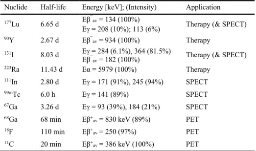
The choice of radionuclide depends on the purpose of the radiopharmaceutical’s application (Table 1.1).
The type of decay plays a critical role in this regard. Radionuclides used for SPECT imaging emit γ- radiation with a tissue range up to several centimeters [2,3]. PET radioisotopes decay by emission of positrons (β+), which annihilate with electrons in the surrounding tissue. The annihilation results in emission of two γ-rays of 511 keV energy that travel at opposite directions. Importantly, due to this defined geometry of annihilation photons, PET does not require additional collimator, and therefore has higher sensitivity as compared to SPECT in a clinical setting [2,3].
The radionuclides decaying by the emission of electrons (β¯,Auger or conversion electrons) or alpha particles are suitable for therapeutic application, since this type of particle radiation destroys tumor cells.
So far β¯-emitters are the most frequently used radionuclides for targeted radionuclide therapy in clinics [4]. Compared to α-particles and Auger electron, β¯-particles emitted by medical radionuclides can have a tissue range of up to ten millimeter, which results in irradiation of a cluster of adjacent tumor cells.
This observation is commonly referred to as “crossfire” effect [5,6]. Alpha-emitters and Auger electron- emitters are suitable for the treatment of small metastases and single tumor cells, due to the short tissue range and high linear energy transfer (LET), which is the energy deposited per unit of distance. In contrast to β¯-emitters, these properties allow for minimizing the damage to the normal tissues and
delivering a high dose-burden to the tumor, since the “crossfire” effect is not existent or reduced, respectively [1,7].
In an ideal case, pre-therapeutic imaging and therapy would be performed using radionuclides of the same chemical element enabling radiotheragnostic application with chemically identical tumor- targeting radiopharmaceuticals, e.g. iodine radioisotopes. However, commonly, this is accomplished with diagnostic and therapeutic radionuclides of different elements, which provide similar but not the same chelation chemistry.
Table 1.1 Examples of clinically used radionuclides and their physical decay properties. Decay data were provided from NuDat database (version 2.8) [8]
Nuclide Half-life Energy [keV]; (Intensity) Application
177Lu 6.65 d Eβ¯av = 134 (100%)
Eγ = 208 (10%); 113 (6%) Therapy (& SPECT)
90Y 2.67 d Eβ¯av = 934 (100%) Therapy
131I 8.03 d Eγ = 284 (6.1%), 364 (81.5%)
Eβ¯av = 182 (100%) Therapy (& SPECT)
223Ra 11.43 d Eα = 5979 (100%) Therapy
111In 2.80 d Eγ = 171 (91%), 245 (94%) SPECT
99mTc 6.0 h Eγ = 141 (89%) SPECT
67Ga 3.26 d Eγ = 93 (39%), 184 (21%) SPECT
68Ga 68 min Eβ+av = 830 keV (89%) PET
18F 110 min Eβ+av = 250 (97%) PET
11C 20 min Eβ+av = 386 keV (100%) PET
1.1.2. Clinically Used Receptor-Targeted Radiopharmaceuticals for Cancer Diagnosis and Therapy
The first and best established radiopharmaceuticals in the clinics are radiolabeled somatostatin (SST) analogs targeting the somatostatin receptor (SSTR). SSTR is overexpressed on neuroendocrine tumors (NETs) originating from gastroenteropancreatic or pulmonary tract [7]. Somatostatin is a natural peptide hormone with a short blood plasma half-life due to enzymatic degradation [9]. Introduction of D-amino acids and shortening the peptide chain resulted in a number of analogs which are more resistant to degradation while preserving binding properties of the natural SSTR binding peptide. [111In]In-DTPA- octreotide (OctreoScan®) was the first somatostatin analog that was approved for diagnosis and monitoring of patients with SSTR-expressing tumors. Currently, a newer analog linked to a DOTA- chelating system, [68Ga]Ga-DOTATOC is being used for PET/CT [10,11]. The development of SST analogs comprising a DOTA chelator, also enabled the chelation of β¯-emitting radionuclides suitable for therapeutic purposes, among those 90Y and 177Lu. Both [90Y]Y-DOTATOC and [177Lu]Lu- DOTATATE showed encouraging results in peptide receptor radionuclide therapy (PRRT) [12,13]. Owing to the lower energy and range of β¯-particles emitted by 177Lu as compared with 90Y, [177Lu]Lu-
DOTATATE showed less toxicity to normal tissues, including kidneys and bone marrow. Therefore, [177Lu]Lu-DOTATATE has become the most widely used somatostatin analog for PRRT [14]. Recently, PRRT with [177Lu]Lu-DOTATATE was approved in the USA and several European countries for the treatment of patients with progressive metastatic gastroenteropancreatic NETs [15].
Another successful example of radiopharmaceuticals are prostate-specific membrane antigen (PSMA)- targeting radioligands. PSMA is a transmembrane protein, which is overexpressed particularly in metastatic castration-resistant prostate cancer (mCRPC) [16]. PSMA is also present in the healthy prostate, kidneys and brain, but at significantly lower levels. Development of radiopharmaceuticals for prostate cancer resulted in small-molecule-based PSMA inhibitors [17]. Until now, [68Ga]Ga-PSMA-11 and [177Lu]Lu-PSMA-617 are most often used for diagnosis and radionuclide therapy of prostate cancers, respectively, in clinics. [177Lu]Lu-PSMA-617 revealed encouraging results in the treatment of mCRPC patients in terms of efficiency, response rate as well as safety [18,19]. Moreover, it was clinically demonstrated that pre-therapeutic PSMA PET imaging can be used for therapy planning, since the uptake of [68Ga]Ga-PSMA-11 strongly correlated with the tumor response to [177Lu]Lu-PSMA-617 treatment, despite the different structure and radionuclide [20]. Another promising PSMA-targeting theragnostic is [68Ga]Ga/[177Lu]Lu-PSMA I&T, which is currently investigated in clinical trials.
Similarly to [177Lu]Lu-PSMA-617, [177Lu]Lu-PSMA I&T was found effective and safe for the treatment of metastatic prostate cancer patients [21,22].
The diagnostic and therapeutic SSTR-targeting radiopeptides and PSMA-targeting radioligands demonstrated a great clinical potential of receptor-targeted radiopharmaceuticals, and hence, encouraged to apply this concept to other tumor types and promising targets. Recently, radiolabeled fibroblast activation protein (FAP)-targeting agents for PET/CT imaging were tested in patients of various tumor types and demonstrated improved tumor-to-background signal as compared with the current gold standard, [18F]FDG [23]. About two decades ago, the folate receptor (FR) emerged as a potential target for cancer imaging and therapy due to the frequent overexpression by numerous tumors of epithelial origin, including gynecological cancers [24]. Since FR-targeted agents could address the needs of a vast number of oncologic patients, exploiting this target through nuclear medicine is rational.
It would provide new treatment options and open further the prospects for radiopharmaceuticals and personalized medicine.
1.2. Properties of Folates and Mechanisms of Their Cellular Uptake
Folates comprise a large family of structurally related derivatives with vitamin B9 activity [25]. Folic acid (pteroyl-glutamic acid) is the oxidized, synthetic version of folate vitamins, which is not biologically active before enzymatic reduction in the cell (Fig. 1.2a). Physiologically active folates display differential oxidation states, and therefore, are conventionally referred to as “reduced folates”.
5-Methyltetrahydrofolate (5-MTHF) is the most prevalent form of folate in blood plasma (Fig. 1.2b).
Fig. 1.2 Chemical structures of folates: (a) folic acid (oxidized folate; synthetic form); (b) 5-methyl- tetrahydrofolate (reduced folate; biologically active form).
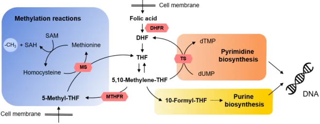
They undergo distinct biochemical processes and, in principle, act as a methyl donors in one-carbon transfer reactions involved in essential metabolic pathways. Examples are e.g. de novo synthesis of purine nucleotides and thymidylate, synthesis of methionine from homocysteine as well as DNA methylation (Fig. 1.3) [26]. Hence, folates play a critical role in proliferation and survival of cells, which consequently, require an efficient transport mechanism to meet the folate demand [25].
Fig. 1.3 Folate metabolism cycle depicting the main molecular species and their essential functions in the cell.
Folic acid is a fully oxidized synthetic form of folate, which upon transport into the cell, needs to be reduced by dihydrofolate reductase (DHFR) to dihydrofolate (DHF) and tetrahydrofolate (THF) that are metabolized to the primary substrate of pyrimidine and purine synthesis, 5,10-methylene-THF. 5,10-Methylenetetrahydrofolate reductase (MTHFR) is a key enzyme in formation of 5-methyl-THF, which is the most abundant form of physiologically active folate, and is a substrate of methionine synthase (MS) and methylation reactions, notably, of DNA. Other abbreviations: TS, tymidylate synthase, dTMP, deoxytymidylate; dUMP, deoxyuridylate; SAM, S-adenosulmethionine, SAH, S-adenosylhomocysteine. Figure was adapted from Liu et al. Adv Genet 2010; 71:79- 121 [26].
1.2.1. Folate Transporters and Receptors
Eukaryotic cells are not capable of producing folate vitamins, which in turn cannot passively cross the cell membrane due to their highly hydrophilic character [27]. For this reason, three distinct molecular carriers are engaged in the cellular uptake of exogenous folates, namely the reduced folate carrier (RFC), the proton-coupled folate transporter (PCFT) and several types of folate receptors (FRs)
(Fig. 1.4). Reduced folates are delivered predominantly by a bidirectional organic anion-exchange mechanism, using the RFC. The RFC is ubiquitous tissue expression in healthy cells of the body and characterized by high transport capacity and, therefore, it plays an important role in folate homeostasis [27].
The PCFT is another carrier protein enabling folate transport which requires a pH-gradient and, thus, acts as a folate-proton symporter [28]. The highest expression levels of PCFT in human and murine tissues were found in the small intestine, kidneys, liver, placenta, retina and brain [28].
Folate uptake can also occur via FR-mediated endocytosis. This uptake mechanism is initiated upon FR- ligand complex formation on the cell surface and followed by release of the folate molecule in the endosome from where it is delivered into the cell cytosol. It was demonstrated that the PCFT is co- expressed with the FR and mediates the export of folates from the acidic endosomes, being created upon FR-mediated endocytosis [29]. FRs exist in four isoforms (FRα, FRβ, FRγ, and FRδ), however, only two of them, the FRα and FRβ, play a significant role in folate uptake. The FRα and FRβ are cell-membrane anchored by a glycosylphosphatidylinositol (GPI) entity. Folic acid displays high affinity to FRs (Kd <
1 nM) [30], whereas the FR-binding affinities of reduced folates are 10- to 100-fold lower [25]. In contrast to RFCs, FRs have limited expression in normal tissues. The FRα is present on the apical surface of epithelial cells of the kidneys, lungs, choroid plexus, placenta, uterus as well as in the salivary glands [31]. Importantly, the FRα is frequently overexpressed in tumors of epithelial origin such as the ovaries, breast, uterus, kidneys, lungs, as well as in cervical and endometrial cancers [32]. Expression of the FRβ is limited to the hematopoietic tissue, like spleen and thymus, as well as monocytes and placenta.
Similarly to the FRα, it can be expressed in tumors, e.g. in cancers of hematological origin, primarily the myelogenous leukemias [32]. FRβ gained a lot of interest in research as a marker of activated macrophages which are significantly involved in numerous inflammatory diseases such as rheumatoid arthritis, psoriasis, Crohn’s disease, atherosclerosis, ulcerative colitis and osteoarthritis [33].
Fig. 1.4 Folate transporters: reduced folate carrier (RFC); proton-coupled folate transporter (PCFT); folate receptor (FR). Abbreviations: OP¯ = organic phosphate; Fol¯ = folate; H+ = proton; GPI = glycosyl- phosphatidylinositol. Visualization of transporters was prepared using Servier Medical Art licensed under a Creative Commons Attribution 3.0 Unported License.
Antifolates have been applied for treatment of cancer and inflammatory diseases, and are used in clinics to date, including methotrexate (MTX), raltitrexed (RTX, TomudexTM) and pemetrexed (PMX, AlimtaTM) [34,35]. Folate uptake mechanisms are utilized by antifolate drugs to enter the affected cells.
Antifolates have commonly a similar structure as folates, however, they inhibit the activity of folate- dependent enzymes, and hence, disrupt the production of essential entities such as building blocks for RNA and DNA which in turn leads to cell death [34,36]. The uptake of most antifolates occurs via RFC and PCFT [28,35], however, since they are expressed in healthy tissues, newer antifolate drugs were tailored in the direction of FR-targeting as this may limit the toxicity to normal cells [37]. Recently, a Phase I clinical trial (NCT02360345) was initiated with FRα-targeted thymidylate synthase inhibitor, CT900 (also known as BGC 945 or ONX-0801) [38].
1.3. Concepts of FR-Targeting Pharmaceuticals
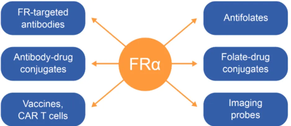
Due to the overexpression on a wide range of tumors and a concurrently limited expression at only a few sites in normal tissue, the FRα has been exploited in the diagnosis and treatment of cancer patients.
In view of therapeutic application, it was targeted with FR-specific antibodies, antibody-drug conjugates (ADCs), vaccine-based approaches, anti-FRα chimeric antigen receptor (CAR) T cells, folate-drug conjugates as well as some antifolates (Fig. 1.5) [24,39]. From numerous FRα-targeting agents that have been developed over the past years, only few reached clinical studies.
Farletuzumab (MORAb-003) is a fully humanized FRα-targeting monoclonal IgG1 antibody which
![Fig. 2.4 Receptor-binding curves for 6R-Aza-5-MTHF, 6S-Aza-5-MTHF and AzaFol (upper panel) as well as for the non-fluorinated analogues, 6S-5-MTHF, 6R-5-MTHF and folic acid (lower panel) using [ 3 H]folic acid as the radioactive tracer (0.82–0.98 nM)](https://thumb-eu.123doks.com/thumbv2/1library_info/3908761.1525977/49.892.175.621.112.477/receptor-binding-curves-azafol-fluorinated-analogues-radioactive-tracer.webp)
![Fig. 2.5 (a) Signal intensities of 6R-[ 18 F]Aza-5-MTHF, 6S-[ 18 F]Aza-5-MTHF and [ 18 F]AzaFol (set as 100%) quantified based on RT16 (left) or D4 (right) autoradiography images](https://thumb-eu.123doks.com/thumbv2/1library_info/3908761.1525977/50.892.206.773.538.1013/signal-intensities-mthf-mthf-azafol-quantified-autoradiography-images.webp)
![Fig. 3.2 Tumor-to-kidney ratios of mice after injection of [ 177 Lu]Lu-folate (5 MBq, 1 nmol)](https://thumb-eu.123doks.com/thumbv2/1library_info/3908761.1525977/66.892.244.727.324.843/fig-tumor-kidney-ratios-mice-injection-folate-mbq.webp)
