AUS DEM LEHRSTUHL FÜR MUND-, KIEFER- UND GESICHTSCHIRURGIE PROF. DR. DR. TORSTEN E. REICHERT
DER FAKULTÄT FÜR MEDIZIN DER UNIVERSITÄT REGENSBURG
DIE LEBENSQUALITÄT VON PATIENTEN MIT MEDIKAMENTEN-ASSOZIIERTER KIEFERNEKROSE STADIUM III VERBESSERT SICH NACH
CHIRURGISCHER THERAPIE
Inaugural – Dissertation zur Erlangung des Doktorgrades
der Zahnmedizin
der Fakultät für Medizin der Universität Regensburg
vorgelegt von Stefan Nikolaus Georg Moll
2020
Dekan: Prof. Dr. Dirk Hellwig
Erster Gutachter: PD. Dr. Dr. Christoph Klingelhöffer, M.Sc.
Zweiter Gutachter: Prof. Dr. Werner Krutsch
Tag der mündlichen Prüfung: 19.02.2021
Deutschsprachige Zusammenfassung der wissenschaftlichen Originalarbeit
„Patients’ quality of life improves after surgical intervention of stage III medication-related osteonecrosis of the jaw”
Von Stefan Moll et al.
publiziert in
Oral and Maxillofacial Surgery 2020
vorgelegt von Stefan Nikolaus Georg Moll
2020
Inhaltsverzeichnis
1. Einleitung und Fragestellung ... 5
2. Material und Methoden ... 6
2.1 Studiendesign und Einschlusskriterien ... 6
2.2 Variablen und Datenerhebung ... 6
2.3 Klinisches Vorgehen ... 8
2.4 Statistik ... 8
3. Ergebnisse ... 9
4. Diskussion ... 13
5. Fazit ... 16
6. Literaturverzeichnis ... 17
7. Danksagung………..……….22
8. Lebenslauf………..………23
Einleitung und Fragestellung
Die Medikamenten-assoziierte Kiefernekrose (engl.: medication-related osteonecrosis of the jaw – MRONJ) stellt seit ihrer Entdeckung durch Marx 2003 sowohl für Patienten als auch für Behandler eine anhaltende Herausforderung dar (1).
Mit einer steigenden Anzahl an Medikamenten, die zu einer Nekrose führen können, und einer letztlich nicht vollständig geklärten Pathologie ist die MRONJ ein viel diskutiertes Thema und die Grundlage andauernder Forschungen. Die MRONJ wurde durch die American Association of Oral and Maxillofacial Surgeons als ein Zustand freiliegenden Knochens definiert, welcher während oder nach einer antiresorptiven oder antiangiogenesen Therapie auftritt und mehr als acht Wochen persistiert. Darüber hinaus darf keine Historie von Kopf- oder Hals-Radiatio oder Tumoren des Kiefers vorhanden sein (2).
Aktuelle Therapieansätze basieren auf Positionspapieren ohne internationale Standardrichtlinien (2–5). Speziell die Therapie von Stadium-III-Patienten führt regelmäßig zu Schwierigkeiten, da diese zwar von einem chirurgischen Therapieansatz profitieren, das Risiko eines Rezidivs jedoch hoch ist und gegebenenfalls mehrere Eingriffe benötigt werden, um eine vollständige mukosale Deckung des Kieferkamms zu erreichen (6–8). Die Patienten leiden oft an einer Vielzahl von Symptomen wie starken Schmerzen, Schluckbeschwerden und einem Gefühl von Unsicherheit im Zusammenhang mit ihren Zähnen. Mit steigendem MRONJ-Stadium nehmen die Beschwerden zu, was zu einer starken Beeinträchtigung der Lebensqualität (engl.: Qualitiy of Life – QoL) führt (9). Da die Verbesserung bzw.
der Erhalt der Lebensqualität eines der Hauptziele der MRONJ-Therapie ist, sollte diese auch bei der Entscheidungsfindung bezüglich der Therapiewahl in Betracht gezogen werden. Zum aktuellen Zeitpunkt gibt es nur eine sehr begrenzte Anzahl an Forschungsarbeiten, die sich mit dem Einfluss der MRONJ-Therapie auf die Lebensqualität auseinandersetzen, insbesondere wenn es um die konkrete Einteilung in MRONJ-Stadien geht (10).
Das Ziel dieser Studie war es, potenzielle Veränderungen der
gesundheitsbezogenen Lebensqualität von Stadium-III-MRONJ-Patienten nach
chirurgischer Therapie festzustellen. Zusätzlich sollte eruiert werden, inwieweit das
Auftreten eines Rezidivs diese Veränderung beeinflusst. Abschließend sollte der
Einfluss von Alter, Geschlecht, Medikation, Dauer der Medikation, Lokalisation der Nekrose und Risikofaktoren auf die Entwicklung der Lebensqualität bestimmt werden.
Material und Methoden
Studiendesign und Einschlusskriterien
Um diese Fragestellung beantworten zu können wurde in der Abteilung der Mund-, Kiefer- und Gesichtschirurgie am Universitätsklinikum Regensburg eine prospektive monozentrische Studie entworfen und durchgeführt. Die Studie wurde von der lokalen Ethikkommission genehmigt (Nr. 16-101-0257). Über einen Zeitraum von mehr als zweieinhalb Jahren (September 2016 bis März 2019) wurden Patienten mit einer Stadium III MRONJ in die Studie eingeschlossen. Die MRONJ wurde diagnostiziert und klassifiziert nach der Definition der American Association of Oral and Maxillofacial Surgeons. Eine Stadium III MRONJ wird als freiliegender nekrotischer Knochen oder als nekrotischer Knochen, welcher sich durch eine intra- /extraorale Fistel sondieren lässt in Kombination mit Schmerzen, einer Infektion und mindestens einem der folgenden Kriterien definiert: der nekrotische Knochen überschreitet den Alveolarknochen mit daraus resultierender pathologischer Unterkieferfraktur, extraoralen Fistel, oronasalen Fistel oder oroantralen Fistel;
Osteolyse bis zum Corpus mandibulae; Osteolyse bis zum Boden des Sinus maxillaris (2). Patienten mit einer radiologischen Bestrahlung im Kopf-/Halsbereich in der Anamnese, einer Persistenz des freiliegenden Knochens oder Fistel von weniger als acht Wochen oder mit Tumoren im Kiefer wurden definitionsgemäß von der Studie ausgeschlossen.
Variablen und Datenerhebung
Die gesundheitsbezogene Lebensqualität wurde mit Hilfe von zwei etablierten
Fragebögen präoperativ (T0), sechs Wochen postoperativ (T1) und sechs Monate
postoperativ (T2) erhoben. Darüber hinaus wurden die anatomische Lokalisation der
Nekrose (Maxilla oder Mandibular), das Alter (≥ 63 <Jahre), das Geschlecht (m/w), die
Einnahmedauer der Medikation (Zeitpunkt der ersten Einnahme bis zur letzten
Einnahme oder Datum der Erstaufnahme im Zusammenhang mit MRONJ), eine Zahnextraktion unmittelbar vor MRONJ (Ja/Nein), der Nikotinabusus (Ja/Nein) und das Auftreten eines Rezidivs dokumentiert. Zur Messung der gesundheitsbezogenen Lebensqualität wurde der EORTC QoL-H&N35-(European Organisation for Research and Treatment of Cancer QoL-H&N35) und der OHIP-G14-(Oral Health Impact Factor- G14) Fragebogen verwendet.
Der EORTC QoL-H&N35 beinhaltet 35 Fragen bezugnehmend auf Symptome und Nebenwirkungen der Behandlung, auf die eigene Körperwahrnehmung und den Einfluss auf Sozialleben und Sexualität. Der Fragebogen beinhaltet sieben Multi-Item- Skalen sowie elf Single-Item-Skalen. Alle Multi-Item-Skalen beinhalten verschiedene Fragen, wobei keine der Fragen mehrmals verwendet wird (Tab. 1). Die Antwortmöglichkeiten (von 1 „not at all“ bis 4 „very much“ oder „Yes/No“) wurden gemäß des offiziellen Auswertungshandbuchs in Werte zwischen 0 und 100 umgerechnet und statistisch ausgewertet (11)(12). Bereits mehrere Studien hatten Probleme bei der Verwendung des EORTC QoL-H&N35-Fragebogens mit fehlenden Daten bezüglich Sexualität (Frage Q 29 und Q 30). Daher wurden bereits vor Beginn der Studie diese Fragen durch Fragen zu Auswirkungen der Erkrankung auf den Alltag der Patienten mit den gleichen Antwortmöglichkeiten ersetzt (13–15). Die Fragen waren für Q29 „do thoughts on your primary disease affect your everyday life?” und für Q30 „do you feel impaired to do physical work (for example, household chores)?“. Sie wurden zur Multi-Item-Skala „Einfluss auf das Alltagsleben “ inkludiert.
Der OHIP-G14 Fragebogen besteht aus 14 Fragen zur Mundgesundheitsbezogenen
Lebensqualität. Die Antwortmöglichkeiten reichten von 1 „never“ bis 4 „very often“ und
wurden summiert zu einem Addierten-OHIP-G14 Wert. Diese Werte wurden statistisch
ausgewertet (16).
Tab.1: EORTC QoL-H&N35 Multi-/Singel-Item-Skalen
Item-Skalen Items Item range* QoL-H&N35 Fragen
Schmerzen 4 3 1-4
Schlucken 4 3 5-8
Empfindungsstörungen 2 3 13-14
Sprachprobleme 3 3 16,23,24
Probleme mit Essen in Gesellschaft 4 3 19-22
Probleme mit sozialen Kontakten 5 3 18,24,28
Sexualität/Einfluss auf das Alltagsleben ** 2 3 29,30
Zähne 1 3 9
Mundöffnung 1 3 10
Trockener Mund 1 3 11
Klebriger Speichel 1 3 12
Husten 1 3 15
Krankheitsgefühl 1 1 17
Analgetika 1 1 31
Nahrungsergänzungsmittel 1 1 32
Ernährungssonde 1 1 33
Gewichtsverlust 1 1 34
Gewichtszunahme 1 1 35
*”Item range” ist die Differenz der maximale mögliche Antwortpunktzahl und der minimal mögliche Antwortpunktzahl
**Fragen zu “Sexualität” wurden ersetzt durch Fragen zu „Einfluss auf das Alltagsleben“
Klinisches Vorgehen
Alle chirurgischen Eingriffe wurden unter nasaler Intubationsnarkose vorgenommen. Nach der Präparation eines Mukoperiostlappens, wurde der nekrotische Knochen mittels einer Knochensäge und piezochirurgischen Instrumenten so weit entfernt bis vitaler Knochen sichtbar wurde. Scharfe Knochenkanten wurden geglättet. Ein spannungsfreier und speicheldichter Wundverschluss erfolgte mittels mehrschichtiger Naht. Alle Patienten erhielten eine Antibiotikatherapie einen Tag präoperativ bis zehn Tage postoperativ. Es wurde Amoxicillin/Clavulansäure verabreicht. In Fällen einer Penicillin-Allergie wurde alternativ Clindamycin verschrieben. Die Nahrungsaufnahme wurde für zehn Tage mittels nasogastraler Sonde sichergestellt. Chlorhexidin (0,12%) wurde als antimikrobielle Mundspüllösung dreimal täglich verwendet. Eine Kontrolluntersuchung fand 14 Tage und sechs Wochen nach der Entlassung statt. Anschließend wurden alle Patienten in ein routinemäßiges halbjährliches Nachsorgeprogramm aufgenommen.
Statistik
Die statistische Auswertung wurden mit SPSS 26 (SPSS Inc. Chicago, IL, USA)
durchgeführt. EOTC QoL-H&N35-Werte wurden anhand des offiziellen
Auswertungshandbuchs analysiert (11). Varianzanalysen (ANOVA) wurden
berechnet, um signifikante Veränderungen in den Umfragewerten feststellen zu können. Kovarianzanalysen (ANCOVA) wurden verwendet, um mögliche signifikante Effekte von Kovariaten auf die Lebensqualität bestimmen zu können. Bei einer Verletzung der Sphärizität, wurde das Greenhouse-Geisser Korrekturverfahren angewandt. Der Exakte Test nach Fisher wurde durchgeführt, um Zusammenhänge innerhalb der Kovariaten zu erkennen. Ein p-Wert von ≤ 0,05 wurde als statistisch signifikant erachtet.
Ergebnisse
Dreiundvierzig Stadium-III-MRONJ-Patienten mit einem Durchschnittsalter von 68 Jahren (40-88 Jahre) wurden operativ behandelt. Der durchschnittliche Einnahmezeitraum des antiresorptiven Therapeutikums lag bei 63 Monaten (3-423 Monate). Weitere Patientencharakteristika sind in Tabelle 2 dargestellt.
Tab. 2:
Patientencharakteristika
*Ein Patient erhielt Pamidronsäure gefolgt von Ibandronsäure
**Im Fall von Bisphosphonatgabe gefolgt von Denosumab, erscheinen Patienten auch in der Kategorie Denosumab oder Bisphosphonat
Patienten 43
•
Männlich 21 48.8%
•
Weiblich 22 51.2%
Maligne Grunderkrankung 36 83.7%
•
Mammakarzinom 11 30.6%
•
Prostatakarzinom 14 38.9%
•
Multiples Myelom 8 22%
•
Lungenkarzinom 2 5.6%
• Leiomyosarkom
1 2.8%
Benigne Grunderkrankung 7 16.3%
•
Osteoporose 7 100%
Orale Bisphosphonate 6 14%
•
Alendronsäure 6 100%
Intravenöse Bishosphonate 33 76.7%
•
Zoledronsäurepamidronsäure 28 84.8%
•
Pamidronsäure* 3 9.1%
•
Ibandronsäure* 3 9.1%
Denosumab-Therapie 13 30.2%
Bisphosphonat gefolgt von Denosumab** 9 20.1%
Lokalisation 43
•
Maxilla 9 20.9%
•
Mandibula 34 79.1%
Alle 43 Patienten füllten die Fragebögen präoperativ und sechs Wochen postoperativ aus. Sechs Monate postoperativ wurden die Fragebögen von 83,7%
(36/43) der Patienten ausgefüllt. Sechs Patienten sind im Laufe der Studie verstorben und ein Patient fühlte sich nicht in der Lage weiter an der Studie teil zu nehmen. Der durchschnittliche Nachsorgezeitraum lag bei 21.9 Wochen mit einem Minimum von sechs Wochen und einem Maximum von sechs Monaten. 34,9% (15/43) der Patienten hatten eine Zahnextraktion unmittelbar vor der MRONJ Erstdiagnose. Während der Nachsorge entwickelten 25,6% (11/43) ein Rezidiv. 63,6% (7/11) der Rezidive traten innerhalb der ersten sechs Wochen nach der Operation auf. 42,9% (3/7) dieser frühen Rezidiven zeigten eine vollständige Ausheilung bis zur Nachuntersuchung nach sechs Monaten. 19,4% (7/36) der Patienten wiesen auch nach sechs Monaten noch ein Rezidiv auf. Präoperativ gaben 20,9% (9/43) der Patienten Nikotinabusus an. Raucher wiesen im Vergleich zu Nichtrauchern ein signifikant höheres Risiko auf auch noch nach sechs Monaten an einem Rezidiv zu leiden (p=0,05). Die Lokalisation der MRONJ zeigte zu keinem Zeitpunkt der Nachsorge einen signifikanten Einfluss auf die Entstehung eines Rezidivs (T1 p=0,624; T2 p=0,652). 80,6% (29/36) der Patienten zeigten eine vollständige mukosale Heilung (T2). In allen Fällen, bei denen keine vollständige Abheilung der Mukosa erreicht wurde, kam es zu einer Verbesserung des MRONJ-Stadiums von III auf I. Ein Patient wurde aufgrund von Extremwerten von allen EORTC QoL-H&N35- und OHIP-G14-Analysen ausgeschlossen. Die Extremwerte waren auf eine neu aufgetretene MRONJ zurückzuführen. Die neue Nekrose befand sich in einer anderen Region als die ursprüngliche Stadium-III-Nekrose und wurde daher nicht als Rezidiv gewertet.
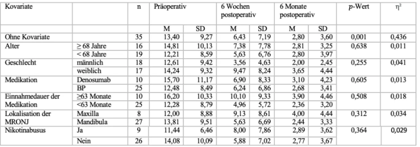
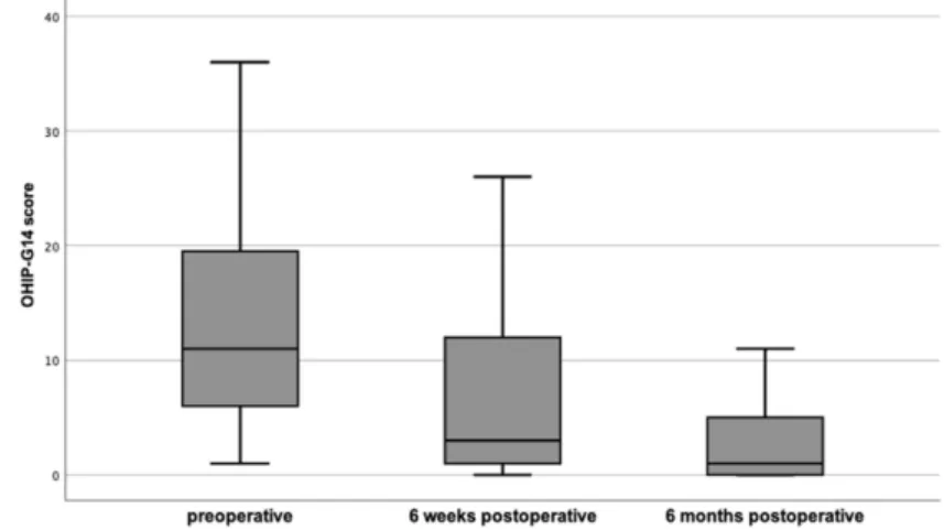
Die OHIP-G14 Werte verringerten sich (Verbesserung der Lebensqualität) signifikant (n=35; p=0,001) zwischen T0 (13,40 ± 9,27) und T1 (6,43 ± 7,19) um 52,02% und zwischen T1 und T2 (2,80 ± 3,60) um weitere 56,45% (Abb. 1). Die erhobenen Kovariaten zeigten keinen signifikanten Einfluss auf die OHIP-G14 Werte, weder präoperativ noch im Verlauf der postoperativen Nachsorgetermine (Tab. 3).
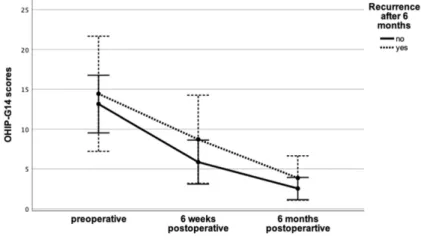
Patienten, die ein Rezidiv entwickelten, zeigten keine signifikant (p=0,181) unterschiedliche OHIP-G14 Werte im Vergleich zu Patienten ohne Rezidiv. Der Rezidiv-Zeitpunkt hatte keinen Einfluss auf die gemessenen Werte (T0-T1 p=0,105;
zwischen T1-T2 p=0,820) (Abb.2).
Abb. 1: Entwicklung der OHIP-G14 Werte (n=35; präoperativ: M 13,40 SD 9,27; sechs Wochen postoperativ: M
6,43 SD 7,19; sechs Monate postoperativ: M 2.80 SD 3.60) p<.001; η²=.44
Abb. 2: Vergleich der OHIP-G14 nach sechs Monaten zwischen Patienten mit und Rezidiv. Es zeigt sich kein
signifikanter Unterschied zwischen den beiden Gruppen (p=.85 η²=.003)
Tab. 3: Zusammenhang zwischen Kovariaten und den 𝑝-Werten der OHIP-G14-Werte
Die einzelnen EORTC QoL-H&N35-Werte wurden miteinander verglichen und zeigten für die Kategorien „Schlucken“ (p=0,007), „Mundöffnung“ (p=0,045),
„Analgetika“ (p=0,005), „Gewichtsverlust“ (p=0,004), „Schmerzen“ (p=0,001),
„Probleme mit Essen in Gesellschaft“ (p=0,001), „Probleme mit sozialen Kontakten“
(p=0,001) und „Zähne“ (p=0,001) eine signifikante Veränderung (Tab. 4). Keiner der EORTC QoL-H&N35-Werte wurde durch das Auftreten eines Rezidivs signifikant beeinflusst. Ebenso zeigten die Kovariaten keinen statistisch messbaren Effekt (Tab.5).
Tab. 4: EORTC QoL-H&N35-Werte
Tab. 5: Zusammenhang zwischen Kovariaten und den 𝑝-Werten der EORTC QoL-H&N35-Werte
Diskussion
Ziel der Studie war es, den Einfluss einer operativen Therapie bei MRONJ Stadium III auf die gesundheitsbezogene Lebensqualität von Patienten zu untersuchen und zu eruieren, ob es dadurch tatsächlich zu einer Verbesserung derselben kommt. Des Weiteren sollte der Effekt eines potenziell auftretenden Rezidivs auf die Entwicklung der Lebensqualität bewertet werden. Durch die Erhebung der gesundheitsbezogenen Lebensqualität präoperativ, sechs Wochen postoperativ (n=43) und sechs Monate postoperativ (n=36) waren wir in der Lage eine signifikante Verbesserung der generellen oralen Mundgesundheit (OHIP-G14) und in Teilen der allgemeinen gesundheitsbezogenen Lebensqualität (EORTC QoL-H&N35) festzustellen. Im Falle einer nicht vollständigen und langfristigen mukosalen Deckung der nekrotischen Areale, zeigten sich postoperativ keine signifikanten Unterschiede in der Entwicklung der Lebensqualität. Dies könnte dem Umstand geschuldet sein, dass obwohl manche Patienten im Verlauf ein Rezidiv entwickelten, sich in allen Fällen das MRONJ-Stadium von III auf I verbessert hat. Diese Veränderung bedeutet zwar für den Patienten weiterhin einen freiliegenden Knochen, jedoch ohne Entzündungszeichen und Schmerzen (2). Die Verbesserung des MRONJ-Stadiums auf Stadium I scheint ein ausschlaggebender Faktor zu sein, da die größte Abnahme der Lebensqualität beim Übergang von Stadium I auf II vorzufinden ist (9).
Schlussfolgernd lässt sich sagen, dass -obwohl das eigentliche Ziel die volle mukosale
Ausheilung ist- die vom Patienten empfundene Verbesserung der Lebensqualität
bereits mit dem Erreichen von Stadium I größtenteils erzielt wurde. Somit ist nach sechs Monaten der Unterschied in der Lebensqualität zwischen Patienten mit Stadium 0 und denen die mit Stadium I nur gering.
Mit einer vollständigen mukosalen Ausheilung nach sechs Monaten in 80,6%
(29/36) der Fälle, sind unsere Ergebnisse mit aktuellen Studien vergleichbar. Hier wird eine studienübergreifende Erfolgsrate bei chirurgischer Therapie eines MRONJ Stadium III von 85% angegeben (17). In allen erwähnten Studien wurde die Nekrose chirurgisch entfernt und der Defekt ohne Verwendung eines mikrovaskulären Lappens gedeckt (6,18–22). Mehr als ein Drittel der Patienten hatten unmittelbar vor der MRONJ-Erstdiagnose eine Zahnextraktion. Studien haben gezeigt, dass Zahnextraktion einen großen Risikofaktor darstellen (23–25). Otto et al. haben in diesem Zusammenhang beschrieben, dass nicht die Zahnextraktion an sich, sondern ein prävalenter infektiöser Zustand im Knochen, zur Erhöhung des MRONJ-Risikos führen könnte. Durch Einhaltung von Leitlinien, welche die perioperative Antibiotikagabe, die atraumatische Extraktion, die Glättung von scharfen Knochenkanten und einen speicheldichten Wundverschluss vorsehen, können Zahnextraktionen risikoärmer durchgeführt werden (26). Aufgrund der Tatsache, dass die Zahnextraktionen nicht am Universitätsklinikum Regensburg durchgeführt wurden, lagen uns keine Information über das genaue Vorgehen bei Extraktionen vor. In den ersten sechs Wochen postoperativ zeigten Raucher kein höheres Rezidivrisiko. Im Falle eines Rezidivs, besteht bei Rauchern ein signifikant höheres Risiko, dass auch nach sechs Monaten noch exponierter Knochen vorliegt. Dies lässt uns vermuten, dass der Nikotinabusus zwar nicht das unmittelbare Risiko eines Rezidivs signifikant erhöht, aber vermutlich zu einer Suppression der sekundären Wundheilung führt (27).
Des Weiteren ist anzunehmen, dass nicht der verbliebene oder der neue nekrotische Knochen das Hauptrisiko für die Entstehung eines Rezidivs ist, sondern das Nichterreichen eines spannungsfreien Wundverschlusses.
Die gesundheitsbezogene Lebensqualität wurde hauptsächlich von zwei
Faktoren beeinflusst. Als größter Einflussfaktor ist die Schmerzsymptomatik zu
nennen. Obwohl einige Patienten keine Schmerzen empfanden, gab der Großteil der
Probanden an starke Schmerzen zu verspüren, welche sich unter mechanischer
Belastung verstärkten. Als Resultat waren hohe Werte (geringe Lebensqualität) in der
Kategorie „Schlucken“ zu beobachten. Diese Kategorie beinhaltete Fragen zum Kauen
von weicher und fester Nahrung. In manchen Fällen gaben die Patienten an, dass sie
aufgrund von starken Schmerzen das Essen regelmäßig unterbrechen mussten.
Dieser Umstand könnte das hohe präoperative Level an „Gewichtsverlust“ in Kombination mit so gut wie keiner „Gewichtszunahme“ erklären. Postoperativ verbesserten sich die „Schmerz“-Werte, ebenso wie die „Schluck“-Werte. Als mögliche Konsequenz zeigten sich weniger Probleme beim Essen, was sich wiederum in einem Anstieg der „Gewichtszunahme“-Werte und einem Abfall der „Gewichtsabnahme“- Werte zeigte. Entsprechend wurde postoperativ ein Rückgang der Einnahme von Analgetika angegeben. Schlussendlich blieb die Anzahl an Patienten, die regelmäßig Schmerzmittel einnahmen, jedoch hoch. An diesem Punkt hatten wir keine Information über die Art und Dosierung der Schmerzmedikation, was die Aussagekraft über den Rückgang der Analgetika limitiert. Auch ist hier anzumerken, dass alle Patienten an einer Grunderkrankung litten, die ebenfalls für eine Schmerzsymptomatik ursächlich sein konnte.
Der zweite Haupteinflussfaktor auf die Lebensqualität schien die Psyche zu sein. Mehrere Patienten beschrieben ein Gefühl der Unsicherheit im Zusammenhang mit ihren Zähnen. Zusätzlich beklagten sich die Patienten über starken Foetor ex ore.
Dieser Umstand in Kombination mit insuffizienter Dentition oder nicht bzw. schlecht sitzender prothetischer Versorgung führte zu hohen Werten in den Multi-Item-Skalen
„Probleme mit Essen in Gesellschaft“- und „Probleme mit sozialen Kontakten“.
Postoperativ kam es zu einer deutlichen Verbesserung der mit dem Sozialleben in Verbindung stehenden Werte. Die Fertigstellung einer neuen prothetischen Versorgung stellte für viele Patienten eine besondere Steigerung der Lebensqualität dar. Die Frage nach dem frühesten Zeitpunkt für eine neue prothetische Versorgung wurde oft gestellt. Abhängig von der Lokalisation der ursprünglichen Nekrose und dem Heilungsprozess, sollte damit mindestens bis nach der sechsten postoperativen Woche gewartet werden.
Nach unserem Wissensstand ist dies die erste Studie, die den Einfluss einer
chirurgischen Therapie auf die gesundheitsbezogene Lebensqualität bei einem
spezifischen MRONJ-Stadium (Stadium III) untersucht hat. Limitation dieser Studie ist
die niedrige Patientenzahl. Diese ist auf die geringe Inzidenz von MRONJ
zurückzuführen (28). Auch wenn wir in der Lage waren signifikante Veränderung in der
Lebensqualität der Patienten festzustellen, so macht die geringe Gruppenstärke die
Analyse bei Unterteilung in Subgruppen unzuverlässig, da hier sehr schnell noch
Kontrollgruppe. Leider konnten wir unsere Ergebnisse nicht mit anderen Gruppen vergleichen, da keine Daten von Patienten mit ähnlicher Grunderkrankung zur Verfügung standen. In Anbetracht der Tatsache, dass die Beeinträchtigung der Lebensqualität von MRONJ-Patienten zu einem großen Teil vom Stadium der Erkrankung abhängig ist, sollten weitere Studien bezogen auf die einzelnen Stadien erfolgen. Dies würde die Vergleichbarkeit der Studien und deren praktischen Nutzen im täglichen klinischen Alltag verbessern.
Fazit
Unsere Ergebnisse lassen darauf schließen, dass Patienten mit MRONJ
Stadium III von einer chirurgischen Therapie profitieren. Die gesundheitsbezogene
Lebensqualität hat sich durch den operativen Eingriff signifikant verbessert. Patienten,
die ein Rezidiv entwickelten, profitierten von einer Verbesserung des Stadiums von III
auf I und zeigten keine signifikant schlechteren Lebensqualitätswerte im Vergleich zu
Patienten mit vollständiger mukosaler Ausheilung. Weitere Studien sind nötig, um den
Einfluss verschiedener Therapieansätze bei unterschiedlichen MRONJ Stadien auf die
Lebensqualität zu untersuchen.
Literaturverzeichnis
1. Marx RE. Pamidronate (Aredia) and zoledronate (Zometa) induced avascular necrosis of the jaws: a growing epidemic. J Oral Maxillofac Surg [Internet].
September 2003 [zitiert 12. Dezember 2020];61(9):1115–7. Verfügbar unter:
https://linkinghub.elsevier.com/retrieve/pii/S0278239103007201
2. Ruggiero SL, Dodson TB, Fantasia J, Goodday R, Aghaloo T, Mehrotra B, u. a.
American Association of Oral and Maxillofacial Surgeons Position Paper on Medication-Related Osteonecrosis of the Jaw—2014 Update. J Oral Maxillofac Surg [Internet]. Oktober 2014 [zitiert 5. Januar 2019];72(10):1938–56. Verfügbar unter: https://linkinghub.elsevier.com/retrieve/pii/S0278239114004637
3. Ruggiero SL, Dodson TB, Assael LA, Landesberg R, Marx RE, Mehrotra B.
American Association of Oral and Maxillofacial Surgeons Position Paper on Bisphosphonate-Related Osteonecrosis of the Jaws—2009 Update. J Oral Maxillofac Surg [Internet]. 1. Mai 2009 [zitiert 19. Februar 2019];67(5, Supplement):2–12. Verfügbar unter:
http://www.sciencedirect.com/science/article/pii/S0278239109001153
4. Otto S, Pautke C, Wyngaert TV den, Niepel D, Schiødt M. Medication-related osteonecrosis of the jaw: Prevention, diagnosis and management in patients with cancer and bone metastases. Cancer Treat Rev [Internet]. 1. September 2018 [zitiert 18. April 2020];69:177–87. Verfügbar unter:
https://www.cancertreatmentreviews.com/article/S0305-7372(18)30101- 4/abstract
5. Khan AA, Morrison A, Hanley DA, Felsenberg D, McCauley LK, O’Ryan F, u. a.
Diagnosis and Management of Osteonecrosis of the Jaw: A Systematic Review and International Consensus. J Bone Miner Res [Internet]. 2015 [zitiert 18. April 2020];30(1):3–23. Verfügbar unter:
https://asbmr.onlinelibrary.wiley.com/doi/abs/10.1002/jbmr.2405
6. Klingelhöffer C, Zeman F, Meier J, Reichert TE, Ettl T. Evaluation of surgical outcome and influencing risk factors in patients with medication-related osteonecrosis of the jaws. J Cranio-Maxillofac Surg [Internet]. Oktober 2016 [zitiert 11. Dezember 2018];44(10):1694–9. Verfügbar unter:
https://linkinghub.elsevier.com/retrieve/pii/S1010518216301639
7. Ristow O, Rückschloß T, Bodem J, Berger M, Bodem E, Kargus S, u. a. Double- layer closure techniques after bone surgery of medication-related osteonecrosis of the jaw – A single center cohort study. J Cranio-Maxillofac Surg [Internet]. 1.
Mai 2018 [zitiert 6. Januar 2019];46(5):815–24. Verfügbar unter:
http://www.sciencedirect.com/science/article/pii/S1010518218300659 8. Otto S, Ristow O, Pache C, Troeltzsch M, Fliefel R, Ehrenfeld M, u. a.
Fluorescence-guided surgery for the treatment of medication-related
osteonecrosis of the jaw: A prospective cohort study. J Cranio-Maxillofac Surg
unter: http://www.sciencedirect.com/science/article/pii/S1010518216300701 9. Miksad RA, Lai K-C, Dodson TB, Woo S-B, Treister NS, Akinyemi O, u. a. Quality
of Life Implications of Bisphosphonate-Associated Osteonecrosis of the Jaw. The Oncologist [Internet]. 1. Januar 2011 [zitiert 5. Januar 2019];16(1):121–32.
Verfügbar unter: http://theoncologist.alphamedpress.org/content/16/1/121 10. Murphy J, Mannion CJ. Medication-related osteonecrosis of the jaws and
quality of life: review and structured analysis. Br J Oral Maxillofac Surg [Internet].
1. April 2020 [zitiert 19. April 2020]; Verfügbar unter:
http://www.sciencedirect.com/science/article/pii/S0266435620300942
11. Fayers P, Aaronson NK, Bjordal K, Groenvold M, Curran D, Bottomley A.
EORTC QLQ-C30 Scoring Manual [Internet]. European Organisation for
Research and Treatment of Cancer; 2001 [zitiert 19. April 2020]. Verfügbar unter:
https://abdn.pure.elsevier.com/en/publications/eortc-qlq-c30-scoring-manual-3rd- edition
12. Aaronson NK, Ahmedzai S, Bergman B, Bullinger M, Cull A, Duez NJ, u. a.
The European Organization for Research and Treatment of Cancer QLQ-C30: A Quality-of-Life Instrument for Use in International Clinical Trials in Oncology. JNCI J Natl Cancer Inst [Internet]. 3. März 1993 [zitiert 6. Januar 2019];85(5):365–76.
Verfügbar unter: https://academic.oup.com/jnci/article- lookup/doi/10.1093/jnci/85.5.365
13. Bjordal K, de Graeff A, Fayers PM, Hammerlid E, van Pottelsberghe C, Curran D, u. a. A 12 country field study of the EORTC QLQ-C30 (version 3.0) and the head and neck cancer specific module (EORTC QLQ-H&N35) in head and neck patients. Eur J Cancer [Internet]. 1. September 2000 [zitiert 19. April
2020];36(14):1796–807. Verfügbar unter:
http://www.sciencedirect.com/science/article/pii/S0959804900001866
14. Kyrgidis A, Triaridis S, Kontos K, Patrikidou A, Andreadis C, Constantinidis J, u. a. Quality of Life in Breast Cancer Patients with Bisphosphonate-related Osteonecrosis of the Jaws and Patients with Head and Neck Cancer: A
Comparative Study Using the EORTC QLQ-C30 and QLQ-HN35 Questionnaires.
Anticancer Res [Internet]. 8. Januar 2012 [zitiert 6. Januar 2019];32(8):3527–34.
Verfügbar unter: http://ar.iiarjournals.org/content/32/8/3527
15. Jensen K, Jensen AB, Grau C. A cross sectional quality of life study of 116 recurrence free head and neck cancer patients. The first use of EORTC H&N35 in Danish. Acta Oncol [Internet]. 1. Januar 2006 [zitiert 19. April 2020];45(1):28–
37. Verfügbar unter: https://doi.org/10.1080/02841860500417536
16. Robinson PG, Gibson B, Khan FA, Birnbaum W. Validity of two oral health- related quality of life measures. Community Dent Oral Epidemiol [Internet]. 2003 [zitiert 19. April 2020];31(2):90–9. Verfügbar unter:
https://onlinelibrary.wiley.com/doi/abs/10.1034/j.1600-0528.2003.00051.x 17. Jasper V, Laurence V, Maximiliaan S, Ferri J, Nicot R, Constantinus P.
Medication-related osteonecrosis of the jaw (MRONJ) stage III: Conservative and
conservative surgical approaches versus an aggressive surgical intervention: A systematic review. J Cranio-Maxillofac Surg [Internet]. März 2020 [zitiert 5. April 2020];S101051822030055X. Verfügbar unter:
https://linkinghub.elsevier.com/retrieve/pii/S101051822030055X
18. Lopes RN, Rabelo GD, Rocha AC, Carvalho PAG, Alves FA. Surgical Therapy for Bisphosphonate-Related Osteonecrosis of the Jaw: Six-Year Experience of a Single Institution. J Oral Maxillofac Surg [Internet]. 1. Juli 2015 [zitiert 17. April 2020];73(7):1288–95. Verfügbar unter:
http://www.sciencedirect.com/science/article/pii/S0278239115000622
19. Favia G, Tempesta A, Limongelli L, Crincoli V, Maiorano E. Medication-related osteonecrosis of the jaw: Surgical or non-surgical treatment? Oral Dis [Internet].
2018 [zitiert 17. April 2020];24(1–2):238–42. Verfügbar unter:
https://onlinelibrary.wiley.com/doi/abs/10.1111/odi.12764
20. Blus C, Giannelli G, Szmukler-Moncler S, Orru G. Treatment of medication- related osteonecrosis of the jaws (MRONJ) with ultrasonic piezoelectric bone surgery. A case series of 20 treated sites. Oral Maxillofac Surg [Internet]. 1. März 2017 [zitiert 17. April 2020];21(1):41–8. Verfügbar unter:
https://doi.org/10.1007/s10006-016-0597-7
21. Berrone M, Florindi FU, Carbone V, Aldiano C, Pentenero M. Stage 3 Medication-Related Osteonecrosis of the Posterior Maxilla: Surgical Treatment Using a Pedicled Buccal Fat Pad Flap: Case Reports. J Oral Maxillofac Surg [Internet]. 1. November 2015 [zitiert 17. April 2020];73(11):2082–6. Verfügbar unter: http://www.sciencedirect.com/science/article/pii/S0278239115009003 22. Pichardo SEC, Kuijpers SCC, van Merkesteyn JPR. Bisphosphonate-related
osteonecrosis of the jaws: Cohort study of surgical treatment results in seventy- four stage II/III patients. J Cranio-Maxillofac Surg [Internet]. 1. September 2016 [zitiert 17. April 2020];44(9):1216–20. Verfügbar unter:
http://www.sciencedirect.com/science/article/pii/S1010518216301044
23. Saad F, Brown JE, Van Poznak C, Ibrahim T, Stemmer SM, Stopeck AT, u. a.
Incidence, risk factors, and outcomes of osteonecrosis of the jaw: integrated analysis from three blinded active-controlled phase III trials in cancer patients with bone metastases. Ann Oncol [Internet]. Mai 2012 [zitiert 13. April
2020];23(5):1341–7. Verfügbar unter:
https://linkinghub.elsevier.com/retrieve/pii/S0923753419346952
24. Vahtsevanos K, Kyrgidis A, Verrou E, Katodritou E, Triaridis S, Andreadis CG, u. a. Longitudinal Cohort Study of Risk Factors in Cancer Patients of
Bisphosphonate-Related Osteonecrosis of the Jaw. J Clin Oncol [Internet]. 5.
Oktober 2009 [zitiert 13. April 2020]; Verfügbar unter:
https://ascopubs.org/doi/pdf/10.1200/JCO.2009.21.9584
25. Barasch A, Cunha-Cruz J, Curro FA, Hujoel P, Sung AH, Vena D, u. a. Risk
Factors for Osteonecrosis of the Jaws: a Case-Control Study from the CONDOR
Dental PBRN. J Dent Res [Internet]. April 2011 [zitiert 13. April 2020];90(4):439–
26. Otto S, Tröltzsch M, Jambrovic V, Panya S, Probst F, Ristow O, u. a. Tooth extraction in patients receiving oral or intravenous bisphosphonate administration:
A trigger for BRONJ development? J Cranio-Maxillofac Surg [Internet]. 1. Juli 2015 [zitiert 14. April 2020];43(6):847–54. Verfügbar unter:
http://www.sciencedirect.com/science/article/pii/S1010518215000931
27. Silverstein P. Smoking and wound healing. Am J Med [Internet]. 15. Juli 1992 [zitiert 29. Juli 2020];93(1, Supplement 1):S22–4. Verfügbar unter:
http://www.sciencedirect.com/science/article/pii/000293439290623J
28. Kuroshima S, Sasaki M, Sawase T. Medication-related osteonecrosis of the jaw: A literature review. J Oral Biosci [Internet]. 1. Juni 2019 [zitiert 15. April 2020];61(2):99–104. Verfügbar unter:
http://www.sciencedirect.com/science/article/pii/S1349007919300477
Danksagung
Besonderer Dank gebührt meinem Doktorvater und Betreuer Prof. Dr. Dr. Christoph Klingelhöffer für den Vertrauensvorschuss, den du mir gegeben hast. Deine bewundernswerte Zielstrebigkeit und stete Professionalität haben mich sehr beeindruckt und motiviert. Ich habe mich von dir zu jedem Zeitpunkt unterstützt gefühlt, wofür ich sehr dankbar bin.
Weiterer Dank geht an alle nicht-namentlich genannten Mitarbeitern der MKG- Poliklinik des Universitätsklinikums Regensburg für die Unterstützung bei den Nachsorgeterminen. Egal wie viele Patienten auch gekommen sind, ihr habt mir immer ein paar Minuten frei geschaufelt, um meine Studienpatienten sehen zu können. Vielen Dank.
Zuallerletzt möchte ich mich bei meinen Eltern, Stefan und Sabine Moll, für die
bedingungslose Unterstützung in allen Lebenslagen bedanken. Ihr seid mir immer ein
großer Rückhalt.
Lebenslauf
Stefan Nikolaus Georg Moll Geboren am 02.08.1992 in München
Schulausbildung
1999-2003 Pater-Rupert-Meyer Volksschule 2003-2004 Samberger Grundschule
2004-2012 Huber-Gymnasium
Januar-Juli 2009 Upton-by-Chester-High School (England) 2012-2013 Bundesgymnasium Biondekgasse (Österreich)
Akademische Ausbildung
2013-2020 Studium der Zahnmedizin Universität Regensburg 2018-2020 Doktorand MKG-Chirurgie Universitätsklinikum
Regensburg
Berufliche Tätigkeit
2020-dato Vorbereitungsassistent in Zorneding bei München
München, 20.12.2020
ORIGINAL ARTICLE
Patients’ quality of life improves after surgical intervention of stage III medication-related osteonecrosis of the jaw
Stefan Moll1&Steffen Mueller1&Johannes K. Meier1&Torsten E. Reichert1&Tobias Ettl1&Christoph Klingelhöffer1
Received: 1 September 2020 /Accepted: 17 November 2020
#Springer-Verlag GmbH Germany, part of Springer Nature 2020 Abstract
PurposeThe treatment of advanced stages of medication-related osteonecrosis of the jaw (MRONJ) remains challenging. In order to improve decision making concerning the therapy, we examined the change of patients’quality of life (QoL) after surgical treatment of MRONJ stage III.
MethodThe primary outcome variable was patients’QoL. It was preoperative (T0), 6 weeks postoperative (T1) and 6 months postoperative (T2) assessed by the European Organisation for Research and Treatment of Cancer QoL-H&N35 (EORTC QoL- H&N35) and the Oral Health Impact Factor-G14 (OHIP-G14) questionnaire in a prospective cohort study. Other variables included location, age, sex, risk factors, and recurrence. Descriptive statistics and general multivariate regression models were calculated.
ResultsForty-three patients with stage III MRONJ underwent surgery. OHIP-G14 scores decreased (improvement) statistically significant (p= .001) by 52.02% (T0-T1) and 56.45% (T1–T2). EORTC QoL-H&N35 showed statistical improvement for
“swallowing”(p= .007),“opening mouth”(p= .045),“painkiller”(.005),“weight loss”(.004),“pain”(p= .001),“trouble with social eating”(p= .001),“trouble with social contact”(p= .001), and“teeth”(p= .001). Patients who developed a recurrence did not show any significant higher (worse) scores in OHIP G14 or EORTC QoL-H&N35 scores compared with patients without recurrence. Twenty-nine out of 36 patients showed full mucosal healing (T2). For patients with no full mucosal healing, a downgrade to stage I was achieved.
ConclusionIn terms of QoL patients with stage III MRONJ do benefit from surgical treatment. The incident of a recurrence seems to have no significant impact on patients QoL.
KeywordsBisphosphonates . Denosumab . Osteonecrosis . Surgery . Recurrence . Quality of life
Introduction
Since the first appearance in 2003 medication-related osteonecrosis of the jaw (MRONJ) remains challenging for clinicians and patients [1]. With the growing number of drugs causing MRONJ and the still not fully resolved pathology, it is part of ongoing controversies. Current treatments rely on po- sition papers as there is no international standard guideline available until today [2–5]. Especially the treatment of stage III patients has shown to be a major challenge because despite
the fact that patients benefit from surgical intervention, the risk of recurrence remains high and it might take more than one attempt to aim full mucosal healing [6–8]. Patients often suffer from pain, impairment of swallowing, or even a feeling of uncertainty regarding their teeth. This affects the quality of life (QoL) and increases with worsening stage [9]. Since a high level of QoL is a major goal of MRONJ treatment, it should be involved in treatment decision making.
Unfortunately, there is very little known about the impact of surgical intervention in patients’QoL especially when it comes to stage III MRONJ [10]. The aim of this study was to determine potential change in stage III MRONJ patients’
QoL after surgery. The null hypothesis was no significant change in QoL after surgical intervention. The specific aims of the study were (1) to measure patients’QoL over time in order to identify which parts of QoL were affected by the disease and whether or not it changed after surgery, (2) to
* Christoph Klingelhöffer christoph.klingelhoeffer@ukr.de
1 Department of Cranio- and Maxillofacial Surgery, Hospital of the University of Regensburg, Franz-Josef-Strauß-Allee 11, 93053 Regensburg, Germany
Oral and Maxillofacial Surgery
https://doi.org/10.1007/s10006-020-00927-7
detect the impact of a recurrence on the change in QoL, and (3) to estimate the effect of covariates such as age, sex, med- ication, duration of medication, location of MRONJ, and risk factors.
Materials and methods Study design and sample
To answer the research question, we designed and implement- ed a prospective monocentric cohort study (Department of Oral and Maxillofacial Surgery, University Hospital Regensburg Germany). The study was approved by the local ethical committee (Nr. 16-101-0257). Over a period of more than two and a half years (September 2016 to March 2019), patients with an established diagnosis of stage III MRONJ were included. MRONJ was diagnosed and classified accord- ing to the American Association of Oral and Maxillofacial Surgeons (AAOMS). The inclusion criteria were“exposed and necrotic bone or a fistula that probes to bone in patients with pain, infection, and one of the following: exposed and necrotic bone extending beyond the region of alveolar bone (i.e., inferior border and ramus in mandible, maxillary sinus, and zygoma in maxilla) resulting in pathologic fracture, extraoral fistula, oral antral or oral nasal communication, or osteolysis extending to inferior border of the mandible or si- nus floor“[2]. Patients were excluded from the study when they had a history of radiation therapy to the head and neck area, exposed bone, or fistula persisted less than 8 weeks or they showed obvious metastatic disease to the jaw.
Variables
The primary predictor variable was time of evaluation. The QoL questionnaires were answered preoperative (T0), 6 weeks postoperative (T1), and 6 months postoperative (T2).
Primary outcome variables were QoL measures. The QoL was assessed by using two established measures: the European Organisation for Research and Treatment of Cancer QoL-H&N35 (EORTC QoL-H&N35) and the Oral Health Impact Factor-G14 (OHIP-G14) questionnaire. The EORTC QoL-H&N35 contains 35 questions assessing symp- toms and side-effects of treatment, social function, body im- age, and sexuality. It contains seven multi-item scales as well as eleven single item measures. All multi item-scales contain a different set of items with no item occurring in more than one scale (Table1). The given answers (1“not at all”to 4“very much”or yes/no) were converted to a range from 0 to 100 and evaluated statistically. High scores represent a higher level of symptomatology [11,12]. Since a number of studies using EORTC QoL-H&N35 had problems with missing data on question (Q) 29 and 30 regarding to the item“sexuality,”we
decided to remove them from the survey [13–15]. We re- placed them with two questions on the impact on daily life with the same range of answers. Q29“do thoughts on your primary disease affect your everyday life?”Q30“do you feel impaired to do physical work? (for example, household chores).”They were combined to the multi-item scale“impact on daily life.”The OHIP-G14 contains 14 questions referring to oral health-related quality of life. The values of the answers range from 1“never”to 4“very often”and were summed up to an additive-OHIP-G14 score. Those scores were statistical- ly compared [16,17].
Other variables were anatomic location of exposed bone or fistula (upper or lower jaw), age (≥63 <years), sex, duration of medication (time from first intake to last intake or first hospitalisation regarding MRONJ in months), dental extrac- tion prior MRONJ, and smoking (present-yes/no).
The secondary outcome variable was the appearance of a recurrence.
Data collection methods
All patients were treated with surgical intervention performed under general anaesthesia using nasal intubation. After dissec- tion of a mucoperiosteal flap necrotic bone was resected with a bone saw and piezo surgery. Sharp bone edges were smooth- ened till visible bleeding was reached. The tension-free and saliva-tight wound closure was accomplished with a multiple layer closure technique. All patients received therapeutic peri- operative antibiotics starting 1 day before till 10 days after surgery. Amoxicillin/clavulanic acid was administered unless patients had a known allergy to penicillin. In that case clindamycin was administered. Food intake was ensured by a nasogastric feeding tube for 10 days. Antimicrobial mouth rinse with chlorhexidine (0.12%) was used 3 times a day.
After patients were discharged from hospital; they had an examination at 14 days and 6 weeks after surgery.
Afterwards, all patients were included in a routine 6-month follow-up program.
Data analyses
Statistical analysis was performed using SPSS 26 (SPSS Inc., Chicago, IL, USA). Scores from the EORTC QoL-H&N35 survey were calculated based on the official scoring Manual [11]. Repeated measure analysis of variance (ANOVA) was performed to detect significant changes in the survey scores. A repeated measure analysis of covariance (ANCOVA) was ex- ecuted to determine whether or not co-variables significantly affected the QoL. In cases of violation of sphericity, the Greenhouse-Geisser adjustment was used. Fisher exact test was used to evaluate whether patients who smoked before, during, and after the surgery are more likely to develop a recurrence. It was also used to evaluate the impact of the Oral Maxillofac Surg
anatomic location on possible relapses. Ap≤.05 was consid- ered as statistically significant.
Results
Forty-three patients with a stage III MRONJ and a mean age of 68 years (range 40–88) underwent surgical intervention.
The mean duration of antiresorptive therapy was 63 months (range 3–423). Further patient characteristics are summarised in Table2. The 6-week survey was accomplished by 43 pa- tients. About 83.7% (36/43) of the patients completed the 6- month follow-up. Six patients passed away, and one patient felt unable to participate in the 6-month follow-up. The mean follow-up period was 21.86 weeks with a minimum of 6 weeks and a maximum of 6 months. About 34.9% (15/43) had dental extraction before MRONJ first occurred. After sur- gery 25.6% (11/43) of all patients developed recurrences with- in the first 6 months. Approximately 63.6% (7/11) of all re- lapses occurred within the first 6 weeks. About 42.9% (3/7) of those early relapses showed full mucosal healing up to the 6- month examination. Approximately 19.4% (7/36) of patients remained with a relapse even after the 6-month follow-up.
Prior surgery 20.9% (9/43) of patients were smokers.
Smokers showed a significant higher risk of developing a relapse that lasts longer than 6 months compared with non- smokers (p= .05). The location of the MRONJ did not show a significant impact on the risk of developing a relapse (T1p= .624; T2p= .652). Twenty-nine out of 36 patients showed full
Table 2 Patients characteristics
Number of patients 43
•Male 21 48.8%
•Female 22 51.2%
Primary malignant disease 36 83.7%
•Mamma carcinoma 11 30.6%
•Prostate carcinoma 14 38.9%
•Multiple myeloma 8 22%
•Lung carcinoma 2 5.6%
•Leiomyosarcoma 1 2.8%
Primary benign disease 7 16.3%
•Osteoporosis 7 100%
Oral bisphosphonate medication 6 14%
•Aledronic acid 6 100%
Intravenous bisphosphonate medication 33 76.7%
•Zoledronic acid 28 84.8%
•Pamidronic acid* 3 9.1%
•Ibandronic acid* 3 9.1%
Denosumab 13 30.2%
Bisphosphonate followed by Denosumab** 9 20.1%
Localisation 43
•Upper jaw 9 20.9%
•Lower jaw 34 79.1%
*One patient received pamidronic acid followed by ibandronic acid
**If patients received bisphosphonate followed by denosumab they also appear as bisphosphonate or denosumab patients
Table 1 EORTC QoL-H&N35
Symptom scales/items Symptom scales/items Number of items Item range* QoL-H&N35 Items
Pain 4 3 1–4
Swallow 4 3 5–8
Sense problems 2 3 13–14
Speech problems 3 3 16,23,24
Trouble with social eating 4 3 19–22
Trouble with social contact 5 3 18,24,28
Sexuality/Impact on daily life** 2 3 29,30
Teeth 1 3 9
Opening mouth 1 3 10
Dry mouth 1 3 11
Sticky saliva 1 3 12
Coughing 1 3 15
Felt ill 1 1 17
Nutritional supplements 1 1 31
Feeding tube 1 1 32
Weight loss 1 1 33
Weight gain 1 1 34
*“Item range”is the difference between the possible maximum and minimum value of individual items
**Questions on“sexuality”were changed to questions on“impact on daily life”
Oral Maxillofac Surg
mucosal healing (T2). In case of non-full mucosal healing a stage improvement from stage III to I was achieved.
One participant was excluded from all analysis regarding QoL (OHIP-G14 and EORTC QoL-H&N35) due to extreme values caused by an additional MRONJ stage II. It appeared in a different location than the previous stage III and therefore was not assessed as recurrence.
OHIP-G14 scores decreased statistically significant (n= 35; after Bonferroni adjustmentp= .001) from T0 (13.40 ± 9.27) to T1 (6.43 ± 7.19) by 52.02% and from T1 to T2 (2.80 ± 3.60) by 56.45% (Fig.1). Co-variables had no significant impact on the improving OHIP-G14 scores over time (Table3). Patients who developed a recurrence did not show significant (p= .181) differences in OHIP-G14 scores at any time, regardless if the recurrence occurred within the first 6 weeks (T1;p= .105) or between T1 and T2 (p= .820) (Fig.2).
EORTC QoL-H&N35 scores were statistically com- pared for T0, T1, and T2. Results are shown in Table4.
The decrease of the score was significant after Bonferroni adjustment for following symptom scales/items:
“swallowing”(p= .007),“opening mouth”(p= .045),
“painkiller”(.005),“weight loss”(.004),“pain”(p= .001),“trouble with social eating”(p= .001),“trouble with social contact”(p= .001), and“teeth”(p= .001). None of the EORTC QoL-H&N35 symptom scales/items showed significant differences between patients with and without recurrence. None of the symptom scales/items showed sig- nificant interactions with any of the tested co-variables.
Correlation between co-variables and thep value of EORTC QoL-H&N35 scores for those symptom scales/
items that improved significantly after surgery are shown in Table5.
Discussion
The aim of this study was to show the impact of surgical treatment on patients suffering from MRONJ stage III in terms of quality of life. The hypothesis was that patients do profit from surgical intervention. Apart from the change in QoL the other special aims were to evaluate the influence of a recur- rence on the process. Furthermore, the impact of co-variables was analysed. By assessing the QoL of 43 patients prior to 6 weeks (n= 43) and 6 months (n= 36) after surgery, we were able to show a significant improvement of the oral health impact factor in general (OHIP-G14) and in some parts of the overall quality of life (EORTC QoL-H&N35).
In cases where long-term wound closure was not obtained, the improvement of QoL did not significantly differ from those patients with full mucosal healing. This might be owed to the fact that even if patients developed a recurrence the MRONJ stage at least improved from stage III to stage I which includes the absence of symptoms and no evidence of infec- tion [3]. That downgrading to stage I appeared to be important since the highest decrease in QoL occurs between stage I and stage II [9]. In conclusion, it can be assumed that although the primary goal is to achieve full mucosal healing, the experi- enced enhancement in terms of QoL is already accomplished by improving from MRONJ stage III to I. The improvement in QoL between a patient with stage 0 and a patient persisting with stage I remains low. None of the co-variables (age, sex, medication, duration of medication, localisation, smoking) showed significant impact on patients’QoL at any time.
With 29 out of 36 patients showing full mucosal healing at the 6-month follow-up, our treatment results are comparable with current studies which mention a full mucosal healing rate of 85% in MRONJ stage III patients [18]. In all studies
Fig. 1Comparison of OHIP-G14 scores (n= 35; preoperative:
mean 13.40 SD 9.27; 6 weeks postoperative: mean 6.43 SD 7.19; 6 months postoperative:
mean 2.80 SD 3.60). Means showed significant differences between each other (p< .001;η2= .44)
Oral Maxillofac Surg
invasive surgery without microvascular flap reconstruction was performed [7,19–23]. Tooth extraction prior to MRONJ diagnosis was reported for more than one third of the patients. Studies have shown that this is a common predis- posing event. [24–26]. On the contrary Otto et al. has de- scribed that not the procedure of extraction leads to the devel- opment of MRONJ but rather a prevailing infectious condition in the bone that may increase the risk. By observing treatment protocols which include perioperative antibiotic prophylaxis, atraumatic surgery, smoothening of sharp bony edges, and saliva tight wound closure, tooth extractions can be performed safely [27]. Due to the fact that none of the tooth extractions were performed by the doctors of our department, we cannot evaluate whether or not those suggestions were obtained. In the first 6 weeks after surgery smoking had no significant impact on the risk of developing a recurrence. On the contrary
after 6 months it was more likely for smokers to remain with exposed bone. This leads us to the assumption that not smoking itself triggers the occurrence of relapses in a signif- icant way but rather suppresses the secondary wound healing process of a persistent relapse [28]. We presume that apart from new necrotic bone one major reason for a higher risk of recurrence is that a tension free wound closure was not obtained.
The QoL appeared to be mainly affected by two differ- ent aspects. One major factor was“pain.”Even though some patients did not feel any pain the majority suffered from constant pain which increased while eating. As a re- sult, we observed high scores (low level of QoL) in
“swallowing”which include problems with chewing and swallowing soft or solid food. In some cases, patients had to interrupt eating because of an aching jaw. This suggests
Fig. 2Comparison of OHIP-G14 scores between patients with and without recurrence after the 6- month follow-up. Groups show no significant differences (p= .85, η2= .003)
Table 3 Correlation between co- variables and thepvalue of OHIP- G14 scores after surgical treatment
Co-variables n Preoperative 6 weeks
postoperative 6 months postoperative
p value η2
Mean SD Mean SD Mean SD
None 35 13.40 9.27 6.43 7.19 2.80 3.60 .001 .436
Age ≥68 years 16 14.81 10.13 7.38 7.78 2.81 3.25 .638 .011
< 68 years 19 12.21 8.59 5.63 6.76 2.80 3.97
Sex Male 18 12.61 9.42 3.56 4.63 2.00 2.45 .255 .041
Female 17 14.24 9.32 9.47 8.24 3.65 4.44
Latest medication
Denosumab 10 15.70 11.17 6.90 8.33 3.10 4.23 .605 .013
Bisphosphonates 25 12.48 8.49 6.24 6.86 2.68 3.41 Duration of
medication
≥63 m 10 16.20 10.33 10.10 9.33 3.90 4.46 .508 .018
< 63 m 25 12.28 8.79 4.96 5.72 2.36 3.20
Location of MRONJ
Upper jaw 8 12.00 8.88 9.13 8.61 4.00 4.44 .312 .034
Lower jaw 27 13.81 9.51 5.63 6.69 2.44 3.33
Smoking Yes 9 11.44 6.46 8.00 7.86 2.89 3.62 .364 .029
No 26 14.08 10.09 5.88 7.02 2.77 3.67
Oral Maxillofac Surg
an influence on the high level of“weight loss”combined with nearly no“weight gain”before surgery. After surgery
“pain”scores decreased and so did“swallowing”prob- lems. In consequence of less eating problems“weight loss”
also showed lower scores with higher scores in“weight gain.”The enhancement was also apparent by the usage
of pain killers. Although there was significant decrease in painkillers intake, the consumption remained high. At this point we did not have information about the dosage of painkillers which limits the validity. Furthermore, patients suffered from an underlaying disease which often caused remaining pain independent from MRONJ.
Table 4 EORTC QLQ-H&N35
scores Symptom scale/item Preoperative 6 weeks
postoperative
6 months
postoperative Δ% p value η2
Mean SD Mean SD Mean SD
Pain 28.10 24.68 9.52 14.73 2.14 4.21 -92.38 .001 .433
Swallowing 10.71 17.10 4.52 7.38 2.38 5.18 -77.78 .007 .165
Senses problems 8.57 25.68 5.24 17.04 10.00 20.29 + 16.- 69
.270 .038
Speech problems 5.71 13.84 6.67 18.90 0.95 4.15 -83.36 .133 .059
Trouble with social eating 24.52 26.50 13.36 20.07 4.76 8.16 -80.59 .001 .263 Trouble with social
contact
9.52 15.28 4.95 12.66 0.38 2.25 -96.01 .001 .194 Impact on daily life 40.48 31.65 32.85 28.15 31.43 31.51 -22.36 .190 .049
Teeth 29.52 35.95 6.67 17.71 2.86 9.47 -90.31 .001 .317
Opening mouth 17.14 35.58 5.71 17.12 2.86 9.47 -83.31 .045 .102
Dry mouth 30.48 39.08 23.81 34.84 20.95 30.34 -31.27 .228 .043
Sticky saliva 18.10 29.53 12.38 24.37 11.43 24.18 -36.85 .209 .046
Coughing 14.29 31.61 8.57 21.91 7.62 16.34 -46.68 .327 .032
Felt ill 21.91 34.25 19.05 29.47 14.29 25.93 -35.05 .513 .019
Pain killers 65.71 48.16 34.29 48.16 42.86 50.20 -34.77 .005 .146 Nutrition supplements 11.43 32.28 11.43 32.28 8.57 28.40 -25.02 .898 .003
Feeding tube 0.00 0.00 0.00 0.00 0.00 0.00 0.00 - -
Weight loss 37.14 49.02 11.43 32.28 8.57 28.40 -76.93 .004 .153
Weight gain 8.57 28.40 20.00 40.58 14.29 35.50 +
66.- 75
.373 .029
Table 5 Correlation between co-variables and thep-value of EORTC QOL-H&N35 scores after surgical treatment
Symptom scale/item None Age Sex Latest medication Duration of
medication
Location of MRONJ
Smoking
≥68 year/< 68 year
male/female Denosumab/Bisphosphonates ≥63 m/< 63 m Upper/lower jaw Yes/No
Pain .001 .712 .481 .561 .650 .649 .535
Swallowing .007 .501 .139 .097 .935 .447 .584
Trouble with social eating
.001 .136 .800 .527 .326 .641 .403
Trouble with social contact
.001 .743 .782 .230 .837 .329 .321
Teeth .001 .428 .536 .258 .241 .483 .119
Opening mouth .045 .703 .364 .480 .751 .229 .566
Pain killers .005 .467 .168 .270 .239 .841 .702
Weight loss .004 .856 .380 .871 .175 .408 .638
Oral Maxillofac Surg