Characterization of the Ubiquitin-Protein Ligase E6-AP by RNA Interference
Inaugural–Dissertation zur
Erlangung des Doktorgrades
der Mathematisch-Naturwissenschaftlichen Fakultät der Universität zu Köln
vorgelegt von
Michael Aloysius D’silva
ausMysore, Indien
Köln, 2005
Referees/Berichterstatter: Prof. Dr. Mats Paulsson Prof. Dr. Jürgen Dohmen
Tag der letzten mündlichen Prüfung: 22.02.06
Acknowledgements
This thesis would not have been completed, if were not for the scientific guidance, encouragement, and patience of Prof. Dr. Martin Scheffner. Thank you very much for giving me this opportunity.
I thank Prof. Dr. Mats Paulsson, Prof. Dr. Siegfried Roth, and Prof. Dr. Jürgen Dohmen for readily accepting to be on my thesis committee. I also thank Dr. Raimund Wagner for agreeing to be the “Beisitzer”.
I specially acknowledge the help, guidance, and contributions of Dr. Arnd Hengstermann to this PhD thesis. Your initial training in cell culture has helped me come to love what I once hated the most.
Special thanks to my lab mate Konstantin Matentzoglo for the help during those “dark days”. The continuous scientific discussions, arguments, and critical reading of the thesis are also highly appreciated. I also thank Dr. Petric Kuballa for having critically read and offering his comments on my thesis.
Special thanks to my lab mates, Ulrike Kogel and Steffanie Lang for their help with all the German translations. In addition, it has been pleasant to have you as friends and lab mates. I extend my thanks to my erstwhile lab mates Dr. Sandra Glockzin and Dr.
Laëtitia Linares for the initial guidance at getting started with my Ph.D work. It has been fun having you’ll as lab mates.
A note of thanks to the rest of my labmates and collegues in Konstanz namely, Hans- Peter Wollscheid, Elvira Weber, Iyappan Saravanankumar, Malte Paulsen, Nicole Richter and Thomas Kapitza. It has been fun working with yourll and thanks for offering a helping hand whenever needed. I also thank Christian Strüh and Iris Andernach for their contributions to this work.
A special word of appreciation and thanks goes to Dr. Brigitte v.Wilcken-Bergmann, Dr. Sebastian Granderath and Mrs. Bettina Lauss, who have been very prompt in helping with all the administrative work.
A PhD can be a long and lonely road to travel, especially if you do not have the right people to give you company. However in my case, my graduate school batch mates especially Bhupendra Shravage, Dr. Palani Murugan, Saline Chakkalakal, Ashwin Kotagiri, Sam Mathew, and Dr. Sunita Shankaran made sure that this was an enjoyable one. It was great having you all as batch mates, right from the time we had to compete for our lab rotations. In addition, I also take this opportunity to specially thank Rajesh Singh, Dr. Neelamegan Dhamodharan, Hemavathi Nandhan, and Phaneeshwara Rao for all the help and support. Your company and friendship during this time will never be forgotten.
I am more than grateful to my Mum, Dad and Sister, who have been a source of motivation and strength. Their sacrifices and hard work has helped me become what I am today.
An acknowledgement page is never complete without thanking the funding agency.
Thus I take this opportunity to thank the state of Nord Rhein Westfalia for sponsoring the International Graduate School for genetics and functional genomics at the University of Cologne and the financial assistance to complete my PhD in Germany. I also thank Prof. Dr. Maria Leptin and the faculty of the graduate school, who I have had the opportunity to interact with. In addition, I also acknowledge the financial assistance obtained from the University of Konstanz and DFG.
Time and again during these past four plus years as a graduate student in Germany, a number of people have been a source of inspiration, help, and motivation. Although I would have loved to acknowledge each one of you, space restrictions are the only limitation. Hence, I take this opportunity to offer my sincere thanks to those “Good Samaritans” who at some point of time have been of some help.
The good times with the “Cologne Indian Gang” and the “Konstanz Indian Gang” will always be treasured.
Finally, it has been a pleasure to have done my PhD in two nice cities in Germany (i.e.
Köln and Konstanz).
Konstanz, Nov 2005 Michael D’silva
Table of contents
Acknowledgements ...i
Table of contents ... iii
Abbreviations ...vii
1. Introduction ...1
1.1 Ubiquitination: A functional perspective ...1
1.2 The ubiquitin-conjugation cascade...2
1.2.1 Ubiquitin-activating enzyme E1...4
1.2.2 Ubiquitin-conjugating enzyme E2 ...4
1.2.3 Ubiquitin-protein-ligase E3...5
1.2.4 Accessory factors ...6
1.3 E6-AP (E6-Associated Protein) ...6
1.3.1 Human papillomaviruses (HPVs) and cervical cancer...7
1.3.2 The E6-E6-AP-p53 interaction...9
1.3.3 E6-dependent substrates of E6-AP...13
1.3.4 Angelman syndrome (AS)...14
1.3.5 E6-independent substrates of E6-AP...15
1.4 RNA interference (RNAi) ...15
1.5 Bimolecular fluorescence assay ...17
2. Aim...19
3. Material and Methods...20
3.1 Materials...20
3.1.1 Buffers...20
3.1.2 Solutions and stocks ...21
3.1.3 Medium ...21
3.1.4 Cell strains and cell lines...22
3.1.5 Mammalian expression vectors...22
3.1.6 Molecular weight markers...24
3.1.7 Antibodies ...25
3.1.8 Primers List ...25
3.1.9 RNA interference ...26
3.1.10 Bimolecular Fluorescence Complementation (BiFC) ...29
3.2 Methods...31
3.2.1 Maintenance of cell lines and bacterial cultures ...31
3.2.2 Cloning and analysis ...31
3.2.3 Transfection of mammalian cell lines ...33
3.2.4 Protein studies ...35
3.2.5 Bimolecular Flourescence Complementation (BiFC) ...36
3.2.6 RNA interference ...37
3.2.7 TUNEL (Tdt mediated dUTP nick end labeling) assay and Immunofluorescence ...38
3.2.8 Microscopy...40
3.2.9 Relative quantitative reverse transcription PCR analysis ...40
3.2.10 Generation of stable cell lines ...41
3.2.11 Colony reduction assay ...42
4. Results ...43
4.1 Identification of potent siRNA/shRNA sequences that target E6-AP mRNA43 4.1.1 siRNA/shRNA design ...43
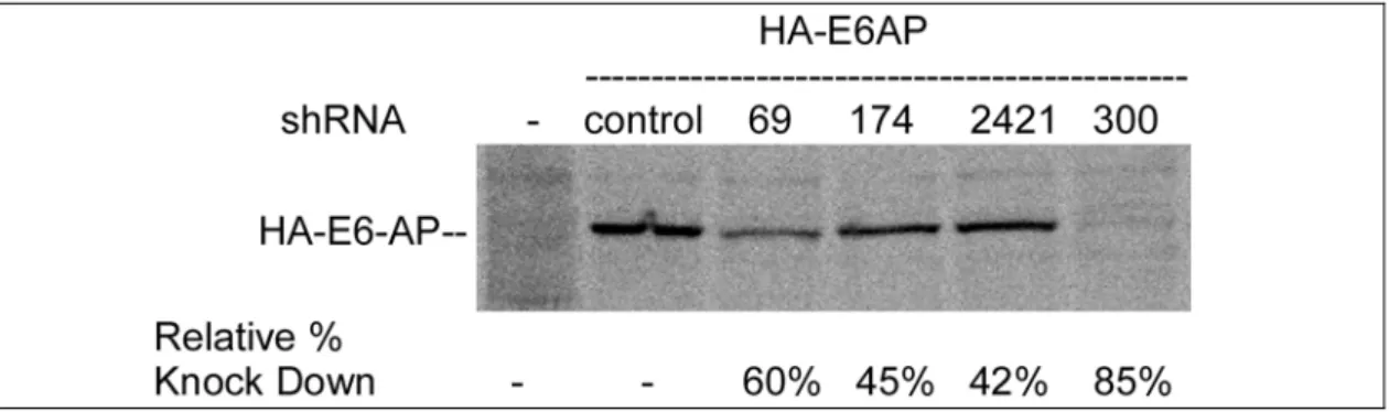
4.1.2 Evaluation of the silencing potency of different shRNAs specific against E6-AP mRNA ...44
4.2 Down-regulation of E6-AP in HPV-positive cell lines by RNAi ...45
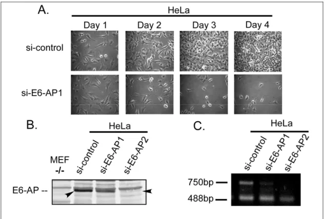
4.2.1 Down-regulation of E6-AP has a growth-suppressive effect in HPV- positive cell lines...45
4.2.2 Down-regulation of E6-AP expression by RNA interference induces accumulation of p53 and interferes with the viability of HPV-positive cancer cell
lines ...48
4.3 Down-regulation of E6-AP in HPV-negative cell lines ...51
4.3.1 RNAi induced down-regulation of E6-AP expression has no growth suppressive effect in HPV-negative tumour cell lines under transient conditions.51 4.3.2 Stable knockdown of E6-AP expression in a H1299, a HPV-negative cell using shRNA expression vectors...53
4.4 Stable suppression of p53...55
4.4.1 Validation of p53 knockdown construct ...55
4.4.2 Generation of stable p53 knockdown cell lines...56
4.4.3 Stress induced activation of p53 in HeLa and SiHa stable clones down- regulated by pSUPER –p53...57
4.5 Down-regulation of E6-AP in p53 null clones HA1 and HC2...60
4.6 RNAi-rescue assays...61
4.6.1 Validation of rescue constructs ...61
4.6.2 Overexpression of E6-AP in HPV-positive cells and HPV-negative cells 64 4.6.3 Transient rescue assay...66
4.7 Visualization of subcellular interactions using Bimolecular fluorescence complementation assay (BiFC) ...68
5. Discussion ...72
5.1 Knockdown of E6-AP in HPV-positive cervical cancer cell lines results in growth suppression...72
5.2 Transient down-regulation of E6-AP in HPV-negative cell lines does not have a growth-suppressive effect as compared to HPV-positive cell lines...75
5.3 Stable knockdown of E6-AP in HPV-negative cell lines...76
5.4 Growth-suppression induced by down-regulation of E6-AP in the HPV-
positive cancer cell line HeLa, depends on p53 expression...77
5.5 RNAi–rescue assays...80
5.6 E6-AP: A potential target for cervical cancer therapy? ...82
5.7 BiFC analysis ...84
6. References ...86
7. Zusammenfasung ...100
8. Abstract ...101
9. Erklärung...102
10. Lebenslauf ...103
Abbreviations
°C Degree celcius A Ampere
AA Amino Acid
AS Angelman Syndrome
ATP Adenosine Tri- Phosphate
bp base pairs
cDNA complementary DNA C-terminus Carboxy terminus DMSO Dimethylsulphoxide DNA Deoxyribonucleic acid DTT Dithiothreitol
E1 Ubiquitin-activating enzyme
E2 Ubiquitin-conjugating enzyme
E3 Ubiquitin-protein ligase EDTA Ethylenediamine-
tetraacetic acid
EtOH Ethanol
FBS Fetal bovine serum HA Haemagglutinin Hr hour
Hrs hours
i.e. id est or that is
IgG Immunoglobulin G IP immunoprecipitation
kDa Kilodalton
M Molar
min minute
mRNA messenger-RNA
mu Murine
n nano N Normal
nm nanometer
nM nanoMolar
nt nucleotide N-terminus Amino-terminius O.D Optical density ONPG Orthonitrophenyl-β-D- galactosidase
ORF Open reading frame PBS Phosphate buffer saline PCR Polymerase chain reaction RNA Ribonucleic acid rRNA ribosomal RNA s second S sedimentation coefficient siRNA small-interfering RNA TAE Tris-acetated-EDTA
Ub ubiquitin
V Volt wt wild-type β-Gal Beta-Galactosidase µ micro
Introduction
1. Introduction
1.1 Ubiquitination: A functional perspective
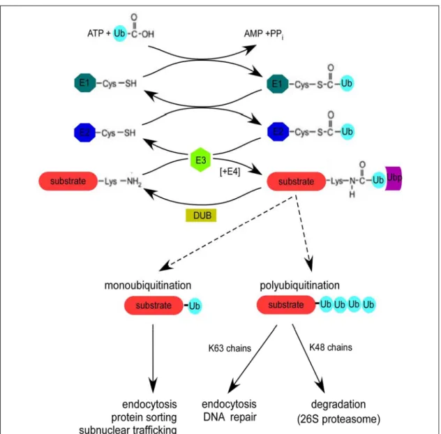
Ubiquitination (also referred to as “ubiquitin conjugation” or “ubiquitylation”) is the post-translational modification of proteins by the covalent attachment of ubiquitin, a 76 amino-acid protein. “Monoubiquitination”, the attachment of a single ubiquitin moiety involves isopeptide bond formation between the α-carboxy group of the C-terminal Glycine residue of ubiquitin and the ε-amino group of a lysine residue in the target protein. In addition to being modified by single ubiquitin moieties, proteins can also be modified by chains of ubiquitin (“polyubiquitination”). Polyubiquitin chain formation takes place by subsequent attachment of ubiquitin moieties linked to one of the lysine residues present in the previously added ubiquitin. Of all the known consequences of ubiquitination, the targeting of proteins for degradation has been best characterized.
Substrates tagged with a polyubiquitin chain are selectively degraded by a multi- subunit ATP-dependent protease known as the 26S proteasome (reviewed in Glickman and Ciechanover, 2002).
Of the seven lysine (K) residues present in ubiquitin (Figure 1), K48-linked polyubiquitin chains are recognized and targeted to the proteasome. Covalent attachment of K48-linked tetraubiquitin chain has been shown to be necessary and sufficient for recognition and degradation of a model substrate by the 26S proteasome in vitro (Thrower et al., 2000). In addition, K11 and K29-linked polyubiquitin chains are also reported to target proteins to the proteasome (Johnson et al., 1995; Liu et al., 1996). The progression of cell cycle (reviewed in Koepp et al., 1999), transcriptional regulation (reviewed in Muratani and Tansey, 2003), signal transduction (reviewed in Welchman et al., 2005), and antigen presentation (reviewed in Rock and Goldberg, 1999), are just a few of the many processes regulated by ubiquitin-proteasome- dependent proteolysis. However, degradation by the proteasome is not the only fate possible for ubiquitin tagged proteins. K63-linked polyubiquitin chains can signal nonproteolytic, reversible events such as in DNA repair (Spence et al., 1995; Hofmann and Pickart, 1999; Hoege et al., 2002), the initiation of the inflammatory response (Deng et al., 2000), ribosomal function (Spence et al., 2000), and the regulation of certain transcription factors (Kaiser et al., 2000). K63-linked polyubiquitin chains
might also act as a signal for endocytosis in some cases (Galan and Haguenauer-Tsapis, 1997; Springael et al., 1999).
Figure 1: Schematic representation of ubiquitin and position of the lysine residues.
Ubiquitin has seven lysine (K) residues, all of which are supposed to be utilized in ubiquitin chain formation. The C- terminal end of ubiquitin has a Glycine residue (G76) through which isopeptide bond formation takes place with the substrate or another ubiquitin moiety.
In contrast to polyubiquitination, monoubiquitination of proteins targets them for endocytosis and lysosomal degradation (reviewed in Hicke and Riezman, 1996;
Haglund et al., 2003). In addition, monoubiquitination can also act as a regulatory modification such as in protein sorting, histone function and transcription (reviewed in Hicke, 2001; Katzmann et al., 2002).
No matter what the final outcome is, the whole process of ubiquitination is made possible through a concerted action of at least three enzymes, in a three-step mechanism referred to as the ubiquitin-conjugation cascade (see below). Since ubiquitination regulates a number of cellular pathways, it is unsurprising that deregulation of the ubiquitin-conjugation system has been implicated as a causative factor in cancer and other human diseases (reviewed in Glickman and Ciechanover, 2002; Ciechanover and Brundin, 2003; Pagano and Benmaamar, 2003).
1.2 The ubiquitin-conjugation cascade
As mentioned earlier, ubiquitination of a substrate protein involves the isopeptide bond formation between the α-carboxy group of the C-terminal Glycine residue (G76) of ubiquitin and the ε-amino group of a lysine of a target protein. More rarely, it can also be conjugated to the α-amino group at the N-terminus (Breitschopf et al., 1998; Aviel et al., 2000; Reinstein et al., 2000; Bloom et al., 2003). For ubiquitination to take place the sequential action of three enzymes, namely the ubiquitin-activating enzyme E1 (UBA), ubiquitin-conjugating enzyme E2 (UBC), and ubiquitin-protein ligase E3 is required. Firstly, the E1 adenylates the C-terminus of ubiquitin and then forms a
Introduction thioester bond between the C-terminus of ubiquitin and a catalytic E1 cysteine residue (Figure 2).
Figure 2: The ubiquitin-conjugation cascade. Free ubiquitin (Ub) is activated in an ATP-dependent manner by the activity of an ubiquitin-activating enzyme (E1), which hydrolyses ATP and forms a thioester bond with ubiquitin. Subsequently, ubiquitin is transferred to one of many distinct ubiquitin-conjugating enzymes (E2). In some reactions, E2s can directly ubiquitinate substrates, whereas others require the help of a ligase (E3). While some E3s have the ability to form ubiquitin thioester during the transfer of ubiquitin to the substrate, others support ubiquitination by recruiting substrates to E2 enzymes. Usually, several ubiquitin molecules, in the form of a polyubiquitin chain, are conjugated to a substrate. In some cases, this requires a specific polyubiquitin chain-assembly factor (E4). Based on the number of ubiquitins (monoubiquitin versus polyubiquitin) and the type of chain linkage (e.g. K48 or K63), the fate of the substrate protein is decided. Ubiquitin modification can simultaneously be removed by deubiquitinating enzymes (DUB). The attachment of ubiquitin to the substrate could also act as signal for the recruitment of ubiquitin-binding proteins (UBP), which could either protect the substrate from deubiquitination and/or act as a bridging factor to transfer polyubiquitinated substrates to the proteasome (Adapted and modified from Passmore and
To be fully active, the E1 must non-covalently bind to, and adenylate a second ubiquitin molecule. Secondly, the thioester-linked ubiquitin is transferred from E1 onto the active site cysteine residue of one of a number of E2s, where it is again linked by a thioester bond. Finally, with the help of a third enzyme, the E3 ligase, ubiquitin is transferred from the E2 to a lysine residue of a substrate protein (reviewed in Passmore and Barford, 2004). This final transfer of ubiquitin to the substrate results in an isopeptide bond between a substrate lysine and ubiquitin. In addition, E3s can also catalyze ubiquitin-ubiquitin conjugation to form a polyubiquitin chain. Although the precise mechanisms vary among the different E3s, they all promote the transfer of ubiquitin, either directly or indirectly, from an E2 to a substrate or another ubiquitin. In addition to the aforementioned components of the ubiquitin conjugation cascade, other conjugation factors, or accessory factors have been identified to play a role in either polyubiquitin chain assembly or in delivering ubiquitinated substrates to the proteasome (see below). Furthermore, concurrent to the conjugation of ubiquitin to substrate proteins, the action of deubiquitinating enzymes (DUBs) antagonizes polyubiquitin chain assembly thus adding another layer of selectivity and regulation (reviewed in Wilkinson, 2000).
1.2.1 Ubiquitin-activating enzyme E1
The first evidence towards understanding the physiological role of ubiquitination was demonstrated by using cells expressing a temperature sensitive mutant of E1 (Finley et al., 1984). Cells expressing the temperature sensitive form of E1 underwent cell cycle arrest at the nonpermissive temperature and failed to degrade short-lived proteins. In yeast, E1 has been shown to be an essential enzyme (McGrath et al., 1991).
Furthermore, in most eukaryotes, a single E1 activates ubiquitin for all downstream processes. The E1 protein is approximately 100 kDa in size and exists as two isoforms in humans (Cook and Chock, 1992).
1.2.2 Ubiquitin-conjugating enzyme E2
More than thirty E2 enzymes have been identified so far, which mediate transfer of ubiquitin from E1 to E3. While the genome of S.cerevisiae encodes 13 E2-like enzymes, there might be up to 96 different E2s in humans (Weissman, 2001; Vaux and Silke, 2005). The family of E2s is structurally well characterized and shares a conserved approximately 150 amino acid core domain through which E2s interact with
Introduction E1. In addition, many E2s have a non-conserved amino or carboxy terminal extension, which is supposedly involved in mediating specific interactions with E3s (Pickart, 2001). While most of the E2s identified are less than 35 kDa, there are few exceptions such as the 523 kDa E2 named BRUCE (BIR Repeat containing ubiquitin conjugating enzyme) (Hauser et al., 1998).
1.2.3 Ubiquitin-protein-ligase E3
Of the three principle enzymes involved in ubiquitin conjugation, it is mainly the E3, which mediates substrate recognition. Based on the presence of conserved protein domains and the mechanistic role they play, E3s can be broadly classified into RING E3s and HECT E3s.
1.2.3.1 RING E3
The RING (Really Interesting New Gene) family of E3s is the largest family of E3s identified so far (Borden, 2000). Members of this family contain a RING finger or RING finger-like (see below) domain and are assumed to function predominantly as molecular scaffolds that bring other proteins together rather than as chemical catalysts.
The RING finger domain consists of a series of histidine and cysteine residues with a characteristic spacing that allows for the coordination of two zinc ions in a cross brace structure.
RING E3s can further be classified into two subtypes; a) single-subunit RING E3s and b) multi-subunit RING E3s. The single-subunit E3s consist of a single polypeptide that possesses the capacity to recognize the ubiquitination signals in their specific substrates through domains that, in general, are structurally distinct from the RING finger. Single- subunit RING E3s include Mdm2, which ubiquitinates p53 and c-Cbl, which is involved in down-regulation of growth factor receptors (reviewed in Pickart, 2001). In the multi-subunit RING E3s, the substrate recognition and the RING finger are on separate subunits. The SCF (Skp1-Cullin-F-box protein), the APC, and the VCB types belong to this class of multi-subunit RING E3s (reviewed in Deshaies, 1999).
In addition to the RING finger domain two closely related domains, the U-box (Hatakeyama et al., 2001) and the PHD/LAP (plant homeodomain or leukemia- associated protein) domains have shown to confer E3 activity (Boname and Stevenson,
conformation and are maintained by salt bridges and hydrogen bonds (Aravind and Koonin, 2000).
1.2.3.2 HECT E3
The family of HECT (Homologous to E6-AP C-Terminus) domain E3s has been named after their founding member E6-AP (further described in section 1.3) (Huibregtse et al., 1995). Members of this family of E3 ligases are characterized by the presence of a conserved catalytically active C-terminal region of approximately 350 amino residues, called the HECT domain. This catalytic activity is mediated by a conserved cysteine residue positioned approximately 35 residues upstream of the C-terminus within the HECT domain, which acts as a site of ubiquitin thioester formation (Huibregtse et al., 1995; Scheffner et al., 1995). HECT domain proteins have a modular structure consisting of the C-terminal HECT domain and a variable N-terminal extension. While the HECT domain is required for interactions with their cognate E2, the N-terminus is presumably involved in determining the substrate specificity of the individual E3. Even though the crystal structure of HECT E3s in complex with their cognate E2s are available (Huang et al., 1999; Verdecia et al., 2003; Zheng, 2003), the mechanism of how exactly ubiquitin is transferred from E2 to E3 and from E3 to the substrate remains ill-defined.
1.2.4 Accessory factors
The conjugation factors labeled as ‘E4s’ help in polyubiquitin chain assembly by extending nascent chains rather than extending ubiquitin chains already attached to a substrate. This family includes the yeast UFD2 protein that has been shown to display E4 activity by binding to ubiquitin moieties of preformed conjugates and catalyzing polyubiquitination (Koegl et al., 1999). In addition to E4 other accessory factors are involved in recognizing polyubiquitin labeled substrates and delivering them to the proteasome. These include distinct families of chaperones or “shuttle factors”
(Hartmann-Petersen et al., 2003).
1.3 E6-AP (E6-Associated Protein)
The cellular ubiquitin-protein ligase E6-AP was originally identified by its ability to interact with the E6 oncoprotein of HPVs associated with cervical cancer and, in complex with E6, to target p53 for ubiquitin-mediated proteasomal degradation
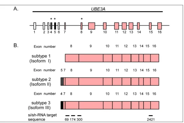
Introduction (Huibregtse et al., 1991; Scheffner et al., 1993). As mentioned earlier, it is also the founding member of the HECT family of ubiquitin-protein ligases. The gene encoding E6-AP (UBE3A) has been localized to the region q11-q13 of chromosome 15 (Nakao et al., 1994). Five mRNA subtypes of E6-AP encoding three potential isoforms have been identified (Yamamoto et al., 1997). These isoforms are approximately 100 kDa in size and vary at their N-terminal region. Whether these isoforms differ in functionality is not known. In addition to its E3 activity E6-AP has also been reported to serve as a transcriptional coactivator for steroid hormone receptors (Nawaz et al., 1999).
Furthermore, these functions have been shown to be separable and independent of one another.
Understanding the physiological role of E6-AP is still of interest because inactivation of UBE3A gene has been associated with Angelman Syndrome, a hereditary neurological disorder (see 1.3.4). Moreover, in the case of cervical cancer, the E6/E6- AP complex not only targets p53 for ubiquitin-mediated degradation, but also targets other proteins (see 1.3.3), which is necessary for HPV-induced cervical carcinogenesis.
1.3.1 Human papillomaviruses (HPVs) and cervical cancer
HPVs are small DNA-viruses that belong to the family Papovaviridae. Productive infection with HPVs has long been known to result in benign squamous epithelial lesions, commonly known as warts. More than 130 HPV types have been isolated from epithelial cells of the skin, anogenital, or oropharyngeal mucosa. Of these, some types of HPV have also been found in samples of patients with cancers of the oral cavity, skin cancers of immunosuppressed patients and, most notably cancer of the uterine cervix (cervical cancer) (reviewed in zur Hausen, 1996).
Cervical cancer represents the second most common form of cancer in women. In greater than 90% of the cases, at least one copy of the HPV viral genome has been found integrated into the host-cell genome. Furthermore, continuous expression of the viral E6 (approximately 150 amino acids) and E7 (approximately 100 amino acids) oncoproteins is required for the malignant phenotype of cervical carcinoma cell lines.
These oncoproteins have been found to possess cell-transforming and cell- immortalizing potentials in cell culture systems. Taken together, the above evidence has led to the causal association of HPV with cervical cancer (reviewed in zur Hausen,
Of around 40 HPV types isolated from the anogenital tract, only few have been associated with malignant lesions. Based on their clinical association with cervical carcinogenesis HPVs can be classified as “high risk” or “low risk” types. High risk HPVs including the type 16, 18, 31, 33, 39, 45, and 52 are associated with malignant lesions. In comparison to high risk HPVs, the low risks HPVs such as type 6 and 11 can be associated with benign lesions such as genital warts (condylomata acuminata).
Studies to elucidate the role of high risk HPVs in cervical carcinogenesis have shown that the E6 protein interacts with the p53 tumour suppressor protein (Werness et al., 1990) and E7 with the retinoblastoma gene product p110RB and the p110RB related proteins p107 and p130 (henceforth collectively referred to as pRB) (Dyson et al., 1989). Subsequently both p53 and pRB have been shown to be targeted for inactivation by ubiquitin-mediated proteasomal degradation (Scheffner et al., 1990; Crook et al., 1991; Dyson et al., 1992; Boyer et al., 1996).
1.3.1.1 E7 and pRB
In normal cells (HPV-negative cells) the pRB family of proteins (also referred to as
“pocket proteins”) regulate progression of the cell cycle from G0/G1 into S phase, by interacting with the E2F/DP family of transcription factors (referred henceforth collectively as “E2F”). E2F is known to transactivate many genes required for DNA replication. Hence, binding of pRB in its active, growth suppressive form to E2F not only interferes with its transactivation properties, but is also said to convert E2F into a transcriptional repressor (Dyson, 1998), thus limiting the cell from transition into S phase. Cell cycle-dependent phosphorylation of pRB leads to disruption of E2F/pRB complex and reverses the pRB-mediated cell cycle arrest resulting in cell cycle progression. In HPV-positive cells, analogous to the phosphorylation of pRB, binding of E7 to pRB and subsequent degradation results in the release of transcriptionally active E2F complex driving cells into S phase. Since HPVs propagate in differentiated non-dividing cells it is imperative that the infected cells are driven into S phase to allow viral replication (Stubenrauch and Laimins, 1999).
The mechanism of E7 mediated degradation of pRB is unclear. Whether E7 in conjugation with another protein acts as an E3 ligase to target pRB for ubiquitination and degradation is not known. Interestingly, E7 has been shown to directly interact with the S4 ATPase subunit of the 19S regulatory complex of the 26S proteasome
Introduction suggesting that E7 could directly target pRB for degradation without prior ubiquitination (Berezutskaya and Bagchi, 1997). However whether this is true remains to be determined.
1.3.1.2 E6 and p53
The p53 tumour suppressor protein is known to play a critical role in cellular responses to various stress signals (Harris and Levine, 2005). In normal cells (HPV-negative cells), p53 is predominantly regulated by a negative feed-back loop with Mdm2, a RING finger ligase that targets p53 for proteasomal degradation (Haupt et al., 1997;
Kubbutat et al., 1997). However under stress conditions inactivation of the Mdm2 feed- back loop leads to stabilization of p53 levels and increased transcriptional activity, the outcome of which leads to cell-cycle dependent growth arrest, senescence or apoptosis.
In contrast to most cancers, wherein the p53 gene is mutated (greater than 40%), it is rarely mutated in HPV-positive cervical carcinomas (Hainaut et al., 1998). Thus it has been hypothesized that the inactivation of the normal functions of p53 by E6 is similar to the inactivation of p53 by mutation and is a key step in HPV-induced cervical carcinogenesis. In contrast to E7 wherein the biochemical mechanism of pRB degradation remains poorly understood, E6 mediated degradation of p53 has been shown to involve the recruitment of E6-AP (Huibregtse et al., 1991).
1.3.2 The E6-E6-AP-p53 interaction
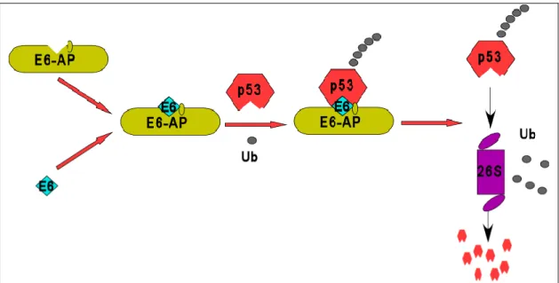
The biochemical mechanism by which high risk E6s target p53 for inactivation is well characterized (Figure 3). In the initial step, E6 forms a complex with E6-AP. This dimeric complex recognizes p53, binds to it and polyubiquitinates p53 with the help of ubiquitin-conjugating enzymes (UbcH5, UbcH7, or UbcH8). In the final step, p53 is recognized and degraded by the 26S proteasome. It should be noted that E6 or E6-AP alone are not able to enter into a complex with p53 and it is still unclear whether E6 or E6-AP or both actually directly contact p53. In comparison to high risk E6s, the low risk E6s are unable to target p53 for degradation.
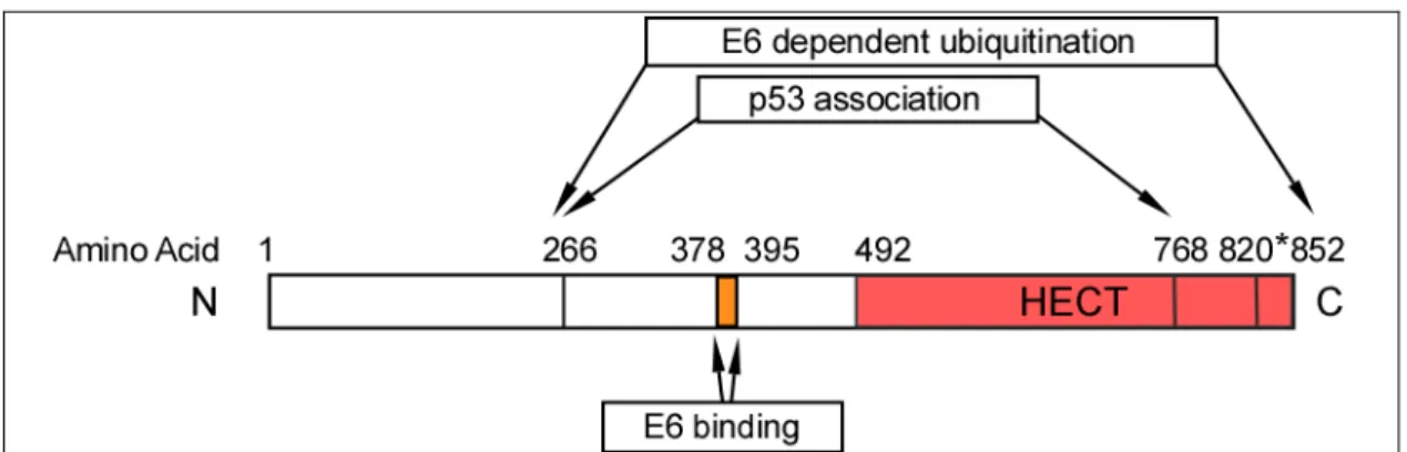
Mutational analysis to study the interaction of E6-AP with E6 has identified three functional domains which are required for 1) Binding with E6, 2) E6 dependent association with p53 and 3) E6 dependent ubiquitination of p53 (Figure 4) (Huibregtse et al., 1993). An 18 amino acid region (amino acids 378-395) within the central portion
of E6-AP was found to be necessary and sufficient for binding with E6 of high risk HPVs. The region that directs E6 dependent association with p53 spans approximately 500 amino acids (amino acids 266-768) and includes the E6 binding domain. E6-AP sequences in addition to those required for formation of a stable ternary complex with E6 and p53 are necessary to stimulate the ubiquitination of p53 (amino acids 768-852).
These sequences lie within the C-terminal 84 amino acids of E6-AP and encompass the HECT domain. Furthermore, a catalytical cysteine residue (C820) within the HECT domain is required for thioester formation (Huibregtse et al., 1995; Scheffner et al., 1995).
Figure 3: Model for the ubiquitination and degradation of p53 in HPV-positive cells. In HPV-positive cervical cancer cells, the E6 oncoprotein forms a complex with the cellular ubiquitin-ligase E6-AP which then targets p53 for polyubiquitination and subsequent degradation by the 26S proteasome. However in normal cells (HPV-negative), p53 is predominantly targeted for degradation by the RING ligase, Mdm2. Ub- ubiquitin.
While the mechanism of E6/E6-AP targeted ubiquitination of p53 is clear and can be reconstituted in vitro using purified enzymes (Scheffner et al., 1993; Rolfe et al., 1995), it remains unclear how exactly polyubiquitinated p53 is delivered to the proteasome for degradation. Recent studies suggest that the human homologue to RAD23 (hHR23) protects polyubiquitinated p53 from deubiquitination and could act as a bridging factor to deliver polyubiquitinated p53 to the proteasome (Glockzin et al., 2003).
Interestingly, hHR23 is also known to interact and is a substrate of E6-AP (see section 1.3.5).
Introduction
Figure 4: Schematic representation of the regions of E6-AP that direct E6 binding, E6 dependent association with p53, and E6 dependent ubiquitination of p53.
Mutational analysis of E6-AP has mapped a central 18 amino acid region that is sufficient for binding to E6 (amino acids 378-395). Amino acid 266-768 is the minimal region required for p53 associated in complex with E6 and includes the E6 binding region. In addition to this, the last 84 amino acids (amino acids 768-852) of the HECT domain is critical for E6 dependent ubiquitination of p53 (Huibregtse et al., 1993).
Even though in HPV-positive cancer cells E6-AP targets p53 for degradation (i.e in presence of E6), E6-AP does not appear to regulate p53 stability in HPV-negative cells (Beer-Romero et al., 1997; Talis et al., 1998; Traidej et al., 2000). Investigation of the MDM2-pathway of p53 regulation in HPV-positive cell lines has indicated that the MDM2-mediated degradation pathway is inactive (Hengstermann et al., 2001). Hence, the degradation of p53 is dependent entirely on the E6/E6-AP pathway in HPV-positive cells. Therefore, studies in which E6 mediated degradation of p53 was inhibited by either using peptide aptamers or RNA interference has led to stabilization of p53 which restores its transactivating properties and tumour suppressor functions in HPV-positive cells (Butz et al., 2000; Butz et al., 2003). Similarly interfering with E6-AP expression using antisense oligonucleotides or ribozyme-based gene inactivation leads to p53 stabilization (Beer-Romero et al., 1997; Kim et al., 2003).
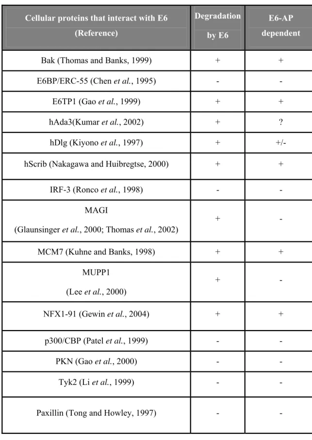
Initially it was believed that the anti-apoptotic properties of high risk E6s were attributed to its ability to target p53 for degradation. However, several lines of evidence later suggested that high risk E6s also possess p53-independent anti-apoptotic properties (Pan and Griep, 1995; Spitkovsky et al., 1996). Search for additional cellular targets of E6 that contribute to its anti-apoptotic potential has led to the identification of several proteins which interact with high risk E6 (Table 1) (reviewed in Munger and Howley, 2002; zur Hausen, 2002). Of these, several have been shown to be targeted for degradation either in an E6-AP-independent or E6-AP-dependent manner.
Cellular proteins that interact with E6 (Reference)
Degradation by E6
E6-AP dependent
Bak (Thomas and Banks, 1999) + +
E6BP/ERC-55 (Chen et al., 1995) - -
E6TP1 (Gao et al., 1999) + +
hAda3(Kumar et al., 2002) + ?
hDlg (Kiyono et al., 1997) + +/-
hScrib (Nakagawa and Huibregtse, 2000) + +
IRF-3 (Ronco et al., 1998) - -
MAGI
(Glaunsinger et al., 2000; Thomas et al., 2002)
+ -
MCM7 (Kuhne and Banks, 1998) + +
MUPP1 (Lee et al., 2000)
+ -
NFX1-91 (Gewin et al., 2004) + +
p300/CBP (Patel et al., 1999) - -
PKN (Gao et al., 2000) - -
Tyk2 (Li et al., 1999) - -
Paxillin (Tong and Howley, 1997) - -
Table 1 Cellular proteins that are known to interact with high risk E6s. Proteins that are degraded by E6 (+) in an E6-AP- independent (-) or E6-AP-dependent manner (+) are indicated. (?) Not known. +/- Conflicting evidence.
Introduction
1.3.3 E6-dependent substrates of E6-AP
As indicated in Table 1, the high risk E6s are known to interact with several proteins that contain PDZ (PSD95/Dlg/ZO-1) domains (i.e., hScrib, MUPP-1, hDlg and MAGI- 1) (Mantovani and Banks, 2001). Multi-PDZ domains form modular structures that are involved in protein-protein interaction with membrane or cytoskeletal proteins. Of the PDZ domain-containing proteins that interact with E6, the human homologue to the Drosophila Scribble (Vartul) tumour suppressor protein (hScrib) has been shown to be targeted for degradation by E6 in an E6-AP-dependent manner (Nakagawa and Huibregtse, 2000; Nakagawa et al., 2004). hScrib localizes to basolateral regions of epithelial cells and is down-regulated by E6/E6AP with the progression of normal uterine cervical cancer tissue to invasive cervical cancer. The ability of E6 to interact with, and possibly degrade PDZ domain proteins is said to contribute to the oncogenic potential of E6 (Kiyono et al., 1997; Mantovani and Banks, 2001). Interestingly, the PDZ domain proteins MAGI-1/2/3 and MUPP1 are targeted for degradation by E6 in an E6-AP independent manner, suggesting that an E3 ligase other than E6-AP may be involved (Glaunsinger et al., 2000; Lee et al., 2000; Thomas et al., 2002).
Earlier studies have indicated that the ability of E6 to immortalize epithelial cells is partly dependent on its ability to increase telomerase activity (Klingelhutz et al., 1996) and this is brought about by induction of expression of the catalytic-rate limiting subunit of telomerase called, hTERT (Kiyono et al., 1998; Gewin and Galloway, 2001;
Oh et al., 2001; Veldman et al., 2001). More recently, this ability of E6 to induce telomerase activity has been shown to be dependent on E6-AP and involves the degradation of NFX1-91 (nuclear factors that binds to the X1 box), a repressor of hTERT (Gewin et al., 2004; Liu et al., 2005). In addition to degrading p53 and inducing of telomerase activity, the ability of high risk E6 to immortalize certain epithelial cells has been found strongly correlate with its ability to bind to and target E6TP1 (E6 targeted protein 1), a Rap GTPase-activating protein (RapGAP) for degradation (Dalal et al., 1996; Kiyono et al., 1998; Gao et al., 1999; Gao et al., 2001).
Degradation of E6TP1 has also been shown to be mediated by E6 in E6-AP dependent manner (Gao et al., 2002).
Thus the presence of E6 redirects the ability of E6-AP to ubiquitinate its normal
promote autoubiquitination and degradation of E6-AP itself thereby reducing its half- life (Kao et al., 2000). Whether dysregulation of E6-AP mediated ubiquitination of its normal substrates also contributes to the transforming function of E6 in HPV-positive cells is unclear.
1.3.4 Angelman syndrome (AS)
Angelman syndrome is a neurological disorder which has an occurrence of 1:15000 births and is characterized by severe mental retardation, ‘puppet-like’ ataxic gait with jerky arm movements, seizures, EEG abnormalities, hyperactivity, and bouts of inappropriate laughter. (Williams et al., 1995; Kishino et al., 1997; Matsuura et al., 1997). AS is one of the best studied cases of genetic disorders, in which imprinting plays a role. In most tissues, the UBE3A gene displays bi-allelic expression but is maternal-specific in certain brain areas due to paternal imprinting (Rougeulle et al., 1997). Thus loss or inactivation of the maternal allele of UBE3A in brain causes AS.
Studies conducted on AS patients indicate that approximately 70% involve maternal deletions at chromosome 15q11-q13, 2% involve paternal uniparental disomy (UPD) at this region, and 2-3% result from imprinting mutations that alter the methylation pattern at this region. The remaining 25% of the cases show biparental inheritance without methylation abnormalities or deletions (Kishino et al., 1997). As mentioned earlier, E6-AP has two separable functions, one as an E3 ligase and another as a transcriptional coactivator. To determine the pathogenesis of AS, mutants of E6-AP obtained from AS patients have been studied. In majority of these cases loss of E3 ligase activity of E6-AP has been detected whereas the coactivator function is found to be intact (Nawaz et al., 1999; Cooper et al., 2004), thus suggesting that deregulated degradation of one or more of its substrates could be responsible for the pathogenesis of AS.
In order to study the causal relationship between inactivation of UBE3A and Angelman syndrome, mouse models have also been constructed in which UBE3A has been inactivated (Jiang et al., 1998; Miura et al., 2002). Mice with maternal deficiency (m- /p+) for UBE3A resembles human AS with motor dysfunction, inducible seizures, and a context dependent learning deficit. Mouse models of AS like the human counterparts display imprinted expression in hippocampus and purkinje neurons (Albrecht et al., 1997).
Introduction
1.3.5 E6-independent substrates of E6-AP
Since loss in E3 ligase activity is supposedly responsible for pathogenesis of AS patients, several labs have tried to identify potential substrates of E6-AP which can be bound and ubiquitinated in an E6-independent manner. Interestingly, E6-AP itself is found to serve as a substrate undergoing autoubiquitination by a mechanism which is predominantly intermolecular (Nuber et al., 1998). In addition, the human homologs of S. cerevisiae RAD23, (HHR23A and HHR23B) (Kumar et al., 1999), the Src tyrosine non receptor kinase Blk (B lymphocyte specific receptor kinase) (Oda et al., 1999), and the multicopy maintenance protein (Mcm) 7 subunit of the replication licensing factor- M (Kuhne and Banks, 1998) have been found to be ubiquitinated by E6-AP. However, of the identified substrates of E6-AP none have been correlated to pathogenesis of AS.
1.4 RNA interference (RNAi)
Till recently, the functional characterization of a particular mammalian gene or gene product involved either elimination by gene knock-out strategies in mouse models, or inactivation using ribozymes, antisense or overexpression of a dominant-negative form of the protein product in cell culture systems. The recent discovery of the natural process termed RNA interference (RNAi) offers an alternate tool to functionally characterize a gene product (Fire et al., 1998).
RNAi is an antiviral post-transcriptional gene silencing defense mechanism in animals and plants caused by the introduction of double-stranded RNA homologous to the silenced gene (reviewed in Paddison and Hannon, 2002; Denli and Hannon, 2003). This mechanism of RNAi has been found to be conserved in most organisms with the notable exception of the yeast, Saccharomyces cerevisiae (Hutvagner and Zamore, 2002). RNAi involves the cleavage of the double stranded RNA (Figure 5B) by an RNase III enzyme (DICER) into smaller fragments of 21-23 nucleotides (nt) in length with characteristic dinucleotide 3’ overhangs referred to as small interfering RNA (siRNA) (Figure 5A) (Zamore et al., 2000). The siRNA is then recruited into a multienzyme complex called RNA induced silencing complex (RISC) which binds specifically to complementary mRNA transcript to target it for cleavage and degradation.
Figure 5: RNA interference. A. Schematic representation of a synthetic 21 nucleotide duplex small interfering RNA (siRNA) with 2 nucleotide overhangs at each 3 prime end. B.
Schematic representation of RNAi pathway. Long double stranded siRNA is cleaved into short siRNAs by RNase III enzyme complex (DICER) in an ATP dependent manner. The antisense strand of the siRNA duplex is then recruited into a RNA induced silencing complex (RISC) which specifically recognizes and cleaves the complementary mRNA (adapted from Dykxhoorn et al., 2003)
Initially, RNAi was not used in mammalian studies, because RNA duplexes larger than 30bp are known to trigger generalized cellular responses, predominantly through activation of dsRNA dependent-protein kinases. However, this limitation has been overcome by the use of synthetic double stranded small interfering RNA (siRNA) of 21 nucleotides in length with overhanging 3’ dinucleotides (Elbashir et al., 2001). While this approach overcomes the cellular non-specific response to a certain extent, it is limited to transient studies due to the fact that the siRNA gets depleted at each cell division. In order to overcome the transient nature of synthetic siRNA, DNA based vector systems have been created whereby it is possible to bring about stable suppression of gene expression (summarized in Paddison and Hannon, 2002). These vectors utilize RNA polymerase III based promoters (e.g. U6 and HI RNA) to express short hairpin like RNA (shRNA) transcripts with a dinucleotide 3’ overhang that can act as siRNA molecules.
Introduction Recently, a class of endogenous non-coding RNA molecules called microRNAs (miRNAs) have been found in most organisms (reviewed in Dillon et al., 2005). These miRNAs, like siRNA can also be recruited into RISC, but unlike siRNAs predominantly lead to translational repression by binding to the 3’untranslated region (UTR). In comparison to siRNAs, miRNA have been found to require weak complementation with the target sequence to facilitate translational repression (Grishok et al., 2001; Hutvagner et al., 2001; Ketting et al., 2001). Functionally miRNA play an important role in developmental regulation, and normal physiology. Interestingly, misexpression of miRNAs has been observed in many cancers, suggesting that miRNAs may also have a role to play in carcinogenesis (Calin et al., 2002; Calin et al., 2004). The finding that miRNAs can also function as siRNA suggests that the eventual mechanism of post-transcriptional genes silencing, i.e. translational repression or mRNA degradation, is probably decided by the extent of complementarity between the regulatory RNA and its target (Doench et al., 2003; Zeng et al., 2003).
1.5 Bimolecular fluorescence assay
Recent evidence suggests that localization of substrate and enzyme complexes to distinct organelles or compartments could give another dimesion of regulation and substrate specificity (reviewed in Pines and Lindon, 2005). Even though the biochemical properties of E6-AP have been well characterized, the subcellular localization of individual protein interactions is poorly characterized. Studies to determine the subcellular localization of E6-AP indicated it to be distributed equally in the nucleus and cytoplasm (Hatakeyama et al., 1997). Although a number of potential partners which can interact with E6-AP in an E6 dependent/independent manner have been identified, it is unclear if localization plays a role in substrate preference.
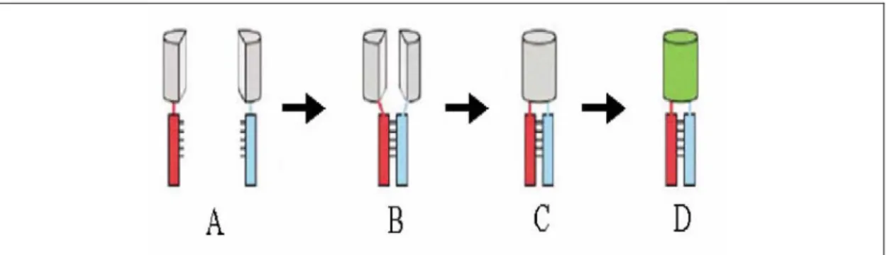
Recently, a new technique called Bimolecular Fluorescent Complementation (BiFC) was reported for visualizing protein complexes in living cells (Hu et al., 2002). This approach involves creating fusion proteins of non-fluorescent N-terminal or C-terminal fragments of the yellow fluorescent protein (YFP) with individual interacting partners of the protein complex to be studied (Figure 6). Co-expression of the N and C –terminal YFP fragments alone within cells does not lead to fluorescence. However, when fused to two separate interacting partners, the interaction between the two proteins leads to
the advantage of not requiring sophisticated instruments in comparison to other techniques for visualizing protein interactions (e.g. FRET). In addition, BiFC has been used for visualizing multiple protein interactions and to study the relative efficiencies of complex formation of the various interacting partners (Hu and Kerppola, 2003;
Grinberg et al., 2004).
Figure 6: Bimolecular fluorescence assay. Two potential interacting proteins (indicated in Red and blue) are tagged with the two non fluorescent fragment of the yellow fluorescent protein (YFP) (indicated in grey). In order to reconstitute the fluorophore, the two fragments should come into close proximity which is made possible by the interacting proteins.
Fluorescence reconstitution can be used as an indicator to study complex formation and localization (adapted from Hu et al., 2002)
Aim
2. Aim
E6-AP interacts with the E6 oncoprotein of cancer-associated HPVs and, in complex with E6 targets the tumour suppressor p53 for proteasomal degradation. In contrast to most cancers wherein the p53 gene is mutated or not expressed, it is rarely mutated in HPV-positive cervical carcinomas. This has led to the assumption that targeted inactivation of p53 by the E6/E6-AP complex is functionally equivalent to the inactivation of p53 by mutation. Thus, the aim of this study was to determine the effect of E6-AP inactivation in HPV-positive cervical cancer cells and whether E6-AP contributes to the known anti-apoptotic properties of E6. In order to do so, RNAi was employed to down regulate E6-AP expression in HPV-positive cervical cancer cell lines and its consequences on cell growth were studied.
Loss of E6-AP function has been associated with the development of Angelman Syndrome, a neurological disorder in humans. It has been suggested that deregulated degradation of the substrates of E6-AP could be involved in the pathogenesis of the disease. However, the identities of the substrate(s) are presently unknown. To study the biological importance of E6-AP in HPV-negative cells, E6-AP expression was also down-regulated by RNAi in various HPV-negative tumour cell lines and its essentiality for cell viability was studied.
While E6-AP is biochemically well characterized, little is known about it subcellular localization and/or whether substrate specificity is dependent on localization to specific subcellular organelles/compartments. To determine the subcellular location of the interactions of E6-AP with its substrates and to study the dynamics of these interactions an attempt was made to establish the bimolecular fluorescence complementation assay.
3. Material and Methods
3.1 Materials
3.1.1 BuffersBuffer Z
( β galactosidase assay)
100 mM NaH2PO4 (pH 7.0), 10 mM KCl, 1 mM MgSO4, 50 mM β- Mercaptoethanol (storage at - 20°C)
DNA stop buffer (10X) 60% Saccharose, 0.25M EDTA, small quantity of Bromophenolblue
Stacking gel buffer 0.5M Tris pH-6.8, 0.4% SDS Separating gel buffer 1.5M Tris pH-8.8, 0.4% SDS
Laemmlli running buffer (10X) 250 mM Tris-HCl pH 8.4, 2 M Glycine,1% SDS Laemmlli stop buffer (2X) 62.5 mM Tris-HCl pH 6.8, 2% SDS, 10% Glycerin,
100 mM DTT, 0.001 % Bromophenol blue (storage at -20°C)
Phosphate buffer saline (PBS) 137mM NaCl, 2.7mM KCl, 10.1mM NaH2PO4, 1.8mM KH2PO4, (pH 7.4)
TAE-buffer (50X) 2M Tris-HCl,, 950 mM Acetic acid, 50mM EDTA TNE-T (wash buffer) 10mM Tris-HCL pH 7.5, 2.5mM EDTA, 50mM
NaCl, 0.1% Tween 20
TNN lysis buffer 0.1 M Tris-HCl pH 8.0, 0.1 M NaCl, 1% NP-40 (IGEPAL), 1mM DTT, 1µg/ml Aprotinin and Leupeptin, 1mM PefaBloc SC(Roche, Mannheim) Transfer buffer 12.5mM Tris HCl, 100mM Glycine, pH 8.3
HBS-buffer 20 mM HEPES (pH 7,4), 150 mM NaCl
Material and Methods
3.1.2 Solutions and stocks
4% Para formaldehyde fixation solution: 4 g Para formaldehyde was dissolved in 20 ml ddH2O by stirring at 40°C and adding 3-4 drops of 2M NaOH. After dissolving, the volume was adjusted to 90 ml with distilled water. The pH was adjusted to 6.5 and volume was made upto 100 ml.
Gelvatol: 2.4 g of polyvinyl alcohol (Mw 30,000-70,000; Sigma) was added to 6 g of glycerol in a 50 ml centrifuge tube and mixed by stirring. To the mixture, 6 ml of distilled water was added and the mixture was incubated at room temperature. After several hours of incubation at room temperature, 12 ml of 0.2 M Tris/HCl, pH 8.5, was added and the mixture was heated to 50°C for 10 min with occasional mixing to completely dissolve polyvinyl alcohol. The solution was centrifuged at 5,100 rpm for 15 min. After centrifugation, 2.5% of diazabicyclo-octane (DABCO), an anti-oxidant agent, was added to reduce the bleaching of the fluorescence. The solution was aliquoted in 1.5 ml micro centrifuge tubes and stored at -20°C.
Cycloheximide: 60mg/ml in methanol Actinomycin D: 1mg/ml in DMSO
DAPI (4',6-Diamidine-2'-phenylindole dihydrochloride): 1µg/ml in PBS
3.1.3 Medium
3.1.3.1 Medium for E.coli
Luria Broth medium (LB): 10g/L NaCl, 5g/L yeast extract, 10g/L Bacto-Tryptone (pH-7.5)
SOC-medium: 20g/L Tryptone, 5g/L yeast extract, 20mM glucose, 10mM NaCl, 2.5mM KCl, 10mM MgCl2, 10mM MgSO4 (pH 7.5)
3.1.3.2 Medium for mammalian cell culture
For culturing of mammalian cell lines, either RPMI-1640 (Sigma) or DMEM (Sigma) supplemented with 10% of Fetal Bovine Serum (FBS) was utilized.
3.1.4 Cell strains and cell lines
3.1.4.1 Bacterial strains
E. coli DH5α, Genotype: F- φ80d lacZ∆M15 ∆(lac) U169 deo R rec A1 hsdR17 (rk-mk+) supE44 λ- thi-1 gyrA96 rel A1 (Gibco BRL)
E. coli BL 21 (DE3), Genotype: F- ompT hsdSb (rb-mb-)βgal dcm (DE3) (Novagen)
3.1.4.2 Cell Lines
-RKO Rectal colon carcinoma, wt p53 (A. Pause) -U2OS Osteosarcoma, wt p53 (T. Rothmann)
-MCF7 Mammary carcinoma, wt p53 (F. Hoppe-Seyler) -H1299 Non small lung carcinoma, p53 null (H. Oie) -SAOS-2 Osteosarcoma, p53-null (M. Scheffner)
-C33A Cervical carcinoma, mutant p53 (R273C), HPV-negative (M. Scheffner) -SiHa Cervical carcinoma, wt p53, HPV-16 positive (M. Scheffner)
-CaSki Cervical carcinoma, wt p53, HPV-16 positive (M. Scheffner) -HeLa Cervical carcinoma, wt p53, HPV-18 positive (M. Scheffner) 3.1.5 Mammalian expression vectors
Vector Characteristics Reference
pRc/CMV
5.5 kb; Ampicillin resistance; Neomycin (G418, Geneticin) resistance; CMV promoter; for in vitro translation and stable protein expression in mammalian cell lines ( HPV cell lines exceptional)
Invitrogen
pCDNA3.0.HA
5.4 Kb; Ampicillin resistance; CMV promoter;
HA-tag at N-terminal; for in vitro translation, transient protein expression in mammalian cell lines
D. Roth, MPI, Frankfurt
Material and Methods
pEF/V5- His
6.2 Kb; Ampicillin Resistance; Neomycin (G418,Geneticin) resistance; EF promoter; for in vitro expression, transient and stable protein expression in mammalian cell lines
Invitrogen
Table 2: Vectors used for cloning and expression in mammalian cells
3.1.5.1 pRC/CMV constructs:
β-Gal A. Hengstermann p53 M. Scheffner HPV-16 E6 M. Scheffner HPV-18 E6 M. Scheffner
3.1.5.2 Miscellaneous constructs
pd2YFP ClontechpeGFP-C2 Clontech
pHA-CMV.bFos.YC155 Hu et al, Mol.Cell, 2002 pFLAG-CMV2.bjun.YN155 Hu et al, Mol.Cell, 2000
3.1.5.3 pcDNA3-HA constructs
Protein Amino acids Restriction sites 5’,3’
Oligonucleotides used
E6-AP 30-852 Kpn I, Apa I MD1, MD4
E6-AP ∆E6 * ∆ 378-401 Kpn I, Apa I
MD1, siRNA2mut sense, MD4
E6-AP * 1-852 Kpn I, Apa I
siRNA2mut sense, siRNA2mut anti
* contains silent mutations at RNAi binding site of siE6-AP1
HA-E6-AP S. Glockzin
HA-E6-AP C820A S. Glockzin
3.1.5.4 pEF/V5 His constructs
All E6-AP pEF constructs were generated by sub cloning from pcDNA3-HA.
Respective HA-E6-AP constructs were digested with BamHI and ApaI, klenow filled finally ligated into pEF/V5 His.neo digested with BamHI-PmeI.
Protein Amino acids
E6-AP 1-852
E6-AP 30-852
E6-AP.mut1 1-852 (with silent mutation at si/sh-E6- AP1 target site)
E6-AP.mut2 1-852 (with silent mutation at si/sh-E6- AP2 target site)
E6-AP ∆E6 ∆ 378-401
E6-AP ∆E6.mut.pEF/V5 His.hygro K. Martentzoglu and C. Strüh 3.1.6 Molecular weight markers
DNA-Marker:
- λ (Lambda)-DNA Hind III-Marker (Invitrogen):
23130, 9416, 6557, 4361, 2322, 2027, 545, 125 [bp]
- 100 bp ladder (New England Biolabs):
1517, 1200, 1000, 900, 800, 700, 600, 500, 400, 300, 200, 100 [bp]
Protein marker:
- Prestained Protein Ladder (MBI Fermentas):
180, 130, 100, 73, 54, 48, 35, 24, 16, 10 [kDa]
Material and Methods - MARK12TM Unstained Standard (Invitrogen):
200, 116.3, 97.4, 66.3, 55.4, 36.5, 31, 21.5, 14.4, 6 [kDa]
3.1.7 Antibodies
Antibody Characteristics Dilution Brand
Anti-p53 (IgG Do-1)
Mouse monoclonal 1:1000 (WB) 1:200 (IF)
Dianova
Anti-Hdm2 (SMP14)
Mouse monoclonal 1:1000 (WB) Santa Cruz
Anti-p53 (1-393) Rabbit polyclonal 1:1000 (WB) Santa Cruz anti-rabbit Peroxidase conjugated, goat 1:5000 (WB) Dianova anti-mouse peroxidase conjugated, goat 1:5000 (WB) Dianova anti-rabbit IgG AlexaFluor 568, goat 1:1000 (IF) Molecular
probes anti-mouse IgG AlexaFluor 568, goat 1:1000 (IF) Molecular
probes anti-HA IgG Mouse monoclonal 1:1000 (WB) Covance WB-western blotting, IF-immunofluorescence
3.1.8 Primers List
Name Sequence 5’ or 3’ Protein
siRNA2mut sense
GGCTGTGGAAATGAAGCATGCA
CCAATGAGTTTTGTGCTTC 5’ E6-AP
siRNA2mut anti
GAAGCACAAAACTCATTGGTGC
ATGCTTCATTTCCACAGCC 3’ E6-AP
si5mute1 TACTACCACCAGTTAACTGAGG GCTGTGGAAATGAAGCATGTAC
CAATGAATTCTG 5’ E6-AP
si5mute2 GCGGGATCCATGAAGCGAGCAG CTGCAAAGCATCTAATAGAACG
CTACTACCACC 3’ E6-AP
MD1 CGCGGATCCATGTGTGCTTCCT
GTCCAAC 5’, BamH I E6-AP
MD2 GGGGTACCGTATGGCGTACCCA
TACGACGTC 5’, Kpn I HA-tag
MD3 TAATGGGCCCGCAGCATGCCAA
ATCCTTTG 3’, Apa I E6-AP
MD4 GACTAGTGGGCCCTTACAGCAT
GCCAAATCCTTTG 3’, Apa I E6-AP
MD8 ACGCGTCGACCATGAAGCGAGC
AGCTGC 5’, Sal I E6-AP
MD9 CGGGGTACCCAGCATGCCAAAT
CCTTTGG 3’, Kpn I E6-AP
MD10 CGGGGTACCAATGAAGCGAGC
AGCTGC 5’, Kpn I E6-AP
3.1.9 RNA interference
3.1.9.1 Synthetic siRNA
Synthetic siRNA were ordered either from Dharmacon, USA or MWG-Biotech AG, Germany. siRNAs were ordered as 19-nt RNA duplexes with 2-nt oligo dT 3’
overhangs. In case of siRNA purchased from Dharmacon, individual sense and antisense strands were ordered and annealed according to manufacturers instructions.
siRNA from MWG biotech were purchased as annealed oligos and resolubilised in supplied annealing buffer. Stock solutions of 20 µM concentration were stored as aliquots at -20°C. The sequences of the various siRNA are given in Table 3.
Material and Methods
Name Target mRNA target sequence
si-E6-AP1 E6-AP TGAAGCCTGCACGAATGAG
si-E6-AP2 E6-AP AGATGTGACTTACTTAACA
si-Hdm2 Hdm2 CAAGAGACCCUGGUUAGAC
si-Control Renilla luciferase AAACAUGCAGAAAAUGCUG Table 3 Sequence of synthetic siRNA used for transfection into mammalian cells
3.1.9.2 shRNA vectors
Vector characteristics Reference
pSUPER shRNA vector driven by H1 RNA polmerase III promoter. Ampicillin resistance for selection in bacteria. No resistance marker for selection in mammalian cell lines
(Brummelkamp et al., 2002b)
pRetro.SUPER.puro 6.4 kb. derived from the Murine embryonic Stem Cell Virus (pesky). shRNA vector driven by H1 RNA polymerase III promoter.
Ampicillin resistance for selection in bacteria. No resistance marker for selection in mammalian cell lines. Can be used to generate virus by transfecting into a suitable packaging cell line or can be transfected into mammalian cell lines.
(Brummelkamp et al., 2002a)
3.1.9.3 Synthetic oligonucleotides used for construction of shRNA expression vectors
Name Sequence
suprp53fwd
AGCTTTTCCAAAAAGACTCGAGTGGTAATCT ACTCTCTTGAAGTAGATTACCACTGGAGTCG GG
suprp53rev
GATCCCCGACTCCAGTGGTAATCTACTTCAA GAGAGTAGATTACCACTGGAGTCTTTTTGGA AA
suprE6AP174fwd
GATCCCCACTCTGTGATCCTCATCCCTTCAA GAGAGGGATGAGGATCACAGAGTTTTTTGG AAA
suprE6AP174rev
AGCTTTTCCAAAAAACTCTGTGATCCTCATC CCTCTCTTGAAGGGATGAGGATCACAGAGTG GG
suprE6AP202fwd GATCCCCGGAGCAAGCTCAGCTTACCTTCAA GAGAGGTAAGCTGAGCTTGCTCCTTTTTGGA AA
suprE6AP202rev AGCTTTTCCAAAAAGGAGCAAGCTCAGCTTA CCTCTCTTGAAGGTAAGCTGAGCTTGCTCCG GG
suprE6AP2421fwd GATCCCCTGGCCCAGACACAGAAAGGTTCA AGAGACCTTTCTGTGTCTGGGCCATTTTTGG AAA
suprE6AP2421rev AGCTTTTCCAAAAATGGCCCAGACACAGAA AGGTCTCTTGAACCTTTCTGTGTCTGGGCCA GGG
3.1.9.4 pSUPER constructs (without selection marker)
pSUPER constructs were generated by annealing the respective oligonucleotides (given in the table on the next page) and ligating it into pSUPER vector digested with HindIII–
BglII according to manufacturers instructions (Oligoengine).
pSUPER-luc (Renilla luciferase) F. Hoppe seyler
pSUPER-18E6 F. Hoppe seyler
Material and Methods
Protein target mRNA
Start-end (nt)
Oligonucleotides used
h-p53 910-928 suprp53fwd,
suprp53rev
hE6-AP 69-87 suprE6APfwd,
suprE6APrev
hE6-AP 174-192 suprE6AP174fwd,
suprE6AP174rev
hE6-AP 2421-2439 suprE6AP2421fwd,
suprE6AP2421rev
hE6-AP 300-318 MD41,MD42c
3.1.10 Bimolecular Fluorescence Complementation (BiFC)
BiFC control vectors bFos.pHA.YC155 and bJun.pFLAG.YN155 and the cloning vectors pHA-CMV.YC155 and pFLAG-YN155 were obtained from Hu et al., 2002.
3.1.10.1 pHA-CMV YC155
In order to see the interaction of E6-AP and HERC2 within cells fusion constructs of E6-AP or RCC1B with the C-terminal fragment of YFP were generated by PCR amplification using the primers as given in the following table. The PCR fragment and pHA-CMV.YC155 vector was digested with the indicated enzymes and then the digested PCR fragment ligated into the digested vector using standard molecular biology protocols. Vectors were sequenced and confirmed for expression of the fusion product within cells.
Protein Amino acids Restriction
sites (5’,3’) Oligonucleotides used
RCC1b 2960-3328 EcoR I, Kpn I MD12, MD13
E6-AP 1-852 Sal I, Kpn I MD8, MD9
3.1.10.2 pFLAG-CMV2 YN155
Fusion construct of RCC1B (amino acids 2960-3328) of HERC2 with the N-terminal fragment of YFP was generated by PCR amplification using the primers MD14 and MD15. The PCR fragment was digested with EcoR I and Kpn I and cloned into the pHA-CMV.YN155 vector.
3.1.10.3 pcDNA3.0 YN155 constructs
pcDNA3.0 YN155 vector was generated by PCR amplication of the YN155 fragment from pFLAG.CMV YN155 using primers MD28 and MD37 which contains an additional linker sequence and cloned into pcDNA3.0-HA. This PCR product was digested Hind III and Kpn I and ligated into the Hind III - Kpn I sites of pcDNA3.0.HA to generate pcDNA3.0. YN155HA. wt-E6-AP and inactive mutant (C820A) along with an N-terminal HA tag were cloned via Kpn I–Apa I restriction sites into this vector individually.
3.1.10.4 pcDNA3.0 YC155 constructs
pcDNA3.0 YC155 vector was generated by PCR amplifying the YC155 fragment from pFLAG.CMV.YC155 using primers MD27 and MD36 which contains as additional linker sequence. This PCR product was digested Hind III and Kpn I and ligated into the Hind III - Kpn I sites of pcDNA3.0.HA to generate pcDNA3.0 YC155. wt-E6-AP and inactive mutant (C820A) along with an N-terminal HA tag were cloned via Kpn I–Apa I restriction sites individually.
Material and Methods
3.2 Methods
3.2.1 Maintenance of cell lines and bacterial cultures
3.2.1.1 Bacterial cultivation and preparation of glycerol stocks
To maintain glycerol stocks of plasmids, the respective plasmid was transformed into DH5α cells and cultured overnight in LB medium containing respective antibiotic at 37°C with shaking at 220 rpm. Glycerol stocks were prepared by mixing 150 µl of cooled sterile glycerol to 850 µl of overnight culture in a cryovial. The contents were mixed and stored at -80 °C.
3.2.1.2 Cultivation and maintenance of mammalian cell lines
All mammalian cell lines were incubated at 37°C under 95% humidity and 5% Carbon- di oxide. H1299 and RKO cells were cultured in RPMI-1640 (Sigma) medium supplemented with 10% (vol/vol) FBS. HeLa, CaSki, SiHa, MCF-7 were cultured in DMEM (Sigma) medium supplemented with 10% (vol/vol) FBS.
3.2.1.3 Freezing of cell lines in liquid nitrogen
Approximately 70% confluent plate of the particular cell line was trypsinized. The cells were released from the plate with 4 ml medium and centrifuged at 1000 rpm for 2 min at 4°C. The cell pellet resuspended in 2 ml freezing medium (5.9 ml growth medium, 1.7 ml FCS, 1 ml DMSO) was aliquoted out into two cryotubes and then transferred to –80°C freezer. The next day the tubes were transferred to a liquid nitrogen tank.
3.2.2 Cloning and analysis
3.2.2.1 Purification of plasmid DNA from bacteria
For the screening of clones, extraction of plasmid DNA from bacteria by mini-preps (2ml) was performed by alkaline-lysis method of Holmes and Quigley (1981).
Plamid DNA for sequencing and transfection assays was extracted from 100ml bacterial cultures (midi- preps) using Macherey-Nagel AX-100 plasmid extraction kit according to manufacturer’s instructions
Plasmid concentration was determined spectrophotometrically using a Bio-Rad Smart-
3.2.2.2 Polymerase chain reaction
PCR were setup using either Pfu-polymerase (Stratagene) or Triple-Mastermix (Eppendorf) according to manufacturer’s instructions.
3.2.2.3 Generation of point mutations by PCR based site directed mutagenesis
Silent point mutations in E6-AP were generated using Pfu polymerase by the quick change site directed mutagenesis method (Stratagene). Complementary primers containing the required mutations were designed and PCR-amplified. The nicked circular PCR product was digested with Dpn I for 1 hr to get rid of the template DNA and 5 µl of the digest was used to transform into E.coli.
3.2.2.4 Restriction Digestion
Typically 30-50 µl digestion reactions were set up. All restriction enzymes were obtained from NEB or Invitrogen and the digestions were performed in the buffer systems and temperature conditions according to manufacturers’ instructions. Digestion were set up for 2-4hrs.
3.2.2.5 Agarose Gel electrophoresis
Agarose gel electrophoresis was to the method described by Sambrook et al. (1989).
Electrophoresis was typically performed with 0.8 % (w/v) agarose gels in 1x TAE buffer submerged in a horizontal electrophoresis tank containing 1x TAE buffer at 1-5 V/cm. Only for resolving fragments less than 1,000 bp, 1-2% (w/v) agarose gels in 1x TAE buffer were used. Gels were analyzed under a UV Tran illuminator and photographed using Gel documentation system (from Fuji or MWG biotech)
3.2.2.6 Purification of DNA fragments and PCR products from Agarose gels
DNA fragments from restriction digestion or from PCR reactions were separated by agarose gel electrophoresis. The gel piece containing the desired DNA fragment was carefully excised while observing the ethidium bromide stained gel under a UV Tran illuminator. The DNA fragment was then purified from the excised gel piece using the Qiagen gel extraction kit according to manufacturer’s instructions.
Material and Methods
3.2.2.7 Ligation
Ligations were set up in 20µl reactions at room temperature for 2hrs. For PCR cloning, the digested PCR product and the appropriate linearized plasmid were mixed in a equimolar ratio of 1:3. T4 DNA ligase (Gibco) was added according to manufacturers instructions. In addition, 1µl of 0.1M DTT was added. For cloning of sh-RNA constructs, dephosporylated vector and annealed and phosphorylated oligonucleotides were used in ligation reactions.
3.2.2.8 Transformation by CaCl
2method
Plasmid DNA (5 µl of a ligase reaction or ~100 ng of a purified plasmid) was mixed with 100-200 µl of CaCl2 -competent E.coli cells and incubated on ice for 30 min. The cells were heat-shocked at 42ºC for 60-90s and immediately transferred to ice for 5 minutes. 1 ml of pre-warmed (at 37ºC) SOC medium was added to the above tube and incubated at 37ºC with shaking for 1 hr. Finally, the transformation mix, or an appropriate dilution, was plated onto selection plates and the transformants were allowed to grow overnight at 37ºC.
3.2.2.9 Estimation of DNA concentration
Concentration of purified plasmid DNA was estimated using a UV spectrophotometer.
(Biorad Smart spec Plus). Measurements were performed in quartz cuvettes using the standard 1O.D280 1.0 = 50µg/ml DNA. The ratio O.D260/OD280 was determined to assess the purity of the sample.
3.2.3 Transfection of mammalian cell lines
Cells were either transfected for transient assays or for generation of stable cell lines using either Lipofectamine 2000 (Invitrogen) (3.2.3.1) or DOTAP (Roche) (3.2.3.2). In transient transfections, transfection efficiency was normalized by co-transfecting 200ng of an expression construct expressing β-galactosidase (pRc/CMV-β gal).
To generate stable cell lines, cells were transfected like transient transfection, but however in the absence of pRC/CMV- β gal construct. For constructs lacking a selection marker, a vector encoding a resistance marker was cotransfected at a ratio of 1:10. 24 hrs after transfection, the cells were trypsinized and plated onto a larger plate