FADD and caspase-8 regulate gut homeostasis and inflammation by controlling MLKL- and GSDMD-mediated death of intestinal epithelial cells
Robin Schwarzer1,2, Huipeng Jiao1,2, Laurens Wachsmuth1,2, Achim Tresch2,3,4 & Manolis Pasparakis1,2,5,6,*
1Institute for Genetics, University of Cologne, 50674 Cologne, Germany
2Cologne Excellence Cluster on Cellular Stress Responses in Aging-Associated Diseases (CECAD), University of Cologne, 50931 Cologne, Germany
3Institute of Medical Statistics and Computational Biology, Faculty of Medicine, University of Cologne, Bachemer Str. 86, 50931, Cologne, Germany.
4Center for Data and Simulation Science (CDS), University of Cologne, Cologne, Germany.
5Center for Molecular Medicine (CMMC), University of Cologne, 50931 Cologne, Germany
6Lead contact
*Correspondence: pasparakis@uni-koeln.de Summary
Pathways controlling intestinal epithelial cell (IEC) death regulate gut immune homeostasis and contribute to the pathogenesis of inflammatory bowel diseases. Here we show that caspase-8 and its adapter FADD act in IECs to regulate intestinal inflammation downstream of Z-DNA binding protein 1 (ZBP1)- and tumor necrosis factor receptor-1 (TNFR1)-mediated receptor interacting protein kinase 1 (RIPK1) and RIPK3 signaling. Mice with IEC-specific FADD or caspase-8 deficiency developed colitis dependent on mixed lineage kinase-like (MLKL)-mediated epithelial cell necroptosis. However, MLKL deficiency fully prevented ileitis caused by epithelial caspase-8 ablation, but only partially ameliorated ileitis in mice lacking FADD in IECs. Our genetic studies revealed that caspase-8 and gasdermin-D (GSDMD) were both required for the development of MLKL-independent ileitis in mice with epithelial FADD deficiency. Therefore, FADD prevents intestinal inflammation downstream of ZBP1 and TNFR1 by inhibiting both MLKL-induced necroptosis and caspase-8-GSDMD-dependent pyroptosis-like death of epithelial cells.
Introduction
The intestinal epithelium plays an important role for the maintenance of gut immune homeostasis by forming a physical and chemical barrier separating luminal microbes and mucosal immune cells (Turner, 2009). Impaired epithelial barrier function has been reported in patients presenting with inflammatory bowel disease (IBD) and their healthy first-degree relatives, thus identifying epithelial permeability defects as possible causal factors in IBD (Buhner et al., 2006; Katz et al., 1989; Kiesslich et al., 2012; Turner, 2009). Therefore, mechanisms regulating the survival and death of IECs are critical for immune homeostasis and the pathogenesis of inflammatory diseases in the gut. Indeed, studies in genetic mouse models provide evidence that death of IECs triggers severe chronic inflammatory pathologies (Dannappel et al., 2014; Eftychi et al., 2019; Gunther et al., 2011; Nenci et al., 2007; Takahashi et al., 2014; Vlantis et al., 2016; Welz et al., 2011).
Fas associated with death domain (FADD) is an adapter essential for caspase-8 activation downstream of death receptors (Chinnaiyan et al., 1996; Muzio et al., 1996). FADD-mediated activation of caspase-8 is essential for death receptor-induced apoptosis, but also for inhibiting receptor interacting protein kinase 3 (RIPK3)-MLKL-dependent necroptosis, as shown by the rescue of the embryonic lethality of Fadd-/- or Casp8-/- mice by crossing to Ripk3-/- or Mlkl-/- animals (Alvarez-Diaz et al., 2016; Kaiser et al., 2011; Oberst et al., 2011; Zhang et al., 2011).
Mice lacking FADD in IECs (Faddfl/fl x Vil1-cre, FaddIEC-KO) develop chronic inflammatory colitis and ileitis accompanied with pronounced loss of Paneth cells, specialized anti-microbial peptide-producing secretory cells of the intestinal crypt (Welz et al., 2011). Mice with IEC-
specific caspase-8 deficiency (Casp8fl/fl x Vil1-cre, Casp8IEC-KO) have been reported to develop ileitis with loss of Paneth cells (Gunther et al., 2011), as well as colitis depending on the microbiota (Stolzer et al., 2020). Ubiquitous RIPK3 deficiency prevents colitis and ileitis in FaddIEC-KO mice, suggesting that epithelial cell necroptosis drives the pathology in both the small and large intestine (Welz et al., 2011). Consistently, RIPK3 deficiency prevents ileitis in Casp8IEC-KO mice (Wittkopf et al., 2013) as well as intestinal inflammation caused by inducible caspase-8 ablation (Weinlich et al., 2013). Together, these studies support that epithelial FADD or caspase-8 deficiencies cause intestinal inflammation by sensitizing IECs to RIPK3- dependent necroptosis. However, in addition to inducing MLKL-dependent necroptosis, RIPK3 can also trigger FADD-caspase-8-mediated apoptosis, and is implicated in cell death- independent functions including inflammatory cytokine production in mucosal dendritic cells as well as inflammasome regulation (Lawlor et al., 2015; Moriwaki et al., 2017; Moriwaki et al., 2014). Therefore, the mechanisms driving intestinal inflammation in FaddIEC-KO and Casp8IEC-
KO mice and particularly the contribution of MLKL-dependent necroptosis have remained unclear. TNF deficiency strongly inhibits colitis in FaddIEC-KO mice (Welz et al., 2011), suggesting that TNF critically contributes to colon inflammation likely by inducing RIPK3- MLKL-dependent IEC necroptosis. However, TNF or TNFR1 deficiency does not prevent ileitis in FaddIEC-KO (Welz et al., 2011) or Casp8IEC-KO mice (Wittkopf et al., 2013) respectively, suggesting that ileitis and Paneth cell loss caused by IEC-specific FADD or caspase-8 deficiency is driven by TNF-TNFR1-independent pathways that have remained elusive.
Here, we have shown that Z-DNA binding protein 1 (ZBP1, also known as DAI or DLM1) acted together with TNFR1 to trigger both colitis and ileitis in FaddIEC-KO mice. Whereas colitis pathogenesis depended on MLKL-mediated necroptosis, ileitis development in FaddIEC-KO mice was induced by both MLKL-dependent necroptosis and caspase-8-dependent activation of Gasdermin-D (GSDMD)-mediated pyroptosis of FADD-deficient IECs.
Results
Mice lacking FADD or caspase-8 in IECs develop colitis and ileitis
To study the role of FADD and caspase-8 in intestinal inflammation, we analyzed side by side FaddIEC-KO and Casp8IEC-KO mice. As shown previously (Welz et al., 2011), FaddIEC-KO mice develop spontaneous colitis characterized by death of IECs, epithelial erosion and hyperplasia, as well as increased infiltration of CD45+ immune cells in the mucosa and/or submucosa (Figures 1A and 1B). Colitis severity varied between individual mice, with about 60% of the animals developing ulcers that were usually found in the middle section of the colon (Figure 1C and data not shown). Casp8IEC-KO mice also developed spontaneous colitis although the pathology was overall less severe compared to FaddIEC-KO mice (Figures 1A-C). Microarray profiling revealed gene expression changes characteristic of pronounced inflammation in the colons of FaddIEC-KO and Casp8IEC-KO mice, with the latter showing an overall milder phenotype consistent with the histological analysis (Figure 1D). Inflammatory signature genes were amongst the most notably induced genes in both genotypes (Figure 1E and 1F).
In line with previous reports (Gunther et al., 2011; Welz et al., 2011), FaddIEC-KO and Casp8IEC-KO mice showed loss of Paneth cells in the ileum, revealed by strongly reduced numbers of lysozyme+ cells and decreased mRNA expression of Paneth cell-associated genes including Ang4, Lyz1 and Defa-rs2 (Figure S1A-B). Furthermore, both FaddIEC-KO and Casp8IEC-KO mice spontaneously developed ileitis characterized by epithelial hyperplasia, IEC death, increased immune cell infiltration in the lamina propria, as well as altered gene expression profiles indicative of intestinal inflammation (Figure S1C-F). As in colitis, ileitis in FaddIEC-KO mice was more severe compared to Casp8IEC-KO (Figure S1E-F), indicating that FADD deficiency elicits additional responses compared to loss of caspase-8 that contribute to intestinal tissue pathology.
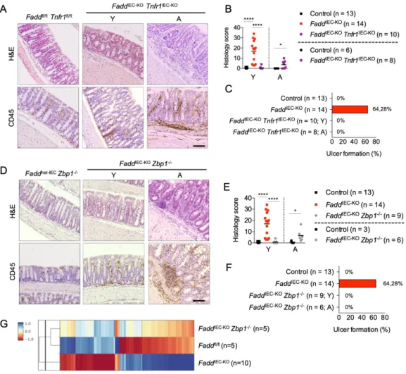
TNFR1 and ZBP1 promote colitis development in FaddIEC-KO mice
Ubiquitous TNF deficiency prevents colitis in FaddIEC-KO mice (Welz et al., 2011), suggesting that TNF may cause the disease by triggering necroptosis of FADD-deficient IECs. To address the epithelial cell intrinsic role of TNFR1, we generated and analyzed Faddfl/fl x Tnfr1fl/fl x Vil1- cre (FaddIEC-KO Tnfr1IEC-KO) and Casp8fl/fl x Tnfr1fl/fl x Vil1-cre (Casp8IEC-KO Tnfr1IEC-KO) mice.
Consistent with elevated TNF expression, IEC-specific ablation of TNFR1 strongly inhibited colitis development and prevented ulcer formation in both FaddIEC-KO and Casp8IEC-KO mice (Figure 2A-C and S2A-D). However, both Casp8IEC-KO Tnfr1IEC-KO and FaddIEC-KO Tnfr1IEC-KO mice showed focal mild immune cell infiltration in the colon at the age of 10-12 weeks (Figure 2A-B and S2B-C), suggesting that, although TNFR1 plays a major role in driving colitis development, TNFR1-independent mechanisms may also be involved. Indeed, analysis of mice at the age of 7-12 months revealed that about half of the FaddIEC-KO Tnfr1IEC-KO mice developed mild colon inflammation, which was however less severe compared to the lesions observed in young FaddIEC-KO mice (Figure 2A-C). Similar to epithelial TNFR1 deficiency, inhibition of RIPK1 kinase activity by crossing with Ripk1D138N/D138N mice expressing kinase inactive RIPK1 (Polykratis et al., 2014) strongly suppressed colitis development in both models (Figure S2E-G). However, some FaddIEC-KO Ripk1D138N/D138N mice developed colon inflammation later in life (Dannappel et al., 2014), suggesting that also RIPK1-independent mechanisms are involved. Thus, TNFR1-RIPK1 signaling plays an important role but also TNFR1-RIPK1-independent mechanisms contribute to the pathogenesis of colitis in FaddIEC-
KO and Casp8IEC-KO mice.
In an effort to elucidate the TNF-independent mechanisms driving colitis in FaddIEC-KO mice, we compared microarray gene expression profiles with histological colitis scores within individual mice to identify genes the expression of which correlated best with disease severity.
Among the top 25 genes for which increased expression correlated with increased colitis severity, we identified the gene encoding ZBP1 (Figure S2H). Increased expression of ZBP1 protein in colonic IECs of FaddIEC-KO mice was confirmed by immunoblotting (Figure S2I). ZBP1 activates RIPK3 in a RIP homotypic interaction motif (RHIM)-dependent manner and has been shown to trigger RIPK3-dependent cell death and inflammation in response to infection with certain viruses but also in the skin of mice lacking RIPK1 or expressing RIPK1 with mutated RHIM (Kuriakose et al., 2016; Lin et al., 2016; Newton et al., 2016b; Nogusa et al., 2016; Upton et al., 2012). We therefore postulated that ZBP1 may contribute to RIPK3-mediated epithelial cell death and intestinal inflammation in FaddIEC-KO mice. Indeed, histological assessment and microarray gene expression profiling of colon tissue from FaddIEC-KO Zbp1-/- mice at the age of 10-12 weeks revealed that ZBP1 deficiency strongly inhibited colitis development (Figure 2D- G). However, similarly to FaddIEC-KO Tnfr1IEC-KO mice, histological examination of colon tissue from 9-10 month old FaddIEC-KO Zbp1-/- mice revealed low grade inflammatory lesions (Figure 2D-F). Collectively, these results identified ZBP1, in addition to TNFR1, as a critical factor that drives colitis development in FaddIEC-KO mice.
TNFR1 and ZBP1 act redundantly to cause ileitis in FaddIEC-KO mice
Having established TNFR1 and ZBP1 as major drivers of colitis in FaddIEC-KO mice, we wondered whether they also contribute to the pathogenesis of ileitis. Consistent with earlier results in FaddIEC-KO Tnf-/- mice (Welz et al., 2011), IEC-specific TNFR1 ablation did not considerably suppress ileitis in FaddIEC-KO mice, although a slight trend towards reduced severity was noted (Figure 3A-B). In contrast to a previous study suggesting a critical role for TNFR1 (Gunther et al., 2011), IEC-specific TNFR1 deficiency did not prevent ileitis development and Paneth cell loss in Casp8IEC-KO mice (Figure S3A-B).
Histological assessment and microarray gene expression analysis of small intestinal tissue revealed that ablation of ZBP1 did not considerably prevent ileitis in FaddIEC-KO mice, although FaddIEC-KO Zbp1-/- mice showed a mild overall amelioration of the pathology (Figure 3A-C).
Intrigued by the similar effect of ZBP1 or TNFR1 deficiencies in mildly ameliorating ileitis in FaddIEC-KO mice, we wondered whether the two pathways might exhibit functional redundancy.
Indeed, histological examination of intestinal sections from FaddIEC-KO Tnfr1IEC-KO Zbp1-/- mice
revealed that combined deficiency of TNFR1 and ZBP1 strongly ameliorated - but did not fully prevent - ileitis development, as shown by overall restored tissue architecture, reduced numbers of dying IECs, partial re-appearance of Paneth cells and reduced CD45+ immune cell infiltrates (Figure 3A-B, D). The redundant role of TNFR1 and ZBP1 in driving ileitis as opposed to their synergistic effect in driving colitis could be related to their increased expression in the ileum compared to the colon (Figure S3F and S2I). We hypothesized that TIR domain- containing adapter protein inducing IFN-beta (TRIF, also known as TICAM-1), an adapter protein mediating signaling downstream of TLR3 and TLR4 that contains a RHIM and can activate RIPK3 (Pasparakis and Vandenabeele, 2015), might also contribute to the pathology, therefore we generated and analyzed Faddfl/fl x Tnfr1fl/fl x Ticam1fl/fl x Vil1-cre x Zbp1-/- mice (FaddIEC-KO Tnfr1IEC-KO Ticam1IEC-KO Zbp1-/-). Additional epithelial ablation of TRIF fully restored the numbers of lysozyme+ Paneth cells (Figure 3B), but could not fully prevent ileitis as these mice still showed mild epithelial hyperplasia and small numbers of dying epithelial cells (Figure 3A, 3B and 3E). Notably, ablation of TRIF did not show any additive effect when combined with TNFR1 deficiency in the presence of Zbp1 heterozygosity (Figure 3A). Collectively, these results revealed that TNFR1 and ZBP1 act in a redundant fashion to induce IEC death and ileitis development in FaddIEC-KO mice, while TRIF also contributes particularly to the loss of Paneth cells.
Inhibition of RIPK1 kinase activity strongly suppressed ileitis development in both FaddIEC-KO Ripk1D138N/D138N and Casp8IEC-KO Ripk1D138N/D138N mice (Figure S3C-E). The kinase inactive Ripk1D138N/D138N had a similar effect in suppressing ileitis compared to combined TNFR1+ZBP1 deficiency (Figure S3C and S3E), suggesting that RIPK1 acts downstream of both receptors.
Considering that RIPK1 has been shown to inhibit ZBP1-mediated necroptosis and inflammation in the skin (Lin et al., 2016; Newton et al., 2016b), we explored the role of RIPK1 in ZBP1-mediated cell death in cellular systems. Doxycycline-inducible expression of ZBP1 in mouse embryonic fibroblasts (MEFs) caused cell death in the presence, but not in the absence, of the caspase inhibitor emricasan, which was strongly inhibited by the RIPK1-specific inhibitor necrostatin-1s (Nec-1s) (Figure 3F, G). To assess whether endogenous ZBP1 also mediates RIPK1-dependent cell death, we employed primary lung fibroblasts (LFs). Emricasan treatment induced cell death in WT LFs, that was strongly, but not completely, suppressed in Tnfr1-/- LFs, suggesting that this is induced by autocrine TNF signaling (Figure 3H). Emricasan- induced cell death was only partially inhibited in Zbp1-/- LFs, but was fully prevented by combined deficiency of TNFR1 and ZBP1 (Figure 3H). We reasoned that the minor role of ZBP1 in this setting might be due to its very low expression and repeated the experiment in cells that were prestimulated with IFNg for 24 hours to induce ZBP1 expression (Figure 3I).
LFs lacking both TNFR1 and ZBP1 were fully protected by IFNg+emricasan-induced cell death, whereas single TNFR1 or ZBP1 deficiencies offered little protection (Figure 3H), demonstrating that ZBP1 and TNFR1 exhibit redundant functions in driving IFNg+emricasan-induced cell death, in line with our findings that ZBP1 and TNFR1 act redundantly to induce ileitis in FaddIEC-
KO mice. Furthermore, LFs from Ripk1D138N/D138N mice were considerably protected from IFNg+emricasan-induced death, showing that RIPK1 kinase activity drives cell death downstream of TNFR1 and ZBP1 (Figure 3H). Consistently, IFNg+emricasan treatment caused phosphorylation of RIPK1 and MLKL that were abolished in Tnfr1-/- Zbp1-/- LFs (Figure 3I and 3J). Moreover, Nec-1s treatment inhibited phosphorylation of MLKL in IFNg+emricasan treated Tnfr1-/- LFs, demonstrating that ZBP1-mediated necroptosis required RIPK1 kinase activity (Figure 3I and 3J). Together, these results showed that TNFR1 and ZBP1 act redundantly to induce necroptosis and intestinal inflammation when FADD-caspase-8 signaling is impaired.
MLKL-dependent and -independent mechanisms cause ileitis in FaddIEC-KO mice
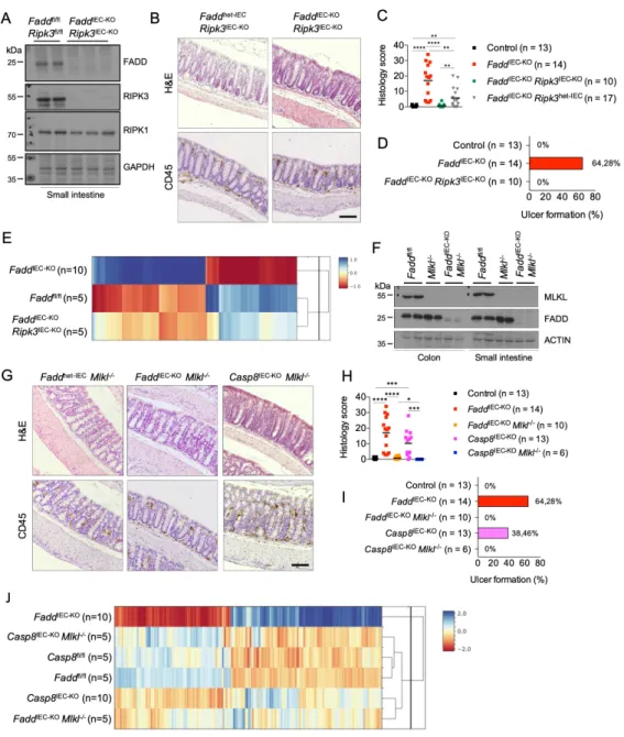
Ubiquitous RIPK3 deficiency prevents colitis and ileitis in FaddIEC-KO mice (Welz et al., 2011) suggesting that necroptosis of Fadd-deficient IECs causes intestinal inflammation. However, since RIPK3 has been reported to regulate intestinal inflammation by acting on immune cells independently from necroptosis (Moriwaki et al., 2014), to address the role of epithelial RIPK3 we generated Faddfl/fl x Ripk3fl/fl x Vil1-cre mice (FaddIEC-KO Ripk3IEC-KO). IEC-specific RIPK3 ablation prevented colitis development in FaddIEC-KO mice, as shown by histological and gene
expression analysis (Figure 4A-E). To exclude the involvement of non-necroptotic functions of RIPK3 and unequivocally assess the contribution of necroptosis in colitis development, we generated and analyzed FaddIEC-KO Mlkl-/- as well as Casp8IEC-KO Mlkl-/- mice (Figure 4F).
Indeed, histological analysis and microarray gene expression profiling revealed that MLKL deficiency prevented colitis development in FaddIEC-KO and Casp8IEC-KO mice (Figure 4G-J).
Taken together, these results showed that RIPK3-MLKL-dependent IEC necroptosis causes colitis in FaddIEC-KO and Casp8IEC-KO mice.
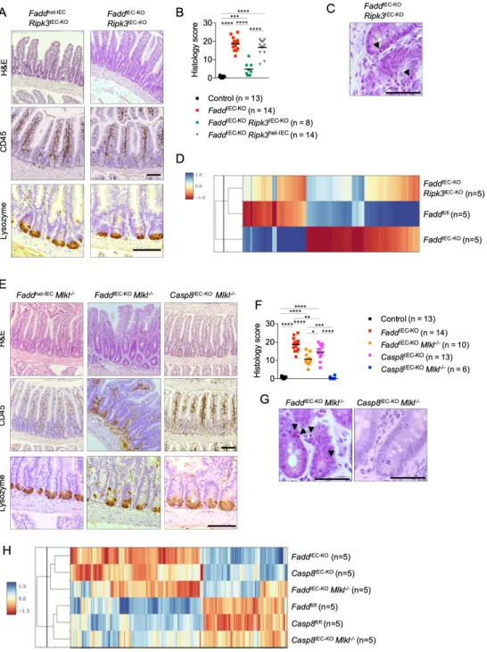
Histological analysis of ileal sections from FaddIEC-KO Ripk3IEC-KO mice revealed that IEC- specific RIPK3 deletion strongly suppressed epithelial hyperplasia, cell death and tissue inflammation, and largely restored the appearance of lysozyme+ Paneth cells in FaddIEC-KO mice (Figure 5A-B). However, mild epithelial hyperplasia, small numbers of dying IECs as well as mildly decreased Paneth cell numbers were observed in FaddIEC-KO Ripk3IEC-KO mice (Figure 5A-C). Furthermore, microarray profiling revealed that RIPK3 epithelial deficiency largely restored but did not completely normalize gene expression in the ileum of FaddIEC-KO mice (Figure 5D). Therefore, IEC-specific RIPK3 deficiency strongly inhibited but did not completely prevent ileitis in FaddIEC-KO mice. To unequivocally assess the role of necroptosis in ileitis development we analyzed ileal tissues from Casp8IEC-KO Mlkl-/- and FaddIEC-KO Mlkl-/- mice.
MLKL deficiency fully prevented the development of ileal pathology in Casp8IEC-KO mice, as assessed by immunohistological and gene expression analysis (Figure 5E-H), demonstrating that MLKL-dependent IEC necroptosis causes ileitis in these animals. In contrast, analysis of FaddIEC-KO Mlkl-/- mice revealed that MLKL-deficiency only partially inhibited ileitis in FaddIEC-KO mice. Examination of ileal tissues from FaddIEC-KO Mlkl-/- mice revealed increased cellularity in the mucosa, crypt hyperplasia, dying IECs, increased CD45+ cell infiltration and decreased number of Paneth cells compared to Faddfl/fl Mlkl-/- littermates (Figure 5E-G). Moreover, microarray analysis showed that the gene expression profiles of FaddIEC-KO Mlkl-/- mice were more similar to Casp8IEC-KO and FaddIEC-KO mice than to the floxed control mice (Figure 5H).
Collectively, these results showed that epithelial caspase-8 deficiency causes ileitis by sensitizing IECs to MLKL-dependent necroptosis, but epithelial FADD deficiency causes ileitis by triggering MLKL-dependent necroptosis and by additional MLKL-independent mechanisms.
Caspase-8 drives MLKL-independent ileitis in FaddIEC-KO mice
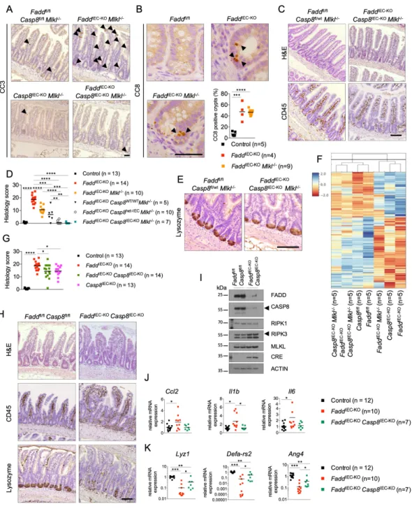
To explore the mechanisms causing MLKL-independent ileitis we examined intestinal sections from FaddIEC-KO Mlkl-/- mice for signs of cell death by immunohistochemical staining for activated cleaved caspase-3 (CC3). Indeed, increased numbers of CC3+ cells were detected in the small intestinal epithelium of FaddIEC-KO Mlkl-/- but not in Casp8IEC-KO Mlkl-/- mice (Figure 6A). Increased numbers of cleaved caspase-8+ (CC8+) cells were also detected primarily in ileal crypts of FaddIEC-KO and FaddIEC-KO Mlkl-/- mice (Figure 6B). Prompted by the presence of CC8+ IECs in FaddIEC-KO Mlkl-/- mice combined with the fact that MLKL deficiency fully prevented ileitis in Casp8IEC-KO mice, we hypothesized that caspase-8 might trigger IEC death and ileitis in FaddIEC-KO Mlkl-/- mice. Hence, we generated and analyzed FaddIEC-KO Casp8IEC-KO Mlkl-/- mice. Indeed, additional IEC-specific deletion of caspase-8 fully prevented the small intestinal pathology observed in FaddIEC-KO Mlkl-/- mice, as shown by normal tissue architecture with absence of infiltrating CD45+ mucosal immune cells and CC3+ IECs, as well as restored numbers of Lysozyme+ Paneth cells (Figure 6A and 6C-E). In agreement with the histological assessment, microarray analysis revealed that the ileal gene expression profile of FaddIEC-KO Casp8IEC-KO Mlkl-/- mice clustered in close proximity to Casp8IEC-KO Mlkl-/- and Faddfl/fl mice (Figure 6F). We then wondered whether caspase-8 contributed to ileitis development in FaddIEC-KO mice also in the presence of MLKL. Indeed, analysis of intestinal tissues from FaddIEC-KO Casp8IEC-KO mice revealed mildly decreased tissue inflammation and epithelial hyperplasia compared to FaddIEC-KO mice, with an overall histological pathology score similar to that of Casp8IEC-KO mice (Figure 6G-I). Moreover, the expression of Il1b, Il6 and Ccl2 mRNAs was decreased in the ileum of FaddIEC-KO Casp8IEC-KO compared to FaddIEC-KO mice, further supporting that caspase-8 deficiency reduced inflammation (Figure 6J). Analysis of the mRNA expression of Paneth cell specific marker genes also supported the partial restoration of
Paneth cells in FaddIEC-KO Casp8IEC-KO mice (Figure 6K). Together, these results showed that caspase-8 promotes ileitis in FaddIEC-KO mice both in the absence and in the presence of MLKL.
Our genetic studies showing that caspase-8 triggers MLKL-independent ileitis in FaddIEC-KO mice raised the question how is caspase-8 activated in the absence of its bona-fide adapter FADD. To gain insight into possible upstream pathways regulating caspase-8, we examined caspase-8 activation by immunostaining for CC8 in the ileum of the different genetic lines crossed to FaddIEC-KO mice. Similar numbers of intestinal crypts containing CC8+ cells were detected in ileal sections from FaddIEC-KO Tnfr1IEC-KO compared to FaddIEC-KO mice, while FaddIEC-KO Zbp1-/- and FaddIEC-KO Tnfr1IEC-KO Zbp1-/- had mildly decreased numbers of CC8+ crypts (Figure S4A and S4B). Moreover, neither deletion of ZBP1 nor inhibition of RIPK1 kinase activity could suppress ileitis and the appearance of CC8+ crypts in FaddIEC-KO Mlkl-/- mice (Figure S4A-D). However, considerably reduced numbers of CC8+ crypts were detected in FaddIEC-KO Ripk3IEC-KO and FaddIEC-KO Tnfr1IEC-KO Zbp1-/- Ticam1IEC-KO samples (Figure S4A and S4B), consistent with the overall stronger protection of these mouse strains from ileal tissue pathology (see Figure 3, Figure 5 and Table S1). Taken together, these results showed that TNFR1, ZBP1 and TRIF redundantly contribute to triggering RIPK3-mediated activation of caspase-8 in IECs from FaddIEC-KO mice. However, a small number of CC8+ cells were still detected even in FaddIEC-KO Ripk3IEC-KO and FaddIEC-KO Tnfr1IEC-KO Zbp1-/- Ticam1IEC-KO mice, suggesting that while this pathway clearly contributes to caspase-8 activation, also other pathways are involved in activating caspase-8 to a lesser extent. Apoptosis-associated speck- like protein containing a CARD (ASC) has been reported to associate with caspase-8 through PYRIN domain (PYD)-death effector domain (DED)-dependent heterotypic interactions (Masumoto et al., 2003; Sagulenko et al., 2013), and activate caspase-8-dependent cell death downstream of inflammasome receptors (Lee et al., 2018; Man et al., 2013; Mascarenhas et al., 2017; Pierini et al., 2012; Rauch et al., 2017; Sagulenko et al., 2013). We therefore hypothesized that ASC-dependent signals could activate caspase-8 in FADD-deficient IECs.
Upon activation, ASC forms speck-like structures that act as platforms for the activation of downstream mediators. Immunostaining of small intestinal sections with an ASC-specific antibody revealed the presence of ASC specks in the epithelium of FaddIEC-KO Mlkl-/- but not Mlkl-/- mice (Figure S5A), indicating that ASC might be involved in driving caspase-8 activation in these mice. However, IEC-specific ASC ablation did not prevent IEC death and the development of ileitis in FaddIEC-KO or FaddIEC-KO Mlkl-/- mice (Figure S5B-F), showing that ASC is not essential for activation of caspase-8 in FADD-deficient IECs and the development of MLKL-independent intestinal inflammation.
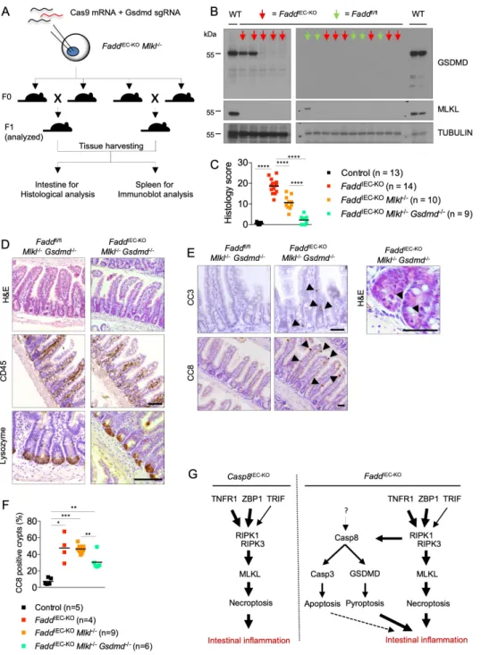
Caspase-8 triggers GSDMD-dependent ileitis in FaddIEC-KO Mlkl-/- mice
The presence of increased numbers of CC8+ and CC3+ cells in FaddIEC-KO Mlkl-/- but not in FaddIEC-KO Casp8IEC-KO Mlkl-/- mice suggested that caspase-8 may cause ileitis by inducing apoptosis of IECs. However, caspase-8 was recently reported to also mediate Gasdermin-D (GSDMD)-dependent cell death (Orning et al., 2018; Sarhan et al., 2018). Considering that apoptosis is generally believed not to cause inflammation while GSDMD triggers inflammatory lytic cell death, we hypothesized that caspase-8 might trigger ileitis in FaddIEC-KO Mlkl-/- mice by activating GSDMD. To address the role of GSDMD, we generated FaddIEC-KO Mlkl-/- Gsdmd-/- mice using CRISPR-Cas9-mediated targeting of the Gsdmd gene in fertilized oocytes from FaddIEC-KO Mlkl-/- mice. The obtained F0 generation was intercrossed and mice from the F1 generation were analyzed for ileitis development (Figure 7A). To confirm GSDMD deficiency, we assessed the expression of GSDMD protein in spleen protein extracts from all F1 mice that were analyzed for intestinal pathology (Figure 7B). FaddIEC-KO Mlkl-/- Gsdmd-/- mice showed restored tissue architecture, re-appearance of Lysozyme+ Paneth cells and absence of infiltrating immune cells, showing that GSDMD deficiency inhibited ileitis development in FaddIEC-KO Mlkl-/- mice (Figure 7C and 7D and Table S1). However, CC8+ and CC3+ cells were still detected in small intestinal crypts from FaddIEC-KO Mlkl-/- Gsdmd-/- mice, suggesting that, in the absence of GSDMD, caspase-8 triggers caspase-3-dependent apoptosis of FADD- deficient IECs, which however is not sufficient to cause ileitis (Figure 7E, 7F, 7G). Taken
together, these results showed that caspase-8 promotes ileal inflammation in FaddIEC-KO Mlkl-
/- mice by inducing GSDMD-dependent death of IECs.
Discussion
Earlier studies suggested that RIPK3-dependent IEC necroptosis causes colitis and ileitis in mice lacking FADD or caspase-8 in the intestinal epithelium (Weinlich et al., 2013; Welz et al., 2011; Wittkopf et al., 2013). Indeed, our results confirmed that MLKL-dependent necroptosis of IECs causes colitis and ileitis in Casp8IEC-KO mice. Considering that the only function known for FADD is to act as an adapter facilitating the activation of caspase-8, it was therefore unexpected that MLKL deficiency rescued colitis but only partially ameliorated ileitis development in FaddIEC-KO mice. Our genetic experiments demonstrating that caspase-8 causes MLKL-independent ileitis in FaddIEC-KO mice, revealed not only that activation of caspase-8 in this context does not require FADD but on the contrary it is inhibited by FADD.
The detection of IECs staining positive for CC3 and CC8 in FaddIEC-KO Mlkl-/- mice suggested that caspase-8 could trigger ileitis by inducing epithelial cell apoptosis. However, taking into account that apoptosis is generally considered a non-inflammatory type of cell death (Pasparakis and Vandenabeele, 2015), we explored an alternative hypothesis. Based on recent studies showing that caspase-8 could induce activation of GSDMD-dependent pyroptosis (Orning et al., 2018; Sarhan et al., 2018), we hypothesized that caspase-8 may activate GSDMD-dependent cell death in IECs of FaddIEC-KO Mlkl-/- mice triggering inflammation. Indeed, our genetic studies showing that GSDMD deficiency inhibited ileitis in FaddIEC-KO Mlkl-/- mice provided experimental evidence supporting that caspase-8-dependent activation of GSDMD drives the pathology. Nevertheless, CC3+ and CC8+ epithelial cells could still be detected in FaddIEC-KO Mlkl-/- Gsdmd-/- mice, suggesting that, in the absence of GSDMD, caspase-8 triggers caspase-3-dependent apoptosis, which is however not sufficient to trigger inflammation. This finding is consistent with the generally accepted notion that apoptotic cell death is immunologically silent in contrast to the highly inflammatory nature of lytic cell death such as necroptosis or pyroptosis (Pasparakis and Vandenabeele, 2015). Therefore, our data provide in vivo experimental evidence that activation of caspase-8 can trigger both non- inflammatory apoptosis but also inflammatory lytic cell death in IECs depending on the downstream effectors that are activated to induce cell death execution.
While it remains to be experimentally assessed whether GSDMD deficiency also ameliorates ileitis in FaddIEC-KO mice in the presence of MLKL, our results showing that caspase-8 deficiency ameliorated ileitis in FaddIEC-KO mice suggest that the caspase-8-GSDMD- dependent axis also operates in the presence of MLKL. It is currently unclear whether caspase- 8 induces GSDMD activation in FaddIEC-KO Mlkl-/- IECs by direct proteolytic processing or indirectly, e.g. via caspases 1 and 11. However, the finding that ASC deficiency could not prevent ileitis in FaddIEC-KO Mlkl-/- mice argues against an indirect activation of GSDMD by caspase-8 through ASC-caspase-1. Two recent studies report that catalytically inactive caspase-8 induces MLKL-independent, ASC-caspase-1-dependent intestinal pathology and perinatal lethality in mice (Fritsch et al., 2019; Newton et al., 2019). These results suggest that caspase-8 acts as a scaffold to activate the ASC-caspase-1 inflammasome and that caspase- 8 catalytic activity prevents inflammasome activation. However, GSDMD deficiency did not prevent the MLKL-independent perinatal lethality of gene targeted mice expressing catalytically inactive caspase-8, arguing against a role of pyroptosis (Newton et al., 2019).
Therefore, the mechanism by which caspase-8 drives MLKL-independent intestinal pathology in FaddIEC-KO mice, which depends on GSDMD but not on ASC, is different from the ASC- caspase-1-dependent but GSDMD-independent pathway induced by inhibition of caspase-8 catalytic activity. Yet, RIPK3 contributes to the MLKL-independent functions of caspase-8 both in FaddIEC-KO mice and in mice expressing catalytically inactive caspase-8 (Fritsch et al., 2019;
Newton et al., 2019), although the mechanism by which RIPK3 engages caspase-8 in the absence of FADD remains elusive.
In addition to TNFR1, we identified ZBP1 as a key driver of RIPK3-dependent intestinal inflammation in FaddIEC-KO mice. Whereas deficiency of either TNFR1 or ZBP1 alone could
inhibit colitis, only combined loss of both pathways could suppress ileitis, suggesting that TNFR1 and ZBP1 exhibit redundant functions in the ileum but synergize to drive severe inflammation in the colon. Differential expression of ZBP1 and TNFR1 in the two tissues could explain the redundant functions in the small intestine as opposed to the synergistic effect in the colon. Because they show higher expression in the small intestine, either TNFR1 or ZBP1 alone could drive RIPK3 activation above a critical threshold causing ileitis, therefore only combined ablation of both suppresses the pathology. In the colon however, TNFR1 and ZBP1 are expressed at lower amounts and need to synergize to drive RIPK3 activation above the threshold required to cause IEC necroptosis and colitis, therefore ablation of one of the two is sufficient to considerably suppress colon inflammation. TRIF also contributed to epithelial cell death and Paneth cell loss, although to a lesser extent compared to the role of TNFR1 and ZBP1.
Taken together, our results revealed that TNFR1 and ZBP1 drive intestinal inflammation by inducing MLKL-dependent necroptosis and caspase-8-GSDMD-mediated pyroptotic-like death of FADD-deficient IECs. This mechanism could be relevant for the pathogenesis of intestinal inflammation during infections with enteropathogenic bacteria that express effectors targeting FADD, such as the arginine glycosyltransferase NleB1 that glycosylates FADD in the death domain inhibiting its function (Pearson et al., 2013; Scott et al., 2017). Furthermore, our results suggest that ZBP1 could be involved in driving intestinal inflammation in human patients with mutations in the CASP8 gene, who were reported to develop severe forms of very early onset IBD that was refractory to anti-TNF treatment (Lehle et al., 2019). Moreover, our findings that RIPK1 kinase activity drives ZBP1-mediated necroptosis when caspase-8 is inhibited suggest that RIPK1 kinase inhibitors might be effective in these patients. Based on the identified functional redundancy between ZBP1 and TNFR1 in driving intestinal inflammation in our mouse models, it will also be important to explore the potential role of ZBP1 in inflammatory pathologies that are independent or only partially dependent on TNF.
STAR Methods Resource Availability
Lead Contact
Further information and reasonable requests for resources and reagents should be directed to and will be fulfilled by the Lead Contact, Manolis Pasparakis (pasparakis@uni-koeln.de).
Materials Availability
Faddfl/fl, Mlkl-/-, Zbp1-/-, Ticam1fl/fl and Ripk1D138N/D138N mice can be obtained from Manolis Pasparakis, Institute for Genetics, Cologne Excellence Cluster on Cellular Stress Responses in Aging-Associated Diseases (CECAD), University of Cologne, 50931 Cologne, Germany.
Email address: pasparakis@uni-koeln.de. Casp8fl/fl mice can be obtained from Stephen M.
Hedrick, Division of Biological Sciences, University of California, San Diego, La Jolla, CA 92093, USA. Tnfr1fl/fl mice can be obtained from George Kollias, Biomedical Sciences Research Center "Alexander Fleming," Vari, GR-16672 Greece. Ripk3fl/fl mice can be obtained from Kim Newton, Department of Physiological Chemistry, Genentech, South San Francisco, CA 94080. Vil1-cre mice can be obtained from Jackson Laboratories, JAX# 004586. Ascfl/fl mice can be obtained from Amir Yazdi, Department of Dermatology and Allergology, Aachen.
Data availability
The MicroArray data have been deposited in the Gene Expression Omnibus (GEO) database under the accession number GEO: GSE137281 and GSE137280.
Acknowledgments
We thank J. Kuth, C. Uthoff-Hachenberg, E. Stade, B. Kühnel and E. Gareus for technical assistance, P. Wagle and H. Nolte for help on microarray data analysis, and B. Zevnik and the CECAD Transgenic Core Facility for CRISPR/Cas9-assisted generation of Gsdmd-/- mice. We also thank K. Newton for Ripk3fl/fl, G. Kollias for Tnfr1fl/fl, S. Hedrick for Casp8fl/fl and J. Tschopp for Ascfl/fl mice. Research reported in this publication was supported by funding from the European Research Council (grant agreements no. 323040 and 787826) and the Deutsche Forschungsgemeinschaft (DFG, German Research Foundation; projects SFB1403 (project no.
414786233), PA 1476/8-1 (project no. 411102043), SFB1218 (project no. 269925409), and CECAD (project no. 390661388). R.S. was supported by a PhD fellowship from the International Graduate School in Development, Health and Disease at the University of Cologne.
Author contributions
R.S. performed all experiments and analyzed data. A.T. contributed to the analysis of microarray gene expression data sets. H.J. and L.W. contributed to the cell death assays in Figure 3G and 3H. M.P. coordinated the project and wrote the paper together with R.S.
Declaration of Interests
M.P. received consulting and speaker fees from Genentech, GSK, Boehringer and Sanofi.
References
Adolph, T.E., Tomczak, M.F., Niederreiter, L., Ko, H.J., Bock, J., Martinez-Naves, E., Glickman, J.N., Tschurtschenthaler, M., Hartwig, J., Hosomi, S., et al. (2013). Paneth cells as a site of origin for intestinal inflammation. Nature 503, 272-276.
Alvarez-Diaz, S., Dillon, C.P., Lalaoui, N., Tanzer, M.C., Rodriguez, D.A., Lin, A., Lebois, M., Hakem, R., Josefsson, E.C., O'Reilly, L.A., et al. (2016). The Pseudokinase MLKL and the Kinase RIPK3 Have Distinct Roles in Autoimmune Disease Caused by Loss of Death- Receptor-Induced Apoptosis. Immunity 45, 513-526.
Beisner, D.R., Ch'en, I.L., Kolla, R.V., Hoffmann, A., and Hedrick, S.M. (2005). Cutting edge:
innate immunity conferred by B cells is regulated by caspase-8. J Immunol 175, 3469-3473.
Buhner, S., Buning, C., Genschel, J., Kling, K., Herrmann, D., Dignass, A., Kuechler, I., Krueger, S., Schmidt, H.H., and Lochs, H. (2006). Genetic basis for increased intestinal permeability in families with Crohn's disease: role of CARD15 3020insC mutation? Gut 55, 342-347.
Chinnaiyan, A.M., Tepper, C.G., Seldin, M.F., O'Rourke, K., Kischkel, F.C., Hellbardt, S., Krammer, P.H., Peter, M.E., and Dixit, V.M. (1996). FADD/MORT1 is a common mediator of CD95 (Fas/APO-1) and tumor necrosis factor receptor-induced apoptosis. J Biol Chem 271, 4961-4965.
Dannappel, M., Vlantis, K., Kumari, S., Polykratis, A., Kim, C., Wachsmuth, L., Eftychi, C., Lin, J., Corona, T., Hermance, N., et al. (2014). RIPK1 maintains epithelial homeostasis by inhibiting apoptosis and necroptosis. Nature 513, 90-94.
Drexler, S.K., Bonsignore, L., Masin, M., Tardivel, A., Jackstadt, R., Hermeking, H., Schneider, P., Gross, O., Tschopp, J., and Yazdi, A.S. (2012). Tissue-specific opposing functions of the inflammasome adaptor ASC in the regulation of epithelial skin carcinogenesis. Proc Natl Acad Sci U S A 109, 18384-18389.
Eftychi, C., Schwarzer, R., Vlantis, K., Wachsmuth, L., Basic, M., Wagle, P., Neurath, M.F., Becker, C., Bleich, A., and Pasparakis, M. (2019). Temporally Distinct Functions of the Cytokines IL-12 and IL-23 Drive Chronic Colon Inflammation in Response to Intestinal Barrier Impairment. Immunity 51, 367-380 e364.
Fritsch, M., Gunther, S.D., Schwarzer, R., Albert, M.C., Schorn, F., Werthenbach, J.P., Schiffmann, L.M., Stair, N., Stocks, H., Seeger, J.M., et al. (2019). Caspase-8 is the molecular switch for apoptosis, necroptosis and pyroptosis. Nature 575, 683-687.
Gunther, C., Martini, E., Wittkopf, N., Amann, K., Weigmann, B., Neumann, H., Waldner, M.J., Hedrick, S.M., Tenzer, S., Neurath, M.F., and Becker, C. (2011). Caspase-8 regulates TNF- alpha-induced epithelial necroptosis and terminal ileitis. Nature 477, 335-339.
Jiao, H., Wachsmuth, L., Kumari, S., Schwarzer, R., Lin, J., Eren, R.O., Fisher, A., Lane, R., Young, G.R., Kassiotis, G., et al. (2020). Z-nucleic-acid sensing triggers ZBP1-dependent necroptosis and inflammation. Nature 580, 391-395.
Kaiser, W.J., Upton, J.W., Long, A.B., Livingston-Rosanoff, D., Daley-Bauer, L.P., Hakem, R., Caspary, T., and Mocarski, E.S. (2011). RIP3 mediates the embryonic lethality of caspase-8- deficient mice. Nature 471, 368-372.
Katz, K.D., Hollander, D., Vadheim, C.M., McElree, C., Delahunty, T., Dadufalza, V.D., Krugliak, P., and Rotter, J.I. (1989). Intestinal permeability in patients with Crohn's disease and their healthy relatives. Gastroenterology 97, 927-931.
Kiesslich, R., Duckworth, C.A., Moussata, D., Gloeckner, A., Lim, L.G., Goetz, M., Pritchard, D.M., Galle, P.R., Neurath, M.F., and Watson, A.J. (2012). Local barrier dysfunction identified by confocal laser endomicroscopy predicts relapse in inflammatory bowel disease. Gut 61, 1146-1153.
Kuriakose, T., Man, S.M., Malireddi, R.K., Karki, R., Kesavardhana, S., Place, D.E., Neale, G., Vogel, P., and Kanneganti, T.D. (2016). ZBP1/DAI is an innate sensor of influenza virus triggering the NLRP3 inflammasome and programmed cell death pathways. Sci Immunol 1.
Lawlor, K.E., Khan, N., Mildenhall, A., Gerlic, M., Croker, B.A., D'Cruz, A.A., Hall, C., Kaur Spall, S., Anderton, H., Masters, S.L., et al. (2015). RIPK3 promotes cell death and NLRP3 inflammasome activation in the absence of MLKL. Nat Commun 6, 6282.
Lee, B.L., Mirrashidi, K.M., Stowe, I.B., Kummerfeld, S.K., Watanabe, C., Haley, B., Cuellar, T.L., Reichelt, M., and Kayagaki, N. (2018). ASC- and caspase-8-dependent apoptotic pathway diverges from the NLRC4 inflammasome in macrophages. Sci Rep 8, 3788.
Lehle, A.S., Farin, H.F., Marquardt, B., Michels, B.E., Magg, T., Li, Y., Liu, Y., Ghalandary, M., Lammens, K., Hollizeck, S., et al. (2019). Intestinal Inflammation and Dysregulated Immunity in Patients With Inherited Caspase-8 Deficiency. Gastroenterology 156, 275-278.
Lin, J., Kumari, S., Kim, C., Van, T.M., Wachsmuth, L., Polykratis, A., and Pasparakis, M.
(2016). RIPK1 counteracts ZBP1-mediated necroptosis to inhibit inflammation. Nature 540, 124-128.
Madison, B.B., Dunbar, L., Qiao, X.T., Braunstein, K., Braunstein, E., and Gumucio, D.L.
(2002). Cis elements of the villin gene control expression in restricted domains of the vertical (crypt) and horizontal (duodenum, cecum) axes of the intestine. J Biol Chem 277, 33275- 33283.
Man, S.M., Tourlomousis, P., Hopkins, L., Monie, T.P., Fitzgerald, K.A., and Bryant, C.E.
(2013). Salmonella infection induces recruitment of Caspase-8 to the inflammasome to modulate IL-1beta production. J Immunol 191, 5239-5246.
Mascarenhas, D.P.A., Cerqueira, D.M., Pereira, M.S.F., Castanheira, F.V.S., Fernandes, T.D., Manin, G.Z., Cunha, L.D., and Zamboni, D.S. (2017). Inhibition of caspase-1 or gasdermin-D enable caspase-8 activation in the Naip5/NLRC4/ASC inflammasome. PLoS Pathog 13, e1006502.
Masumoto, J., Dowds, T.A., Schaner, P., Chen, F.F., Ogura, Y., Li, M., Zhu, L., Katsuyama, T., Sagara, J., Taniguchi, S., et al. (2003). ASC is an activating adaptor for NF-kappa B and caspase-8-dependent apoptosis. Biochem Biophys Res Commun 303, 69-73.
Mc Guire, C., Volckaert, T., Wolke, U., Sze, M., de Rycke, R., Waisman, A., Prinz, M., Beyaert, R., Pasparakis, M., and van Loo, G. (2010). Oligodendrocyte-specific FADD deletion protects mice from autoimmune-mediated demyelination. J Immunol 185, 7646-7653.
Moriwaki, K., Balaji, S., Bertin, J., Gough, P.J., and Chan, F.K. (2017). Distinct Kinase- Independent Role of RIPK3 in CD11c(+) Mononuclear Phagocytes in Cytokine-Induced Tissue Repair. Cell Rep 18, 2441-2451.
Moriwaki, K., Balaji, S., McQuade, T., Malhotra, N., Kang, J., and Chan, F.K. (2014). The necroptosis adaptor RIPK3 promotes injury-induced cytokine expression and tissue repair.
Immunity 41, 567-578.
Muzio, M., Chinnaiyan, A.M., Kischkel, F.C., O'Rourke, K., Shevchenko, A., Ni, J., Scaffidi, C., Bretz, J.D., Zhang, M., Gentz, R., et al. (1996). FLICE, a novel FADD-homologous ICE/CED- 3-like protease, is recruited to the CD95 (Fas/APO-1) death--inducing signaling complex. Cell 85, 817-827.
Nenci, A., Becker, C., Wullaert, A., Gareus, R., van Loo, G., Danese, S., Huth, M., Nikolaev, A., Neufert, C., Madison, B., et al. (2007). Epithelial NEMO links innate immunity to chronic intestinal inflammation. Nature 446, 557-561.
Newton, K., Dugger, D.L., Maltzman, A., Greve, J.M., Hedehus, M., Martin-McNulty, B., Carano, R.A., Cao, T.C., van Bruggen, N., Bernstein, L., et al. (2016a). RIPK3 deficiency or catalytically inactive RIPK1 provides greater benefit than MLKL deficiency in mouse models of inflammation and tissue injury. Cell Death Differ 23, 1565-1576.
Newton, K., Wickliffe, K.E., Maltzman, A., Dugger, D.L., Reja, R., Zhang, Y., Roose-Girma, M., Modrusan, Z., Sagolla, M.S., Webster, J.D., and Dixit, V.M. (2019). Activity of caspase-8 determines plasticity between cell death pathways. Nature 575, 679-682.
Newton, K., Wickliffe, K.E., Maltzman, A., Dugger, D.L., Strasser, A., Pham, V.C., Lill, J.R., Roose-Girma, M., Warming, S., Solon, M., et al. (2016b). RIPK1 inhibits ZBP1-driven necroptosis during development. Nature 540, 129-133.
Nogusa, S., Thapa, R.J., Dillon, C.P., Liedmann, S., Oguin, T.H., 3rd, Ingram, J.P., Rodriguez, D.A., Kosoff, R., Sharma, S., Sturm, O., et al. (2016). RIPK3 Activates Parallel Pathways of MLKL-Driven Necroptosis and FADD-Mediated Apoptosis to Protect against Influenza A Virus.
Cell Host Microbe 20, 13-24.
Nolte, H., MacVicar, T.D., Tellkamp, F., and Kruger, M. (2018). Instant Clue: A Software Suite for Interactive Data Visualization and Analysis. Sci Rep 8, 12648.
Oberst, A., Dillon, C.P., Weinlich, R., McCormick, L.L., Fitzgerald, P., Pop, C., Hakem, R., Salvesen, G.S., and Green, D.R. (2011). Catalytic activity of the caspase-8-FLIP(L) complex inhibits RIPK3-dependent necrosis. Nature 471, 363-367.
Orning, P., Weng, D., Starheim, K., Ratner, D., Best, Z., Lee, B., Brooks, A., Xia, S., Wu, H., Kelliher, M.A., et al. (2018). Pathogen blockade of TAK1 triggers caspase-8-dependent cleavage of gasdermin D and cell death. Science 362, 1064-1069.
Pasparakis, M., and Vandenabeele, P. (2015). Necroptosis and its role in inflammation. Nature 517, 311-320.
Pearson, J.S., Giogha, C., Ong, S.Y., Kennedy, C.L., Kelly, M., Robinson, K.S., Lung, T.W., Mansell, A., Riedmaier, P., Oates, C.V., et al. (2013). A type III effector antagonizes death receptor signalling during bacterial gut infection. Nature 501, 247-251.
Pierini, R., Juruj, C., Perret, M., Jones, C.L., Mangeot, P., Weiss, D.S., and Henry, T. (2012).
AIM2/ASC triggers caspase-8-dependent apoptosis in Francisella-infected caspase-1- deficient macrophages. Cell Death Differ 19, 1709-1721.
Polykratis, A., Hermance, N., Zelic, M., Roderick, J., Kim, C., Van, T.M., Lee, T.H., Chan, F.K.M., Pasparakis, M., and Kelliher, M.A. (2014). Cutting edge: RIPK1 Kinase inactive mice are viable and protected from TNF-induced necroptosis in vivo. J Immunol 193, 1539-1543.
Rauch, I., Deets, K.A., Ji, D.X., von Moltke, J., Tenthorey, J.L., Lee, A.Y., Philip, N.H., Ayres, J.S., Brodsky, I.E., Gronert, K., and Vance, R.E. (2017). NAIP-NLRC4 Inflammasomes Coordinate Intestinal Epithelial Cell Expulsion with Eicosanoid and IL-18 Release via Activation of Caspase-1 and -8. Immunity 46, 649-659.
Sagulenko, V., Thygesen, S.J., Sester, D.P., Idris, A., Cridland, J.A., Vajjhala, P.R., Roberts, T.L., Schroder, K., Vince, J.E., Hill, J.M., et al. (2013). AIM2 and NLRP3 inflammasomes activate both apoptotic and pyroptotic death pathways via ASC. Cell Death Differ 20, 1149- 1160.
Sarhan, J., Liu, B.C., Muendlein, H.I., Li, P., Nilson, R., Tang, A.Y., Rongvaux, A., Bunnell, S.C., Shao, F., Green, D.R., and Poltorak, A. (2018). Caspase-8 induces cleavage of gasdermin D to elicit pyroptosis during Yersinia infection. Proc Natl Acad Sci U S A 115, E10888-E10897.
Scott, N.E., Giogha, C., Pollock, G.L., Kennedy, C.L., Webb, A.I., Williamson, N.A., Pearson, J.S., and Hartland, E.L. (2017). The bacterial arginine glycosyltransferase effector NleB preferentially modifies Fas-associated death domain protein (FADD). J Biol Chem 292, 17337- 17350.
Stolzer, I., Kaden-Volynets, V., Ruder, B., Letizia, M., Bittel, M., Rausch, P., Basic, M., Bleich, A., Baines, J.F., Neurath, M.F., et al. (2020). Environmental Microbial Factors Determine the Pattern of Inflammatory Lesions in a Murine Model of Crohn's Disease-Like Inflammation.
Inflamm Bowel Dis 26, 66-79.
Takahashi, N., Vereecke, L., Bertrand, M.J., Duprez, L., Berger, S.B., Divert, T., Goncalves, A., Sze, M., Gilbert, B., Kourula, S., et al. (2014). RIPK1 ensures intestinal homeostasis by protecting the epithelium against apoptosis. Nature 513, 95-99.
Turner, J.R. (2009). Intestinal mucosal barrier function in health and disease. Nat Rev Immunol 9, 799-809.
Upton, J.W., Kaiser, W.J., and Mocarski, E.S. (2012). DAI/ZBP1/DLM-1 complexes with RIP3 to mediate virus-induced programmed necrosis that is targeted by murine cytomegalovirus vIRA. Cell Host Microbe 11, 290-297.
Van Hauwermeiren, F., Armaka, M., Karagianni, N., Kranidioti, K., Vandenbroucke, R.E., Loges, S., Van Roy, M., Staelens, J., Puimege, L., Palagani, A., et al. (2013). Safe TNF-based antitumor therapy following p55TNFR reduction in intestinal epithelium. J Clin Invest 123, 2590-2603.
Vlantis, K., Wullaert, A., Polykratis, A., Kondylis, V., Dannappel, M., Schwarzer, R., Welz, P., Corona, T., Walczak, H., Weih, F., et al. (2016). NEMO Prevents RIP Kinase 1-Mediated Epithelial Cell Death and Chronic Intestinal Inflammation by NF-kappaB-Dependent and - Independent Functions. Immunity 44, 553-567.
Weinlich, R., Oberst, A., Dillon, C.P., Janke, L.J., Milasta, S., Lukens, J.R., Rodriguez, D.A., Gurung, P., Savage, C., Kanneganti, T.D., and Green, D.R. (2013). Protective roles for caspase-8 and cFLIP in adult homeostasis. Cell Rep 5, 340-348.
Welz, P.S., Wullaert, A., Vlantis, K., Kondylis, V., Fernandez-Majada, V., Ermolaeva, M., Kirsch, P., Sterner-Kock, A., van Loo, G., and Pasparakis, M. (2011). FADD prevents RIP3- mediated epithelial cell necrosis and chronic intestinal inflammation. Nature 477, 330-334.
Wittkopf, N., Gunther, C., Martini, E., He, G., Amann, K., He, Y.W., Schuchmann, M., Neurath, M.F., and Becker, C. (2013). Cellular FLICE-like inhibitory protein secures intestinal epithelial cell survival and immune homeostasis by regulating caspase-8. Gastroenterology 145, 1369- 1379.
Zhang, H., Zhou, X., McQuade, T., Li, J., Chan, F.K., and Zhang, J. (2011). Functional complementation between FADD and RIP1 in embryos and lymphocytes. Nature 471, 373- 376.
STAR Methods Key Resources Table
REAGENT or RESOURCE SOURCE IDENTIFIER
Antibodies
CD45 BD Biosciences Cat# 560510, RRID:AB_1645208
cleaved-Caspase-3 Cell Signaling Cat# 9661, RRID:AB_2341188
Lysozyme Dako Cat# F0372
cleaved-Caspase-8 Cell Signaling Cat# 8592, RRID:AB_10891784
Anti-rabbit alexa 488 ThermoScientific Cat# A-11008, RRID:AB_143165 Anti-rabbit-IgG, HRP conjugated GE Healthcare Cat# NA934, RRID:AB_772206 Anti-mouse-IgG, HRP conjugated GE Healthcare Cat# NA931, RRID:AB_772210 Anti-rat-IgG, HRP conjugated Jackson Laboratories Cat# 112-035-003,
RRID:AB_2338128 Anti-goat-IgG, HRP conjugated Jackson Laboratories Cat# 705-035-003,
RRID:AB_2340390
mFADD Millipore Cat# 05-486, RRID:AB_2100627
mMLKL Millipore Cat#MABC604
mRIPK1 BD Biosciences Cat# 610459, RRID:AB_397832
mRIPK3 Enzo LifeSciences Cat# ADI-905-242-100,
RRID:AB_2039527
mGAPDH Novus Biologicals Cat# NB300-221,
RRID:AB_10077627
mGSDMD Abcam Cat# 219800
CRE Novagen Cat# 69050-3, RRID:AB_2314229
mCaspase-8 (total) Alexis Cat# ALX-804-447,
RRID:AB_2243830
mZBP1 Adipogen Cat# AG-20B-0010,
RRID:AB_2490191
mACTIN SantaCruz Cat# sc-1616, RRID:AB_630836
Alpha-Tubulin Sigma Cat# T6074, RRID:AB_477582
mASC (Immunoblot) Adipogen Cat# AG-25B-0006,
RRID:AB_2490440
mASC (Immunohistochemistry) Santa Cruz Cat#sc-22514-R,
RRID:AB_217874
Phosphorylated RIPK1 (S166) Cell Signaling Cat# 31122, RRID:AB_2799000 Phosphorylated MLKL (S345) Cell Signaling Cat# 37333, RRID:AB_2799112 Chemicals
Doxycycline Sigma Aldrich Cat#D9891
Necrostatin 1s BioCat Cat#2263-5-BV
Puromycin Sigma Aldrich Cat#P8833-25MG
Polybrene Sigma Aldrich Cat#H9268
YOYO™-1 Iodide (491/509) ThermoScientific Cat#Y3601
Emricasan Selleckchem Cat#S7775
Recombinant mouse interferon gamma ImmunoTools Cat#12343537 Plasmids
pMD2.G Addgene Cat#12259
psPAX2 Addgene Cat#12260
pCW-sFLAG-ZBP1 this paper N/A
Critical Commercial Assays
X-tremeGENE HD DNA Transfection Reagent Roche Cat#06366236001 NucleoSpin RNA isolation kit Macherey Nagel Cat#740955.250 SuperScript III Reverse Transcriptase ThermoScientific Cat# 18080044
Clariom-S-Assay ThermoScientific Cat#902930
Deposited Data
RNA MicroArray Data Colon This paper https://www.ncbi.nlm.nih.
gov/geo/query/acc.cgi?acc
=GSE137281
RNA MicroArray Data Ileum This paper https://www.ncbi.nlm.nih.
gov/geo/query/acc.cgi?acc
=GSE137280 Experimental Models: Organisms/Strains
Mouse: Faddfl/fl, C57BL/6N (Mc Guire et al., 2010) N/A Mouse: Mlkl-/-, C57BL/6N (Dannappel et al.,
2014) N/A
Mouse: Casp8fl/fl, C57BL/6N (Gunther et al., 2011) N/A Mouse: Tnfr1fl/fl, C57BL/6N (Van Hauwermeiren et
al., 2013) N/A
Mouse: Ascfl/fl, C57BL/6N (Drexler et al., 2012) N/A Mouse: Ripk3fl/fl, C57BL/6N (Newton et al., 2016) N/A Mouse: Zbp1-/-, C57BL/6N (Jiao et al., 2020)
Mouse: Ticam1fl/fl, C57BL/6N (Dannappel et al.,
2014) N/A
Mouse: Ripk1D138N/D138N, C57BL/6N (Polykratis et al.,
2014) N/A
Mouse: Vil1-Cre: B6.Cg-Tg(Vil1-cre)997Gum/J The Jackson Laboratory
JAX# 004586
Mouse: Gsdmd-/-, C57BL/6N This paper N/A
Oligonucleotides
murine Gsdmd sgRNA1:
5’-GCAGTATACACACATTCATGG-3’
this paper N/A
murine Gsdmd sgRNA2:
5’-GCGTGTGACTCAGAAGACCT-3’
this paper N/A
TagMan Probes
Ccl2 ThermoScientific Mm00441242_m1
Il1b ThermoScientific Mm00434228_m1
Il6 ThermoScientific Mm00446190_m1
Lyz1 ThermoScientific Mm00657323_m1
Defa-rs1 ThermoScientific Mm00655851_gH
Ang4 ThermoScientific Mm03647554_g1
Tbp ThermoScientific Mm00446973_m1
Software and Algorithms ThermoScientific
Prism 6.0 Graphpad Inc RRID: SCR_002798
Instant Clue (Nolte et al., 2018) http://www.instantclue.uni-
koeln.de/
References for STAR Methods Key Resources
Dannappel, M., Vlantis, K., Kumari, S., Polykratis, A., Kim, C., Wachsmuth, L., Eftychi, C., Lin, J., Corona, T., Hermance, N., et al. (2014). RIPK1 maintains epithelial homeostasis by inhibiting apoptosis and necroptosis. Nature 513, 90-94.
Gunther, C., Martini, E., Wittkopf, N., Amann, K., Weigmann, B., Neumann, H., Waldner, M.J., Hedrick, S.M., Tenzer, S., Neurath, M.F., and Becker, C. (2011). Caspase-8 regulates TNF- alpha-induced epithelial necroptosis and terminal ileitis. Nature 477, 335-339.
Jiao, H., Wachsmuth, L., Kumari, S., Schwarzer, R., Lin, J., Eren, R.O., Fisher, A., Lane, R., Young, G.R., Kassiotis, G., et al. (2020). Z-nucleic-acid sensing triggers ZBP1-dependent necroptosis and inflammation. Nature 580, 391-395
Mc Guire, C., Volckaert, T., Wolke, U., Sze, M., de Rycke, R., Waisman, A., Prinz, M., Beyaert, R., Pasparakis, M., and van Loo, G. (2010). Oligodendrocyte-specific FADD deletion protects mice from autoimmune-mediated demyelination. J Immunol 185, 7646-7653.
Newton, K., Dugger, D.L., Maltzman, A., Greve, J.M., Hedehus, M., Martin-McNulty, B., Carano, R.A., Cao, T.C., van Bruggen, N., Bernstein, L., et al. (2016). RIPK3 deficiency or catalytically inactive RIPK1 provides greater benefit than MLKL deficiency in mouse models of inflammation and tissue injury. Cell Death Differ 23, 1565-1576.
Nolte, H., MacVicar, T.D., Tellkamp, F., and Kruger, M. (2018). Instant Clue: A Software Suite for Interactive Data Visualization and Analysis. Sci Rep 8, 12648.
Polykratis, A., Hermance, N., Zelic, M., Roderick, J., Kim, C., Van, T.M., Lee, T.H., Chan, F.K.M., Pasparakis, M., and Kelliher, M.A. (2014). Cutting edge: RIPK1 Kinase inactive mice are viable and protected from TNF-induced necroptosis in vivo. J Immunol 193, 1539-1543.
Van Hauwermeiren, F., Armaka, M., Karagianni, N., Kranidioti, K., Vandenbroucke, R.E., Loges, S., Van Roy, M., Staelens, J., Puimege, L., Palagani, A., et al. (2013). Safe TNF-based antitumor therapy following p55TNFR reduction in intestinal epithelium. J Clin Invest 123, 2590-2603.
Experimental model and subject details Mice
Faddfl/fl (Mc Guire et al., 2010), Mlkl-/- , Ticam1fl/fl (Dannappel et al., 2014), Casp8fl/fl (Beisner et al., 2005), Ripk1D138N/D138N mice (Polykratis et al., 2014), Vil1-cre (Madison et al., 2002), Tnfr1fl/fl (Van Hauwermeiren et al., 2013), Ascfl/fl (Drexler et al., 2012) and Ripk3fl/fl (Newton et al., 2016a) were described previously. Zbp1-/- mice were generated using CrispR/Cas9 mediated gene disruption in fertilized oocytes (Jiao et al., 2020). Gsdmd-/- mice were generated in this study. Mice of the indicated genotype were assigned at random to groups. Mouse studies were performed in a blinded fashion. All mice were maintained in C57BL/6N background. Mice in other than C57BL/6N genetic background were first backcrossed for at least ten generations onto the C57BL/6N background except for FaddIEC-KO Mlkl-/- Gsdmd-/- mice as described in Figure 7. Mice were maintained at the SPF animal facilities of the CECAD Research Center, University of Cologne, under a 12 hour light cycle and given water ad libitum and a regular chow diet (Harlan, diet no. 2918). All animal procedures were conducted in accordance with European, national, and institutional guidelines, and protocols were approved by local government authorities (Landesamt für Natur, Umwelt und Verbraucherschutz Nordrhein- Westfalen). Animals requiring medical attention were provided with appropriate care. No other exclusion criteria existed.
Targeting of Gsdmd in mouse zygotes
Gsdmd-/- mice were generated using CrispR/Cas9 technology as previously described (Eftychi et al., 2019). Fertilized oocytes obtained from breedings of Faddfl/fl Mlkl-/- Vil1-cretg/wt male mice with Faddfl/fl Mlkl-/wt or Faddfl/fl Mlkl-/- female mice were microinjected with two short guide (sg)RNAs targeting the Gsdmd gene (5’- GCAGTATACACACATTCA-3’ and 5’- GCGTGTGACTCAGAAGACCT-3’) together with Cas9 mRNA (TriLink). Mice obtained in the F0 generation were sequenced for Gsdmd allele mutations and mice with targeted inactivation of Gsdmd were intercrossed to generate FaddIEC-KO Mlkl-/- Gsdmd-/- mice in the F1 generation.
F1 mice were screened for GSDMD and MLKL deficiency using immunoblot analysis of spleen lysates.
Method details Cell culture
Cell lines were maintained at 37°C and 5% CO2. All cell lines were cultured in 10%FCS, 1%
P/S, 1% L-Glutamate, 1mM Sodium Pyruvate (ThermoFisher Scientific, #11360) and DMEM- Dulbecco’s Modified Eagle Medium (ThermoFisher, #41965-039).
Lentiviral production
HEK293 cells were transfected with pCW-sFLAG-mZBP1(10µg), psPAX2 (5µg) (Addgene,
#12260) and pMD2.G (5µg) (Addgene, #12259) plasmid to produce lentivirus pCW-sFLAG- mZBP1. In detail, plasmids were mixed with 500µl 0.25M CaCl2. Then, 500µl 2xHBS was added slowly while stirring the solution using pipette tip. After 5 min of incubation (RT), the CaCl2-HBS-plasmid solution was added dropwise to HEK293 cells cultured in 10ml culture medium. HEK293 cells were fed with fresh medium 1h prior to transfection. After 24h (day1), medium was replaced with fresh culture medium. Supernatant containing viruses were collected on day 2 and 3.
Generation of immortalized mouse embryonic fibroblasts with doxycycline-inducible ZBP1 expression
Immortalized mouse embryonic fibroblasts (iMEFs) overexpressing mouse ZBP1 (ENSMUST00000029018.13) were generated using lentiviral gene transfer. 48h prior to transduction, 200.000 cells/well were seeded in a 6-well plate (Sigma Aldrich, # CLS3335).
Cells were incubated with 1.5ml fresh medium + 750µl Virus-Supernatant + 8µg/ml Polybrene (Sigma Aldrich, #H9268) for 24h. On the next day, virus containing medium was removed and