Ecient comprehensive scoring of docked protein complexes - a machine learning approach
Inaugural - Dissertation zur
Erlangung des Doktorgrades
der Mathematisch-Naturwissenschaftlichen Fakultät der Universität zu Köln
vorgelegt von
Oliver Sven Martin
aus Kaiserslautern
Köln 2006
Tag der mündlichen Prüfung: 12. Juni 2006
Meinen Eltern
Science has explained nothing; the more we know the more fantastic the world becomes and the profounder the surrounding darkness.
Aldous Huxley, 1894-1963
Acknowledgements
Writing a decent acknowledgement has been among the hardest parts of my thesis work nding the task for a perfect acknowledgement to be a crucial question. Whom to acknowledge at which point in which order and in how much detail? Too many acknowledgements are preferable to too few, but who wants to read page after page of acknowledgements? What about those people who feel aggrieved after reading this acknowledgement and not nding their name listed here or listed in the wrong order?
Instead of only thanking those people which I commemorate easiest due to physical or temporal vicinity, I would rather like to thank generically all those who contributed to this work. All those who helped to built the fundamentals for this work, who contributed to the development of new ideas, who motivated and inspired me and who oered their support and friendship; all those forgotten! May each of you nd your own name in this acknowledgement in the order you think appropriate. Be assured that I feel obliged to you and have thought long about you and the appropriate way of expressing my gratitude to you.
- My sincere thanks -
I
Abstract
Biological systems and processes rely on a complex network of molecular interactions.
The association of biological macromolecules is a fundamental biochemical phenomenon and an unsolved theoretical problem crucial for the understanding of complex living systems. The term protein-protein docking describes the computational prediction of the assembly of protein complexes from the individual subunits. Docking algorithms generally produce a large number of putative protein complexes. In most cases, some of these conformations resemble the native complex structure within an acceptable degree of structural similarity. A major challenge in the eld of docking is to extract the near- native structure(s) out of this considerably large pool of solutions, the so called scoring or ranking problem. It has been the aim of this work to develop methods for the ecient and accurate detection of near-native conformations in the scoring or ranking process of docked protein-protein complexes. A series of structural, chemical, biological and physical properties are used in this work to score docked protein-protein complexes.
These properties include specialised energy functions, evolutionary relationship, class specic residue interface propensities, gap volume, buried surface area, empiric pair potentials on residue and atom level as well as measures for the tightness of t. Ecient comprehensive scoring functions have been developed using probabilistic Support Vector Machines in combination with this array of properties on the largest currently available protein-protein docking benchmark. The established scoring functions are shown to be specic for certain types of protein-protein complexes and are able to detect near-native complex conformations from large sets of decoys with high sensitivity. The specic complex classes are Enzyme-Inhibitor/Substrate complexes, Antibody-Antigen complexes and a third class denoted as "Other" complexes which holds all test cases not belonging to either of the two previous classes. The three complex class specic scoring functions were tested on the docking results of 99 complexes in their unbound form for the above mentioned categories. Dening success as scoring a 'true' result with a p-value of better than 0.1, the scoring schemes were found to be successful in 93%, 78% and 63% of the examined cases, respectively. The ranking of near-native structures can be drastically improved, leading to a signicant enrichment of near- native complex conformations in the top ranks. It could be shown that the developed scoring schemes outperform ve other previously published scoring functions.
III
Zusammenfassung
Biologische Systeme beruhen auf komplexen Netzwerken molekularer Interaktionen.
Die Interaktion biologischer Makromoleküle stellt ein fundamentales biochemisches Phänomen dar, sowie ein ungelöstes theoretisches Problem von herausragender Bedeutung für das Verständnis komplexer lebender Systeme. Als Protein-Protein Docking wird die computergestütze Vorhersage der Assoziation von Proteinkomplexen aus den individuellen Untereinheiten bezeichnet. Dockingalgorithmen produzieren im Allgemeinen eine sehr hohe Anzahl hypothetischer Komplexanordnungen, von denen meist nur einige wenige der korrekten, nativen Lösung ähnlich sind. Eine der grossen Herausforderungen im Bereich des Dockings besteht im Herausltern der wenigen nahe-nativen Strukturen aus der grossen Menge von Lösungsvorschlägen.
Dieses wird auch als Scoring- oder Rankingproblem bezeichnet. Ziel dieser Arbeit war es, Methoden zur ezienten und akkuraten Detektion von nahe-nativen Lösungen während der Bewertungsphase von gedockten Proteinkomplexen zu entwickeln.
Eine Reihe von strukturellen, chemischen, biologischen und physikalischen Parame- tern wurde verwendet, um Komplexanordungen, wie sie als Lösungsvorschläge eines Dockingalgorithmus enstehen, zu bewerten. Diese Bewertungsschemata beinhalten spezialisierte Energiefunktionen molekularer Fragmente, evolutionäre Verwandtschaft, komplexklassenspezische Wahrscheinlichkeitsverteilungen von Residuen, Lückenvolu- men, die Grösse der verborgenen Oberäche, emprische Paarpotentiale auf atomarer und Aminosäurebene sowie ein Mass für die Festigkeit der Bindung. Unter Verwendung des derzeit grössten Datensatzes von Protein-Protein Docking Testfällen wurden Ver- fahren des überwachten maschinellen Lernens in Form von probabilistischen Support Vector Machines trainiert,s um umfassende eziente Bewertungsfunktionen für drei spezische Klassen von Proteinkomplexen zu erstellen. Bei diesen Dockingklassen handelt es sich um Enzym-Inhibitor bzw. Enzym-Substrat und Antikörper-Antigen Komplexe sowie eine dritte Klasse, der alle weiteren Testfälle zugordnet werden, die keiner der beiden bisherigen Kategorien angehören. Die entwickelten Bewertungs- funktionen sind hochspezisch für die einzelnen Kategorien von Proteinkomplexen und in der Lage, nahe-native Lösungen mit hoher Sensitivität aus einer grossen Anzahl potentieller Komplexanordnungen heraus zu erkennen. Eine Sortierung der
Lösungsvorschläge durch Anwendung der Bewertungsfunktionen führt zu einer sig- nikanten Anreicherung von nahe-nativen Komplexen in den oberen Rängen. Die drei entwickelten spezischen Bewertungsfunktionen wurden an Dockingergebnissen für 99 Testfälle erprobt, bei denen versucht wird, native Komplexe aus den ungebunden Strukturen der einzelnen Untereinheiten vorherzusagen. Deniert man ein korrektes Ergebnis über einen Wahrscheinlichkeitswert (p-value) von 0,1 oder besser, so sind die entwickelten Bewertungsfunktionen in 93%, 78% und 63% der untersuchten Fälle erfolgreich. Ein Vergleich mit fünf publizierten Bewertungsfunktionen für Protein- Protein Docking zeigt, dass die komplexklassenspezischen Bewertungsfunktionen den jeweils einzelnen Methoden in der Anwendung überlegen sind.
VI
Denitions and abbreviations
ASA accessible surface area
Ångstrøm 1 Å=10−10m
spec− specictiy; reliability of false/negative predictions spec+ specicity; reliability of true/positive predictions
acc accuracy
ACE atomic contact energies; an atom-atom pair potential
AUC area under the curve
avg. average
avgTF scoring scheme based on average temperature factor in the interface area
BSV Bounded Support Vectors
BurSurf buried surface area; are occluded from solvent by contact surfaces of complexed proteins
CAPRI Critical Assessment of PRedicted Interactions; academic challenge for blind protein interaction predictions
Cons protein docking scoring scheme based on amino acid conservation scores ConsOE protein docking scoring scheme based on amino acid conservation scores
with an over emphasis on residues with a high interface propensity
fn false negative
fp false positive
fval f-value; harmonic average between sensitivity and specicity
GapVol gap volume; volume inbetween and delimited by contact surface of two or more proteins
geo score/rank according to geometric t
IF improvement factor
mcc Matthews correlation coecient
NhcR number of highly conserved residues NhvR number of highly variable residues
P probability
PairPot atom-atom pair potential
pred score/rank according to SVM predictor
red. reduction
RIP residue interface propensities
RIPAA residue interface propensities for Antibody-Antigen complexes
RIPEI residue interface propensities for Enzyme-Inhibitor/Substrate complexes RIPU N I residue interface propensities; universally applicable
RMSD root mean square deviation; a measure for structural similarity
VII
RMSDiCα root mean square deviation of interface C-alpha atoms
ROC Receiver Operator Characteristics; plot of true positive against false posi- tive rate
Rpscore an empiric residue-residue pair potential
SE solvent eect
sens sensitivity
SV Support Vectors
SVM Support Vector Machines; a machine learning method
SVMAbAg SVM-based scoring scheme developed for Antibody-Antigen complexes SVMEI SVM-based scoring scheme developed for Enzyme-Inhibitor/Substrate cim-
plexes
SVMOth SVM-based scoring scheme developed for complexes of type "Other" (non- Antibody-Antigen and non-Enzyme-Inhibitor/Substrate complexes)
tn true negatve
ToF Tightness of Fit; a scoring scheme for docked protein-complexes ToFU N I Tightness of Fit; universally applicable
ToFEI Tightness of Fit; specialised for Enzyme-Inhibitor/Substrate complexes ToFAA Tightness of Fit; specialised for Antibody-Antigen complexes
tp true positive
Aminoacids
Alanine ALA A
Cysteine CYS C
Aspartate ASP D
Glutamate GLU E
Phenylalanine PHE F
Glycine GLY G
Histidine HIS H
Isoleucine ILE I
Lysine LYS K
Leucine LEU L
Methionine MET M
Asparagine ASN N
Proline PRO P
Glutamine GLN Q
Arginine ARG R
Serine SER S
Threonine THR T
Valine VAL V
Tryptophane TRP W
Tyrosine TYR Y
VIII
List of Tables
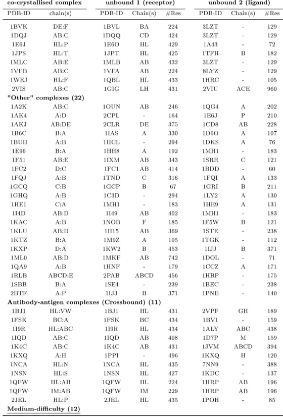
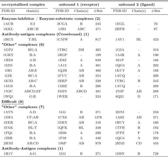
2.1 Unbound-unbound protein-protein docking examples from literature . . 37
2.2 Protein-protein docking benchmark 2.0 (Mintseris et al., 2005). . . 39
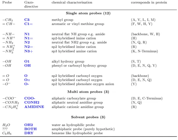
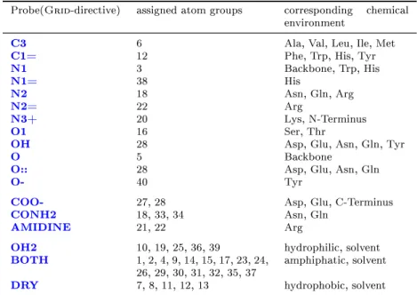
2.3 Grid probes . . . 48
2.4 Grid probes and corresponding atom types . . . 51
2.5 Residue interface propensities . . . 55
3.1 Number of near native solutions for docking benchmark 2.0 . . . 83
3.2 Number of near native solutions for test cases from literature . . . 85
3.3 Composition of training and testing datasets . . . 89
3.4 Feature names and corresponding F-score values . . . 92
3.5 SVM training characteristics . . . 93
3.6 SVM testing . . . 95
3.7 Scoring performance of SVM-based ranking (SVMEI) . . . 97
3.8 Scoring performance of SVM-based ranking (SVMAbAg) . . . 100
3.9 Scoring performance of SVM-based ranking (SVMOth) . . . 103
IX
1.1 Selected examples for protein-protein interaction types I . . . 4
1.2 Selected examples for protein-protein interaction types II . . . 5
2.1 Atomtypes as proposed by Melo and Feytmans (1997) . . . 49
2.2 Schematic illustration of the Grid score calculation method I . . . 50
2.3 Schematic illustration of the Grid score calculation method II . . . 52
2.4 Reglar and inverse transformation applied to binary protein complex . 53 2.5 Atomtypes according to Zhang et al.(1997) . . . 59
2.6 Illustration of the gap volume calculation method . . . 65
2.7 Software owchart . . . 70
2.8 A binary classication toy problem . . . 73
2.9 The kernel trick as basic idea of SVMs . . . 74
2.10 Denotations used in quality measures for binary classication . . . 77
3.1 F-scores as used for feature selection . . . 91
3.2 ROC curves for SVM-training . . . 94
3.3 Enrichment plots of SVMEI scoring function . . . 98
3.4 Enrichment plots of SVMAbAg scoring function . . . 101
3.5 Enrichment plots of SVMOth scoring function . . . 105
3.6 Specicity of SVM-based scoring functions . . . 107
3.7 Comparison of scoring performance for various scoring methods . . . . 108
4.1 Problematic docking test cases from benchmark 2.0 . . . 113
X
Contents
Prex I
Acknowledgement . . . I Abstract . . . III Zusammenfassung in deutscher Sprache . . . V List of abbreviations . . . VII List of tables . . . IX List of gures . . . X
1 Introduction 1
1.1 Protein-protein assemblies . . . 2
1.1.1 Types of protein-protein interactions . . . 2
1.1.2 Specicity of protein-protein interactions . . . 5
1.1.3 Evolution of protein-protein interactions . . . 7
1.2 Protein-protein interfaces . . . 8
1.2.1 Structural characteristics of protein interfaces . . . 8
1.2.1.1 Geometric properties of interface patches . . . 8
1.2.1.2 Physico-chemical properties of interface patches . . . . 8
1.2.1.3 Composition of interface patches . . . 10
1.2.1.4 Native interfaces versus crystal contacts . . . 10
1.3 Characterisation of protein-protein interactions . . . 11
1.3.1 Experimental methods . . . 11
1.3.1.1 Determination of interaction restraints . . . 12
1.3.1.2 Determination of the complex structure . . . 14
1.3.1.3 Quantication of the binding force . . . 15
1.3.2 Theoretical computational methods . . . 17
1.3.2.1 Methods based on genetic information . . . 17
1.3.2.2 Sequence based methods . . . 18
1.3.2.3 Structure based methods . . . 20
1.4 Protein-protein docking . . . 22
1.4.1 The rigid body approach . . . 22
1.4.2 Principle steps of a docking procedure . . . 23
1.4.2.1 Representation of the system . . . 23
1.4.2.2 Conformational space search . . . 24
1.4.2.3 Scoring and ranking of potential solutions . . . 26
1.4.2.4 Renement of accepted solutions . . . 30
1.4.3 Incorporation of exibility into protein-protein docking . . . 31
1.4.4 Docking problems and challenges . . . 33
1.5 Aim of work . . . 35
2 Methods 36 2.1 Data fundamentals . . . 36
2.2 Docking algorithm . . . 41
2.2.1 Ckordo . . . 41
2.2.2 RMSD calculations . . . 43
2.3 The Grid software package . . . 44
2.4 Employed scoring schemes . . . 47
2.4.1 Grid based scoring schemes . . . 47
2.4.2 Residue interface propensities . . . 53
2.4.3 Residue-residue pair potential . . . 55
2.4.4 Tightness of t . . . 56
2.4.5 Atom-atom pair potential . . . 57
2.4.6 Atomic contact energies . . . 58
2.4.7 Evolutionary relationship . . . 60
2.4.8 Temperature factors . . . 62
2.4.9 Approximation of the buried surface area . . . 62
2.4.10 Calculation of the gap volume . . . 64
2.5 Comprehensive scoring of protein-protein docking solutions . . . 66
2.5.1 Theoretical approaches to the merging of postlter scores . . . . 66
2.5.1.1 Consecutive application of the individual scores . . . . 66
2.5.1.2 Combined application of the individual scores . . . 67
2.5.1.3 Using machine learning methods to combine scores . . 68
2.6 Classication of docking results . . . 68
CONTENTS XIII
2.7 Postlter software development . . . 69
2.8 Machine learning . . . 71
2.8.1 Support Vector Machines . . . 72
2.8.1.1 Probabilistic Support Vector Machines . . . 75
2.8.2 Performance measures for machine learning . . . 76
2.8.3 Feature selection strategies . . . 78
2.9 Evaluation of scoring performance . . . 79
3 Results 82 3.1 Primary docking and postlter results . . . 82
3.2 Training and testing of probabilistic Support Vector Machines . . . 87
3.2.1 Selection of training and testing data . . . 87
3.2.2 Feature selection . . . 90
3.2.3 Results on training and testing data sets . . . 92
3.3 SVM-based scoring functions . . . 95
3.3.1 Performance of SVM-based scoring functions . . . 96
3.3.1.1 Enzyme-Inhibitor/Substrate complexes . . . 96
3.3.1.2 Antibody-Antigen complexes . . . 99
3.3.1.3 "Other" complexes . . . 102
3.3.2 Specicity of SVM-based scoring functions . . . 106
3.3.3 Comparison to other scoring functions . . . 106
4 Discussion 110 4.1 General comparability of docking results . . . 110
4.2 Limitations of data fundamentals and docking software . . . 112
4.3 Quality of the developed comprehensive scoring functions . . . 115
4.3.1 Eects of feature selection on specicity of scoring functions . . 115
4.3.2 Eects of training data selection on quality of scoring functions 117 4.4 Support Vector Machines as black box . . . 117
4.5 Versatility of the developed method . . . 118
5 Conclusion and outlook 119 5.1 Future developments . . . 119
References 121
A Appendix 136
A.1 Armation . . . 136 A.2 CV . . . 137
1 Introduction
"He has half the deed done who has made a beginning."
Horace, 65-8 B.C.
According to the conventional denition of life, an organism in question must exhibit the following ve stages of a living system at least once during their existence: growth, motion, reproduction, metabolism, and response to stimuli. These parameters alone, however, may be inadequate for proper classication without further specication.
For example, a mule is a living system, yet it cannot reproduce. Conversely, a non- living entity such as re may experience all ve stages on some level. Biochemistry focuses specically on the aspect of metabolism to dene a living system and implies that the energy gained through metabolism is utilised to maintain the living state by owing into a coordinated regulatory network of molecular interactions. This network is the fundamental basis for reactivity and all the other phenomena used in the conventional denition of a living system. Implicitly, a huge quantity of the ongoing processes in every living organism are based on, regulated or mediated by molecular recognition mechanisms, thus the activity of a living cell can be portrayed as a network of interactions. Such an interaction network could never be coordinated without a high level of specicity. The specicity is mostly provided by the enormous structural and physico-chemical variability of biological macromolecules like proteins and nucleic acids, that are involved in the transfer of biological information.
Protein-protein interactions play a signicant role in these processes for example in signal cascades or gene regulation. In the proteomics era, where experimental high- throughput methods like e.g. the yeast two-hybrid system yield growing amounts of putative protein interaction data, the large quantity of data can no longer be handled by experimental methods alone. Instead it requires the computer aided simulation methods of bioinformatics to complement this knowledge. The exploration, under- standing and detailed knowledge of complete protein interaction networks can only be achieved by combining the often time consuming experimental methods like structure solution by X-ray crystallography or NMR spectroscopy with the data management
facilities and theoretical predictions provided by bioinformatics. Predictive methods for protein-protein interactions are of special interest and importance where experimental methods fail, e.g. for such short-term transient interactions which are not accessible by the mentioned experimental methods due to their low stability and short half-life (Eisenstein and Katchalski-Katzir, 2004).
1.1 Protein-protein assemblies
Protein-protein interactions play diverse roles in biology and dier based on the com- position, anity and half-life of the association. In vivo, the localisation, concentration and local environment of a protomer (subunit of an oligomeric protein complex) can aect the interaction between protein domains and are vital to control the composition and oligomeric state of protein complexes. Since a change in quaternary structure is often coupled with biological function or activity, transient protein-protein interactions are important biological regulators.
1.1.1 Types of protein-protein interactions
Protein-protein interactions are often categorised into distinct types according to their composition, in vivo stability and lifetime (Nooren and Thornton, 2003a):
• Homo- and hetero-oligomeric complexes
Protein-protein interactions occur between identical or non-identical chains (i.e. homo- or hetero-oligomers). Oligomers of identical or homologous protomers can be organised in an isologous or heterologous way. An isologous association involves the same surface on two monomers forming an interface with matching surfaces (e.g. Arc repressor and lysin; Figure 1.1 (a) and (c)), related by a 2-fold symmetry axis. In contrast to an isologous association that can only further oligomerise using a dierent interface (e.g. form a dimer of dimers with three 2-fold axes of symmetry), heterologous assemblies use dierent interfaces that, without a closed (cyclic) symmetry, can lead to innite aggregation (cf. Figure 1.2 (a,b)).
1.1 Protein-protein assemblies 3
• Non-obligate and obligate complexes
As well as composition, two dierent types of protein-protein complexes can be distinguished on the basis of whether a complex is obligate or non-obligate. In an obligate protein-protein interaction, the protomers are not found as stable structures on their own in vivo.
Such complexes are generally also functionally obligate; for example, the Arc repressor dimer (Figure1.1(a)) is essential for DNA binding. Many of the hetero- oligomeric structures in the Protein Data Bank (PDB) (Berman et al.,2000) involve non-obligate interactions of protomers that exist independently, such as intracellular signaling complexes and antibody-antigen, receptor-ligand and enzyme-inhibitor (e.g. thrombin-rhodniin; Figure 1.1 (e)) complexes. The components of such protein-protein complexes are often initially not co-localised and thus need to be independently stable. However, some homo-oligomers, which by denition are co-localised, can also form non-obligate assemblies (e.g. sperm lysin; Figure 1.1 (c)).
• Transient and permanent complexes
Protein-protein interactions can also be distinguished based on the lifetime of the complex. In contrast to a permanent interaction that is usually very stable and thus only exists in its complexed form, a transient interaction associates and dissociates in vivo. One can distinguish between weak transient interactions that feature a dynamic oligomeric equilibrium in solution, where the interaction is broken and formed continuously (e.g. lysin; Figure1.1 (c)), and strong transient associations that require a molecular trigger to shift the oligomeric equilibrium.
Structurally or functionally obligate interactions are usually permanent, whereas non-obligate interactions may be transient or permanent.
It is important to note that many protein-protein interactions cannot be classied according to such unique distinct types. Rather, a continuum exists between non- obligate and obligate interactions (Nooren and Thornton, 2003b), and the stability of all complexes is highly dependant on the physiological conditions and environment.
An interaction may be mainly transient in vivo but become permanent under certain
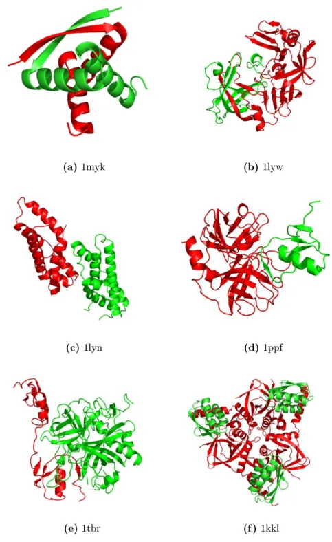
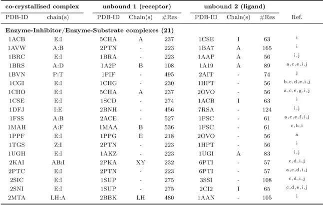
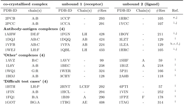
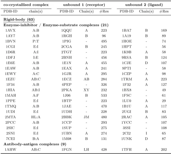
(a) 1myk (b) 1lyw
(c) 1lyn (d) 1ppf
(e) 1tbr (f) 1kkl
Figure 1.1: Selected examples for protein-protein interaction types (4-letter PDB- identier given): (a) obligate homomeric complex: P22 ARC repressor, (b) obligate heteromeric complex: human cathepsin D, (c) non-obligate homomeric complex:
sperm lysin, (d) non-obligate heteromeric complex: human leukocyte elastase / turkey ovomucoid inhibitor, (e) non-obligate permanent heteromeric complex: thrombin / rhodniin inhibitor, (f) non-obligate transient heteromeric complex: L. casei protein kinase Hprk / B. subtilis Hpr (obligate permanent interaction in Hprk trimer (red), transient binding to Hpr (green)).
1.1 Protein-protein assemblies 5
(a) 1cwp (b) 1cwp-assumed biological unit
Figure 1.2: Selected examples for protein-protein interaction types (4-letter PDB- identier given): (a) trimeric unit of viral coat protein: cowpea chlorotic mottle virus (b) assumed biological unit of viral coat protein: spherical virus capsid (consisiting of 60 trimeric units as depicted in (a), identical colour coding)
cellular conditions (Nooren and Thornton, 2003a). Folding data, as well as data on the dynamics of the assembly at dierent physiological conditions or environments, are often not available. However, the sub cellular localisation of subunits and the function of the protein will often suggest the biologically relevant type of interaction.
For example, interactions in intracellular signaling are expected to be transient, since their function requires a ready association (Rittinger et al., 1997b,a). Ultimately, all interactions and complexation processes are driven by the concentration of the components and the free energy of the complex relative to alternate states.
1.1.2 Specicity of protein-protein interactions
The specicity of protein-protein interaction is composed of two major factors: the possibility of forming a more or less stable binding to its predestined binding partner(s) and the potentially lower possibility of association to other protomers in an equally stable and favorable way. It is well known that the binding specicity of protein- protein interactions is mostly accomplished by relatively small structural changes in the contact area of the binding partners - the so called interface region - rather than spacious structural rearrangements (Sear, 2004). A single point mutation leading to
an amino acid change in the interface region of any of the binding partners can cause a complete loss or change of binding specicity as well as create an entirely new one.
The consequences of such a change in binding specicity can be quite drastic as the following prominent examples of human hereditary diseases show:
(i) An example for a reduction or loss in binding specicity due to a single amino acid change is osteogenesis imperfecta, commonly known as the "brittle bone"
disorder, a genetic disorder characterised by bones that break easily, often from little or no apparent cause. The molecular cause for this disease is a defective collagen assembly (Vogel et al., 1987). Collagen is a family of related structural proteins which are vital to the integrity of many tissues including skin and bones.
The mature collagen molecule is comprised of three peptide chains wound in a triple helix. In order to form the triple helix, collagen peptide chains have a special repeating structure consisting of a specic three amino acid pattern. A point mutation which, by changing a single amino acid, disrupts that pattern, will either disturb the association of chains or prevent the triple helix formation and may have very severe consequences. One mutant chain can disrupt a triple helix with two wild type chains, eectively disturbing the functional unit in its stabilising ecacy.
(ii) The single substitution of valine for glutamic acid at position six of the beta- globin polypeptide chain in human haemoglobin gives rise to sickle cell anaemia in homozygote individuals. The modied chain reveals an extended binding specicity to itself and therefore develop a tendency to crystallise at low oxygen concentrations, forming threads of haemoglobin molecules which in turn evoke the sickle-like shape of erythrocytes that gave the disease its name (Rodgers, 1997).
Often, proteins are only biologically active in the complexed oligomeric state. A loss of the ability to form the relevant oligomer therefore can lead to a loss in biological activity, as it has been described by Bennett et al. (1994) for a single amino acid substitution in dimeric proteins.
Single mutations on primary structure level that lead to a loss of the binding anity of transcription factors can even lead to gene knockout (Rausa et al., 2004). The ability of introducing an entirely new binding specicity by a single point mutation without
1.1 Protein-protein assemblies 7 taking the intermediate stage of a non-specic interface is a critical pre-requisite for the evolution of interfaces (Xu et al., 1998) (see section1.1.3).
1.1.3 Evolution of protein-protein interactions
The structure and anity of a protein-protein interaction is tuned to its biological function as well as the physiological environment and control mechanism. Protein- protein interactions presumably evolve to optimise functional ecacy. Not necessarily are strong interactions involved, since weak transient interactions that are eciently controlled are of similar importance in cellular processes. Obligate complexes may simply reect the need for stability or the evolution of a function that requires both protomers. For example, symmetric DNA-binding modules, designed to bind to an equally symmetric macromolecule, or inter-subunit active sites with catalytic residues on dierent subunits, that would be inactive as separate proteins. While some oligomerisations are obligate from a functional perspective, others may seem incidental to function (e.g. oligomerisation of cytokines whose primary function lies in receptor binding as a monomer). It might seem that such an interaction survives because there is simply no selective pressure to reject it from the evolutionary path, but on the other hand redundancy is also an evolutionary principle, providing a backup in the case of malfunctions of the rst instance. The evolution of a protein-protein interaction may also be related to folding, especially in the case of obligate complexes, where folding of the individual protomers and oligomerisation occur concurrently (Xu et al., 1998). In contrast, in non-obligate interactions, each protomer folds independently and the interaction site has presumably evolved on the surface of the stable monomer.
Some oligomers may evolve through domain swapping that involves a rearrangement of domains where inter-domain interactions are replaced by inter-monomer interactions (Bennett et al., 1994). Varying oligomeric states or structures within a homologous protein family can give further hints on the evolution of the family. For a conserved oligomeric state, the residues at the interface are preferentially conserved compared with the rest of the surface (Valdar and Thornton, 2001). However, in large families that have members with varying oligomeric states or structures, these residues are found to be less conserved, as expected (Nooren and Thornton,2003b).
1.2 Protein-protein interfaces
Many studies have been accomplished in order to gain knowledge on the general nature of the interacting surface areas involved in protein-protein interactions, the so called protein-protein interfaces (Bogan and Thorn, 1998; Larsen et al., 1998; Jones and Thornton,1996). The individual aspect of such studies, ranging from the size and shape of interfaces to their composition and physico-chemical properties will be discussed in the following subsections.
1.2.1 Structural characteristics of protein-protein interfaces
The interface of a - notional binary - protein complex depicts those parts of the surface area of the protomers where they are in close contact to each other. Those contacting sections of the protomers' surface are often denoted as interface patches.
1.2.1.1 Geometric properties of interface patches
Areas on the surface of the individual protomers of a protein-protein complex which are in close proximity to each other and presumably involved in establishing the interaction are denoted as interface areas. The interface areas consist of one or more areal contiguous fractions of the proteins' surfaces denoted as interface or recognition patches. In statistical surveys, Janin and Chothia (1990) list the average interface area with 1600 ± 400 Å2 giving an estimate for the standard interface size. This is equivalent to 170±39 surface atoms, or 85 atoms per recognition patch. Often, but not necessarily, a protein-protein interface constitutes of a single interface patch.
Chakrabarti and Janin (2002) rened the statistical analysis and showed that multi patch interfaces can be considerably larger than single patch interfaces but generally contain at least one pair of patches that is equivalent in size to a single patch interface.
While Jones and Thornton (1997) have noted, that protein-protein interfaces tend to be planar, Chakrabarti and Janin (2002) found out, that some are clearly non-planar.
1.2.1.2 Physico-chemical properties of interface patches
By screening a large number of alanine mutants for which the change in free energy of binding upon mutation to alanine has been measured, Bogan and Thorn (1998)
1.2 Protein-protein interfaces 9 discovered that the free energy of binding is not evenly distributed across interfaces.
Instead, there are so called hot spots of binding energy, made up of a small subset of residues in the dimer interface. These hot spots are enriched in hydrophobic amino acids, and are surrounded by energetically less important residues that most likely serve to occlude bulk solvent from the hot spot. Occlusion of solvent was found to be a necessary condition for highly energetic interactions.
Lo Conte et al. (1999) as well as Chakrabarti and Janin (2002) consequently distin- guished two regions of interface patches: the core region, which constitues of those atoms that are solvent accessible in the unbound state and will loose their contact to the solvent within the transition to the complexed state, and the rim region, which is composed of those atoms that remain at least partially solvent accessible in the complexed state. The idea behind such a distinction is that the rim region of a recognition patch acts like a sealing, shielding the core region of the interface from the solvent and thus enabling a drastic change in the medium that transmits interaction forces.
Further studies showed, that energetic hot spots correlate well with sequentially highly conserved residues (Hu et al., 2000; Ma et al., 2001, 2003; Halperin et al., 2004). If it is only a limited number of residues that make up for most of the binding free energy of a functional protein-protein complex and if protein-protein interactions evolve to optimise functional ecacy, these residues should clearly be conserved during evolution.
Lijnzaad and Argos (1997) found in their studies, that hydrophobicity plays an important role in complex formation by detecting hydrophobic surface areas of the protomers (Lijnzaad et al.,1996) and a subsequent statistical analysis of these patches in interface and non-interface regions of a set of protein-protein complexes. In 90%
of the cases, the largest or second largest hydrophobic surface patch was overlapping with the interface region. The fraction and distribution of hydrophobic patches vary signicantly with the type of protein-protein complex. Large hydrophobic contact areas are predominant mostly in homomeric obligate permanent complexes. This gives also an explanation for their permanent and obligate binding: Exposing such large hydrophobic surface areas directly to the aqueous environment of a living cell would destabilise the protomers beyond means.
1.2.1.3 Composition of interface patches
Since interface regions of protein-protein complexes seemingly dier from the rest of the protein surface in their physico-chemical properties, investigations aiming for the detection of a dierence in the composition of their primary building units - the amino acids - lie at hand. Jones and Thornton (1997) and Lo Conte et al. (1999) calculated propensities for each of the 20 proteinogen amino acids to be part of an interface region on dierent datasets of protein-protein complexes and came to similar results:
The by far highest interface propensities were assigned to the large hydrophobic amino acids Tryptophane and Tyrosine, followed by Methionin and Phenylalanine. Cysteine obtained a rather high value as well due to its ability to form highly stabilising disulde bridges, followed by Histidine, Isoleucine and Leucine. A propensity around zero was assigned to the smallest amino acid Glycine, while the highly hydrophilic amino acids Asparagine and Aspartate, Glutamine and Glutamate, Lysine, Proline, Serine and Threonine were assigned negative values, clearly being least abundant in interface areas.
Chakrabarti and Janin (2002) were able to further rene these interface propensities by splitting the interface patches into a core and rim region, clearly indicating that the more hydrophilic an amino acid is, the higher the dierence in propensity between the core and the rim region becomes. This forties the theory of a hydrophobic core surrounded by a slightly more hydrophilic rim on interface patches.
1.2.1.4 Native interfaces versus crystal contacts
While native protein-protein interfaces are highly specic, thus making complex for- mation a directed process, non-native interfaces, as they occur e.g. in protein crystals, are often randomly induced. This is among others caused by the fact, that proteins are dynamic structures and that the concentration of protein in a crystal is signicantly higher than under natural conditions. In order to distinguish native interfaces from crystal-packing contacts, geometric and physical chemical properties can be consulted as distinction criteria:
The size of random interfaces is consistent with those of native interfaces, as an analysis of non-native random protein-protein associations generated by computer aided sim- ulations yielded (Janin and Rodier, 1995). Even though the overall interface area of non-specic interfaces does not distinguish them from functional contact surfaces, the
1.3 Characterisation of protein-protein interactions 11 size of the individual interface patches can be used to discriminate highly fragmented crystal contact areas (45% of which show sizes of less than 100 Å2; Carugo and Argos (1997)) from functional interfaces. Furthermore, non-specic interfaces are found to be less compact in terms of atomic packing. The chemical and amino acid compositions of large crystal-packing interfaces resemble the protein solvent-accessible surface. These interfaces are less hydrophobic than in homodimers and contain much fewer fully buried atoms. Using a residue propensity score and a hydrophobic interaction score to assess preferences seen in the chemical and amino acid compositions of the three dierent types of interfaces, as well as indexes to evaluate the atomic packing, Bahadur et al.
(2004) were able to distinguish crystal contacts from native protein-protein interfaces with accuracies up to 95%.
1.3 Characterisation of protein-protein interactions
With the amount of data available on genetic interactions, a lot of attention has been drawn on systems biology, in particular biomolecular interactions. Even though a large number of methods has been developed to detect, examine, predict and quantify protein-protein interactions, it is still not possible to determine the full interaction network of a complete cell. These methods dier with respect to their aim as well as the nature and the amount of details their results provide. The most common goals are the determination of binding partners, the determination of the complex structure, the quantication of the binding force, and the examination of binding kinetics.
1.3.1 Experimental methods
Biochemical and biophysical experiments are widely used to gain insight into biomolec- ular interactions. The following section gives a brief overview on the basic working principles and the information gained by selected experimental methods. The methods can be classied according to the detail level of information they provide about protein complexes which reach from the determination of individual binding residue pairs to the complete determination of complex structures in atomic detail and the quantication of the binding force.
1.3.1.1 Determination of binding partners, binding regions or interaction restraints
• Protein anity chromatography
A protein can be covalently coupled to a matrix such as Sepharose under controlled conditions and be used to select ligand proteins that bind and are retained from an appropriate extract. A particular clever and useful variety of this method is the so called Tandem Anity Purication (TAP) (Rigaut et al., 1999), where, via systematic Polymerase Chain Reaction (PCR) mutagenesis at the 3'-end of the gene, the protein is provided with a specic tag. This tag allows for the purication using an appropriate adapted anity column. Using the right conditions for eluations makes the purication of complex partners of the tagged protein(s) possible (Gavin et al., 2002), implicitly providing the information which binding partners are involved.
• Anity Blotting
In a procedure analogous to the use of anity columns, proteins can be fractionated by Polyacrylamid Gel Electrophoresis (PAGE), transferred to a nitrocellulose membrane, and identied by their ability to bind a protein, peptide, or other ligand (Vasilescu et al., 2004).
• Immunoprecipitation
Co-immunoprecipitation is a classical method of detecting protein-protein inter- actions and has been used frequently in experiments. For this method cell lysates are generated, antibody is added, the antigen is precipitated and washed, and bound proteins are eluted and analysed (Masters, 2004).
• Chemical cross-linking
The procedure of chemical cross-linking involves three steps. First, the complex (presumably of units P and P') is reacted with a cleavable bi-functional reagent containig a disulde bridge of the form RSSR', and the R and R' groups react with susceptible amino acid side chains in the protein complex PP'. This reaction forms adducts of the form P-RSSR'- P'. Second, the proteins are fractionated on an SDS-gel in the absence of reducing agents. The gel separates the proteins
1.3 Characterisation of protein-protein interactions 13 based on molecular weight, and cross-linked proteins of the form P-RSSR'-P' migrate as species of greater molecular weight. Third, a second dimension of the SDS-gel is run after treatment of the gel with a reducing agent to cleave the central disulde bond. Un-cross-linked species align along the diagonal of the 2D-gel, because their molecular weights do not change after reduction. Cross- linked proteins migrate o the diagonal because they migrated as P-RSSR'-P' in the rst dimension and as molecules of the form P-RSH and P'-R'SH in the second dimension. The cross-links are identied by their size, which corresponds to that of the un-cross-linked species P and P' (Fancy, 2000).
• Protein probing
A labeled protein can be used as a probe to screen an expression library in order to identify genes encoding proteins interacting with this probe. Interactions occur on nitrocellulose lters between an immobilised protein and the labeled probe protein. This procedure was automised in large scale on what is known as protein microarrays or proteome chips (Kawahashi et al., 2003).
• Phage display
Smith(1985) rst demonstrated that an E. coli lamentous phage can express a fusion protein bearing a foreign peptide on its surface. These foreign amino acids were accessible to antibody, such that the "fusion phage" could be enriched over ordinary phage by immunoanity purication. Smith suggested that libraries of fusion phage might be constructed and screened to identify proteins that bind to a specic antibody. There have been numerous developments in this technology to make it applicable to a variety of protein-protein and protein-peptide interactions.
• Yeast two-hybrid system
The two-hybrid system (Fields and Song, 1989) is a genetic method that uses transcriptional activity as a measure of protein-protein interaction. It relies on the modular nature of many site-specic transcriptional activators, which consist of a DNA-binding domain and a transcriptional activation domain. The DNA-binding domain serves to target the activator to specic genes that will be expressed, and the activation domain contacts other proteins of the transcriptional machinery to enable transcription. The two-hybrid system is based on the observation
that the two domains of the activator need not be covalently linked and can be brought together by the interaction of any two proteins. The application of this system requires that two hybrids are constructed: a DNA-binding domain fused to protein X, and a transcription activation domain fused to protein Y.
These two hybrids are expressed in a cell containing one or more reporter genes.
If X and Y interact, they create a functional activator by bringing the activation domain into close proximity with the DNA-binding domain. This can be detected by expression of the reporter genes.
• Mass Spectrometry
There has been increasing interest in Mass Spectrometry as a tool in structural biology in general, but particularily to obtain information about biomolecular complexes. One approach used is Hydrogen/Deuterium exchange. With this method, the rate of exchange provides information about the accessibility of a residue in question. Rate dierences between free and bound forms indicate that a given residue is protected on complex formation and thus probably involved in the interaction (Lanman and Prevelige, 2004). Another possibility is cross- linking, where residues close in space are detected by rst covalently linking two molecules by the use of a cross-linking reagent, and then subjecting the resulting material to peptide mass ngerprinting or other protein identication methods (Back et al., 2003).
1.3.1.2 Determination of the complex structure
• X-ray crystallography
Protein X-ray crystallography provides a detailed picture of the atomic structure of a protein-protein complex. Most of the complex structures known so far have actually been determined by this method which uses the diraction of X-rays by periodically composed protein crystals. From the resulting diraction patterns, the relative position of the protein backbone, side chains, down to the individual atoms (depending on the resolution attained) can be calculated. As mono- crystals of the respective protein are needed for this process, this also limitates the method, since especially large and hydrophobic proteins (e.g. membrane com- plexes) are dicult and often time consuming to crystallise. Another problematic
1.3 Characterisation of protein-protein interactions 15 feature of X-ray diraction patterns is the fact that they provide a single "frozen"
snapshot of the dynamic protein structure in an articial environment (cf. section 1.2.1.4 on page 10). Though one might argue, that protein crystals can consist of up to 70% of crystal water. The concentration of protein in such a crystal is such comparable to the cytosolic protein concentration (up to 25%).
• Nuclear Magnetic Resonance spectroscopy (NMR)
NMR spectroscopy relies on the absorption and emission of radio-frequency radiation by the nuclei of certain atoms when they are placed in a magnetic eld and facilitates the measurement of inter atomic distances and connectivities. In contrary to the X-ray crystallography, this method allows proteins to be studied in solution, giving full access to the molecules' dynamics via a whole time series of snapshots of the molecule, without the inuence of crystal contacts. The method is limitated, due to its complexity, by the size of the molecules for which the rel- ative positions of the atoms can be determined. The "classical" approach, based on the use of intermolecular Nuclear Overhauser Eects (NOE), in combination with Residual Dipolar Couplings (RDC) allows for the determination of protein structures of a sequence length up to a maximum of 300 amino acids. Novel methodologies like Transverse Relaxation Optimised Spectroscopy (TROSY) and Chemical Shift Perturbations (CSP) have alleviated the size limitations for the determination of biomolecular structures in solution up to a mass of 50 kDA (Bonvin et al., 2005). Based on the average mass of the twenty proteinogen amino acids (118.9 DA), this equals an average sequence length of 420 amino acids.
1.3.1.3 Quantication of the binding force
• Isothermal Titration Chromatography (ITC)
Isothermal Titration Chromatography (ITC) is the most quantitative means available to measure the thermodynamic properties of protein-protein interaction.
The procedure is able to determine the stoichiometry of the interaction, the association constant, the free energy, enthalpy, entropy, and heat capacity of binding. ITC measures the binding equilibrium directly by determining the heat
evolved on association of a ligand with its binding partner. In a single experiment, the values of the binding constant, the stoichiometry, and the enthalpy of binding are determined. The free energy and entropy of binding are determined from the association constant. The temperature dependence of the enthalpy parameter, measured by performing the titration at varying temperatures, describes the heat capacity term (Pierce et al., 1999).
• Nanobiotechnology
The recent progress in nanobiotechnology enabled the direct access to inter- molecular forces. One example is the so called Atom Force Microscopy (AFM) which allows to physically measure the absolute binding force between two macro- molecules via capillary springs (Clausen-Schaumann et al., 2000). Besides this direct way to measure the binding anity, there is also the possibility to quantify this force via a comparison of one complex to others posing as a reference, in a procedure known as Congruent Force Intermolecular Test (C-FIT) (Albrecht et al., 2003).
• Surface Plasmon Resonance (SPR)
This method measures complex formation by monitoring changes in the resonance angle of light impinging on a gold surface as a result of changes in the refractive index of the surface up to 300 nm away. A ligand of interest (peptide or protein in this case) is immobilised on a dextran polymer on the gold coated surface.
A protein that interacts with the immobilised ligand is retained on the polymer surface, which alters the resonance angle of impinging light as a result of the change in refractive index brought about by increased amounts of protein near the polymer. Since all proteins have the same refractive index and since there is a linear correlation between resonance angle shift and protein concentration near the surface, this allows to measure changes in protein concentration at the surface due to protein-protein or protein-peptide binding, respectively. Furthermore, the measurements can be done in real time, giving access to the kinetics of the reaction (Malmqvist,1993).
In the area of functional genomics, the rapidly increasing number of completely anno- tated genomes accessible reveals the existence of many proteins for which functional
1.3 Characterisation of protein-protein interactions 17 information is incomplete or absent. Especially the methods mentioned above gained on importance as they can be used to screen whole libraries of proteins and are suitable as high-throughput assays. This arises the question of reliability of such methods. The yeast two-hybrid method for example is known to produce a high number of false positive interactions and the assessed liability is about 50% (Sprinzak et al., 2003), which clearly indicates that so far a combination of dierent methods is needed to uncover the interaction network of a cell. Theoretical methods, which will be attended to in the next section, can further supplement the experimental data.
1.3.2 Theoretical computational methods
Theoretical approaches are used to address the problem of protein-protein interaction prediction. These are classied here according to the information required as input and/or prerequisite.
1.3.2.1 Methods based on genetic information
Computational methods based on genetic information are often used to validate experimental interaction data (e.g. the outcome of yeast two-hybrid experiments) and detect false positive interactions, but can also be used for the prediction of protein function and interaction (Date and Marcotte, 2005).
• Phylogenetic prole comparison
Phylogenetic prole comparison is based on the pattern of the presence or absence of a given gene in a set of genomes, that is, determining in which organisms the gene is present and in which it is not. Similarity of phylogenetic proles can be interpreted as being indicative of the functional need for corresponding proteins to be simultaneously present in order to perform a given function in combination.
However, although this similarity may suggest a related functional role, a direct physical interaction between the proteins is not necessarily implied (Pellegrini et al., 1999). The main limitations of this approach lie in the fact that it can only be applied to complete genomes, as only then it is possible to rule out the absence of a given gene. Similarly, the method cannot be used with essential proteins that are common to most organisms.
• Conservation of gene neighbourhood
The organisation of bacterial genomes into regions that tend to code for function- ally related proteins, such as operons, is a well-known fact. This neighbourhood relationship becomes even more relevant when it is conserved in dierent species.
The adjacency of genes in various bacterial genomes has been used to predict functional relationships between corresponding proteins (Dandekar et al.,1998).
The main limitations of this method is that it is only directly applicable to bacteria, in which the genome order is a relevant property.
• Gene fusion events
Interactions between proteins can be deduced from the presence in dierent genomes of the same protein domains, which either form part of a single polypeptide chain (multi-domain protein) or act as independent proteins (single domains). Methods based on recursive sequence searches and multiple sequence alignments have been combined in order to detect such domain fusion events (Marcotte et al., 1999; Enright et al., 1999). By denition, this approach is restricted to shared domains in distinct proteins, a phenomenon whose true extent is still unclear, especially in prokaryotic organisms.
• Similarity of phylogenetic trees
Based on the assumption that interacting protein pairs coevolve, the correspond- ing phylogenetic trees of the interacting proteins should show a greater degree of similarity or symmetry than noninteracting proteins would be expected to show. This so called mirrortree method, essentially an extended version of the phylogenetic prole comparison, can be used to identify potentially interacting proteins (Pazos and Valencia, 2001).
1.3.2.2 Sequence based methods
According to Annsen's hypothesis that "protein sequence determines structure deter- mines function" (Annsen,1973), many of the properties of a protein can be predicted if only they are known for other proteins of homologous sequence. To a certain amount, this is also true for the identication of protein-protein interfaces, in particular when specialised binding motifs or domains have evolved due to a higher amount of selective
1.3 Characterisation of protein-protein interactions 19 pressure on functional parts of the protein surface which exist in various proteins and communicate certain binding properties. Prominent examples are the Leucine zipper motif or the SH2 and SH3 protein-protein interaction domains. The Leucine zipper is a protein-protein interaction motif in which there is a cyclical occurrence of Leucine residues every seventh residue over short stretches of a protein in an alpha-helix. These Leucine residues project into an adjacent Leucine zipper repeat by interdigitating into the adjacent helix, forming a stable coiled-coil (Landschulz et al., 1988). The SH2 domain has been recognised as a common motif involved in protein-protein interactions in a signicant number of proteins. They share a motif of about 100 amino acids that is involved in the recognition of proteins and peptides containing phosphorylated tyrosines. Many proteins have been shown to have an SH3 domain, which varies between about 55 and 75 amino acids in length. Like the SH2 domain, the SH3 domain binds simple peptides with a high degree of sequence specicity and a high anity. As judged on a qualitative basis, a 10-amino-acid Proline-rich sequence within the domain is responsible for strong binding (Koch et al., 1991).
Supervised machine learning methods have been applied in order to recognise inter- actions based solely on primary structure (Bock and Gough, 2001; Ofran and Rost, 2003). Using a combination of dierent machine learning techniques while focusing on sequence neighbours of a target residue,Yan et al.(2004) were able to identify interface residues on the basis of sequence information with an averaged accuracy of 72%.
Since, as previously claimed, the selective pressure on functional surface regions are known to be quite high, amino acids that contribute predominantly to the binding force should be highly conserved if the interacting function of an interface region is to be maintained during evolution. Comparing protein sequences among dierent species gives hint to so called evolutionary traces which can be used for interface prediction (Lichtarge et al., 1996; Lichtarge and Sowa, 2002).
The co-evolution of interacting proteins can be tracked closely by quantifying the degree of co-variation between pairs of residues from these proteins (correlated mu- tations). These positions may correspond to compensatory mutations that stabilise the mutations in one protein with changes in the other in order to further ensure the interaction function. Those correlated mutations can be detected by species-spanning sequence comparisons and used for the prediction of binding partners and the amino acids involved in interactions (Pazos et al., 1997; Valencia and Pazos, 2003; Bradford
and Westhead, 2003). The main limitation of this sometimes called "in silico two- hybrid" approach is the need for complete alignments with a good coverage of species common to the two proteins under study. This limitation arises as a direct consequence of the hypothesis of co-evolution, which naturally requires the simultaneous study of the corresponding protein pairs in each genome.
1.3.2.3 Structure based methods
Dierent varieties of approaching the problem of theoretical protein-protein interaction prediction depend on the amount of data available as input and the nal aim of the prediction. The common feature of these methods is that they require the knowledge of structural data in order to calculate position specic geometrical, physical or chemical properties of the proteins in question.
• Prediction of interaction sites/interface regions
These methods require the structural data of a single protein as input and will in return predict those residues or areas of the surface which are most likely part of an interface to other proteins.
The bioinformatics tool Ispred (Fariselli et al.,2002) uses evolutionary conserva- tion along with surface disposition as descriptors to train a neural network (NN) based system. The NN is nally able to detect in average 73% of the residues involved in protein-protein interactions correctly within a selected database of heterodimers.
The protein interaction prediction programs Promate (Neuvirth et al., 2004) and PPI-pred (Bradford and Westhead, 2005) follow a dierent approach, aiming for the prediction of contiguous interface regions rather than interface residues.
Promate has been developed using an extensive optimisation procedure in order to create a contiguous scoring function from individual scoring schemes created for each surface patch examined. The individual scoring schemes in use are based on amino acid propensities, pairwise amino acid distribution, evolutionary conservation, secondary structure information, sequence distance, distribution of temperature factors, the number of water molecules in the crystal structure
1.3 Characterisation of protein-protein interactions 21 and hydrophobicity. The nal predictor is able to correctly predict 70% of the interfaces for a dataset of transient dimeric complexes.
PPI-pred uses evolutionary conservation and surface disposition (respectively solvent accessibility) along with further criteria such as the interface propensity of individual residue types, electrostatics, hydrophobicity and surface topography to train a Support Vector Machine (SVM) on this classication problem. For 76%
of the interfaces of a selected dataset of homo- and heterodimeric complexes, a surface patch could be correctly predicted, showing at least 20% of correctly predicted interface residues while covering a minimum of 50% of the interface.
• Prediction of the complex structure
Since the experimental determination of the complex structure (cf. 1.3.1.2 on page 14) is often disproportionate in diculty to the determination of the protomers, the computational prediction of the 3D structure of a protein-protein complex from structural information of the protomers is of great interest. The process of the computational prediction of the complex structure, respectively to the prediction of the orientation of the complex subunits relative to each other in 3D space, starting from the structures of the protomers is called protein-protein docking. The search for candidate solutions in a docking problem is addressed in two essentially dierent approaches:
(1) full solution space search
This approach scans the entire solution space in a predened systematic manner. Since an exhaustive search in the six dimensional conformational space (three degrees of freedom for rotation and translation each) using fully detailed information would be computationally too expensive, all existing approaches rely on reduced representations of the individual proteins.
(2) gradual guided progression through solution space
Only a part of the solution space is scanned in a partially random and par- tially criteria-guided manner. This approach consists mainly of simulations using Monte Carlo (MC), simulated annealing, molecular dynamics (MD), as well as evolutionary algorithms such as genetic algorithms (GA) and Tabu
search. Again, the simulation of many particle systems such as a protein- protein complex in solution over a sucient amount of time is limited by currently available computation power.
Despite the high computational cost for these two general approaches above, all those methods rely on scoring or tness functions to either evaluate the generated conformations (1) or guide the search (2).
Protein-protein docking using a full solution space search is particularly impor- tant for this work and is thus decribed in greater detail in the next section (section1.4).
1.4 Protein-protein docking
The term protein-protein docking refers to the computational prediction of how two proteins interact; more precisely to the prediction of the orientation of the complex subunits relative to each other in 3D space. The fundamental basis from which all docking approaches emerged and still vastly rely on is the assumption of complementar- ity. Besides the question which properties complement each other, the usability of such complementaries in a docking procedure heavily depends on their nature. Of particular importane is the question whether the respective complementarity is implicitly present before the formation of the complex structure or whether it is induced by or during the association of the complex partners (see also subsection1.4.2.3 on page 26).
1.4.1 The rigid body approach
Although there is no doubt that proteins are dynamical biological macromolecules, a large number of docking procedures published so far treat the individual proteins as rigid bodies in what is known as the rigid body approach or rigid body docking.
Docking is computationally dicult because there are various ways of assembling two molecules (three translational and three rotational degrees of freedom). The number of possibilities grows exponentially with the size of the components. This is because a similar exponential growth is given for every additional degree of freedom that is introduced into the molecule in order to allow for internal movements, thus representing
1.4 Protein-protein docking 23 protein exibility. The combinatorial problem increases rapidly to such an amount that implementing full conformational exibility into a search stage of a docking process is infeasible. The computational problem is even more profound when considering protein exibility and the increasing demand to screen large databases.
There have been various approaches to incorporate protein exibility into docking procedures which will be discussed in section 1.4.3 on page 31.
1.4.2 Principle steps of a docking procedure
Each docking method can be divided into four major steps (Halperin et al., 2002) consisting of
(i) representation of the system, (ii) conformational space search,
(iii) scoring and ranking of potential solutions and (iv) renement of accepted solutions
which will be individually addressed to in the following subsections.
1.4.2.1 Representation of the system
Since interactions between proteins are mainly transmitted by those amino acids lying on the surface of the complex partners, any representation for the docking problem likewise focuses on descriptions of the protein surface. The basic description of the protein surface is given by the atomic representation of exposed residues. Such a representation in "atomic detail" is generally avoided because most algorithms scale with the number of representative points in three dimensional space and therefore, mathematical models of surface representation have been developed which oer a sparse distribution of surface points while simultaneously storing as much information as possible.
One frequently used approach originated from the pioneering work of Katchalski- Katzir et al. (1992) and Jiang and Kim (1991), where the proteins in question are mapped on a three dimensional grid of dened spacing with the spacing of the grid
determining the level of detail of the resulting lattice representation of the protein.
Another popular approach is the surface represented by its geometric features for which Connolly (Connolly, 1983) laid the foundation with the developed method of protein surface analysis that bears his name since. The Connolly surface consists of that part of the van der Waals surface of the atoms that is accessible to the probe sphere (contact surface) connected by a network of convex, concave, and saddle shape surfaces that smooths over the crevices and pits between the atoms. Based on the Connolly analysis, the surface may be described by sparse critical points(Lin et al., 1994), dened as the projection of the gravity center of a Connolly face.
Parallel slices of the Connolly analysis can be transformed into a polygon to be used in a rigid surface matching (Ausiello et al., 1997). Jiang and Kim (1991) combine two representations of the molecule: surface dots with attached surface normals as proposed by Connolly, and volume (interior) and surface cubes, the latter containing two to three surface dots each.
Furthermore, volumetric and surface-based techniques for computing shape properties of molecular surfaces can be used. Several scalar and vector surface properties are gained, such as the Gaussian and mean curvature, principal curvatures, and principal curvature directions (Duncan and Olson, 1993). An extension to these methods is given by the description of protein surfaces using spherical harmonic functions where each protein surface shape is represented by a "double skin" model that describes thin regions of space exterior and interior to the molecular surface. Each skin is represented as a Fourier series expansion of real orthogonal radial and spherical harmonic basis functions (Ritchie and Kemp,2000).
1.4.2.2 Conformational space search
Once the proteins, or rather their surfaces have been transformed into a mathematical surface representation, all possible orientations of the two individual subunits to each other have to be generated. The 3D structures of protein complexes reveal a close geometric and chemical match between those parts of the molecular surfaces that are in contact. Hence, the shape and other physical characteristics of the surfaces largely determine the nature of the specic interaction. Furthermore, in many cases the 3D structures of the components of the complex closely resemble those of the molecules in