Novel Lineages in Cercozoa and Their Feeding Strategies
Inaugural – Dissertation
zur
Erlangung des Doktorgrades
der Mathematisch-Naturwissenschaftlichen Fakultät der Universität zu Köln
vorgelegt von
Kenneth Dumack
aus Dormagen Stadtteil Hackenbroich
Berichterstatter:
Prof. Dr. Michael Bonkowski Prof. Dr. Hartmut Arndt
Vorsitz der Prüfung:
Prof. Dr. Tim Mansfeldt
Beisitzer:
Dr. Frank Nitsche
Tag der mündlichen Prüfung: 24. 01. 2017
Zusammenfassung ... 6
Introduction ... 8
Protists ... 8
Heterotrophic protists in soil biology and their feeding types ... 9
Culturing heterotrophic protists and the difficulties ... 11
Cercozoa ... 14
What we know about fungivores and hypothesized evolutional adaptations ... 15
Aims ... 19
Chapters in three parts ... 20
Results ... 21
Part 1 - Characterization of the unusual bacterivorous amoeba Kraken carinae. ... 21
Chapter 1: A Novel Lineage of ‘Naked Filose Amoebae’; Kraken carinae gen. nov. sp. nov. (Cercozoa) with a Remarkable Locomotion by Disassembly of its Cell Body. ... 21
Chapter 2: Cercomonad or archaic Imbricatea? On the hunt for the true taxonomy of the scale-bearing Kraken (incertae sedis, Cercozoa, Rhizaria): Combining ultrastructure data and a two-gene (SSU + LSU) phylogeny. ... 33
Part 2 - Eukaryvorous amoebae of the Thecofilosea, Cercozoa. ... 51
Chapter 3: Description of Lecythium terrestris sp. nov. (Chlamydophryidae, Cercozoa), a Soil Dwelling Protist Feeding on Fungi and Algae. ... 51
Chapter 4: A bowl with marbles: Revision of the thecate amoeba genus Lecythium (Chlamydophryidae, Tectofilosida, Cercozoa, Rhizaria) including a description of four new species and an identification key. . 65
Chapter 5: Shedding light on the polyphyletic genus Plagiophrys: The transition of some of its species to Rhizaspis (Tectofilosida, Thecofilosea, Cercozoa) and the establishment of Sacciforma gen. nov. (Cryomonadida, Thecofilosea, Cercozoa). ... 86
Chapter 6: Polyphyly in the thecate amoeba genus Lecythium (Chlamydophryidae, Tectofilosida, Cercozoa), redescription of its type species L. hyalinum, description of L. jennyae sp. nov. and the establishment of Fisculla gen. nov. and Fiscullidae fam. nov. ... 116
Part 3 - Eukaryvorous protists, their capabilities and dispersal. ... 142
Chapter 7: What does it take to eat a fungus? A case study with the eukaryvorous amoeba Fisculla terrestris. ... 142
Chapter 8: The soil food web revisited: Diverse and widespread mycophagous soil protists. ... 158
Discussion ... 168
Unknown Diversity in Cercozoa ... 168
Culturing of ‘unculturable’ protists ... 169
What is the ecological impact and dispersal of eukaryvorous protists? ... 171
Remaining questions and perspective for future investigations ... 172
References ... 175
Acknowledgements ... 184
Subpublications in Three Parts and Record of Achievement ... 185
Erklärung (gemäß § 4 Abs 1 Punkt 9 der Prüfungsordnung) ... 187
Lebenslauf ... 188
4
Summary
The term protist describes an informal grouping of unicellular eukaryotic organisms that do not form tissues. With a tremendous diversity in morphology and ecology, they represent the vast majority of eukaryotic heterogeneity of which only a small fraction is yet known. Ubiquitously dispersed in marine, freshwater and terrestrial habitats they occupy various ecological niches as e.g. primary producers, osmotrophs, bacterivores, fungivores, algivores, omnivores, predators or parasites of e.g.
animals and plants. However, methodological drawbacks in culturing impeded research of protists.
Research focused on the easily culturable taxa, especially bacterivores and algae, leading to a skewed image of protist diversity and many ‘unculturable’ protist taxa are still unknown to science.
Therefore we focused on protists with unusual feeding types (in particular bacterivorous sit-and-wait predators and eukaryvorous predators) in the phylum Cercozoa. The Cercozoa CAVALIER-SMITH 1998, were discovered to be closely related based on molecular analyses although being highly divergent in morphology and ecology. Molecular surveys revealed a high genetic diversity in the Cercozoa of which only a small fraction can yet be linked to morphological data. Based on culture material, cell morphology, feeding processes and life history stages of several cercozoan amoebae have been studied by us. This was achieved by using mainly light microscopy and time-lapse photography but also ultra structure data was obtained. Genetic markers, e.g. SSU rDNA and LSU rDNA were subjected to phylogenetic analyses to draw conclusions on cercozoan evolution.
Based on six isolates from German and Spanish soils a novel lineage of bacterivorous amoebae was described. Kraken carinae gen. nov. sp. nov. is an amoeba distinguished by a scale bearing cell body (usually <10 µm in diameter) and a network of filopodia (up to 0.5 mm in diameter). K. carinae is one of the few known sit-and-wait predators in the Cercozoa, preying on bacteria that get in contact with its large filopodia network. Unlike other cercozoan amoebae that usually use the filopodia to drag prey to their cell bodies for ingestion, K. carinae ingests bacteria directly at the point of contact and transports them through the filopodia to the cell body for digestion. SSU rDNA phylogeny showed an affinity to the order Cercomonadida in the class Sarcomonadea with only weak support, but a concatenated approach, by combining SSU rDNA and LSU rDNA sequences, confirmed the results with higher (though still moderate) support, in particular with the family Paracercomonadidae.
However, Kraken carinae still remains incertae sedis as ultrastructure revealed the presence of scales, a morphological character predominantly known from the class Imbricatea, contradicting the phylogenetic results.
By combining literature research with phylogenetic examination focusing on Lecythium (HERTWIG et LESSER, 1874) and its family the Chlamydophryidae (DE SAEDELEER 1934) we were able to clarify the confusing taxonomy of genera like Plagiophrys, Lecythium, Rhizaspis and others. All of these amoebae bear a flexible organic theca, branching and anastomosing filopodia. However, they differ in cell shape and show species (or strain) specific feeding preferences. SSU rDNA phylogenies reflected the phenotypic differences between those genera but also revealed surprising results: The genera Lecythium (Novel Clade 4) and Rhizaspis (Tectofilosida) were polyphyletic and had to be separated, resulting in a secession of Fisculla gen. nov. (Tectofilosida) from Lecythium and Sacciforma gen. nov. (Cryomonadida) from Rhizaspis.
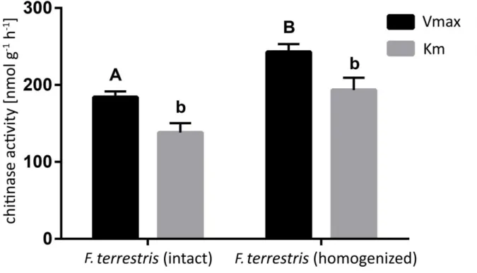
5 As these thecofilosean amoebae, similar to the predominantly known eukaryvores in the Cercozoa, the Vampyrellida, have been shown to be eukaryvorous, we further focused on eukaryvorous protists in terrestrial habitats, by investigating (a) their feeding preferences (b) their physiological requirements to consume eukaryotic prey and (c) their dispersal in terrestrial and freshwater systems. This was achieved by conducting thorough sampling, observing individuals in their unaltered sample (if possible) and performing experiments on feeding preference, chemical sensing, and enzyme production with several omnivorous or eukaryvorous Cercozoa. In laboratory experiments, we could show that the eukaryvorous protist Fisculla terrestris is able to sense and select its preferred prey and produce a battery of enzymes needed to digest cell wall compounds of eukaryotes, such as chitin. F. terrestris preferred fungal prey (in particular Saccharomyces cerevisiae) and only fed to a small extend on algae. To get more insight into the dispersal of eukaryvorous protists in terrestrial systems, we screened metatranscriptomes of different terrestrial habitats for the eukaryvorous Vampyrellida and Grossglockneriidae, showing high dispersal, since they were present in all screened habitats, with up to 3% of total reads.
The phenotypic, phylogenetic and ecologic data on the investigated cercozoan amoebae resulted in a comprehensive characterization of the Thecofilosea and the novel lineage Krakenidae. Based on intensive literature research and a critical evaluation of it, first steps for a phylogeny-based taxonomy of these cercozoan lineages were made. Finally, this thesis provides an evaluation of the hidden diversity of eukaryvorous Cercozoa in terrestrial and freshwater habitats.
6 Zusammenfassung
Der Begriff Protist beschreibt einzellige, eukaryotische Organismen die kein Gewebe ausbilden. Mit einer enormen Mannigfaltigkeit in Morphologie und Ökologie repräsentieren sie die Mehrheit der eukaryotischen Diversität, von der bisher nur ein kleiner Teil bekannt ist. Weltweit verbreitet, in marinen, limnischen und terrestrischen Habitaten, besetzen sie unzählige ökologische Nischen, z. B.
als Primärproduzenten, Osmotrophen, Bakterivoren, Fungivoren, Algivoren, Omnivoren, Prädatoren oder Parasiten z. B. von Tieren und Pflanzen. Probleme im Kultivieren von Protisten sorgten allerdings für eine bevorzugte Erforschung von Bakterivoren und Algen, was zu einem verzerrten Abbild der Protistendiversität führte. Viele „nicht kultivierbare“ Protisten Taxa sind der Forschung noch immer unbekannt.
Deshalb befassten wir uns mit Protisten mit unüblichen Ernährungsweisen (insbesondere bakterivore Lauerjäger und eukaryvore Räuber) in den Cercozoa. Die Cercozoa CAVALIER-SMITH 1998, wurden aufgrund von molekularen Untersuchungen als phylogenetische Entität entdeckt obwohl sie starke Unterschiede in Morphologie und Ökologie aufweisen. Molekulare Untersuchungen deckten eine hohe Diversität in den Cercozoa auf, von der bisher nur ein geringer Bruchteil mit morphologischen Daten verknüpft werden kann. Auf Basis von angelegten Kulturen wurden Zellmorphologie, Ernährungsweise und Zellzyklen von verschiedenen cercozoen Amöben untersucht. Dafür wurden hauptsächlich Lichtmikroskopie und Zeitraffer-Mikrofotographie und auch durch Ultrastrukturaufnahmen Daten erhoben. Genetische Marker, d. h. SSU rDNA und LSU rDNA, wurden sequenziert und für phylogenetische Untersuchungen genutzt, um Rückschlüsse über die Evolution der Cercozoa zu ziehen.
Von einer unbeschriebenen bakterivoren Amöbe wurden sechs Stämme aus deutschen und spanischen Böden isoliert. Kraken carinae gen. nov. sp. nov. ist eine Amöbe, unterteilt in einen schuppentragenden Zellkörper (normalerweise <10 µm im Durchmesser) und ein Netzwerk aus Filopodien (bis zu 0,5 mm im Durchmesser). K. carinae ist einer der seltenen Lauerjäger der Cercozoa, sie bewegt sich nur selten und erbeutet Bakterien, die mit ihrem Netzwerk aus Filopodien in Kontakt kommen. Anders als andere amöboide Vertreter der Cercozoa, die üblicherweise ihre Filopodien nutzen, um ihre Beute zum Zellkörper zu ziehen und dann zu phagozytieren, ingestiert K. carinae Bakterien direkt am Kontaktpunkt der Beute mit den Filopodien und transportiert diese dann intrazellulär zum Zellkörper um sie dort zu verdauen. SSU rDNA Phylogenie zeigte eine nähere Verwandtschaft mit den Cercomonadida auf, wenn auch nur mit mäßiger statistischer Unterstützung. Ein weiterer Anlauf, diesmal mit verketteten SSU rDNA und LSU rDNA Sequenzen, bestätigte die Verwandtschaft zu den Cercomonadida, insbesondere mit den Paracercomonadidae, mit erhöhter (aber dennoch nur moderater) statistischer Unterstützung. Allerdings bleiben die genaue Verwandtschaftsverhältnisse von Kraken carinae immernoch unklar, da die Ultrastrukturdaten konträr zu den phylogenetischen Ergebnissen, auf eine nähere Verwandtschaft mit den Imbricatea hinweisen, da Kraken carinae genau wie sie Schuppen auf dem Zellkörper trägt.
Durch die Kombination von intensiver Literaturrecherche mit phylogenetischen Untersuchungen von Lecythium (HERTWIG et LESSER, 1874) und seiner Familie, der Chlamydophryidae (DE SAEDELEER 1934) konnten wir die unklare Taxonomie von Gattungen wie (Plagiophrys, Lecythium, Rhizaspis, …) enträtseln. All diese Amöben haben gemeinsam, dass sie eine hyaline flexible Schale tragen und verästelnde und anastomisierende Filopodien aufweisen, aber haben auch klare Unterschiede zueinander. SSU rDNA Phylogenien haben die phänotypischen Unterschiede zwischen den Gattungen
7 widergespiegelt, bargen aber auch Überraschungen: Die Gattungen Lecythium (Novel Clade 4) und Rhizaspis (Tectofilosida) waren beide polyphyletisch und mussten daher geteilt werden. Daher wurden Fisculla gen. nov. (Tectofilosida) von Lecythium und Sacciforma gen. nov. (Cryomonadida) von Rhizaspis abgetrennt. Weiterhin zeigen diese Schalenamöben, ähnlich wie die meistbekannten Eukaryvoren in den Cercozoa, die Vampyrellida, eukaryvores Verhalten. Deshalb führten wir weitere Studien über die Ökologie von eukaryvoren Protisten in terrestrischen Habitaten durch. Vor allem geben wir Einsicht auf (a) ihre Nahrungspräferenzen, (b) den physiologischen Voraussetzungen, um eukaryotische Beute zu konsumieren und (c) ihrer Verbreitung in terrestrischen und limnischen Systemen. Dazu wurden umfassende Probenahmen, gefolgt von Experimenten durchgeführt, welche Fütterungsversuche und Experimente über die chemische Wahrnehmung und Enzymproduktion mit diversen omnivoren oder eukaryvoren Cercozoa umfassten. Wir konnten zeigen, dass die eukaryvore Amöbe Fisculla terrestris fähig ist, die Anwesenheit ihrer bevorzugten Beute (Saccharomyces cerevisiae) wahrzunehmen und darauf zu selektieren. Außerdem produzieren sie eine Auswahl an Enzymen zum Abbau der Zellwand der Beute, wie z. B. Chitin. Um mehr Einblick auf die Verbreitung von terrestrischen Eukaryvoren zu erhalten, durchsuchten wir Metatranskriptom-Datenbanken von verschiedenen terrestrischen Habitaten nach den eukaryvoren Vampyrellida und Grossglockneriidae, die auf eine weite Verbreitung von Vampyrellida und Grossglockneriidae hinwiesen, da diese nicht nur in allen überprüften Habitaten nachgewiesen werden konnten, sondern auch bis zu 3% der absoluten Sequenzen ausmachten.
Die phänotypischen und phylogenetischen Daten der untersuchten Amöben der Cercozoa resultieren in einer umfassenden Charakterisierung der Thecofilosea und der neuen Familie Krakenidae. Auf Grundlage einer intensiven Literaturrecherche und einer kritischen Auswertung dieser im Kontext der selbstständig durchgeführten Arbeiten wurden erste Schritte in Richtung einer phylogeniebasierten Taxonomie gemacht. Diese Doktorarbeit umfasst weiterhin eine Diskussion über die noch unerforschte Diversität der Cercozoa in terrestrischen und limnischen Habitaten.
8
Introduction
Protists
The term protist defines an informal grouping of unicellular eukaryotes that do not form tissues. Protists represent the vast majority of eukaryotic diversity of which only a small fraction is yet known. Despite their estimated 60.000 - 300.000 species (Foissner 2008; Mora et al. 2011) with a tremendous diversity in ecology and morphology, they were traditionally assigned to the same single eukaryotic kingdom, Protista in the Eukaryota, next to plants, animals and fungi (Haeckel 1866; Whittaker 1969). The Protista accommodated therefore organisms of fundamentally different lifestyles, like autotrophic and mixotrophic algae, heterotrophic bacterial grazers and parasites, but also organisms of diverse appearances, like ciliates, flagellates, naked amoebae and testate amoebae. That is why protists were shared as a field of research among zoologists, botanists and mycologists. The most animal- like protists (i.e. heterotrophic protists feeding by means of phagocytosis) were called protozoa (proto= first; zoa= animals) and studied by zoologists, whereas pigmented protists were adopted by the botanists as protophytes (phytes= plants) and fungi-like protists (like the fruiting body forming myxomycetes or the osmotrophic filamentous oomycetes) were studied by mycologists. Researchers were well aware that even morphological highly similar taxa (e.g. the dinoflagellates or euglenids) comprised heterotrophic protists and also photosynthetic algae, but were unable to resolve this issue in a widely accepted taxonomy.
Since the 18
thand 19
thcentury where protists were most often studied by light microscopy, novel methods were established: In the early 20
thcentury electron microscopical techniques were developed to acquire more detailed morphological data for protist taxonomy, in the late 20
thcentury molecular methods enabled the comparison of genetic markers for phylogeny.
The molecular methods led finally to a widely accepted consensus in protist taxonomy (Adl
et al. 2005, Adl et al. 2012, Baldauf 2008). Protists turned out to be paraphyletic, instead of
being separated kingdoms, with multicellular eukaryotes nestled in between. Although
ciliates show monophyletic origin (Lynn and Sogin 1988; Sogin and Elwood 1986), flagellates,
naked and testate amoebae are dispersed all over the eukaryotic tree of life showing
paraphyly and polyphyly. For instance, testate amoebae evolved independently in at least
three different lineages, the Amoebozoa, Cercozoa and Stramenopiles (Kosakyan et al. 2016;
9
Nikolaev et al. 2005; Wylezich et al. 2002). The search for the last eukaryotic common ancestor is still ongoing and a highly discussed controversy. Currently it is only accepted that the eukaryotic ancestor was probably a heterotrophic flagellate of unknown affiliation, but concepts differ about the most basal protist taxon (Baldauf 2008; Stechmann & Cavalier- Smith 2002). Cavalier-Smith (1981) established the Archezoa, but this grouping of lineages he believed to be the most basal protists turned out to be an artifact of long branch attraction (Philippe and Germot 2000). Based on rare genetic events like gene fusions and fissions it is assumed that the eukaryotes are divided into two groups, the unikonts (Opisthokonts and Amoebozoa) and bikonts (the remaining eukaryotic diversity). However, still no consensus whether to root the eukaryotes in the unikonts or bikonts could be achieved (Baldauf 2008). Nevertheless, it is widely accepted that during eukaryotic evolution several lineages lost independently their flagella and evolved locomotion by amoeboid movement and/ or gained (or secondarily lost again) autotrophy by endocytobiosis, leading to the intermingled ecology and physiology, sometimes even between closely related protist taxa (Nowack 2014; Rogers et al. 2007; Stechmann & Cavalier-Smith 2002).
Heterotrophic protists in soil biology and their feeding types
Protists, especially in soil ecology, were commonly considered to represent the major bacterial grazers, thereby channeling the carbon flow to higher trophic levels (Bonkowski 2004, Crotty et al.
2011, de Ruiter et al. 1995, Hunt et al. 1987).
However, these assumptions were derived from simple model calculations, based on laboratory experiments with few selected species. Although molecular methods gradually revealed the enormous phenotypic and genetic diversity of heterotrophic protists, models on protist functional roles have not changed in soil biology (Holtkamp et al. 2011, Banašek-Richter et al. 2009). For instance Glücksman et al. (2010) and Weisse et al. (2001) showed that prey (bacteria) communities have been altered by protist grazing in a species-specific manner, but still by far most information on the impact of protist grazing have been obtained in studies either with whole microbial communities in which measured effects can not be traced back to the causing species or in abstract assemblages with just one or few selected organisms. Moreover, such studies were most often conducted only with exclusively bacterivorous protists. The non-bacterivorous protists have been addressed in very few, most often taxonomic studies (Bass et al. 2009; Berney et al. 2013; Foissner 1980; Petz and Foissner 1985). Their ecology and functional importance is often not known.
10
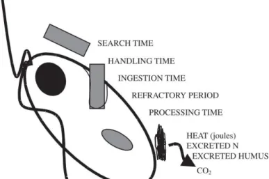
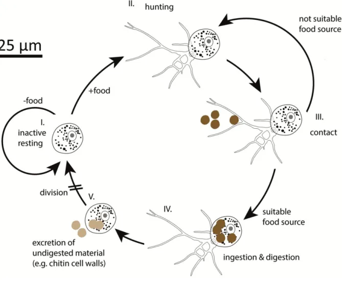
Heterotrophic feeding in general needs many evolutionary adaptations. The functional response of a heterotrophic protist
is basically composed of search time, handling time, ingestion and digestion (Fig. 1). The presumed most widespread and maybe simplest method of preying (eukaryotic and prokaryotic) cells is the process of phagocytosis: this includes the incorporation of the whole prey into a food vacuole, adjusting the milieu to unsuitable conditions (e.g. increased or
decreased pH; Laybourn-Parry, 1984) and bombarding this environment with a battery of enzymes degrading various compounds (Bowers and Korn 1973; Laybourn-Parry, 1984). The two most important advantages of this method are a probably low cost for degrading enzymes, as they do not have to be excreted, where they would be highly diluted and once ingested prey (usually) can be digested while the predator already can search for a novel food source.
Feeding on eukaryotic cells can be viewed as an additional evolutionary hurdle, as it requires a series of adaptations: First of all (and maybe most important), food size relative to its own is a crucial factor. Many eukaryotic cells have evolved (sometimes inducible) ways to prevent their ingestion by increasing their size by the formation of colonies or filaments (Lampert et al. 1994; Van Donk et al. 2011).
The protection of the cell body by a rigid covering is another common defense strategy that repeatedly evolved. For instance such coverings are e.g. cell walls (usually completely enclosing the cells, composed of chitin or cellulose, being found mainly in green algae), tests (usually with one or two openings, most often built by scales or by agglutinated foreign materials, like sand grains embedded in an organic cement, most prominently known from testate amoebae) or frustules (two rigid, porous, silica-composed and overlapping sections covering the cell, found in diatoms). Those coverings may even (a) bear spines, rods or horns
Figure 1: Simplified feeding process of a heterotrophic protist.
Extracted from Adl and Gupta 2006
11
which may also prevent the ingestion by predators or (b) be enveloped by mucilage which may prevent adhesion of predatory cells or even impede prey recognition (Van Donk et al.
2011).
Next to such morphological defense mechanisms, protists also evolved behavioral defense strategies that might be inducible and aggressor-specific, like (a) the production of chemical compounds, e.g. toxins; (b) flight, often aided by special organelles, like extrusomes or trichocysts, that can be ‘fired’ to the possible predator; (d) metabolic movement that enables already ingested prey to stretch and burst food vacuoles, like in
Euglena mutabiliswhen being consumed by Actinophrys sp. or a yet undescribed species in the Leptophryidae (Hausmann 1978; Potin et al. 1999, Van Donk et al. 2011; Hess pers. comm.; own observations unpublished).
Some specialized predators co-evolved strategies to overcome such defense mechanisms.
These adaptations are often highly specialized: Large cells, like filaments, even with a thick cell wall might be an easy prey if the predator is adapted by morphology and behavior, e.g.
some ciliates (e.g. Grossglockneriidae; Foissner 1980, 1999) are equipped with a feeding tubus for the lysis of fungal cell walls; or some cercozoans (e.g. Vampyrellida;
Viridiraptoridae; Hess et al. 2012, Hess & Melkonian 2013) are able to perforate algal or fungal cell walls by extracellular enzymatic degradation. Moreover, trichocysts or extrusomes might also be used by attacking cells (Hausmann 1978).
Research in protistology is astonishing and still full of surprises; we therefore focus in this thesis on heterotrophic protists that show interesting and unusual feeding types.
Culturing heterotrophic protists and its difficulties
In early times of protistology, protists were predominantly studied by direct observations of
fresh sampled material. Marine and freshwater samples do not require any preparation thus
being intensively studied (Hertwig & Lesser 1874; Penard 1890). Soils however, due to being
opaque, can not be observed directly but need preparation like dilution. Diverse methods
have been developed, like the most probable number (MPN; Darbyshire et al. 1974) or liquid
aliquot method (LAM; Butler and Rogerson 1995), all of which have advantages and
disadvantages. Still today, there is no method that enables an objective unbiased view on
12
soil microorganisms and soil protists are mainly studied by indirect observation of cultured material extracted from soil samples. Despite the abstract nature of such observations, culturing, i.e. the long term maintenance of single species or strains in an artificial and controlled environment, enabled researchers to study complex life cycles, for instance the amoebozoan genus Pelomyxa GREEFF 1874, comprises about 20 described species of which the validity is still highly discussed (Frolov et al. 2004, 2005, 2006 and 2011). It is currently not clear whether all described species are just different stages of the life cycle of Pelomyxa
palustris (the type species of the genus; Chapman-Andresen 1978, 1982), or how many true Pelomyxa species exist (2004, 2005, 2006 and 2011). Another example for protists withcomplex life cycles are the fruiting-body forming amoebae where in many cases the identity of fruiting bodies and trophozoites can not be assigned to each other (Tice et al. 2016). Even in well studied amoebae, like the Acanthamoebidae, novel life history stages (like fruiting- bodies) can be discovered (Tice et al. 2016). Finally, cultures enable the conduction of laboratory experiments, such as an alteration of environmental variables, i.e. biotic or abiotic, enabling conclusions about species autecology, i.e. preferred food sources, necessity of certain chemical compounds in the environment and in general conclusions might be drawn about several biological aspects, such as their physiology and ecology.
For bacteria it is estimated that barely 1% of their diversity can be cultured (see for instance:
Ferrer et al. 2009; Lee et al. 2010). Although many (bacterivorous) protist lineages are easily culturable by basically transferring single individuals into water with a carbon source, the vast majority has highly specific requirements (Page 1976). Therefore it would not be surprising to find a similar pattern for protists; and indeed culturing protists differs in the degree of difficulty and is strongly depended of the requirements of the targeted species.
Besides abiotic conditions like medium composition and temperature, it seems obvious that heterotrophic protists need a suitable food source in a sufficient concentration: e.g.
fungivorous protists need fungi; bacterivorous protists need bacteria and so on. Culturing
bacterivorous protists is often quite easy, often non-toxic strains of bacteria are added or
maybe even easier, co-transferred bacteria are fed with solved carbon, the bacteria then
grow fast, even under nutrient limited conditions and subsequently can be grazed by the
protists.
13
In contrast to often easily culturable and fast growing bacteria as food source, the culturing of protists that have to be fed with comparatively slow growing and difficult to maintain eukaryotes as food source harbours additional difficulties: At the time of observation of the desired protist predator in a natural sample, the required food source has to be already available in a suitable concentration that does not only enable the growth of the predator but further the sustained growth of the food source. Since eukaryotes are more complex and usually larger than bacteria, they grow slow in comparison to many bacteria and can not be easily stored by drying, cooling or freezing (although research is currently making progress in that field). The amount of cultures maintained by single researchers are therefore very limited, this emphasises the importance of culture collections in protistology. Moreover, many eukaryotes, like algae and fungi (whether as food source or the desired cultured protist itself) produce metabolites that in cultures accumulate and finally may lead to extinction of one or more co-cultured eukaryotes; a frequent subcultivation is therefore necessary, increasing operating expenses (Andersen 2005).
However, according to own experiences, it is difficult to find a suitable food source. If the
process of feeding in a natural sample is not observed, which especially in opaque
environments like soils is difficult, researchers might not even know what the presumed
eukaryvorous predator actually feeds on. Knowledge on protist taxonomy and phylogeny
might enable a ‘good guess’ about the required needs of the desired protist, but often those
needs are not only lineage specific but furthermore species or strain specific requiring trial
and error. For instance there are generalists, like
Amoeba spp. that feed on bacteria orvarious eukaryotes (Prescott and James 1955) and specialists, like the Vampyrellida,
Viridiraptoridae or the endobiotic phytomyxids. For instance the SSU-sequences of the
Vampyrellida reveal, whether the desired protist groups within fungivores, “filamentous
algae feeders” or “unicellular algae feeders” clades and enables to draw conclusions about
the feeding preferences of the sequenced Vampyrellida species (Hess et al. 2012); the
parasitic phytomyxids show co-evolution with their hosts, although host shifts seem to occur
(Neuhauser et al. 2014); and the monophagous
Hatena arenicola feeds not only on a singlespecies of
Nephroselmis algae but even only on distinct genetic lineages (Okamoto andInouye 2006; Yamaguchi et al. 2014).
14
However, long-time maintained cultures enable research on a professional basis and are therefore imperative for taxonomic and autecological research.
Cercozoa
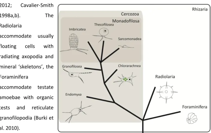
The first discovered protist phylum Cercozoa CAVALIER-SMITH 1998, accommodates morphological and ecological divergent organisms that were found to be closely related based on molecular analyses. Together with the Radiolaria and Foraminifera, both with a more conserved morphology, they constitute the eukaryotic supergroup Rhizaria (Adl et al.
2012; Cavalier-Smith
1998a,b). The
Radiolaria
accommodate usually floating cells with radiating axopodia and mineral ‘skeletons’, the Foraminifera
accommodate testate amoebae with organic tests and reticulate granofilopodia (Burki et al. 2010).
The divergent Cercozoa consist predominantly of naked amoebae, flagellates and amoeboflagellates (Bass et al. 2009a,b; Hess & Melkonian 2013; Hess et al. 2012). Nestling between those, it comprises several polyphyletic testate amoeba lineages: e.g. the order Euglyphida with tests made of siliceous plates (Cavalier- Smith 1998a, b), the family Rhizaspididae (being here renamed Rhogostomidae) with organic thecae (Howe et al. 2011) and the family Pseudodifflugiidae with tests composed of agglutinated foreign material embedded in an organic cement, the latter branching in the Thecofilosea (Wylezich et al.
2002). Environmental surveys of terrestrial, limnic and marine systems have shown a high genetic diversity of Cercozoa (and particular the little studied Thecofilosea) that can not yet
Figure 2: Hypothesized and strongly simpflified sketch of cercozoan evolution (after Cavalier-Smith and Chao 2003).
15
be connected to morphological and autecological data (Bass and Cavalier-Smith 2004), leading to our first hypothesis that there are still many lineages in Cercozoa to be discovered.
The Thecofilosea were established by Cavalier-Smith and Chao (2003) to accommodate thecate flagellates and amoebae. On a morphological basis it was assumed that the thecate amoeba family Chlamydophryidae might branch as well in the Thecofilosea, but molecular data was lacking (Howe et al. 2011).
The family Chlamydophryidae was established by de Saedeleer (1934) (then as a subfamily of Gromiidae) to accommodate genera like
Chlamydophrys CIENKOWSKI, 1876 and LecythiumHERTWIG ET LESSER, 1874. Species of both genera are all filose amoebae with a hyaline test or theca, but they divide in different ways (Meisterfeld 2002; for a discussion see Chapter 3).
The predominantly bacterivorous Chlamydophrys could easily be cultured and was therefore well characterized long ago (Belar 1921). In contrast, no
Lecythium (Hertwig and Lesser1874) was successfully cultured, leading to sometimes poorly described species and therefore a confusing taxonomy. We propose therefore the second and third hypotheses that
Lecythium, which has been reported to feed on algae, but being unculturable, can becultured with the right techniques and further groups on basis of molecular data in the Thecofilosea.
What we know about fungivores and hypothesized evolutional adaptations
A series of studies has been conducted by Chakraborty et al. (1982, 1983, 1985) showing that several omnivorous species of the Amoebozoa are able to feed on spores and yeasts and further are even able to reduce the colonization of root by mycorrhizal fungi, suggesting that fungi-feeding protists significantly affect the rhizosphere system.
O
nly few specialised fungivorous protists have been described (predominantly the Grossglockneriidae and terrestrial lineages in the Vampryrellida (Cercozoa); Bass et al. 2009;
Foissner 1980). Morphology of those lineages is well characterized, but there are only few
available SSU sequences and nearly nothing is known about their dispersal and feeding
preferences.
16
The terrestrial Vampyrellida have been repeatedly reported from agricultural fields all over the world (predominantly of Germany, Australia and Japan but also reports of Iran and Canada; Hess et al. 2012; Homma et al. 1979; Homma and Ishii 1984). Data about their true nutrition in the field is scarce. In laboratories the amoebae could be cultured mainly on spores of fungi, predominantly with Botryotinia fuckeliania, Gaeumunnomyces graminis and
Cochliobolus sativus and even on the oomycete Pythium debaryanum (Pakzad 2003; Pakzadand Schlösser 1998; Chakraborty and Old 1982; Chakraborty et al. 1983).
Feeding on hyphae, as they are large filaments that can not be completely enveloped by a food vacuole, seems more difficult to achieve. Next to the spore feeding Vampyrellida, some have been reported to feed on fungal hyphae, probably by external degradation as found in filamentous algae feeding Vampyrellida; Hess et al. 2012; Homma et al. 1979; Homma and Ishii 1984). The Grossglockneriidae is a family of ciliates with several described genera (Grossglockneria, Mykophagophrys and Pseudoplatyophyra) with specific oral structures enabling them to feed on filament forming fungi (Foissner 1980; Lynn et al. 1999; Dunthorn et al. 2008). They have been repeatedly studied from Austrian forest litters, out of twelve tested fungi
Grossglockneria only fed on three: Aspergillus sp., Mucor mucedo and a notfurther determined Mucoraceae sp. (Foissner 1980; Petz et al. 1985). The latter two representing typical “sugar fungi”, those fast growing fungi are usually the first to colonize novel food sources and as a trade-off produce only few secondary metabolites (Frankland 1998; Torres et al. 2005).
Nevertheless, in comparison to the very few studies focussing on fungivores, there are more studies, old and recent, targeting especially algivorous protist lineages. The question arises why algivores are more intensely studied than fungivores.
There are two possible explanations: [1] fungivores are just rare, less abundant and less diverse than e.g. algivores; [2] soil hampers work with protists and prevents direct observation, thus fungivores are just not recognized. Resulting in the forth of our hypotheses: Protist functions are understudied in terrestrial systems; especially the functional roles of fungivores have been underestimated.
The few studied terrestrial relatives of algivorous protists most often show fungivorous
feeding habits. It is therefore likely, that the shift from being a freshwater algivorous protist
17
to a terrestrial fungivorous protist (and maybe vice versa) is accompanied by few and/ or simple adaptations.
The shift from freshwater to terrestrial (soil) habitats might include the following adaptions:
(1) Since soils are often referred to as harsh environments with a huge variability in moisture and therefore salinity, protists should be able to cope with droughts. The ability to form cysts (a life history stage that usually is defined as a partial dehydration and the secretion of a more or less rigid cell envelope, usually composed of cellulose, chitin, proteins or a combination of those) is necessary in terrestrial protists as it enables the survival in dry periods or heatwaves that occur frequently (see Adl and Gupta 2006).
The development of tests was seen as an additional adaptation to dry conditions, since the test might reduce water loss and therefore represent an advantage to dry environments. Accordingly a smaller test opening should be of advantage in drier environments. Schönborn (1992) and Bobrov and Mazei (2004) have shown that the size of the opening of the test varies and can be adapted to drought within few generations. Series of physiological changes are likely, including an adaptation to rapid changing osmotic pressure and very different chemical compositions of the environment;
(2) As space is limited in soils many soil protists show a reduction in size compared to their freshwater relatives. For instance,
Parvularia is smaller than its closestfreshwater relative
Nuclearia (Dirren et al. 2014; Torruella pers. comm.), terrestrialGlissomonads are smaller than the freshwater inhabiting Viridiraptoridae (Hess and Melkonian 2013). Behavioural adaptations, especially in locomotion, might be of advantage when changing to a space limited environment like soils. For instance the exclusively limnic
Vampyrella predominantly float, whereas the closely related Theratromyxa and Platyreta mainly creep (Bass et al. 2009, Hess et al. 2012), similarevolutionary process seem to be found in Nuclearia and the closely related Parvularia although there is not much data yet (Dirren et al. 2014; Dirren and Posch 2016;
Torruella pers. comm.), the described Viridiraptoridae show the ability to swim, a
behaviour not known from the terrestrial glissomonads but one (i.e.
Proleptomonas faecicola, see Hess & Melkonian 2013).18
(3) Since most soil systems harbour less algae than fungi (Damon et al. 2012, Cutler et al.
2013), algivorous protists might take advantage of novel food sources like fungi (i.e.
becoming fungivorous), this should lead to an alteration of the set of digestive enzymes, since fungi mainly build their cell walls from chitin, in contrast to (green-) algae that use mainly cellulose, it is likely that the composition of the enzyme cocktail needed to degrade cell walls changes either in concentrations of compounds, by evolving novel enzymes or by obtaining enzymes by lateral gene transfer from the prey itself (Adams 2004; Popper et al. 2011; Xu et al. 2016).
Accordingly we hypothesize that life in terrestrial habitats requires special adaptations in
‘aquatic‘ protists, and we may detect adaptations of terrestrial protists, for example in the
predominantly algivorous Tectofilosida.
19
Aims
The central aim of this thesis was to give insight into eukaryvorous protists, in particular Cercozoa, in freshwater and terrestrial systems, in order to contribute to a deepened understanding of cercozoan evolution. We cultured, investigated and characterized selected
‘unculturable’ protists of the phylum Cercozoa in terms of morphology, behaviour and phylogeny.
The following hypotheses were proposed:
1. Many still unknown lineages exist in the Cercozoa.
2. The algivorous genus Lecythium groups in the Thecofilosea.
3. Significant culturing efforts are needed to link protist morphotypes and functions to environmental sequences.
4. Protist functions are understudied in terrestrial systems; especially the functional roles of fungivores have been underestimated.
5. Life in terrestrial habitats requires special adaptations in ‘aquatic‘ protists, and we may
detect adaptations in terrestrial protists that are lacking in closely related freshwater
inhabitants, for example in the predominantly algivorous Tectofilosida.
20
Chapters in three parts
Part 1 -
Characterization of the unusual bacterivorous amoeba Kraken carinae.
Chapter 1: A Novel Lineage of ‘Naked Filose Amoebae’;
Kraken carinae gen. nov. sp. nov.(Cercozoa) with a Remarkable Locomotion by Disassembly of its Cell Body.
Chapter 2: Cercomonad or archaic Imbricatea? On the hunt for the true taxonomy of the scale-bearing
Kraken (incertae sedis, Cercozoa, Rhizaria): Combining ultrastructure data anda two-gene (SSU + LSU) phylogeny.
Part 2 -
Eukaryvorous amoebae of the Thecofilosea, Cercozoa.
Chapter 3: Description of Lecythium terrestris sp. nov. (Chlamydophryidae, Cercozoa), a Soil Dwelling Protist Feeding on Fungi and Algae.
Chapter 4: A bowl with marbles: Revision of the thecate amoeba genus
Lecythium(Chlamydophryidae, Tectofilosida, Cercozoa, Rhizaria) including a description of four new species and an identification key.
Chapter 5: Shedding light on the polyphyletic genus Plagiophrys: The transition of some of its species to
Rhizaspis (Tectofilosida, Thecofilosea, Cercozoa) and the establishment of Sacciforma gen. nov. (Cryomonadida, Thecofilosea, Cercozoa).Chapter 6: Polyphyly in the thecate amoeba genus
Lecythium (Chlamydophryidae,Tectofilosida, Cercozoa), redescription of its type species
L. hyalinum, description of L.jennyae sp. nov. and the establishment of Fisculla gen. nov. and Fiscullidae fam. nov.
Part 3 -
Eukaryvorous protists, their capabilities and dispersal.
Chapter 7: What does it take to eat a fungus? A case study with the eukaryvorous amoeba
Fisculla terrestris.Chapter 8: The soil food web revisited: Diverse and widespread mycophagous soil protists.
21
Results
Part 1 - Characterization of the unusual bacterivorous amoeba Kraken carinae.
Chapter 1: A Novel Lineage of ‘Naked Filose Amoebae’; Kraken
carinae gen. nov. sp. nov. (Cercozoa) with a Remarkable Locomotion
by Disassembly of its Cell Body.
22
23
24
25
26
27
28
29
30
31
32
33
Chapter 2: Cercomonad or archaic Imbricatea? On the hunt for the true taxonomy of the scale- bearing Kraken (incertae sedis, Cercozoa, Rhizaria):
Combining ultrastructure data and a two-gene
(SSU + LSU) phylogeny.
34 Authors Kenneth Dumacka, Alexander P. Mylnikovb & Michael Bonkowskia
Corresponding author Kenneth Dumack
Phone: +49-(0)221-470-6635 Fax: +49-(0)221-470-5038
a University of Cologne, Department of Terrestrial Ecology, Institute of Zoology, Zülpicher Str. 47b, 50674 Köln, Germany
b Institute for the Biology of Inland Waters, Russian Academy of Sciences, Borok, Yaroslavskaya Obl.
152742, Russia
kenneth.dumack@uni-koeln.de, ap.mylnikov@rambler.ru, m.bonkowski@uni-koeln.de
Title
Cercomonad or archaic Imbricatea? On the hunt for the true taxonomy of the scale-bearing Kraken (incertae sedis, Cercozoa, Rhizaria): Combining ultrastructure data and a two-gene (SSU + LSU) phylogeny.
Keywords
Sarcomonadea, Imbricatea, scales, flat cristae, cercomonads, electron mircoscopy Abstract
The genus Kraken represents a morphologically distinct lineage of filose amoebae within the Cercozoa. Currently only a single species, Kraken carinae, has been described. SSU phylogeny showed an affiliation with the Cercomonadida, branching with weak support at its base, close to Paracercomonas, Metabolomonas, and Brevimastigomonas. Light microscopical analyses showed several unique morphological and behavioral features of the genus Kraken, but ultrastructure data was lacking. In this study, K. carinae has been studied by electron microscopy, this data conjoined with a concatenated SSU and LSU phylogeny was used to give more insight into Kraken taxonomy.
The data confirmed the absence of flagella, but also showed novel characteristics, like the presence of extrusomes, osmiophilic bodies, mitochondria with flat cristae and, surprisingly, the presence of single-tier scales, which are carried by cell outgrowths, much of what is expected of the last common ancestor of the class Imbricatea. The phylogenetic analyses however confirmed previous results, indicating Kraken carinae as a sister group to Paracercomonas within the Sarcomonadea with an increased but still moderate support of 0.98/63. Based on the unique features of the Kraken we establish the Krakenidae fam. nov. that we, due to contradicting results in morphology and phylogeny, assign incertae sedis, Cercozoa.
35 Introduction
Recently, the genus Kraken was described as a new lineage of cercozoan filose amoebae inhabiting soil and freshwater (probably sediment) ecosystems (Dumack et al. 2016a). Small subunit (SSU) phylogeny indicated with low support an affiliation with Filosa (Cercozoa), in particular to Paracercomonas, Metabolomonas, and Brevimastigomonas (Paracercomonadidae, Cercomonadida).
Cercozoa is a phylum of high morphological diversity, comprising naked amoebae (Hess et al. 2012), testate amoebae (Dumack et al. 2016b,c) but also flagellates and amoeboflagellates (Bass et al.
2009a,b; Hess and Melkonian 2013). The Cercomonadida are a taxon of relatively well studied amoeboflagellates with quite conserved morphology (Bass et al. 2009a). They are currently assigned to the Sarcomonadea, CAVALIER-SMITH, 1993. The Sarcomonadea are, next to the Thecofilosea and Imbricatea, one of the as yet known major classes of Filosa, Cercozoa. The latter two mainly accommodate test- or scale-bearing protists, whereas the Sarcomonadea were established to unite cercozoan free-living, heterotrophic and naked (amoebo-) flagellates (Cavalier-Smith 1993), although they might be of polyphyletic origin (Cavalier-Smith and Karpov 2012). They comprise the orders Pansomonadida, Glissomonadida and Cercomonadida (Cavalier-Smith and Karpov 2011). The latter two are very common, diverse and abundant in terrestrial and freshwater systems and are commonly referred to as the predominant protistan bacteria grazers (Glücksman et al. 2010).
Light microscopic observation of Kraken did not specifically support the affinity to cercomonads, although its dimensions and general appearance was not inconsistent with the diversity of body forms within the Cercomonadida (Dumack et al. 2016a). However, all as yet described species of the Cercomonadida are biflagellate, naked cells and some may be able to form short filopodia. The Kraken in contrast bears no flagella but has instead a huge network of filopodia that exceeds the size of the cell body by a factor of 50. Kraken differs therefore markedly from the currently known Cercomonadida. Since SSU phylogeny could not resolve its direct affiliation, ultrastructure as well as more genetic data, enabling a more detailed comparison with other Cercomonadida and other cercozoan amoebae or amoeboflagellates, are desirable.
Two-gene phylogenies have been shown to resolve low support in some questionable clades of single gene phylogenies (Chantangsi et al. 2010; Wylezich et al. 2010). Although there is a considerable amount of SSU data of cercozoans available, only few strains have been characterized by additional genes, mostly large subunit sequence (LSU) data (Chantangsi et al. 2010; Wylezich et al. 2010).
Additionally various ultrastructural traits, obtained by SEM (e.g. overall cell shape, scales and tests) or TEM (e.g. flagellar apparatus) display group-specific patterns and may reveal valuable
36 morphological characteristics for comparison (Cavalier-Smith and Karpov 2012; Hess & Melkonian 2014).
We therefore decided to combine a concatenated SSU and LSU phylogeny with ultrastructure data to get more insight into Kraken taxonomy. In particular, two questions arise: Does a concatenated (SSU+LSU) phylogeny confirm the phylogenetic placement of Kraken carinae presented in Dumack et al. (2016a) and does it further provide an enhanced support? Will ultrastructure data show characteristics typical for cercomonads, its class Sarcomonadea or other cercozoans?
We were in particular interested to find a possible rudimentary flagellar apparatus, and to obtain data of the cell (surface) architecture. Our data show the ultrastructure of a very unusual filosan amoeba and may lead to a re-interpretation of cercozoan evolution.
Results
Ultrastructure of Kraken carinae Cell shape and content
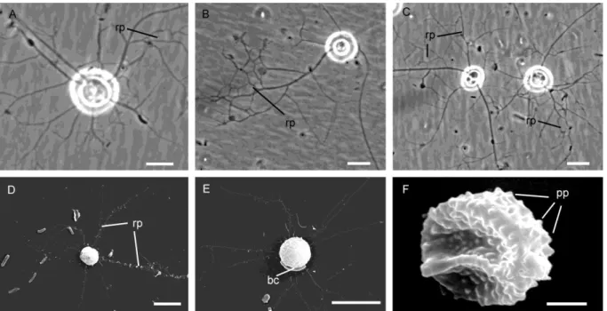
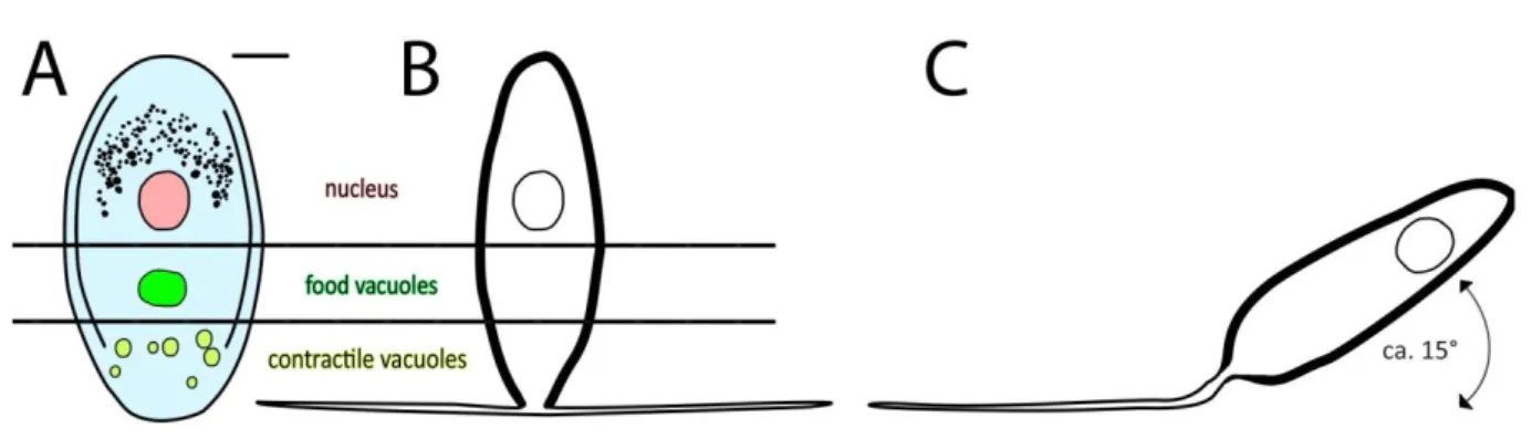
The light microscopical observations presented in Dumack et al. (2016a) correspond to the ultrastructural data. Briefly, the overall shape of the cell body is spherical with an invagination (Dumack et al. 2016a called it ‘ring-like structure’) at the basal end from which branching and anastomosing filopodia arise (Fig. 1A-E). Cysts and elongated cells (during cell body transport) have not been studied in detail and are not presented here. The fragile cells lost filopodia during centrifugation and therefore the cell body and filopodia are shown here separately.
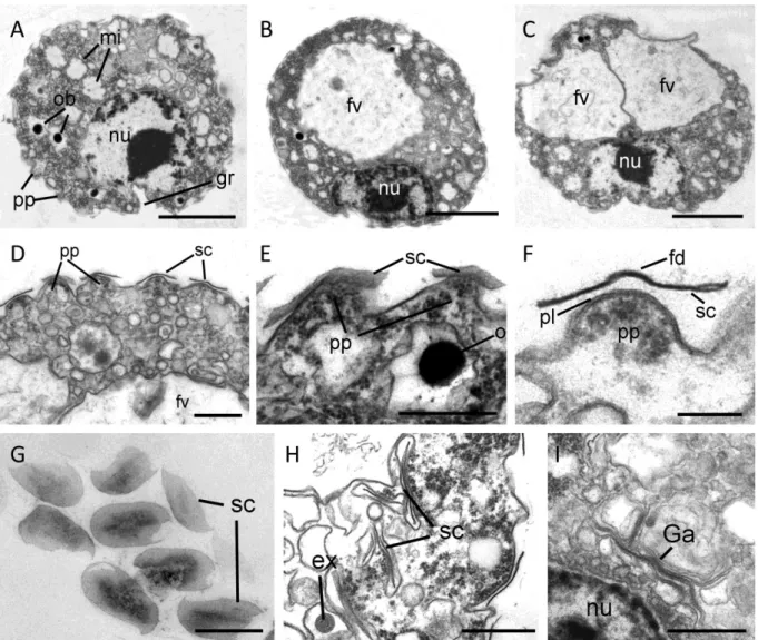
The cells contain a single, eccentric nucleus (2.4 – 3.2 µm), usually located at the basal end of the cell body close to the invagination, with a homogenously granular nucleolus in its center (Fig2A-C). The invagination reaches usually into the cell body, deforming the nucleus spherical shape. The shape of the invagination can be seen in the selected serial sections of a single cell (Fig. 3A-F). Golgi stacks are found in association to the nucleus (Fig. 2I, 4A). The cells contain various amounts of spherical mitochondria (0.5 - 0.6 µm) with an electron-translucent central matrix (Fig.2A). The plate-like (flattened) cristae lie on the internal periphery of the mitochondria (Figs. 4B, 4C). Sections of tips of the cristae are sometimes roundish (Fig. 4C, arrow). The contractile vacuole has irregular cell walls during systole (Fig. 4D).
37 In the periphery of the cell body extrusomes are located (Fig. 4). The extrusomes are reminiscent of the kinetosomes of cercomonads and glissomonads (Mylnikov, 1988; Fig. 4G,I). They are enveloped by a small vesicle and consist of a capsule with an internal cylinder. After discharging the cylinder is partly exposed (Fig. 4J).
Additionally to the medium stained extrusomes, the cells contain a huge amount of small vesicles which are either electron-translucent, resembling reserve granules (Fig. 4L) or highly stained, resembling osmiophilic bodies (Fig. 2A, 2E) and bundles of microfilaments (Figs 5A,B). Kraken cells do not contain flagella, kinetosomes or a flagellar root system.
The filopodia are more electron-translucent than the cell body. Aggregations of osmiophilic amorphous material, small electron-translucent vesicles and microtubules were seen inside the filopodia (Figs 5D-G, selected serial sections). Additionally the filopodia contain small vacuoles with single bacteria (Fig. 4E). According to light microscopical observations on the feeding process of the Kraken, these bacteria were caught immediately before the fixation and were in the process of transportation to the cell body. Within the cell body one (rarely two) food vacuoles are located in the apical end (for the ejection of such a food vacuole during defecation, see Fig. 4F). No endocytobiotic bacteria were observed.
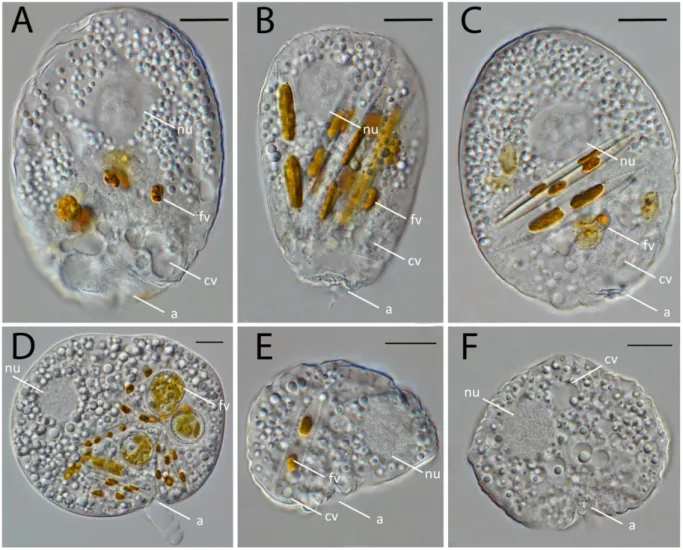
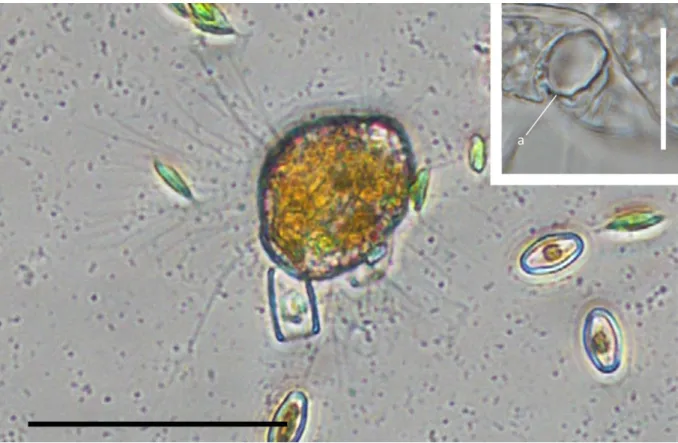
Fig. 1. General view of the cells. A-C – light microscopy pictures. Roundish cells forming anastomosing reticulopodia. D-F – scanning electron microscopy pictures; D-E cells form branching pseudopodia, F – the cell is covered papillae, bc – attached bacterium. Scale bars: A – 5 µm; B-E – 10 µm; F – 1 µm
38 Cell surface and scales
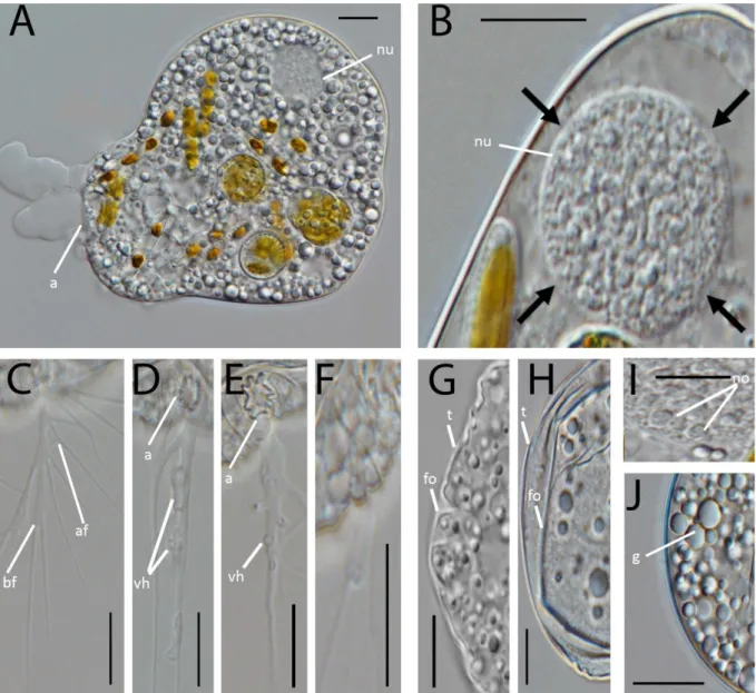
The surface of the cell body appears at smaller magnification rough (Figs 1F, 2A-2D). At higher magnification pyramidal papillae (outgrowths) covering the cell surface are observable. Every papilla bears a thin unadorned and oval scale (0.3-0.35 µm in width and 0.6 µm in length) and is densely filled by ribosomes (Figs 2D-2G). The scales are evenly distributed over the cell body and rarely overlap, but were never observed on the filopodia. Probably, the scales are formed in cisternae of the Golgi apparatus (Fig. 2I), which lies close to the nucleus and from which they are transported in vesicles to the cell surface (Fig. 2H).
Fig. 2. Cell sections (TEM). A-C – sections of whole cell, nucleus (nu) lies eccentric, close to the basal invagination, 1-2 food vacuoles (fv), roundish mitochondria (mi) and osmiophilic bodies (ob) are seen. D-E – arrangement of the papillae (pp) and single-tier scales (sc) on the cell surface. F, G - shape of the scales (sc). H – location of young scales and extrusomes inside vesicles in cytoplasm. I – possible formation of the scale inside cisterns of Golgi apparatus (Ga). Scale bars: A-C- 2 µm; D, G, H, I - 0.5 µm, E -0.4 µm; F - 0.1 µm
39 Phylogenetic analyses
The maximum likelihood tree shows the position of the Kraken within the cercozoan subphylum Filosa (Fig. 6). The Filosa comprises currently published sequences of the Monadofilosa and Granofilosea and as an outgroup the Chlorarachnea. The Monadofilosa are comprised of the Sarcomonadea, Imbricatea and Thecofilosea. As in previous analyses using SSU and LSU rDNA sequences for phylogeny, the chlorarachnean outgroup was highly supported, the Granofilosea were of moderate support and the Monadofilosa were highly supported (Chantangsi et al. 2010).
The Sarcomonadea, accommodating the Kraken sequences, separate from the Thecofilosea and Imbricatea with full support. Within the Sarcomonadea the three following well known clades were resolved: (1) the Glissomonadida HOWE, BASS VICKERMAN CHAO & CAVALIER-SMITH, 2009; (2) the Cercomonadidae KENT, 1880; (3) the Paracercomonadidae CAVALIER-SMITH, 2011; all with maximal support. The Cercomonadida were not monophyletic in our analysis, similar to previous studies in which they were either not monophyletic (Chantangsi et al. 2010) or the monophylum of little support (Wylezich et al. 2010). Within the Sarcomonadea, Kraken forms a sistergroup to the Paracercomonadidae with moderate support of 0.98/63. The genus Kraken is represented by the three currently known (and cultured) different genotypes of the species Kraken carinae. All strains formed a monophylum with maximal support. Similar to the SSU phylogenetic analyses (Dumack et al. 2016a) the morphologically similar strains KJ0003 and KD0092 grouped closely to each other, and the morphologically slightly different (smaller cell body) strain KD0248 basal to both.
40 Fig. 3.Selected serial sections (TEM) of the same cell showing the location of the groove (gr) as invagination of the nucleus (nu) and cell surface, also papillae (pp) and extrusomes (ex) are seen. Scale bar 2 µm.
41 Discussion
Ultrastructure data
Kraken carinae has very unique ultrastructure. Important ultrastructure characteristics for the taxonomy of protists (if present) are: flagella, the flagellar apparatus and the structure of scales (Cavalier-Smith & Karpov 2012; Hess and Melkonian 2014; Scoble and Cavalier-Smith 2014).
However, the complete loss of flagella and the flagellar apparatus in K. carinae prevent the comparison with Sarcomonadea.
The loss of flagella is likely a derived character, since Kraken cells usually feed as a sit-and-wait predator and move rarely. We suggest that Kraken inhabits small soil (or sediment) pores and only emerges from that with their long filopodia. Kraken has a peculiar way of movement by disassembling its own cell body (thereby reducing its cell diameter), possibly used when moving form pore to pore. A similar way of movement is known from the chlorarachniophyte Bigelowiella (Ota et al. 2007), leading to the question whether this is an ancestral trait or an adaption to life in sediments that evolved independently.
Due to is unique form of the locomotory structures Kraken can not be compared to the group specific
‘tails’ of ‘other’ Cercomonadida (Bass et al. 2009a).
Mitochondria with tubular cristae are common in Filosa (Bass et al. 2009a; Cavalier-Smith and Karpov 2012; Hess and Melkonian 2014; Shiratori et al. 2014). In contrast, K. carinae bears flat cristae. Only very few Cercozoa differ from tubular cristae, e.g. anaerobic Cercomonadida with cristae-lacking mitochondria or for instance some granofiloseans, such as the genus Limnofila which groups more basal in the Cercozoa and bears, similar to the Kraken, flat cristae (Bass 2009b; Cavalier-Smith and Chao 2003, Mikrjukov and Mylnikov 1995). Limnofila further shows some remarkable similarities with Kraken on the morphological level. Apart from flat cristae, both taxa are slow-moving filose amoebae with a huge filopodial network and a uninuclear cell body (Mikrjukov and Mylnikov 1998). Moreover the extrusomes which this study found in the Kraken, show similarity to the extrusomes of Limnofila which also contain a cylinder that is being exposed when the extrusome is charged (Mikrjukov and Mylnikov 1995). However distinct differences of Limofila compared to the Kraken are: A less defined form of the naked cell body of Limnofila, granules within the filopodia and flagella (or flagellate stages), apart from its well defined phylogenetic position (Bass 2009b).
Moreover the Kraken bears scales, a character unknown from any Sarcomonadea or Granofilosea species.
42 Fig. 4. Some organelles of the cell (TEM). A – Golgi apparatus (Ga) lies close to the nucleus (nu), B, C – mitochondria with plate-like cristae (cr), sometimes the roundish profile of the crista tip is seen (arrow). D – contractile vacuole (cv) of stage systole. E – small food vacuole (fv) contains bacterium (bc) inside reticulopodium. F – ectoproct (ec) eject the content of the food vacuole. G-J – extrusomes (ex) (G and I - longitudinal and cross sections, J – extrusome outside of the cell after its discharging). L – reserve substance. Scale bars: A, D, E – 0.5 µm; F – 1.0 µm ; B, C – 0.4 µm; G-I - 0.1 µm; L – 0.2 µm
Scales however are predominantly known from the Imbricatea, where the two major orders, the Thaumatomonadida and Euglyphida, both (usually) build scales. The Thaumatomonadida bear two- tier scales, the Euglyphida build whole rigid tests of overlapping single-tier silica scales (for an extensive and detailed discussion see Scoble and Cavalier-Smith 2014). However, due to their absence in some lineages, e.g. Esquamula or the Spongomonadida, they are considered as a paraphyletic character (Scoble and Cavalier-Smith 2014; Shiratori et al. 2012). Whole tests are considered as polyphyletic, as they are found in distant related cercozoan taxa, indicating that the development of a protective test has evolved multiple times in Cercozoa (Dumack et al. 2016 b,c;
Kosakyan et al. 2016; Wylezich et al. 2002).
Scoble and Cavalier-Smith (2014) assumed that the Imbricatean ancestor probably bore unadorned oval single-tiered scales; much as the scales of the Kraken. Accordingly, the Kraken might resemble an archaic ancestor of the Imbricatea. Since tests apparently evolved multiple times in the Cercozoa, it is not unlikely that also scales evolved multiple times. It seems unlikely that the last common
43 ancestor of the Filosa already carried scales, the Thecofilosea may have developed from those their organic tests and the Imbricatea further developed two-tier scales (Thaumatomonadida) or whole rigid tests composed of scales (Euglyphida), but were lost in most Sarcomonadea, except in the possible sarcomonadean Kraken.
Phylogenetic analyses
The phylogeny of the Sarcomonadea is still controversial (see Cavalier-Smith & Karpov 2012 for a detailed discussion). It is still unclear whether Sarcomonadea are monophyletic, and especially the phylogenetic position of the Paracercomonadidae (extremely long branch, changing position in trees) contributes to this uncertainty. Since Kraken groups with SSU and concatenated SSU+LSU phylogeny next to Paracercomonas, this long branch might (a) interfere with good support or (b) may even indicate questionable results due to long branch attraction. Nevertheless the constructed two-gene phylogeny confirms the SSU phylogeny published in Dumack et al. (2016a). Although still moderate, the phylogenetic support was significantly enhanced (from 0.6/29 to 0.98/63) in this study. Since only few cercozoan LSU sequences are currently available we were not able to achieve a highly supported phylogeny. With an increase of (a) taxon sampling of Cercozoa and especially the Paracercomonadidae (in particular Metabolomonas and Brevimastigomonas) or (b) gene sampling, in particular whole transcriptomes the phylogenetic positions of the Kraken and Paracercomonadidae within Cercozoa might be resolved.
The Kraken remains incertae sedis
The ultrastructure data give some indications that the Kraken resembles a direct descendant of the scale-bearing last common ancestor of the Imbricatea. The two-gene phylogeny indicates, however with moderate support, an affiliation with the Cercomonadida in the Sarcomonadea. Due to these contradicting results we are not able to clearly assign the Kraken to either the class Sarcomonadea or Imbricatea.
The characters of the Kraken have not been found in any other cercozoan family. We therefore establish a new family Krakenidae fam. nov., but still hesitate to establish a novel order that should be called Krakenida if it will be shown to be necessary in future studies. To resolve this issue, ultrastructure data (of e.g. Discomonas), SSU sequencing data (of e.g. Zoelucasa; Nicholls 2012), or large scale multi-gene transcriptome analyses are needed in future studies.
44 Fig. 5. Microfilaments and microtubules in the cells (TEM). A, B – arrangement of the microfilament bundles (mf) in the cytoplasm, C – microtubules (mt) inside cell, D-F – arrangement of the osmiophilic concentration of amorphous material (oc) inside reticulopodia, G – microtubules (mt) inside reticulopodia. Scale bar: C – 0.2 µm; A, B, D – G – 0.5 µm
45 Fig. 6. Phylogeny of the Filosa, Cercozoa with Chlorarachnea sequences as outgroup based on concatenated SSU rDNA and LSU rDNA sequences. Classes are indicated by vertical bars, the Sarcomonadea are highlighted in colour, newly obtained sequences are in bold. The support values of the Bayesian and PhyML analyses are shown on the respective branches (BI/ML). Support values
<50% (bootstrap values) and <0.95 (posterior probabilities) are only shown if necessary for interpretation of direct Kraken relationship, otherwise not shown (-). Branches with bootstrap values
>95% are presented in bold. Interrupted branches (//) show 20% of their original length. The scale bars represent 0.02 nucleotide substitutions per site. Given accession numbers refers to the strains LSU sequences.