Apoptoseaktivierung und
Zellzyklusarrestregulation durch das p14 ARF Tumorsuppressorgen
D i s s e r t a t i o n
zur Erlangung des akademischen Grades d o c t o r r e r u m n a t u r a l i u m
(Dr. rer. nat.) im Fach Biologie eingereicht an der
Mathematisch-Naturwissenschaftlichen Fakultät I der Humboldt-Universität zu Berlin
von
Diplom-Biologe Tim Overkamp
Präsident der Humboldt-Universität zu Berlin Prof. Dr. Jan-Hendrik Olbertz
Dekan der Mathematisch-Naturwissenschaftlichen Fakultät I Prof. Dr. Andreas Herrmann
Gutachter: 1. Prof. Dr. Harald Saumweber 2. Prof. Dr. Peter Daniel 3. PD Dr. Jürgen Eberle Arbeit eingereicht am: 31. Januar 2012
Tag der mündlichen Prüfung: 11. Oktober 2012
Abstract
BH3-only proteins, a pro-apoptotic subgroup of the Bcl-2 family of proteins, are central mediators of apoptosis signals by regulating the intrinsic apoptosis pathway. We have recently shown, that apoptosis triggered by the p14ARF tumour suppressor protein is mediated by the p53-dependent activation of the BH3-only protein Puma/Bbc3. Nevertheless, expression of p14ARF in p53-family deficient cells is capable of inducing both cell cycle arrest and apoptosis, but the signalling pathways initiated remain elusive. Here, we report that the BH3- only protein Bmf (Bcl-2 modifying factor) is involved in cell death in p53-deficient cells triggered by p14ARF. Expression of p14ARF leads to the induction of the PERK kinase, subsequent phosphorylation of eIF2α and activation of transcription factors ATF4 and CHOP.
This signalling cascade is usually part of the ‘unfolded protein response’ (UPR), which is activated upon ER stress to reduce the amount of misfolded proteins by reduction of global protein translation initiation and upregulation of chaperones. Of note, p14ARF does not induce ER stress but activates the PERK‒CHOP pathway. ATF4 and CHOP transcription factors directly bind to the promotor region of bmf and induce its transcription. These data suggest that the PERK‒eIF2α‒ATF4‒CHOP signalling pathway may play a substantial role in mediating p14ARF-triggered apoptosis. This pathway could play the role of a ‘fail-safe’
mechanism that allows cells, even after loss of p53, to undergo apoptosis induced by upregulation of p14ARF by oncogenes.
Keywords:
apoptosis, BH3-only, Bcl-2 modifying factor (Bmf), p14ARF, p53, unfolded protein response (UPR), endoplasmic reticulum (ER)
Zusammenfassung
BH3-only Proteine, eine pro-apoptotische Untergruppe der Bcl-2 Proteinfamilie, sind zentrale Mediatoren von apoptotischen Signalen durch die Regulierung intrinsischer Apoptose- signalwege. Unsere Arbeitsgruppe hat vor kurzem gezeigt, dass Apoptose, die durch den p14ARF Tumorsuppressor induziert wird über die p53-abhängige Aktivierung des BH3-only Proteins Puma/Bbc3 vermittelt wird. Interessanterweise induziert p14ARF aber auch in p53 defizienten Zellen Zellzyklusarrest und Apoptose. Die dahinterliegenden Signalwege sind jedoch nicht bekannt. In dieser Arbeit berichten wir, dass das BH3-only Protein Bmf (Bcl-2 modifying factor) beim p14ARF-induzierten Zelltod in p53 defizienten Zellen eine wichtige Rolle spielt. Expression von p14ARF führt zu einer Induktion der PERK Kinase, daran anschließender Phosphorylierung von eIF2α sowie Aktivierung der stromabwärts liegenden Transkriptionsfaktoren ATF4 und CHOP. Diese Signalkaskade ist normalerweise Teil einer zellulären Antwort auf fehl- oder ungefaltete Proteine im Endoplasmatischen Retikulum (ER), der sogenannten ‘unfolded protein response’ (UPR), die zum einen durch verminderte Translationsinitiation und Hochregulierung von Chaperonen die Menge der fehlgefalteten Proteine reduzieren soll. Allerdings induziert p14ARF keinen ER Stress, sondern den PERK‒
CHOP Signalweg. Die Transkriptionsfaktoren ATF4 und CHOP binden direkt in der Promotorregion von bmf und sind für dessen transkriptionelle Regulation verantwortlich.
Unsere Daten zeigen, dass der PERK‒eIF2α‒ATF4‒CHOP Signalweg eine wesentliche Rolle bei der Induktion von Apoptose durch p14ARF spielt. Dieser Weg könnte ein Sicherungsmechanismus sein, der es den Zellen auch nach Verlust von p53 erlaubt Apoptose einzuleiten, nachdem p14ARF durch Onkogene hochreguliert wurde.
Stichwörter:
Apoptose, BH3-only, Bcl-2 modifying factor (Bmf), p14ARF, p53, unfolded protein response (UPR), Endoplasmatisches Retikulum (ER)
Table of Contents
Abstract ... ii
Zusammenfassung ... iii
Table of Contents ... iv
1. Introduction ... 1
1.1 Apoptosis pathways ... 2
1.1.1 Death receptor pathway ... 3
1.1.2 Mitochondrial pathway ... 3
1.1.3 Endoplasmic reticulum pathways ... 5
1.2 Bcl-2 family of proteins ... 8
1.2.1 BH3-only proteins ... 10
1.3 The p14ARF tumour suppressor ... 11
1.4 The p53 family of proteins ... 14
2. Aim of the study ... 16
3. Materials and Methods ... 17
3.1 Materials ... 17
3.1.1 Tools ... 17
3.1.2 Chemicals ... 17
3.1.3 Solutions, buffers and media ... 18
3.1.4 Kits ... 19
3.1.5 Markers ... 20
3.1.6 Antibodies ... 20
3.1.7 Enzymes ... 20
3.1.8 Oligonucleotides ... 21
3.1.9 Vectors and Plasmids ... 22
3.1.10 Adenoviruses ... 25
3.1.11 Bacteria ... 26
3.1.12 Cell lines ... 26
3.1.13 Software ... 27
3.2 Methods ... 28
3.2.1 Polymerase chain reaction (PCR) ... 28
3.2.2 DNA Electrophoresis ... 28
3.2.3 Gel extraction... 28
3.2.4 Isolation of plasmid DNA ... 29
3.2.5 Measurement of DNA concentration ... 29
3.2.6 Enzymatic restriction of DNA ... 29
3.2.7 Ligation of DNA fragments ... 29
3.2.8 Mini- and Maxi-Prep of plasmid DNA from bacteria ... 30
3.2.9 DNA sequencing ... 30
3.2.10 RNA extraction from cells ... 31
3.2.11 cDNA synthesis from total RNA ... 31
3.2.12 Quantitative real-time PCR (qRT-PCR) ... 31
3.2.13 Transfection of bacterial cells (heat shock) ... 32
3.2.14 Transfection of eucaryotic cells (electroporation) ... 32
3.2.15 Protein assay (Bradford) ... 32
3.2.16 SDS polyacrylamide gel electrophoresis ... 33
3.2.17 Immunodetection of proteins - Western blot analysis ... 33
3.2.18 Analysing BAX/BAK N-terminal conformational change by flow cytometry 34 3.2.19 Detection of genomic DNA fragmentation with propidium iodide (PI)... 34
3.2.20 AnnexinV-FITC/PI staining ... 35
3.2.21 Measuring breakdown of the mitochondrial membrane potential (ΔΨm) ... 35
3.2.22 Cytochrome c release ... 35
3.2.23 Chromatin Immunoprecipitation (ChIP) ... 36
3.2.24 Luciferase Assay ... 38
3.2.25 siRNA ... 38
3.2.26 Statistics ... 39
4. Results ... 40
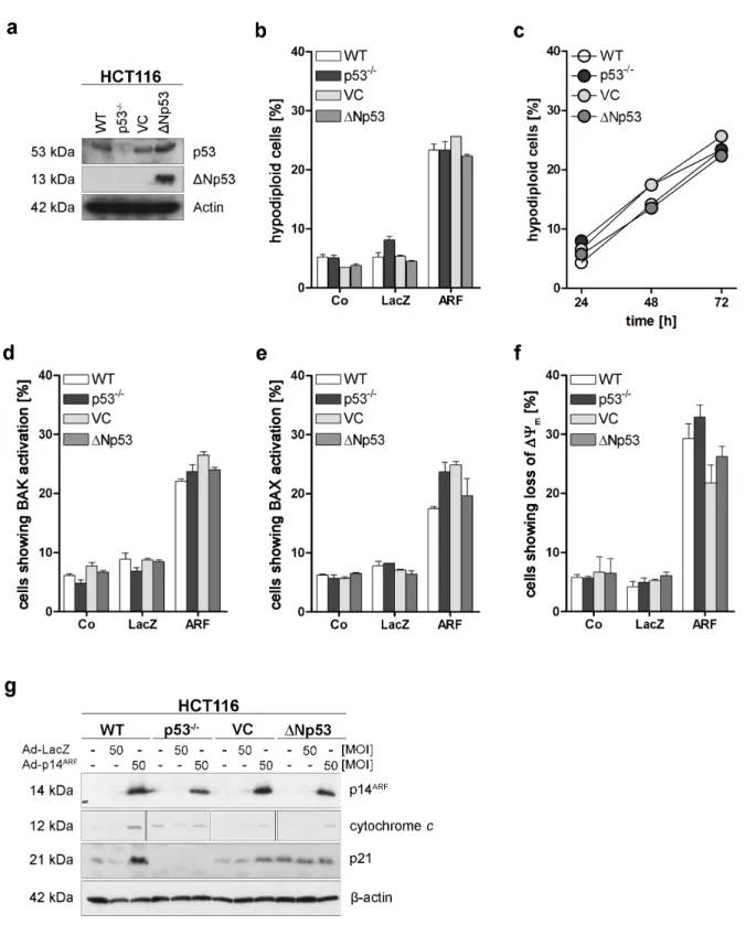
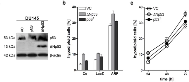
4.1 p53 family independent induction of apoptosis by p14ARF ... 40
4.2 Involvement of cellular organelles in p14ARF induced apoptosis ... 43
4.3 Induction of BH3-only proteins by p14ARF ... 45
4.4 Induction of genes triggering the UPR by ER stress and p14ARF ... 48
4.5 Inhibition of apoptosis by dominant negative mPERKΔC ... 52
4.6 Inhibition of apoptosis by PERK and ATF4 siRNA ... 53
4.7 Specific phosphorylation of eIF2α by p14ARF ... 54
4.8 Induction of apoptosis by ER stressors, Ad-ATF4(Tet) and Ad-CHOP(Tet) ... 55
4.9 Induction of BH3-only proteins by ER stressors, Ad-ATF4(Tet) and Ad-CHOP(Tet) ... 57
4.10Inhibition of apoptosis by Bmf siRNA in p53 pro- and deficient HCT116 cells ... 60
4.11Bmf translation after phosphorylation of eIF2α ... 61
4.12bmf promoter binding studies and transcriptional activity ... 62
5. Discussion ... 67
5.1 p14ARF induces p53 family independent apoptosis ... 67
5.2 p14ARF-induced apoptosis utilises pathways of the mitochondria and the ER ... 68
5.3 Induction of the PERK‒CHOP pathway by p14ARF ... 68
5.4 Role of BH3-only proteins in ER-mediated induction of apoptosis ... 70
5.5 Bmf gene regulation ... 71
5.6 Involvement of the ER as an energy saving mechanism ... 72
5.7 A cap-independent network regulating apoptosis ... 73
5.8 Model of p14ARF induced activation of apoptosis ... 73
5.9 Outlook ... 75
6. Summary ... 76
References ... 77
7. Appendix... 92
7.1 Abbreviations ... 92
7.2 Bcl-2 modifying factor (Bmf) promoter region ... 96
Acknowledgements ... 97
Eidesstattliche Erklärung ... 98
1. Introduction
The concept of a natural instead of a pathological form of cell death was described as early as 1842 by Carl Vogt in studies about the development of the midwife toad Alytes obstetricans (Vogt, 1842), only three years after Schwann and Schleiden’s cell theory in 1839 (Schwann, 1839). More than a century later, the term apoptosis (from greek από [apo; from] and πτωσις [ptosis; falling, a fall]; like leaves or petals falling from a plant) had been coined in order to describe morphological processes leading to controlled cellular self-destruction and was first introduced in a publication by Kerr, Wyllie and Currie (Kerr et al., 1972).
Apoptosis, or Type I cell death, can be described as an active and evolutionary defined process which plays an important role in the development of multicellular organisms and in the regulation and maintenance of the cell homeostasis in tissues upon physiological and pathological conditions. One type of apoptosis designated as ‘anoikis’ (from greek άν [an;
without], οίκ [oik; house], and ις [is; extracted from apoptosis]; the state of being without a home) is induced by detachment or inadequate and inappropriate cell-matrix interactions (Frisch and Francis, 1994). Apoptosis is the most frequent form of programmed cell death, followed by autophagy/autophagocytosis (from greek αυτό [auto; self] and φαγία [phagia; to eat), or Type II cell death, where the cell’s own components are catabolic degraded through its lysosomal machinery. Autophagy was initially described as a cellular response to starvation by Christian de Duve in 1963 (de Duve, 1963). Next to these types of programmed cell death, other, non-apoptotic types, e.g. caspase-independent programmed cell death, are also of biological significance (Leist and Jäättelä, 2001).
Programmed cell death mechanisms differ from necrosis (from greek νεκρός [necros; death, dead]), or Type III cell death, which is an accidental and unordered cell death where the release of cellular contents causes an inflammatory response. In contrast, apoptosis leads to shrinkage and fragmentation of the nucleus, condensation of chromatin and blebbing of the cell membrane due to breakdown of the cytoskeleton. These constricted particles, so called apoptotic bodies, are eventually taken up by phagocytes and the components are recycled.
Autophagy and apoptotic processes are involved in development, starting in ontogenesis, when gastrulation of the early embryo is made possible by programmed death of cells within the blastula, later on during digit formation when web cells die off and disappear (Zuzarte- Luís and Hurlé, 2002) and throughout the life of the organism. In the immune system, apoptosis is responsible for negative selection of autoreactive B- and T-cells as well as for removing virus-infected cells. In the human body about 100,000 cells are produced every
second by mitosis and meiosis and a similar number die by apoptosis (Vaux and Korsmeyer, 1999). This homeostasis between the regulation of cell cycle and apoptosis is achieved by a tight regulation of pro- and anti-apoptotic proteins (Krammer et al., 1994; Daniel, 2000). One of the key regulators is the tumour suppressor p53, also termed “guardian of the genome”
(Lane, 1992). This protein was identified in 1979 and is known to be activated upon different types of stress, e.g. DNA damage, oxidative shock and deregulated oncogene expression (Finlay et al., 1989; Han et al., 2008). Oncogenic stress activates the INK4b-ARF-INK4a locus that gives raise to proteins that on the one hand arrest the cell in the cell cycle and on the other hand induce p53. Disruption of cell cycle pathways and programmed cell death deregulation participates in the pathogenesis of several diseases. Uninhibited cell proliferation leads to cancer or auto-immune diseases, while upregulation of apoptosis leads to neurodegenerative diseases such as Creutzfeldt Jakob, Alzheimer’s and Parkinson’s disease (Yuan and Yankner, 2000; Rathmell and Thompson, 2002; Daniel in Ganten and Ruckpaul, 2007).
1.1 Apoptosis pathways
Apoptosis is executed by a network of genetically encoded components. It can be triggered by various stimuli from outside or inside the cell, leading to the activation of the extrinsic or death receptor pathway and the intrinsic or mitochondrial pathway. Every cellular organelle or subcellular component, e.g. the endoplasmic reticulum (ER) or the nucleolus, can sense stressful and pathogenic alterations and initiate local or global responses leading to adaptation or, once a critical threshold of damage has been reached, cell death (Ferri and Kroemer, 2001). The activation of cell death is mediated by cysteine-dependent aspartate-specific proteases (caspases). These are synthesised as inactive zymogenes (procaspases) and are located in the cytosol and the nucleus. Activation of the so called caspase cascade requires the prior cleavage of upstream caspases called initiator caspases, such as caspase-8 and -9, that are autocatalytically cleaved and activated through recruitment by adaptor proteins that share death-effector domains (DED) or caspase activation and recruitment domains (CARD) with these initiator caspases (Nicholson, 1999). Downstream effector (or executioner) caspases-3, -6, and -7 will then cleave multiple structural and repair proteins within the cell to trigger apoptosis (Slee et al., 2001).
Caspases-12 (murine) and -4 (human) have been proposed to function as initiator caspases that are activated by the endoplasmic reticulum (ER). The human caspase-12 gene has aquired several nonsense mutations, leading to a premature translational stop or a loss-of function mutation (Rocher et al., 1997; Fischer et al., 2002). Human caspase-4 is partially localised to the ER and is selectively activated in response to ER stress (Hitomi et al., 2004b), although events upstream of its activation remain poorly defined (Heath-Engel et al., 2008).
Nevertheless, compared to the murine system, other factors than caspases seem to play a role in the human system linking the ER to induction of apoptosis (Hitomi et al., 2004a).
1.1.1 Death receptor pathway
The death receptor pathway is mediated by transmembrane receptors, a subfamily of the TNF-R family (tumour necrosis factor-receptor), located in the plasma membrane.
Trimerisation of these death receptors by death ligands, e.g. TNFα or TRAIL (TNF-related apoptosis inducing ligand), leads to recrution of adaptor proteins, e.g. FADD (fas associated protein with death domain), TRADD (TNF-R type 1-associated death domain protein), RIP (receptor interacting protein 1) or RAIDD (RIP-associated ICH-1/Ced3 homologous protein with a death domain) via their death domains (DD) and formation of the death-inducing signalling complex (DISC) (Banner et al., 1993; Boldin et al., 1995). Here, procaspase-8 binds via its death effector domain (DED) and is cleaved into its active form that subsequently activates downstream effector caspases -3, -6, and -7. Activation of caspase-8 is also achieved by a membrane independent, cytosolic complex induced by activation of TNF-R1. This so called ‘complex II’ consists of pro-caspase-8, FADD, TRADD and RIP1-kinase and is able to induce self-processing of caspase-8 and NF-κB (Micheau and Tschopp, 2003).
Cytosol localised FLIP (FADD-like interleukin-1β converting enzyme (FLICE) inhibitory protein) proteins carry two tandem DEDs which can bind FADD and procaspase-8 thereby forming a proteolytically inactive heterodimer, inhibiting activation of caspase-8 (Krueger et al., 2001; Golks et al., 2005).
1.1.2 Mitochondrial pathway
The mitochondria-initiated apoptotic pathway is tightly regulated by Bcl-2 family proteins that mediate diverse cellular stress signals, e.g. DNA damage, hypoxia, endoplasmatic reticulum (ER) or nutritive stress (Daniel et al., 2003). Pro-apoptotic members Bax (Bcl-2
associated X protein) and Bak (Bcl-2 antagonist/killer) are activated by a variety of these apoptotic stimuli, leading to oligomerisation and insertion into the mitochondrial outer membrane. This leads to a loss of mitochondrial membrane potential (ΔΨm) and release of cytochrome c and other apoptotic factors that are normally sequestered in the mitochondrial intermembrane space. Cytochrome c and d(ATP) bind to cytosolic apoptotic protease activating factor 1 (Apaf-1), also recruiting procaspase-9 which together build the mitochondrial apoptosome (Perkins et al., 1998). Here, procaspase-9 is activated and functions as initiator caspase upon which the caspase cascade is started.
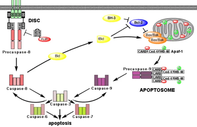
The death receptor and the mitochondrial pathway are connected by caspase-8 and the BH3-
Figure 1. Extrinsic or death receptor pathway (left) and intrinsic or mitochondrial pathway (right). The extrinsic pathway is induced by binding of death ligands and assembly of the death-inducing signalling complex (DISC) that leads to cleavage of procaspase-8 to active caspase-8. This initiator caspase then activates effector caspases -3, -6, -7 and thereby eventually apoptosis. FLIP inhibits caspase-8 activation at the DISC by forming proteolytically inactive heterodimer with procaspase-8. The intrinsic pathway is activated by stress signals that are relayed by BH3-only proteins, which inactivate anti-apoptotic Bcl-2 family proteins, thereby releasing Bax and Bak. Activation of the mitochondria leads to the release of cytochrome c (depicted in green), (d)ATP (depicted in red) and several pro-apoptotic factors. Cytochrome c and d(ATP) bind to cytosolic Apaf-1, also recruiting procaspase-9 which together build the mitochondrial apoptosome. Procaspase-9 is cleaved autocatalytically and the caspase cascade is activated. Both pathways are connected by the BH3-only protein Bid, which is cleaved by caspase-8 into a truncated form (tBid) that can activate the mitochondrial pathway.
Abbreviations: death domain (DD), death-effector domain (DED), caspase recruitment domain (CARD), Caenorhabditis elegans homology domain (Ced-4), repeating sequence at C-terminus (WD-40).
only protein Bid (BH3 interacting domain death agonist). Bid is cleaved into its active truncated form tBid by caspase-8. tBid then activates the mitochondrial pathway. After formation of the apoptosome, active caspase-9 cleaves and activates caspase-3 that subsequently, in a feedback amplification loop, activates caspase-8 (Crompton, 2000). In so called “type I cells” processed caspase-8 directly activates effector caspases, while in “type II cells” caspase-8 first needs to activate the mitochondria via tBid to induce the apoptosome which then activates effector caspases through caspase-9 (Scaffidi et al., 1998).
1.1.3 Endoplasmic reticulum pathways
The endoplasmic reticulum (ER) is the place for synthesis and folding of secreted and membranous protein and lipid biosynthesis. It is the major organelle involved in intracellular calcium ion (Ca2+) homeostasis and signalling. ER and mitochondria cooperate in cell death induction by Ca2+ signalling, which, released from the ER, can trigger cytochrome c release from mitochondria (Rong and Distelhorst, 2008).
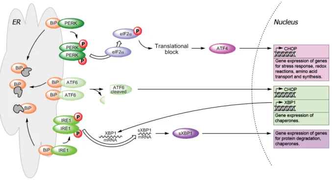
Perturbances in ER homeostasis, protein folding and ER calcium concentrations will also result in a cytoprotective response called the unfolded protein response (UPR). UPR is mediated by three ER transmembrane proteins: protein kinase RNA-dependent-like ER kinase (PERK), activation transcription factor 6 (ATF6) and inositol requiring ER-to-nucleus signal kinase 1 (IRE1):
PERK is activated by dimer formation and is then autophosphorylated (Zhou et al., 2006). It will phosphorylate the eucaryotic translation initiation factor 2 α (eIF2α), which together with eIF2β and eIF2γ is part of the heterotrimer eIF2. Under normal conditions eIF2 mediates binding of tRNAmet to the 40 S subunit of the ribosome in a GTP-dependent manner to form the 43 S preinitiation complex. Phosphorylation of eIF2α results in sequestration of eIF2B, the GDP/GTP exchange factor, thereby inhibiting formation of the 43 S complex and stopping global cap-dependent translation. The ‘cap’ is a specifically altered nucleotide, i.e.
7-methylguanylate (m7G), on the 5’ end of most nuclear mRNAs. Certain mRNAs carrying regulatory sequences in their 5’ untranslated regions, e.g. the internal ribosomal entry site (IRES), can bypass the eIF2α-dependent translational block (Schröder and Kaufman, 2005).
Activation transcription factor 4 (ATF4) contains two upstream open reading frames (uORFs) in front of the ORF that codes for the protein itself, which facilitate ribosome scanning and reinitiation at the next uORF, an inhibitory element, that blocks ATF4 expression under
normal conditions. Phosphorylation of eIF2α delays reinitiation and initiates at the ATF4- coding region (Vattem and Wek, 2004). ATF4 is a transcription factor, which belongs to the CREB/ATF family of bZIP (basic leucine zipper domain) proteins (Rutkowski and Kaufman, 2003). Targets of ATF4 include genes involved in amino-acid metabolism, resistance to oxidative stress and the pro-apoptotic transcription factor C/EBP (CCAAT/enhancer-binding protein) homologous protein (CHOP) also known as C/EBPζ (Ma et al., 2002).
C/EBPζ (CHOP) is also a target of ATF6, a second transducer, which is cleaved at the Golgi apparatus by site 1 and site 2 (S1/S2) proteases into its active form that moves to the nucleus and induces the activation of genes with an ER stress response element (ERSE) in their promoter (Schröder and Kaufman, 2005). Other targets of ATF6 include BiP and X box binding-protein 1 (XBP1) which is important in IRE1 signalling.
On activation, transducer number three, IRE1 will remove a 26-nucleotide intron from the XBP1 mRNA and the frameshift splice variant (sXBP1) encodes a stable, active transcription factor (Yoshida et al., 2001) which targets different ER chaperones and some members of the HSP40 family. Via binding to the TNF-receptor-associated factor 2 (TRAF2), the IRE1- TRAF2 complex can recruit the apoptosis-signal-regulating kinase 1 (ASK1), which is a mitogen-activated protein 3-kinase (MAP3K) that has been shown to relay various stress signals to the downstream MAPK c-Jun N-terminal kinase (JNK) (Nishitoh et al., 1998).
Figure 2. Unfolded protein response (UPR) pathway. Release of transducer proteins PERK, ATF6 and IRE1 by BiP upon aggregation of unfolded proteins (depicted in grey) within the ER leads to the activation of transcription factors and gene expression of genes for stress response, protein degradation and chaperones.
Prolonged UPR eventually leads to apoptosis (modified after Szegezdi et al., 2009).
Activation of JNK is known to influence the cell death machinery through the regulation of Bcl-2 family proteins, e.g. by phosphorylating Bcl-2 its anti-apoptotic activity is suppressed while phosphorylating Bim enhances its pro-apoptotic potential (Davis, 2000).
Under physiological conditions all transducer proteins are bound to BiP (immunoglobulin heavy chain binding protein; also known as GRP78, glucose-regulated protein 78 kDa) and are released when unfolded proteins accumulate within the ER lumen because of a higher affinity of BiP to misfolded proteins. BiP is an abundant protein under all growth conditions, but its synthesis is markedly induced under conditions that lead to the accumulation of unfolded polypeptides in the ER (Kozutsumi et al., 1988).
Phosphorylation of eIF2α is a critical convergence point of the integrated stress response (ISR), which supports eukaryotic cellular adaptation to diverse environmental and engogenous stress signals, including ER stress, amino acid deprivation, infection with double- stranded RNA viruses, osmotic stress, UV light exposure, heme deficiency, and oxidative stress (Lu et al., 2001; Deng et al., 2002; Ron, 2002; Zhang et al., 2002). Next to arrest of global cap-dependent translation, numerous stress-triggered cytoprotective genes are induced.
Also, short half life proteins, e.g. cyclin D1, a regulator of G1 to S-phase cell cycle transition and an important cofactor for several transcription factors in numerous cell types, as well as anti-apoptotic proteins disappear by degradation in the proteasome (Brewer et al., 1999;
Scheuner et al., 2006), thereby giving the cell time to react to stressful conditions and/or enabling it to undergo apoptosis.
Accumulation of small aggregates from unfolded or misfolded proteins are thought to be highly toxic, as they impair the ubiquitin proteasome pathway (Bence et al., 2001).
Malfunctions of the ER stress responses caused by aging, genetic mutations, or environmental factors can result in various diseases such as diabetes, inflammation, and neurodegenerative disorders including Alzheimer’s and Parkinson’s disease (Yoshida, 2007).
Patients with multiple myeloma, a haematological cancer that results from the malignant transformation of plasma cells, show high expression levels of XBP1. Because plasma-cell development and survival depend on an intact UPR, this signalling pathway is also an intriguing target for novel treatments of myeloma.
1.2 Bcl-2 family of proteins
One of the first proteins identified to be involved in apoptosis was the second B-cell lymphoma gene (bcl-2; described by Vaux et al., 1988). Initially characterised as a proto- oncogene, overexpression of bcl-2 was found to increase the survival of haematopoietic cells by reducing the sensitivity to apoptosis (Tsujimoto and Croce, 1986). Today more than 20 human proteins of the Bcl-2 family are known, that are able to regulate the permeability of intracellular membranes to ions and proteins (Sharpe et al., 2004).
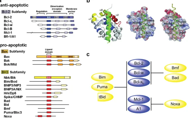
The Bcl-2 family can be divided into the anti-apoptotic subfamily and the pro-apoptotic subfamily, which can be subdivided into the Bax and BH3-only subfamilies (Figure 3). These proteins contain highly conserved domains, referred to as BH (Bcl-2 homology) domains (BH1-4), which are important for complex formation. In response to death signals, such as cytotoxic agents or radiation, the BH3-only proteins antagonises the function of the antiapoptotic proteins. The BH3 domains of these proapoptotic molecules form an amphipathic α-helical fold when bound to a groove lined by the BH1, BH2, and BH3 domains of antiapoptotic proteins, a step thought to be important for apoptosis induction (Czabotar et al., 2007).
Figure 3. Bcl-2 family overview. (a) Anti- and pro-apoptotic members of the Bcl-2 family of proteins (excerpt).
(b) X-ray structure of the interaction between Bim and Mcl-1; Bim BH3 peptide in green and the BH1, BH2, and BH3 regions of Mcl-1 in blue, yellow, and red, respectively (left). Bim BH3 complexed with Mcl-1 (middle) and Bcl-xL (right) (Czabotar et al., 2007). (c) Target selectivity of different Bcl-2 family proteins (modified from Chen et al., 2005).
Bcl-2 family proteins can mostly be found at intracellular membranes where they are inserted with their carboxy-terminal hydrophobic tail, while BH3-only proteins are primarily localised in the cytosol (Krajewski et al., 1993; Tanaka et al., 1993). Multidomain protein Bak can be found at both the mitochondrial and ER membranes (Cheng et al., 2003). Nbk, a member of the BH3-only subfamily, is inserted into the ER membrane by its transmembrane segment at the COOH-terminus and is able to induce mitochondrial cytochrome c release from its position at the ER (Germain et al., 2002). Activation of mitochondria by Nbk is mediated only by Bax, because Bax homolog Bak is held in check by anti-apoptotic Mcl-1 (Gillissen et al., 2003, 2007).
Although Bax contains a hydrophobic carboxy-terminal tail, inactive Bax is a cytosolic monomeric protein that, upon activation, changes its conformation. The internalised C-terminal anchor domain is released from the hydrophobic pocket formed by the BH1-3 domains and the protein then translocates to the mitochondrial outer membrane (Hsu et al., 1997). The exact mechanism of Bak and Bax activation by BH3-only proteins is still under discussion (Danial and Korsmeyer, 2004; Youle and Strasser, 2008). Antiapoptotic proteins of the Bcl-2 family (Bcl-2, Bcl-x, Mcl-1, A1, Bcl-w, and Bcl-B) either bind to Bax and Bak and prevent oligomerisation, or are inhibited by binding to proapoptotic BH3-only proteins. The
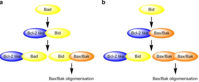
‘direct activation’ hypothesis claims that a direct interaction between BH3-only proteins and Bak or Bax is necessary for their activation (Figure 4a) (Kuwana et al., 2005). Recently, BH3
Figure 4. Models for Bax and Bak oligomerisation. (a) Direct activation model. BH3-only proteins can be divided into ‘sensitizers’ or ‘derepressors’ (e.g. Bad) that bind only to pro-survival proteins and ‘activators’ (e.g.
Bim) that can also directly engage Bax and Bak. ‘Sensitizers/derepressors’ induce apoptosis by displacing
‘activators’ from pro-survival proteins, which then proceed to trigger Bax/Bak activation. (b) Displacement model. Pro-survival proteins inhibit Bax and Bak, perhaps through direct interaction as has been demonstrated for Bak. BH3-only proteins induce apoptosis by neutralizing pro-survival molecules and Bax/Bak activation occurs spontaneously in the absence of pro-survival activity (modified after van Delft and Huang, 2006).
domains of Bmf and Noxa were shown to induce mitochondrial outer membrane permeabilisation and apoptosis similar as the direct activators Puma, Bim, and Bid (Du et al., 2011). The ‘displacement’ model proposes that BH3-only proteins bind to a complex of anti- apoptotic Bcl-2 and Bak or Bax, thereby replacing and activating Bak and Bax (Figure 4b) (Willis et al., 2007).
1.2.1 BH3-only proteins
BH3-only proteins are important components in the regulation and activation of cell death (Daniel et al., 2003). In response to different cell stress stimuli, different BH3-only proteins relay apoptosis signals to the mitochondria to induce apoptosis (Shibue and Taniguchi, 2006).
BH3-only proteins are controlled by transcriptional and post-translational regulation. BH3- only genes under transcriptional control in mammals include hrk (harakiri), noxa (from latin
‘damage’), puma (p53-upregulated mediator of apoptosis), bim (Bcl-2 interacting mediator of cell death), and nbk/bik (natural born killer / Bcl-2-interacting killer). Noxa, puma, and nbk are regulated by the tumour suppressor p53 (Oda et al., 2000; Nakano and Vousden, 2001;
Mathai et al., 2002). BH3-only proteins regulated by post-translational mechanisms comprise of Bad (Bcl-2 antagonist of cell death), which is phosphorylated upon cellular stimulation with growth factors, Bid, that is N-myristoylated and cleaved by activated caspase-8, and Bmf (Bcl-2 modifying factor) and Bim (Bcl-2 interacting mediator of cell death), which are activated by sequestration from cytoskeletal structures inside the cell.
The amphipathic helix formed by the BH3 domain of BH3-only and Bax-like proteins binds to a hydrophobic groove on the surface of the anti-apoptotic Bcl-2 family members (Fesik, 2000). Although this nine amino acid long BH3 domain is highly conserved, it shows a specific target selectivity between pro-apoptotic BH3-only and anti-apoptotic Bcl-2 proteins (Figure 3c).
1.2.1.1 Bcl-2 modifying factor (Bmf)
The human bmf gene is located on chromosome 15q14 and loss of this site has been reported in lung and breast cancer (Wick et al., 1996; Schmutte et al., 1999). Bmf is regarded as a candidate tumour suppressor (Bouillet et al., 2001; Puthalakath and Strasser, 2002). In this line of thought Bmf has been shown to be involved in mammary morphogenesis and to be repressed by oncogenic Ras (Schmelzle et al., 2007). Bmf is constitutively expressed in
healthy tissues and is sequestered to myosin V motor complexes by association with the dynein light chain 2. Disruption of the actin cytoskeleton is thought to trigger release and activation of Bmf, initiating anoikis, i.e. apoptotic processes (Puthalakath et al., 2001).
Monoclonal antibodies specific for mouse Bmf have revealed the presence of multiple isoforms in most hematopoietic tissues with the highest levels found in immature T and B cells (Labi et al., 2008). Until now, it is unclear how these isoforms arise, but alternative splicing of bmf has been reported to regulate its in vivo function. Two additional splice variants (bmf II and bmf III) were found to be expressed in normal and malignant human B cells (Morales et al., 2004).
1.3The p14ARF tumour suppressor
The INK4b-ARF-INK4a locus at chromosome 9p21.3 gives raise to three different proteins.
Two structurally related proteins, p15INK4b and p16INK4a, function as inhibitors of the cyclin- dependent kinase 4 (CDK4). The third transcript, p14ARF, includes an alternate first exon 1β located about 20 kb upstream of the exon 1α, 2, and 3 of p16INK4a and contains an alternative reading frame (ARF), hence its name. Because of the alternative reading frame, p16INK4a and p14ARF do not share any amino acid homology and they have different functions within the cell (Figure 5).
The INK4a-ARF locus is second only to p53 in the frequency of its disruption in human cancer (Haber, 1997). The tumour suppressor p14ARF is frequently inactivated in a wide spectrum of human cancer types, including colorectal, breast and pancreatic adenocarcinomas, malignant glioma, melanoma and non-Hodgkin’s lymphoma (Sharpless and DePinho, 1999;
Burri et al., 2001). Simultaneous inactivation of p53 and p14ARF results in a broader tumour spectrum and more aggressive tumours than are observed with either knockout alone (Weber et al., 2000).
Human (p14ARF) and murine (p19ARF) proteins are composed of 132 and 169 amino acids, respectively. They share about 50 % sequence homology and are both composed of more than 20 % arginine residues conferring them highly basic and having hydrophobic properties.
There are no recognisable structural motifs in ARF proteins and the protein probably needs to form complexes with other molecules, both to be folded and for its charge to be neutralised at physiological pH (Ozenne et al., 2010). Murine and human ARF contain a single internal methionine residue, which are absent in other species. Translational initiation from these
AUG codons produce a short form of the protein (smARF), which, when overexpressed, is supposed to localise to the mitochondria (Reef et al., 2006). Full-length p14ARF possesses two nucleolar localisation signals (NoLS). The first one located in exon 1β plays a key role in the antiproliferative function as its deletion inhibits the ability of p14ARF to activate a checkpoint response via the Hdm-2 (human double minute 2) ‒p53 pathway (Zhang et al., 1998; Rizos et al., 2000; Sherr, 2006). The second one is involved in the ability of p14ARF to promote the sumoylation of its binding partners (Xirodimas et al., 2002).
Ectopic expression of a variety of oncogenes such as Ras, c-myc, E1A and E2F1 upregulates p14ARF expression as part of a checkpoint response that limits cell cycle progression in response to hyperporoliferative signals (Sharpless, 2005). P14ARF expression is also increased after exposure to ionising or ultra violet radiation and genotoxic drugs and contributes to the DNA damage response that eliminates damaged cells from the proliferative pool (Sherr, 2006). It has also been shown that ARF expression is induced by viral infection and acts to reduce viral infectivity (García et al., 2006).
Both mouse and human ARF are relatively stable proteins with estimated half-lifes ranging from approximately 1 to 8 hours. ARF turnover is still not completely understood, although two residues in exon 1β were found to be essential for p14ARF stability (di Tommaso et al.,
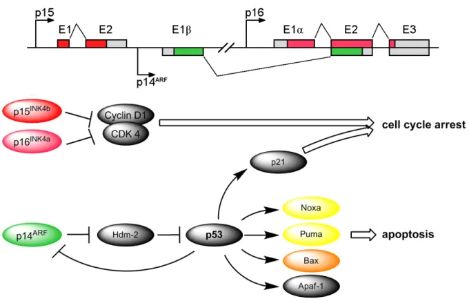
Figure 5. Schematics of the INK4b-ARF-INK4a locus. Activation of different pathways by transcribed genes:
p15 and p16 inhibit the cyclin dependent kinase 4 (CDK4). p14ARF inhibits Hdm-2, thereby stabilising p53, leading to downstream transcription of pro-apoptotic proteins like Noxa, Puma, Bax, Apaf-1 and others.
2009). Some studies have shown, that ARF degradation depends, partially, on the proteasome and that, although it lacks lysine, ARF can undergo N-terminal ubiquitination independent of p53 and Hdm-2 (Pollice et al., 2008). ARF is stable when expressed within the nucleolus, but turns over more rapidly in the nucleoplasm. In the nucleolus, ARF assumes a stable structure thanks to its sequestration by nucleophosmin (NPM1/B23) which prevents its nucleoplasmic degradation (den Besten et al., 2005; Colombo et al., 2006). From within the nucleolus ARF can attenuate translation by inhibiting RNA polymerase I and thereby ribosomal RNA (rRNA) synthesis (Sugimoto et al., 2003).
To suppress aberrant cell growth in response to oncogene activation, ARF activates the transcription factor p53 by neutralising the inhibitory effects of the ubiquitin ligases Hdm-2 and ARF-BP1/Mule (ARF-binding protein 1 / Mcl-1-ubiquitin ligase E3). Both proteins are ubiquitin ligases for p53 and can inhibit its tumour suppressor functions. ARF interacts directly with Hdm-2 and blocks Hdm-2-mediated ubiquinilation, nuclear export and degradation of p53 by the proteasome (Sherr, 2006). P53 then triggers the expression of cell cycle inhibitory and pro-apoptotic genes, e.g. puma to induce apoptosis (Hemmati et al., 2010). Although the ARF‒p53 axis was proposed initially to constitute the main pathway of apoptosis induction, a number of publications clearly indicate, that ARF is able to restrict cell proliferation and induce cell death through p53-independent pathways (Lowe and Sherr, 2003). For example, expression of p19ARF induces a G1 arrest in cells lacking p53 (Carnero et al., 2000; Weber et al., 2000). Loss of the cyclin-dependent kinase inhibitor p21, that is regulated by p53 (see Figure 5), disrupts this p14ARF induced G1 arrest and increases the amount of apoptosis (Hemmati et al., 2005). In double deficient p53/p21 cells, p14ARF expression results in a cell cycle arrest in the G2 phase by targeting p34cdc2 kinase, possibly representing an additional fail-safe mechanism preventing unrestrained proliferation (Normand et al., 2005).
Our group showed, that p53 independent mitochondrial activation and subsequent induction of apoptosis by p14ARF is also independent of pro-apoptotic Bax and dependent of Bak in p53 deficient cells (Hemmati et al., 2002; Müer et al., 2011 submitted). Loss of Bax in cells can be functionally complemented by its homolog Bak, suggesting the induction of different pathways and BH3-only proteins (Hemmati et al., 2006). One possible apoptotic pathway is targeting of C-terminal binding protein (CtBP) by ARF (Paliwal et al., 2006; Kovi et al., 2010).
Next to apoptosis, ARF plays an important role in induction of other types of cell death, e.g.
autophagy. Although autophagy is a pro-survival mechanism where starving cells degrade their own cellular components through the lysosomal machinery for recycling purposes, deregulation of autophagy also plays a critical role in the initiation and progression of tumours (Pimkina and Murphy, 2009). Again, autophagy can be induced by ARF independent of p53 (Abida and Gu, 2008).
p16INK4a/p19ARF genes also appear to be coregulated during aging. In rodents, p19ARF seems to play a predominant role in senescence entry, since in mouse embryonic fibroblasts (MEFs), the loss of the p19ARF/p53, but not of the p16INK4a/pRB axis, leads to spontaneous escape from senescence (Gil and Peters, 2006; Kim and Sharpless, 2006).
1.4The p53 family of proteins
The p53 family consists of the tumour suppressor p53 (or tumour protein 53, TP53) and its two identified homologues, p63 and p73 (Kaghad et al., 1997; Yang et al., 1998). They share homology in their transactivation, DNA-binding, and oligomerisation domains with about 60 % amino acid identity in the DNA-binding domain (Melino et al., 2003). The family members are differentially involved in the regulation of cell cycle and DNA-damage-induced apoptosis (Bénard et al., 2003). Similar to TP53, the TP73 and TP63 genes each have two promoters (P1 and P2), and their transcripts undergo extensive splicing at the NH2- and COOH-termini. Splice variants that retain the N-terminal transactivation domain are named TA isoforms. TAp73, TAp63 and p53 share a set of target genes such as p21waf1, bax, puma, and noxa to induce cell cycle arrest and apoptosis (Harms et al., 2004; Perez and Pietenpol, 2007).
In general, the p53 tumour suppressor protein functions as a transcription factor that regulates the expression of stress response genes and mediates a variety of anti-proliferative processes.
It is known to mediate its effect through the activation of genes regulating cell cycle checkpoints, DNA damage and repair response, and apoptosis (Stewart and Pietenpol, 2001).
In contrast, p63 is a putative oncogene and is required for the development and maintenance of stratified epithelium (Westfall and Pietenpol, 2004). It can bind to p53 DNA consensus sequences and induces apoptosis independent of p53 status when overexpressed in cells by triggering signalling via death receptors and the mitochondria (Gressner et al., 2005).
Endogenous p63 has been shown to be induced by many chemotherapeutic agents and
blocking its function might confer chemoresistance (Petitjean et al., 2005). Interestingly, p14ARF has been shown to inhibit p63 mediated transactivation of p53 responsive genes (Calabrò et al., 2004).
The high homology shared by p73 and p53 and the observation that p73 maps to chromosome 1p36.1, a region frequently deleted in several tumours, including neuroblastoma, colorectal cancer and breast cancer, initially suggested that p73 is a tumour suppressor. It is involved in neurogenesis, neuron survival and the inflammatory response (Irwin and Kaelin, 2001a).
TP73 is directly activated by E2F and can lead to the activation of pro-apoptotic genes in a p53-independent manner (Stiewe and Pützer, 2000).
Mutant p53 proteins form complexes with other transcription factors, such as p63 or p73, and either inactivate them or change their pattern of transcription, altering the properties of the cancer cell. Indeed, mutant p53/p73 complexes have been detected in cancer cells (Irwin et al., 2003), and an altered chemotherapeutic response could be the result of an inactivation of p73 apoptotic functions (Irwin and Kaelin, 2001b). Both p63 and p73 are rarely mutated in cancer cells and therefore could be putative targets for novel anti-cancer therapeutics. Still, many questions remain about their roles in human tumourigenesis as data suggests that p63 and p73 have both tumour suppressive and oncogenic properties (Zaika and El-Rifai, 2006).
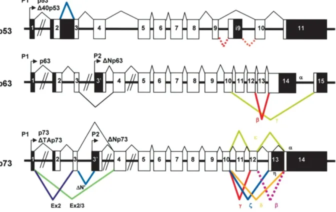
Figure 6. Overview of the p53 protein family. Structure of the human p53, p63, and p73 genes and their multiple splice variants. Coding sequence in white; noncoding sequence in black (modified from Bourdon et al., 2005).
2. Aim of the study
The p14ARF tumour suppressor plays a central role in the regulation of cell cycle arrest, apoptosis, and cellular senescence. Expression of cellular or viral oncogens leads to a rapid upregulation of p14ARF at the mRNA and protein level. Upon upregulation, cell cycle arrest and/or cell death is mediated via the p14ARF/Hdm-2/p53 pathway. Apoptosis by this pathway is mainly executed via p53 mediated upregulation of Puma leading to mitochondrial activation (Hemmati et al., 2010). Unfortunately, this pathway is not functional in most tumours because of mutations or deletions of p53. However, we previously showed, that p14ARF can also induce apoptosis and cell cycle arrest independent of p53 (Hemmati et al., 2002, 2005), but the mechanism remains elusive.
The aim of this study was to analyse this mechanism by different approaches. First, the p53 family members p63 and p73 can be activated by p14ARF and contribute to programmed cell death induction in the absence of p53 (Yao and Chen, 2010; Slade and Horvat, 2011).
Therefore, we wanted to analyse if these p53 homologs are able to compensate for loss of p53 in p14ARF induced apoptosis. Second, cellular organelles other than the mitochondria could be utilised to mediate p14ARF induced apoptosis. Next to the extrinsic and intrinsic mitochondrial pathway, induction of apoptosis via the endoplasmic reticulum (ER) has been reported.
Comparative expression analysis of genes involved in the unfolded protein response (UPR), a mechanism to relay stress signals from the ER, by use of ER stressing drugs and p14ARF should be performed. Third, the BH3-only protein Puma plays an essential role in p14ARF induced apoptosis via p53. BH3-only proteins might also be involved in apoptosis induced by p14ARF in p53 deficient cells. Induction of BH3-only genes by p14ARF in p53 pro- and deficient cell lines and via the ER stress pathway should be addressed.
3. Materials and Methods
3.1 Materials 3.1.1 Tools
ABI7300 Applied Biosystems, Darmstadt, Germany
BD FACScan™ Becton Dickinson, Franklin Lakes, USA
centrifuge 5415C Eppendorf, Hamburg, Germany
centrifuge Varifuge 3.0R Heraeus Sepatech, Hanau, Germany
Curix 60 AGFA, Mortsel, Belgium
electroporator Gene Pulser II Bio-Rad, München, Germany gel apparatus MiniSub Cell Bio-Rad, München, Germany
gel doc imager Bio-Rad, München, Germany
incubator Certomat R / Certomat H B. Braun Biotech International, Allentown, USA laboratory scales BP210S Satorius, Göttingen, Germany
PCR cycler GeneAmp PCR System 2400 Applied Biosystems, Norwalk, USA
pH meter CG825 Schott, Hofheim am Taunus, Germany
photometer GeneQuant II Pharmacia Biotech, San Francisco, USA
Power Pac 300 Bio-Rad, München, Germany
precision scales BP3100S Satorius, Göttingen, Germany
shaker Duomax 1030 Heidolph, Schwabach, Germany
Sonopuls GM70 Bandelin, Berlin, Germany
thermo shaker compact Eppendorf, Hamburg, Germany Vortex Genie II Scientific Industries, Bohemia, USA
3.1.2 Chemicals
acetic acid Merck, Darmstadt, Germany
agarose Biozym, Hessisch-Oldendorf, Germany
ampicillin AppliChem, Gatersleben, Germany
bacto agar BD Biosciences, Heidelberg, Germany
β-mercaptoethanol Carl Roth, Karlsruhe, Germany
boric acid Carl Roth, Karlsruhe, Germany
bromphenol blue Carl Roth, Karlsruhe, Germany
DMEM Gibco, Karlsruhe, Germany
DMSO (dimethylsulfoxid) Merck, Darmstadt, Germany doxycycline hydrochlorid Sigma, Taufkirchen, Germany
EDTA (ethylenediaminetetraacetic acid) Carl Roth, Karlsruhe, Germany
ethanol J. T. Baker, Griesheim, Germany
ethidiumbromid Sigma, Taufkirchen, Germany
FCS (fetal calf serum) Gibco, Karlsruhe, Germany gel loading solution Sigma, Taufkirchen, Germany
glycerol J. T. Baker, Griesheim, Germany
HCl (hydrochloric acid) Merck, Darmstadt, Germany
isopropanol Carl Roth, Karlsruhe, Germany
kanamycin Carl Roth, Karlsruhe, Germany
McCoy’s 5A Gibco, Karlsruhe, Germany
methanol Carl Roth, Karlsruhe, Germany
NaCl (sodium chloride) Carl Roth, Karlsruhe, Germany
Neomycin (G418) Carl Roth, Karlsruhe, Germany
PBS (phosphate buffered saline) Gibco, Karlsruhe, Germany penicillin/streptomycin Biochrom AG, Berlin, Germany phenol-chloroform-isoamylalcohol Carl Roth, Karlsruhe, Germany
puromycin Carl Roth, Karlsruhe, Germany
SDS (sodium dodecyl sulfate) Carl Roth, Karlsruhe, Germany
thapsigargin A.G. Scientific, San Diego, CA, USA
tris base Carl Roth, Karlsruhe, Germany
tris hydrochlorid Carl Roth, Karlsruhe, Germany
trypsin-EDTA Gibco, Karlsruhe, Germany
trypton Carl Roth, Karlsruhe, Germany
tunicamycin A.G. Scientific, San Diego, CA, USA
yeast extract Carl Roth, Karlsruhe, Germany
3.1.3 Solutions, buffers and media
Antibiotics (1000x) 50 mg/ml Ampicillin; 30 mg/ml Kanamycin
LB medium 10 g Bacto-tryptone
5 g Bacto-yeast extract 10 g NaCl
ad 1 l bidest H2O; pH adjusted to 7.2; autoclave
LB agar LB medium
15 g/l Bacto agar; autoclave
6x DNA loading buffer 0.2% Bromphenol blue 60% Glycerol
60 mM EDTA 10x Orange G DNA loading buffer 20 g Sucrose
100 mg Orange G ad 50 ml H2O
10x TBE buffer 108 g Tris base
55 g Boric acid
40 ml 0.5 M EDTA; pH 8.0 ad 1 l H2O
TE buffer 10 mM Tris-HCl
1 mM EDTA; pH 8.0
10x annealing buffer 10 mM Tris-HCl; pH 7.6
100 mM NaCl 1 mM EDTA
4x Laemmli buffer 8g SDS
40 ml Glycerin
40 ml 0.6 M Tris pH 6.8 80 mg bromophenol blue ad 80 ml bidest H2O 20 ml β-mercaptoethanol SDS-PAGE running buffer 25 mM Tris-HCl pH 8.3
190 mM Glycin 0.1% SDS Western blotting running buffer 20 mM Tris
150 mM Glycin 20% methanol 0.08% SDS
3.1.4 Kits
ChIP Kit Diagenode, Liege, Belgium
Invisorb Plasmid Maxi Kit Invitek, Berlin, Germany Invisorb Spin Plasmid Mini Two Invitek, Berlin, Germany QIAquick Gel Extraction Qiagen, Hilden, Germany
3.1.5 Markers
rainbow molecular weight marker GE Life Sciences, Fairfield, CT, USA 1 kb Plus DNA ladder Invitrogen, Karlsruhe, Germany
100 bp DNA ladder Invitrogen, Karlsruhe, Germany
3.1.6 Antibodies
β-actin (Ab-1) Mouse mAB (JLA20) Merck, Darmstadt, Germany
ATF4/CREB-2 (C-20): sc-200 Santa Cruz Biotechnology, Santa Cruz, CA, USA
Bak NT (TC102) Merck, Darmstadt, Germany
Bax NT, #06-499 Millipore, Billerica, MA, USA
BiP/GRP78 #610979 BD Biosciences, Heidelberg, Germany
CHOP/GADD153 (B-3): sc-7351 Santa Cruz Biotechnology, Santa Cruz, CA, USA eIF2α-phospho (Ser51) #9721 Cell Signaling, Frankfurt, Germany
p14ARF (Clone 14P02) Thermo Scientific, Bonn, Germany
p53 #554293 BD Biosciences, Heidelberg, Germany
p53 (PAb421) #OP03 Merck, Darmstadt, Germany
anti goat, #6165-05 Southern Biotech, Birmingham, AL, USA anti mouse #1031-05 Southern Biotech, Birmingham, AL, USA anti rabbit #4050-05 Southern Biotech, Birmingham, AL, USA anti rat #3050-05 Southern Biotech, Birmingham, AL, USA
goat-anti-rabbit IgG (H+L) FITC labeled F(ab)2 Jackson Immuno Research, W. Grove, PA, USA goat-anti-mouse IgG (H+L) FITC labeled F(ab)2 Jackson Immuno Research, W. Grove, PA, USA If not stated otherwise, primary antibodies were used at a dilution of 1:1,000 and the secondary antibody at a dilution of 1:10,000.
3.1.7 Enzymes restriction endonucleases
(AfeI, BamHI, BglII, ClaI, EcoRI, EcoRV, HindIII, KpnI, NdeI, NheI, NotI, PacI, SacII, SpeI, SwaI, XbaI, XhoI)
NEB, Frankfurt am Main, Germany
CrimsonTM Taq DNA polymerase (5 U/µl) NEB, Frankfurt am Main, Germany GoTaq® DNA polymerase (5 U/µl) Promega, Mannheim, Germany
InviTaq polymerase Invitek, Berlin, Germany
T4 DNA Ligase (100 U/µl) Invitrogen, Karlsruhe , Germany
3.1.8 Oligonucleotides
Oligonucleotides for genotyping, sequencing, and ChIP assay were synthesised by BioTez GmbH (Berlin, Germany) or Tib MolBiol (Berlin, Germany). Primer and probes for RT-PCR (TaqMan) analysis were ordered from Tib Molbiol as complete gene expression assay or synthesised by BioTez GmbH.
TaqMan primer:
Name Forward and reverse primer Probe
p53 5‘- AgT gTg gTg gTg CCC TAT gAg C -3‘
5‘- CgC CCA TgC Agg AAC TgT TAC -3‘ 6FAM-Tgg CTC TgA CTg TAC CAC CAT CCA CTA CAA CTA C--TMR p14ARF 5‘- CCC TCg TgC TgA TgC TAC TgA ggA -3‘
5‘- ggC gCT gCC CAT CAT CAT gAC -3‘ 6FAM-AgC gTC TAg ggC AgC AgC CgC TTC CTA gAA--TMR
ATF4 5‘- CAg TCC CTC CAA CAA CAg CAA -3‘
5‘- AAg TCg AAC TCC TTC AAA TCC ATT -3‘ 6FAM-Agg ATg CCT TCT CCg ggA CAg ATT g--TMR
ATF6 5‘- TCT CTT TgC TgA ACT Cgg TTA TTT C -3‘
5‘- AAT TgT TTT CAT ACg TCT CAT TTg CT -3‘ 6FAM-CAg ACA CTg ATg AgC TgC AAT Tgg AA--TMR
CHOP 5‘- ggA AAT gAA gAg gAA gAA TCA AAA AT -3‘
5‘- gTT CTg gCT CCT CCT CAg TCA -3‘ 6FAM-TTC ACC ACT CTT gAC CCT gCT TCT CTg g-TMR
eIF2α 5‘- gAg gAT CAg AAg gAC TgT ACA Tgg T -3‘
5‘- TCT TCC CAg ATT CCC TTg gA -3‘ 6FAM-ATg gAC CAC CAC ATT TTA CAg AAA gCA CAg Tg--TMR
BiP/GRP78 5‘- gCA ACC AAA gAC gCT ggA A -3‘
5‘- TgC CgT Agg CTC gTT gAT g -3‘ 6FAM-ATT gCT ggC CTA AAT gTT ATg Agg A--TMR
IRE1 5‘- AAg CAg gAC ATC Tgg TAT gTT ATT gA -3‘
5‘- CgT ACA Tgg TgA Tgg TgT ATT CTg TT -3‘ 6FAM-TTg TCA TCg gCC TTT gCA gAT AgT CTC Tg--TMR
PERK 5‘- gCA AAC CAg Agg TAT TTg ggA AT -3‘
5‘- ggT CTT ggTCCC ACT ggA AgA -3‘ 6FAM-ATg ATC ATT CCT TCC CTg gAT ggA gCC--TMR
XBP1u 5‘- CAg TgA Agg AAg AAC CTg TAg AAg ATg AC -3‘
5‘- CAg TAg gCA ggA AgA Tgg CTT Tg -3‘ 6FAM-ATC TCA AAT CTg CTT TCA TCC AgC CAC TgC--TMR
XBP1u+s 5‘- CgC TgA ggA ggA AAC TgA AAA A -3‘
5‘- TgT TCC AgC TCA CTC ATT CgA -3‘ 6FAM-AgC TCA gAC TgC CAg AgA TCg AAA gAA--TMR
BAD 5‘- gCA CAg CAA CgC AgA TgC -3‘
5‘- AAg TTC CgA TCC CAC CAg gA -3‘ 6FAM-CCA gCT ggA CgC gAg TCT TCC Ag-TMR
BID 5‘- CCA AgA Agg Tgg CCA gTC A -3‘
5‘- TCC TCA CgT Agg TgC gTA ggT -3‘ 6FAM-ACg CCg TCC TTg CTC CgT gAT gT
BIM 5‘- CCA ggC CTT CAA CCA CTA TCT -3‘
5‘- CCA ATA CgC CgC AAC TCT T -3‘ 6FAM-CTT CAA TgA ggC Agg CTg AAC CTg C--TMR
BMF 5‘- Tgg CAA CAT CAA gCA gAg gT -3‘
5‘- CTg CTg gTg TTg CTg CAC A -3‘ 6FAM-CAg ATT gCC CgA AAg CTT CAg Tg--TMR
BNIP3 5‘- gAg gAA CAC gAg CgT CAT gA -3‘
5‘- Agg TgC Tgg Tgg Agg TTg TC -3‘ 6FAM-CCA TCT CTg CTg CTC TCT CAT TTg CTg g-TMR
HRK 5‘- gCA ggC ggA ACT TgT Agg AA -3‘
5‘- TTT CTC CAA ggA CAC Agg gTT T -3‘ 6FAM-Cgg AgC CgA gAC CCA gCC g-TMR
NBK 5‘- CAC AgC CTg ggT CTg gCT T -3‘
5‘- TTA AgT gTg gTg AAA CCg TCC A -3‘ 6FAM-CAT CCC TgA TgT CCT CAg TCT ggT CgTXT--PH
NOXA 5‘- gCA AgA ACg CTC AAC CgA g -3‘
5‘- gCA gAA gAg TTT ggA TAT CAg -3‘ 6FAM-AAg TCg AgT gTg CTA CTC AA TCA--TMR
PUMA 5‘- AgA CAA gAg gAg CAg CAg Cgg -3‘
5‘- ACC TAA TTg ggC TCC ATC TCg g -3‘ 6FAM-CTC ATC ATg ggA CTC CTg CCC TTA CCC--TMR
SPIKE 5‘- CCT gCA AAg AAT ATg gTC AAg -3‘
5‘- CCC gCT gCT CAT ACA T -3‘ 6FAM-CAg AAA gCC TTg CgA gTT TTA AAg--TMR
ABL 5‘- Tgg AgA TAA CAC TCT AAg CAT AAC TAA Agg T -3‘
5‘- gAT gTA gTT gCT Tgg gAC CCA -3‘ 6FAM-CCA TTT TTg gTT Tgg gCT TCA CAC CAT T--TMR
Primer for ChIP-Assay:
Name Forward primer Reverse primer
NS 5‘- Agg CCA gTg ggA Agg CAg gT -3‘ 5‘- CCC TTg gCA ATg ggg TCC TTT CC -3‘
RE1 5‘- AgA ATC CgC ACT ggC gAC gg -3‘ 5‘- CTC ggg gCA TCC CgC AAA CA -3‘
RE2 5‘- gCA CCC TgC ACC CAC Tgg AC -3‘ 5‘- CCg TCg CCA gTg Cgg ATT CT -3‘
RE3 5‘- TgA ggg CAg ACg CCA ggT TT -3‘ 5‘- AAg Cgg AgC gCT CAA gAA gg -3‘
RE4 5‘- AgA CAg ggT TTC CCg TgT Tg -3‘ 5‘- CTg gAg AAg gCC TCA ggg Ag -3‘
RE5 5‘- TAA gCT CTC CAg CTC AgC AC -3‘ 5‘- TgC CTg TAA TCC CAg CTA CT -3‘
3.1.9 Vectors and Plasmids
For cloning and mutagenesis of genes and protein expression, a collection of vectors and plasmids available from the previous work in our laboratory has been used. All new constructs were analysed and verified by sequencing.
pGEM-T Promega, Madison, WI, USA
pIRESneo3 Clontech, Cat #6988-1
pIRESpuro3 Clontech, Cat #631619
pCMV-p53 Clontech, Cat #6004-1
pCMV-Sport6_ATF4 (IRATp970B0515D) cDNA ImaGenes, Berlin-Buch, Germany pOTB7_DDIT3 (IRAUp969G0819D6) cDNA ImaGenes, Berlin-Buch, Germany
pcDNA3-p14ARF provided by Philipp Hemmati
pAd1-∆1∆3+tTA provided by Bernd Gillissen
pAd2 TrePro provided by Bernd Gillissen. The insert contains a c-Myc epitope tag (EQKLISEEDL).
mPERK.∆C.9E10.pCDNA.amp provided by David Ron, Skirball Institute of New York University’s School of Medicine. The insert contains a c-Myc epitope tag (EQKLISEEDL) on its C-terminal end.
pGL3-Basic Promega, Madison, WI, USA
pRL-TK Promega, Madison, WI, USA
pIRESneo3-revMCS+XhoI and pIRESpuro3-revMCS+XhoI
Oligonucleotides containing restriction sites in a reverse order of pIRESneo3 and one added XhoI-site were designed (Fwd: 5’-CgA TAT CTC Cgg ATT CgA ATT Cgg ATC CAC Cgg TTA ACA ggC CTT AAg CgC TAg CCT CgA ggC-3’, Rev: 5’-ggC CgC CTC gAg gCT AgC gCT TAA ggC CTg TTA ACC ggT ggA TCC gAA TTC gAA TCC ggA gAT AT-3’) annealed and ligated into pIRESneo3 and pIRESpuro3 digested with ClaI and NotI.
pIRESneo3-revMCS+XhoI-mPERKΔC
Vectors mPERK.∆C.9E10.pCDNA.amp and pIRESneo3-revMCS+XhoI were digested with EcoRI and XhoI. The truncated PERK fragment was then ligated into pIRESneo3- revMCS+XhoI.
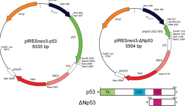
pIRESneo3-p53 and pIRESneo3-ΔNp53
The p53 cDNA was cloned into pIRESneo3 by digesting pCMV-p53 with NdeI and EcoRI and ligated into pIRESneo3, thereby replacing part of the human cytomegalovirus major immediate early promoter (PCMV IE) of pIRESneo3 by the one from pCMV-p53.
Figure 7. Schematics of pIRESneo3-p53 and pIRESneo3-ΔNp53 vectors. ΔNp53 contains amino acids 302- 393 of full length p53. Abbreviations: transactivation domain (TA), DNA binding domain (DBD) and tetramerisation domain (TET).
Based on the work of Shaulian et al. we created a dominant negative p53 (ΔNp53) gene, lacking the transactivation and DNA binding domains (Shaulian et al., 1992). The PCR product was raised by using a forward primer including a NheI site and the startcodon (5’- TAg CTA gCA Tgg ggA gCA CTA AgC gAg C-3’) and a reverse primer including an EcoRI site (5’-ATA gAA TTC TCA gTC TgA gTC Agg CC-3’) on pCMV-p53 as template. It was ligated into pGEM-T vector and analysed by sequencing. Correct clones were digested with EcoRI and NheI and ligated into the multiple cloning site of pIRESneo3.
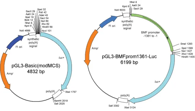
pGL3-Basic(modMCS)
Oligonucleotides containing restriction sites of pGEM-T were designed (Fwd: 5’-CgA TAT CTC Cgg ATT CgA ATT Cgg ATC CAC Cgg TTA ACA ggC CTT AAg CgC TAg CCT CgA ggC-3’, Rev: 5’-ggC CgC CTC gAg gCT AgC gCT TAA ggC CTg TTA ACC ggT ggA TCC gAA TTC gAA TCC ggA gAT AT-3’) annealed and ligated into pGL3-Basic digested with KpnI and HindIII.
Figure 8. Schematics of pGL3-Basic(modMCS) and pGL3-BMFprom1361-Luc. Vectors with bmf promotor inserts from -1114 to -1, -821 to -1, -427 to -1, and -229 to -1 were cloned accordingly.
pGL3-BMFprom1361-Luc
Vector containing basepairs -1361 to -1 of the bmf promoter. PCR product raised from HCT116 wt genomic DNA with primers (Fwd: 5’-TAA gCT CTC CAg CTC AgC AC- 3’, Rev: 5’-AAA ATA CgC CTg CTC ggg gC-3’) was ligated into pGEM-T vector.
This vector was digested with SacII and SpeI and the insert ligated into pGL3- Basic(modMCS).
pGL3-BMFprom229/427/821/1114-Luc
PCR products raised from pGL3-BMFprom1361-Luc with primers (Fwd 229: 5’-AgA ATC CgC ACT ggC gAC gg-3’, Fwd 427: 5’-gCA CCC TgC ACC CAC Tgg AC-3’, Fwd 821: 5’-TgA ggg CAg ACg CCA ggT TT-3’, Fwd 1114: 5’-AgA CAg ggT TTC CCg TgT Tg-3’, and Rev: 5’-AAA ATA CgC CTg CTC ggg gC-3’) were ligated into pGEM-T vector. pGEM-T vectors were then tested by sequence analysis, digested with SacII and SpeI and inserts ligated into pGL3-Basic(modMCS).
pcDNA3.1(+)-CHOP and pcDNA3.1(+)-ATF4
pAd2 Vectors containing chop and atf4 genes (see 2.1.10) were digested with BamHI and XbaI and ligated into pcDNA3.1(+) vector digested accordingly.
pcDNA3.1(+)-p14ARF
pcDNA3-p14ARF vector was digested with BamHI and EcoRI and ligated into pcDNA3.1(+) vector digested accordingly.
3.1.10 Adenoviruses
Ad5-CMV-LacZ provided by Bernd Gillissen
Ad5-CMV-p14ARF provided by Philipp Hemmati
Ad5-CMV-p14ARF(Tet) provided by Antje & Anja Richter Ad5-CMV-ATF4(Tet) and Ad5-CMV-CHOP(Tet)
Oligonucleotides containing BamHI and XbaI restriction sites were designed (ATF4 Fwd:
5’-TTC gAA TTC ggA TCC ATg ACC gAA ATg AgC-3’, ATF4 Rev: 5’-gAg CTC gAg TCT AgA CTA ggg gAC CCT TTT-3’; CHOP Fwd: 5’-TTC gAA TTC
ggA TCC ATg gCA gCT gAg TCA TTg CCT TTC TTC-3’, CHOP Rev: 5’-AgA TCT AgA TCA TgC TTg gTg CAg ATT CAC CAT TCg-3’). PCR products raised by these primers from pCMV-Sport6_ATF4 (IRATp970B0515D) and pOTB7_DDIT3 (IRAUp969G0819D6), respectively, were digested with BamHI and XbaI and ligated into pAd2 vectors digested accordingly. Shuttle plasmids pAd2 containing ATF4 and CHOP inserts were digested with PacI and NotI and recombinated with ClaI digested pAd1-
∆1∆3+tTA vector in E. coli BJ5183 cells. pAd1 containing ATF4 and CHOP inserts were transfected in HEK 293 cell lines by calcium phosphate precipitation. After plaque formation, supernatant was used to infect HEK 293 cells to amplify viruses. All viruses were banded in CsCl gradients, dialysed, and stored in aliquots.
3.1.11 Bacteria
Escherichia coli DH5α (Genotype: fhuA2, Δ(argF-lacZ)U169, phoA, glnV44, Φ80, Δ(lacZ)M15, gyrA96, recA1, relA1, endA1, thi-1, hsdR17), Escherichia coli SCS110 (Genotype: rpsL, thr, leu, endA, thi-1, lacY, galK, galT, ara, tonA, tsx, dam, dcm, supE44,
∆(lac-proAB)) and Escherichia coli JM109 (Genotype: endA1, recA1, gyrA96, thi, hsdR17 (rk–, mk+), relA1, supE44, Δ(lac-proAB), [F´ traD36, proAB, laqIqZΔM15]) were used for transformations and plasmid amplifications.
Escherichia coli BJ5183 (Genotype: endA, scbBC, recBC, galK, met, thi-1, bioT, hsdR (Str)) were used for homologic recombinations.
3.1.12 Cell lines
All cells were kept in an incubator at 37 °C, 5% CO2 and 95% humidity.
HEK293 is derived from human embryonal kidney cells which were transformed by adenovirus 5 (Ad5) (Graham et al., 1977). They constitutively express Ad5 specific E1 proteins which allow replication and transcription of adenoviral DNA. The cell line is used for amplification and titration of human adenoviruses. Cells are grown in DMEM (Dulbecco’s Modified Eagle Medium) supplemented with 10 % FCS (heat inactivated for 30 min at 56 °C), 100 U/ml penicillin and 0.1 μg/ml streptomycin.