Research Collection
Doctoral Thesis
Longitudinal assessment of frailty and osteosarcopenia in an in vivo model of premature aging
Author(s):
Scheuren, Ariane C.
Publication Date:
2020
Permanent Link:
https://doi.org/10.3929/ethz-b-000423860
Rights / License:
In Copyright - Non-Commercial Use Permitted
This page was generated automatically upon download from the ETH Zurich Research Collection. For more information please consult the Terms of use.
ETH Library
Longitudinal assessment of frailty and osteosarcopenia in an in vivo
model of premature aging
A thesis submitted to attain the degree of
DOCTOR OF SCIENCES of ETH ZURICH
(Dr. sc. ETH Zurich)
presented by
Ariane C. Scheuren
M.Sc. Biomedical Engineering, ETH Zurich born on 03.12.1989
citizen of Luxembourg
accepted on the recommendation of
Prof. Dr. Ralph Müller, examiner Prof. Dr. Ilaria Bellantuono, co-examiner
2020
Acknowledgements i
Summary iii
Zusammenfassung vii
1 Introduction 1
1.1 Thesis motivation 2
1.2 Specific aims 5
1.3 Thesis outline 5
2 Background 13
2.1 Bone mechanobiology in mice: toward single-cellin vivomechanomics 14 3 Development of anin vivomodel of premature aging for the longitudinal assessment of
bone and frailty 49
3.1 Optimization of anin vivomultiscale mechanobiology approach 50 3.2 Optimization of longitudinalin vivophenotyping techniques 81 4 Longitudinal assessment of frailty and osteosarcopenia in anin vivomodel of premature
aging 107
4.1 Hallmarks of frailty and osteosarcopenia in prematurely aged PolgAD257A/D257Amice 108
5 Synthesis 155
Curriculum Vitae 171
The work presented in this thesis would not have been possible without the support of a number of people. I would like to express my sincere gratitude to them.
First of all, I would like to thank Prof. Dr. Ralph Müller for giving me the opportunity to work at the Institute for Biomechanics. His motivation, energy and particularly his ability to always find a good solution have been very inspiring to me. I greatly appreciated his guidance, support and experience throughout my PhD, from which I have learnt a lot, not only for my PhD but most definitely also for my future.
Special thanks go to my co-supervisor Gisela Kuhn, who was always there for me whenever I needed help, advice or reassurance in my work. She always offered to help - be it in the lab or by reviewing my chapters for this thesis. I really appreciated her honesty, caring personality and efforts in making the work environment a nicer place.
I would like to thank Prof. Dr. Ilaria Bellantuono not only for being my co-referee but also for welcoming me to her laboratory to learn new techniques. Her inputs on the PolgA mice and on the quantification of the frailty index were very valuable for the progression of my project.
Furthermore, I would like to thank Dr. Gommaar D’Hulst and Prof. Dr. Katrien de Bock for a very inspiring and fruitful collaboration, which brought my PhD to a different level. Their energy and enthusiasm was very encouraging to me and I enjoyed being a part of that during our collaboration.
Likewise, I would like to thank Nicole Grob McDonald, Paul Vallaster and Charlotte Roth for their successful master thesis projects as well as Bryant Schroeder and Ryan Plett for their great work in the LivE imaging project. I also thank Prof. Dr. Yoshitaka Kameo for our collaboration and interesting discussions regarding the first part of this thesis.
I thank the past and current members of the institute of biomechanics. Without their support, this thesis could not have been completed. In particular, I would like to thank Dr. Andreas Trüssel and Dr. Carly Taylor, from whom I took over parts of this project. Special thanks also go to Paul Graeme for numerous collaborative and fruitful projects over the past years. Patrik Christen for his support on the LivE imaging project, Dr. Duncan Tourolle né Betts for his support with any VMS related and technical questions, Dr. Jolanda Vetsch for her wide-ranging
support, Dr. Esther Wehrle for her technical advice concerning animal experiments, Dr. Angad Malhotra for his help in the laboratory and discussions, Nicholas Ohs for his technical help with the IfB framework. I would also like to thank Peter Schwilch and Marco Hitz from the Center for Mechatronics and Innovation (CMI) for their support with the animal loading devices.
Special thanks go to Felicitas Flohr for being a great office mate for multiple years and becoming a very dear friend. We shared many moments together, hopefully there will be many more to come. In this respect, I also thank my next office mate Daniele Boaretti, for the nice coffees we’ve shared and for his help with python issues. Special thanks also go to Caitlyn Collins for always offering to review my work (including chapters in this thesis), no questions asked. I also thank her for being there for me the last few months and offering her advice when needed. In this respect, I am also very grateful to Gian Nutal for reviewing parts of this thesis.
Last but not least, I am extremely thankful to Olivier Del Fabbro, my parents, my sisters Paulina and Annelise, Bruna, who always believed in me, encouraged me and always put up with me no matter my mood.
Finally, I am thankful for financial support of the European Cooperation in Science and Technology (COST Action BM1402: MouseAGE) and the European Research Council (ERC Advanced MechAGE ERC-2016-ADG-741883).
Zurich, March 2020 Ariane Scheuren
Frailty is a geriatric syndrome characterized by increased susceptibility to adverse health outcomes. One major determinant thereof is the gradual weakening of the musculoskeletal system and the associated osteosarcopenia. Although anabolic interventions such as mechanical stimuli are known to promote bone and muscle mass, it remains unclear whether the ability of the musculoskeletal system to sense mechanical signals is maintained with age. A better understanding of the pathophysiology of osteosarcopenia will help to identify interventions to strengthen the musculoskeletal system, which ultimately will be beneficial for the prevention and/or treatment of frailty. With the advent of longitudinal in vivo phenotyping techniques, animal models are of increasing interest in aging studies as disease progression can be monitored over time in multiple tissues of individual animals. This not only provides a more comprehensive analysis of multi-system dysfunctions but also reduces the number of required animals. However, a suitable animal model mimicking frailty and osteosarcopenia is still lacking.
Therefore, the presented thesis has been divided into three aims: (i) To develop an approach to study bone mechanobiologyin vivoacross multiple scales (ii) To develop an approach to study frailty and osteopeniain vivoin a model of premature aging (iii) To longitudinally assess frailty and osteosarcopenia in anin vivomodel of premature aging.
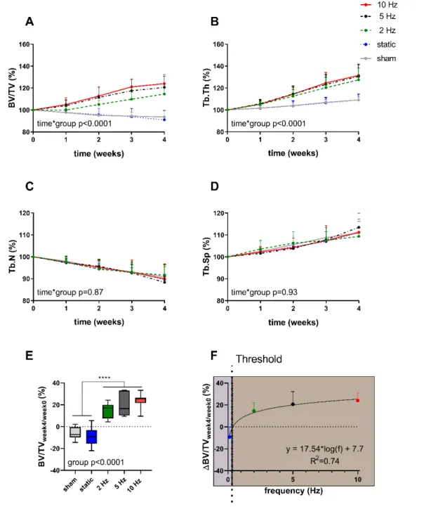
To address the first aim, a previously developed methodological platform known as “Localin vivoEnvironment (LivE) histochemistry” was optimized to investigate the relationship between loading frequency and bone adaptation across multiple scales. Specifically, the combination of longitudinal micro-CT imaging with micro-finite element (micro-FE) analysis of mouse caudal vertebrae subjected to either static or cyclic loading at varying frequencies showed that bone adaptation to load is controlled by local mechanical signals with net bone changes logarithmically dependent on loading frequency. In order to assess whether the link between bone remodeling and the local mechanical environments can also be observed at the cellular level, a rigid 2D-3D registration was used to map osteocytes, the orchestrators of bone remodeling, identified on 2D histology sections to the 3D micro-CT data. Following this, the expression of the anti-anabolic and anti-catabolic signaling factors Sclerostin and RANKL were evaluated both globally and locally in relation to the mechanical and remodeling environments.
Consistent with the anabolic responses observed at the tissue level, cyclic loading resulted in a down-regulation of osteocytic Sclerostin and RANKL expression on a global level. In line with this, the RANKL expression was lower in regions close to bone formation than in regions close to bone resorption, thus suggesting that alterations in protein levels are locally linked to the in vivomicroenvironment.
In a second part of this thesis, a long-term in vivomicro-CT imaging approach was coupled with longitudinal assessments of the clinical mouse frailty index (FI), a tool to quantify the accumulation of health deficits, in order to evaluate the suitability of a mouse model of premature aging as a model for frailty and senile osteoporosis. Furthermore, as the proposed method requires repeated scanning over a long period, potential biasing effects of radiation, anesthesia and handling associated within vivomicro-CT imaging were investigated. Although the long-term imaging approach can lead to small but significant changes in bone morphometric parameters, the comparison between genotypes was not impaired, and the overall health status of the animal (i.e., FI and body-weight) was not affected. Moreover, this study demonstrated that longitudinal designs including baseline measurements already at young age are more powerful at detecting age-related phenotypic changes than those including multiple groups with fewer imaging sessions.
Finally, in order to evaluate the suitability of the PolgA model as a model for frailty and osteosarcopenia, the thus established long-term FI and in vivo micro-CT approach was combined with extensive musculoskeletal phenotyping. Concomitant to a higher rate of deficit accumulation, PolgA mice displayed progressive musculoskeletal deterioration such as reduced bone and muscle mass as well as the functionality thereof. In addition to lower muscle weights and fiber area, PolgA showed impairments in grip-strength and concentric muscle forces.
Longitudinal micro-CT imaging of the 6th caudal vertebrae showed that PolgA had reduced bone micro-architectural integrity as well as lower bone turnover, thus mimicking senile osteoporosis as observed in humans. Lastly, this study showed that PolgA mutation altered the response to various anabolic stimuli in skeletal muscle and bone, indicating that the mechanoregulation of the musculoskeletal system may indeed change with age.
In summary, the application of the multiscale bone mechanobiology approach was successful to improve our understanding of the relationship between loading frequency and trabecular bone adaptation in vivo. Secondly, the application of long-term in vivo micro-CT imaging combined with longitudinal FI assessments was shown to be pivotal for monitoring the
development of frailty and senile osteoporosis in PolgA mice. Lastly, the integration of comprehensive musculoskeletal phenotyping showed that prematurely aged PolgA mice mimic multiple signs of frailty and of osteosarcopenia and thus provide a powerful model to improve our understanding of frailty and the aging musculoskeletal system. Taken together, the development of a multiscale mechanobiology approach as well as the identification of a model of frailty and osteosarcopenia provide the groundwork to elucidate the pathophysiology of osteosarcopenia and to test potential interventions, which ultimately will be constructive towards the prevention and/or treatment of frailty.
Die Gebrechlichkeit, auch Frailty-Syndrom genannt, ist ein altersbedingtes Syndrom, das gekennzeichnet ist von verminderter physiologischer Reserve und verringerter Widerstandsfähigkeit gegen Stressoren. Ein bestimmender Faktor dafür ist die allmähliche Schwächung des Bewegungsapparates und die damit verbundene Osteosarkopenie. Obwohl anabole Eingriffe wie mechanische Reize bekanntlich Knochen- und Muskelmasse fördern, bleibt unklar, ob die Fähigkeit des Bewegungsapparates, mechanische Signale zu erfassen, mit dem Alter erhalten bleibt. Von daher soll ein besseres Verständnis der Pathophysiologie der Osteosarkopenie dazu beitragen, mögliche Interventionen zur Stärkung des muskuloskelettalen Systems zu identifizieren, die letztlich für die Prävention und/oder Behandlung von Gebrechlichkeit von Nutzen sein werden. Mit dem Aufkommen von longitudinalen in vivo Phänotypisierungstechniken sind Tiermodelle von zunehmendem Interesse für Alterungsstudien, da der Krankheitsverlauf über die Zeit in mehreren Geweben einzelner Tiere beobachtet werden kann. Dies ermöglicht nicht nur eine umfassendere Analyse von Multisystem-Dysfunktionen, sondern reduziert auch die Anzahl der benötigten Tiere. Zur Zeit jedoch fehlt in der Forschung ein geeignetes Tiermodell, das die Gebrechlichkeit und Osteosarkopenie simuliert.
Daher wurde die vorliegende Arbeit in drei Ziele gegliedert: (i) Entwicklung eines Ansatzes zur Untersuchung der Knochenmechanobiologie in vivo über mehrere Skalen hinweg (ii) Entwicklung eines Ansatzes zur Untersuchung der Gebrechlichkeit und Osteopenie in vivoin einem Modell des vorzeitigen Alterns (iii) Longitudinale Evaluierung der Gebrechlichkeit und Osteosarkopenie in einemin vivoModell des vorzeitigen Alterns.
Um das erste Ziel zu erreichen, wurde eine zuvor entwickelte methodische Plattform, die als
"Local in vivo Environment (LivE) histochemistry" bekannt ist, optimiert, um die Beziehung zwischen Belastungsfrequenz und Knochenanpassung über mehrere Skalen hinweg zu untersuchen. Insbesondere die Kombination der longitudinalen Mikro-CT-Bildgebung mit der Mikro-Finite-Elemente-Analyse (Mikro-FE) von Schwanzwirbeln einer Maus, die entweder statischen oder zyklischen Belastungen mit unterschiedlichen Frequenzen ausgesetzt worden sind, hat gezeigt, dass die Knochenanpassung an die Belastung durch lokale mechanische Signale gesteuert wird; wobei die Netto-Knochenveränderungen logarithmisch von der
Belastungsfrequenz abhängen. Um zu beurteilen, ob die Verbindung zwischen dem Knochenumbau und den lokalen mechanischen Umgebungen auch auf zellulärer Ebene beobachtet werden kann, wurde eine starre 2D-3D-Registrierung verwendet, um Osteozyten, den Knochenumbau regulierende Zellen, die auf 2D-Histologieschnitten identifiziert wurden, den 3D-Mikro-CT-Daten zuzuordnen. Danach wurde die Expression der antianabolen und antikatabolen Signalfaktoren Sclerostin und RANKL sowohl global als auch lokal in Bezug auf die mechanische und umgebaute Umgebung bewertet. In Übereinstimmung mit den auf der Ebene des Gewebes beobachteten anabolen Reaktionen, führte die zyklische Belastung zu einer Herabregulation der osteozytischen Sclerostin- und RANKL-Expression auf globaler Ebene. In Übereinstimmung damit war die RANKL-Expression in Regionen, nahe der Knochenbildung, geringer als in Regionen in der Nähe der Knochenresorption. Dies deutet darauf hin, dass Veränderungen der Proteinkonzentrationen lokal mit der in vivo Mikroumgebung zusammenhängen.
In einem zweiten Teil dieser Arbeit wurde ein langfristiger in vivo Mikro-CT- Bildgebungsansatz mit longitudinalen Untersuchungen des klinischen Maus-Frailty-Index (FI) gekoppelt. Letzteres ist ein Instrument zur Quantifizierung der Akkumulation von Gesundheitsdefiziten, um die Angemessenheit eines Mausmodells des vorzeitigen Alterns als Modell für Gebrechlichkeit und senile Osteoporose zu bewerten. Da die vorgeschlagene Methode wiederholtes Scannen über einen langen Zeitraum erfordert, wurden darüber hinaus mögliche verzerrende Effekte von Strahlung, Anästhesie und Handhabung im Zusammenhang mit derin vivoMikro-CT-Bildgebung untersucht. Obwohl der Langzeit-Bildgebungsansatz zu kleinen, aber signifikanten Veränderungen von knochenmorphometrischen Parametern führen kann, wurde der Vergleich zwischen den Genotypen nicht beeinträchtigt. Zudem wurde der allgemeine Gesundheitszustand des Tieres (d.h. FI und Körpergewicht) nicht beeinflusst.
Darüber hinaus hat diese Studie gezeigt, dass longitudinale Studien, die bereits in jungen Jahren Basis-Messungen beinhalten, die altersbedingten phänotypischen Veränderungen besser erkennen können als solche die mehrere Gruppen mit weniger Mikro-CT Messungen umfassen.
Um schließlich die Eignung des PolgA-Modells als Modell für Gebrechlichkeit und Osteosarkopenie zu bewerten, wurde der so etablierte langfristige FI- und in vivoMikro-CT- Ansatz mit einer umfangreichen muskuloskelettalen Phänotypisierung kombiniert. Parallel zu einer höheren Defizitakkumulationsrate zeigten PolgA-Mäuse eine progressive Verschlechterung des muskuloskelettalen Systems, wie z.B. reduzierte Knochen- und
Muskelmasse sowie deren Funktionalität. Zusätzlich zu den geringeren Muskelgewichten und der geringeren Faserfläche zeigte PolgA eine Beeinträchtigung der Griffstärke und der konzentrischen Muskelkräfte. Die longitudinale Mikro-CT-Bildgebung des 6. Schwanzwirbels zeigte, dass PolgA-Mäuse reduzierte mikro-architektonische Integrität des Knochens sowie einen geringeren Knochenumsatz aufwiesen und damit die beim Menschen beobachtete senile Osteoporose nachahmte. Schliesslich zeigte diese Studie, dass die PolgA-Mutation die Reaktion auf verschiedene anabole Stimuli in Muskeln und Knochen veränderte, was darauf hindeutet, dass sich die Mechanoregulation des muskuloskelettalen Systems tatsächlich mit dem Alter verändern kann.
Zusammenfassend lässt sich sagen, dass die Anwendung des multiskalaren Knochenmechanobiologie-Ansatzes erfolgreich war, um unser Verständnis der Beziehung zwischen Belastungsfrequenz und der trabekulären Knochenadaption zu verbessern. Zweitens erwies sich die Anwendung der langfristigenin vivoMikro-CT-Bildgebung in Kombination mit longitudinalen FI-Bewertungen als entscheidend für die Überwachung der Entwicklung von Gebrechlichkeit und seniler Osteoporose bei PolgA-Mäusen. Letztlich zeigte die Integration einer umfassenden muskuloskelettalen Phänotypisierung, dass vorzeitig gealterte PolgA-Mäuse mehrere Anzeichen von Gebrechlichkeit und Osteosarkopenie nachahmen und somit ein leistungsfähiges Modell zum besseren Verständnis der Gebrechlichkeit und des alternden muskuloskelettalen Systems darstellen. Zusammengenommen bilden die Entwicklung eines multiskalaren mechanobiologischen Ansatzes sowie die Identifizierung eines Modells der Gebrechlichkeit und der Osteosarkopenie die Grundlage für die Aufklärung der Pathophysiologie der Osteosarkopenie und für das Testen potenzieller Interventionen, die letztlich zur Prävention und/oder Behandlung der Gebrechlichkeit beitragen können.
Introduc�on
1.1 Thesis motiva�on
Life expectancy has improved dramatically over recent decades with the proportion of people aged 65 or above expected to more than double in the next thirty years [1]. At the same time though, the number of people suffering from frailty will also rapidly increase. Although there is no universally accepted definition of frailty [2], it is considered as an age-related syndrome characterized by the decline of multiple physiological functions, leading to the accumulation of health deficits, and thus a higher vulnerability to adverse health outcomes such as morbidity and mortality [3, 4]. One of the most striking age-related physiological declines, in terms of function and structure, is that of the musculoskeletal system, resulting in diseases such as osteoporosis and sarcopenia. Recent evidence has shown that individuals suffering from both osteoporosis and sarcopenia, also known as “osteosarcopenia” [5, 6], are at higher risk of falls, fractures, disability, and frailty [7-9]. Although the exact mechanisms underlying age-related bone and muscle loss are not yet fully understood, it is known that the coupling between anabolic (i.e., bone formation and protein synthesis) and catabolic processes (i.e., bone resorption and protein degradation), which in healthy tissues are evenly balanced out, becomes less efficient with age [10]. This impaired remodeling capability can, at least in part, be explained by altered responses to anabolic stimuli such as mechanical signals [11-17]. In muscle tissue, this phenomenon, also termed “anabolic resistance”, has been linked to reduced protein synthesis due to diminished intracellular signaling through the mechanistic target of rapamycin complex 1 (mTORC1) pathway [12, 13] as well as to the reduction in the number and proliferation capacity of satellite cells [18-21]. The diminished response to mechanical stimuli in bone tissue on the other hand, most likely arises due to reduced mechanosensitivity of osteocytes, the main orchestrators of bone remodeling [22, 23]. However, the exact mechanisms by which mechanical stimuli are transduced into anabolic and catabolic signaling events remain unclear. A better understanding of the pathophysiology of osteosarcopenia will help to identify interventions that strengthen the musculoskeletal system, which ultimately will facilitate either the prevention or treatment of frailty or both.
Although several animal models have provided invaluable information on aspects of frailty and the aging musculoskeletal system [24-27], a suitable model that mimics both frailty and osteosarcopenia is currently still lacking. The PolgA(D257A/D257A)mouse model (referred to as PolgA) represents a desirable candidate. Due to the expression of an exonuclease-deficient
version of the mitochondrial DNA polymerase γ it develops multiple age-related phenotypes (including hearing loss, greying hair, kyphosis, enlarged heart, muscle loss) early in life and thus strongly mimics the multi-system morbidity observed during aging in humans [28, 29].
Intriguingly, subjecting PolgA mice to endurance exercise interventions resulted in a reduced multi-system pathology compared to their sedentary littermates [30, 31]. Even though PolgA mice have been shown to have lower muscle weights and reduced bone mineral density compared to their wild-type littermates (WT) [28-30, 32], little is known about the quality and functionality of their musculoskeletal system. Moreover, the frailty phenotype has not yet been assessed in these mice.
With the advent of tools such as the clinical mouse frailty index (FI), it is now possible to non- invasively quantify the accumulation of health deficits over time in individual animals [33, 34].
Indeed, the establishment of such standardized tools has been highly instrumental in the identification of rodent models of frailty and to test potential therapies to prevent, treat or even revert frailty [24, 25]. Similarly, longitudinal in vivomicro-CT imaging is widely used in pre- clinical studies to monitor changes in the three-dimensional bone micro-architecture over time in individual animals [35]. Furthermore, by registering consecutive images onto one-another, bone formation and resorption activities can be visualized and quantified over time [36], providing the possibility to detect differences in bone remodeling activities in response to various interventions such as ovariectomy [37] and mechanical loading [15-17, 38-40].
Although several animal models have been used to investigate changes in osteogenic responses to loading with age, contradicting results have been reported; while studies using a tibia-loading model have shown a reduced response of bone formation to mechanical loading both in cortical [16, 17] and in trabecular [15, 41] bone with age, trabecular bone adaptation in response to loading of the caudal vertebrae was maintained with age [39]. These differences could be explained by the different skeletal sites which were analyzed [42, 43] or by the differences in the applied loading waveforms (including for example loading frequency, peak strain, number of cycles), which are known to play a crucial role in determining osteogenic responses to load [43, 44]. Hence, an important prerequisite for the investigation of age-related changes in bone mechanosensitivity is a better understanding of how loads applied at the organ scale are sensed at the cellular scale, which ultimately leads to remodeling at the tissue scale. By combining in vivo micro-CT with advanced registration techniques and micro-finite element (micro-FE) analysis, previous studies have shown that bone remodeling activities at the tissue scale are
controlled by local mechanical signals [45, 46], enabling an improved understanding of bone mechanobiology. Moreover, by registering endpoint 2D histological data into the 3D bone volume obtained by micro-CT, an approach termed “Local in vivo Environment (LivE) histochemistry” has been established to link the protein expression of single osteocytes to their local mechanical microenvironment in vivo and to the bone remodeling history. Using this approach, a previous study has shown that the expression of Sclerostin, an inhibitor of bone formation, is correlated to the local remodeling and mechanical environment [47]. As such, investigating load-induced bone adaptation across multiple scales provides a promising approach to better understand changes in mechanosensitivity with age.
Taken together, the overall goal of this thesis was to longitudinally assess the development of frailty and of osteosarcopenia in prematurely aged PolgA mice. In addition, this study aimed to study the effects of various anabolic interventions such as eccentric contractions and mechanical loading on the musculoskeletal system of PolgA mice.
Therefore, prior to evaluating the PolgA mouse model, the first part of this thesis aimed at optimizing and further developing existing techniques to study bone adaptationin vivoacross multiple scales. Specifically, the combination of in vivo micro-CT with micro-FE and immunohistochemistry was used to study the effects of loading frequency on bone adaptation at the tissue and molecular scale. The second aim of this thesis was to develop an approach to longitudinally monitor the development of frailty and senile osteoporosis in an in vivomodel of premature aging. For this study, long-term in vivo micro-CT imaging was combined with longitudinal measurements of FI to monitor individual mice during the process of aging, providing the possibility to capture the onset of osteoporosis and to track other signs of aging.
Furthermore, as aging is accompanied with increased sensitivity to external stimuli, the cumulative effects of radiation, anesthesia and handling associated with micro-CT imaging on bone morphometric parameters as well as on the overall well-being of the animal were assessed.
Once established, the last aim of this thesis was to longitudinally assess frailty and osteosarcopenia in an in vivo model of premature aging. Therefore, in addition to the longitudinal micro-CT and FI measurements as developed in the previous aim, the quality and functionality of PolgA muscles was assessed using various in vivo and ex vivo phenotyping techniques such as the assessment of fore-limb grip-strength, concentric muscle forces and muscle masses, respectively. Lastly, the effects of various anabolic stimuli such as eccentric muscle contractions and mechanical loading in PolgA muscles and bones were investigated.
1.2 Specific aims
The immediate goal of this thesis was to longitudinally assess frailty and osteosarcopenia in an in vivomodel of premature aging. Specifically, the following three aims were defined:
Aim 1: Development of an approach to study bone mechanobiology in vivo across multiple scales.
Aim 2: Development of an approach to study frailty and osteopenia in vivo in a model of premature aging.
Aim 3: Longitudinal assessment of frailty and osteosarcopenia in anin vivomodel of premature aging.
1.3 Thesis outline
The thesis consists of five chapters. In addition to the current chapter outlining the motivation and specific aims, the content of the subsequent chapters is the following:
Chapter 2 provides a background chapter about bone mechanobiology in mice. After introducing existing models and methods to investigate load-induced bone remodeling at different length scales, more integrative approaches that allow to study bone mechanobiology across multiple scales – from the organ down to the molecular scale – are described. Lastly, an
“in vivomechanomics” approach is proposed to locally analyze the transcriptome of single cells with respect to their local 3D mechanicalin vivoenvironment.
Chapter 3 describes the optimization and further development of existing techniques that ultimately will allow to study bone mechanobiology in vivo across multiple scales in aging mice. In the first part, a “Local in vivo Environment (LivE) histochemistry” approach combining micro-CT, micro-FE and immunohistochemistry was used to study the effects of varying loading frequencies on bone adaptation across multiple length scales. Specifically, load applied at the organ scale was linked to local mechanical and remodeling environments at the tissue and molecular scale. In the second part, in vivo micro-CT imaging was optimized for usage in a mouse model of premature aging. For this, long-termin vivomicro-CT imaging over 20 weeks was combined with longitudinal assessments of the clinical mouse frailty index to
evaluate the suitability of the model as a model for frailty and senile osteoporosis. Furthermore, the cumulative effects of radiation, anesthesia and handling associated with long-termin vivo micro-CT imaging were investigated.
Chapter 4 describes the application of the techniques developed in the previous chapter to comprehensively evaluate the relevance of a mouse model of premature aging (PolgA(D257A/D257A)) as a model for frailty and osteosarcopenia. In addition to long-termin vivo micro-CT imaging and longitudinal assessments of the frailty index, we assessed the quantity and quality of bone and muscle tissue. Lastly, using a combination ofin vivo,ex vivo andin vitrotechniques, the effects of various anabolic stimuli such as mechanical loading, eccentric muscle contractions and leucine administration were assessed at various ages.
Chapter 5 is the synthesis of this thesis including the major findings, the limitations of the presented work, and an outlook for future research.
[1] United Nations. World Population Ageing 2019 (ST/ESA/SER.A/444). Department of Economic and Social Affairs, Population Division.www.unpopulation.org, 2020: New York, USA.
[2] Rodríguez-Mañas L, Gonzalez-Colaço Harmand M, Carcaillon L, Scuteri A, Sinclair A, Pelaez M, Van der Cammen T, Beland F, Bickenbach J, Delamarche P, Ferrucci L, Fried LP, Gutiérrez-Robledo LM, Féart C, Rockwood K, Rodríguez Artalejo F, Serviddio G, Vega E, Nicholson C, Mann G, Viña J, Chatterji S, Chodzko-Zajko W, Bergman H. Searching for an operational definition of frailty: a delphi method based consensus statement. The Frailty Operative Definition-Consensus Conference Project.
J Gerontol A Biol Sci Med Sci, 2013; 68(1):62-67.
[3] Whitson HE, Duan-Porter W, Schmader KE, Morey MC, Cohen HJ, Colon-Emeric CS.
Physical resilience in older adults: systematic review and development of an emerging construct.J Gerontol A 2016; 71(4):489-495.
[4] Clegg A, Young J, Iliffe S, Rikkert MO, Rockwood K. Frailty in elderly people.Lancet (London, England), 2013; 381(9868):752-762.
[5] Hassan E,Duque G. Osteosarcopenia: a new geriatric syndrome.Aust Fam Physician, 2017; 46:849-853.
[6] Hirschfeld HP, Kinsella R, Duque G. Osteosarcopenia: where bone, muscle, and fat collide.Osteoporos Int, 2017; 28(10):2781-2790.
[7] Frisoli A, Chaves PH, Ingham SJM, Fried LP. Severe osteopenia and osteoporosis, sarcopenia, and frailty status in community-dwelling older women: results from the Women's Health and Aging Study (WHAS) II.Bone, 2011; 48(4):952-957.
[8] Binkley N,Buehring B. Beyond FRAX®: It's time to consider “Sarco-Osteopenia”.J Clin Densitom, 2009; 12(4):413-416.
[9] Huo YR, Suriyaarachchi P, Gomez F, Curcio CL, Boersma D, Muir SW, Montero- Odasso M, Gunawardene P, Demontiero O, Duque G. Phenotype of osteosarcopenia in older individuals with a history of falling.J Am Med Dir Assoc, 2015; 16(4):290-295.
[10] DiGirolamo DJ, Kiel DP, Esser KA. Bone and skeletal muscle: neighbors with close ties.J Bone Miner Res, 2013; 28(7):1509-1518.
[11] Fry CS, Drummond MJ, Glynn EL, Dickinson JM, Gundermann DM, Timmerman KL, Walker DK, Dhanani S, Volpi E, Rasmussen BB. Aging impairs contraction-induced human skeletal muscle mTORC1 signaling and protein synthesis.Skelet Muscle, 2011;
1(1):11.
[12] Breen L,Phillips SM. Skeletal muscle protein metabolism in the elderly: interventions to counteract the 'anabolic resistance' of ageing.Nutr Metab (Lond), 2011; 8:68.
[13] Francaux M, Demeulder B, Naslain D, Fortin R, Lutz O, Caty G, Deldicque L. Aging reduces the activation of the mTORC1 pathway after resistance exercise and protein intake in human skeletal muscle: potential role of REDD1 and impaired anabolic sensitivity.Nutrients, 2016; 8(1).
[14] Stattin K, Michaëlsson K, Larsson SC, Wolk A, Byberg L. Leisure-time physical activity and risk of fracture: a cohort study of 66,940 men and women.J Bone Miner Res, 2017; 32(8):1599-1606.
[15] Willie BM, Birkhold AI, Razi H, Thiele T, Aido M, Kruck B, Schill A, Checa S, Main RP, Duda GN. Diminished response to in vivo mechanical loading in trabecular and not cortical bone in adulthood of female C57Bl/6 mice coincides with a reduction in deformation to load.Bone, 2013; 55(2):335-346.
[16] Birkhold AI, Razi H, Duda GN, Weinkamer R, Checa S, Willie BM. Mineralizing surface is the main target of mechanical stimulation independent of age: 3D dynamic in vivo morphometry.Bone, 2014; 66:15-25.
[17] Holguin N, Brodt MD, Sanchez ME, Silva MJ. Aging diminishes lamellar and woven bone formation induced by tibial compression in adult C57BL/6.Bone, 2014; 65:83-91.
[18] Nederveen JP, Joanisse S, Snijders T, Ivankovic V, Baker SK, Phillips SM, Parise G.
Skeletal muscle satellite cells are located at a closer proximity to capillaries in healthy young compared with older men.J Cachexia Sarcopenia Muscle, 2016; 7(5):547-554.
[19] Verdijk LB, Koopman R, Schaart G, Meijer K, Savelberg HHCM, van Loon LJC.
Satellite cell content is specifically reduced in type II skeletal muscle fibers in the elderly.Am J Physiol Endocrinol Metab, 2007; 292(1):E151-E157.
[20] Barberi L, Scicchitano BM, De Rossi M, Bigot A, Duguez S, Wielgosik A, Stewart C, McPhee J, Conte M, Narici M, Franceschi C, Mouly V, Butler-Browne G, Musaro A.
Age-dependent alteration in muscle regeneration: the critical role of tissue niche.
Biogerontology, 2013; 14(3):273-292.
[21] Parker MH. The altered fate of aging satellite cells is determined by signaling and epigenetic changes.Front Genet, 2015; 6:59-59.
[22] Klein-Nulend J, van Oers RF, Bakker AD, Bacabac RG. Bone cell mechanosensitivity, estrogen deficiency, and osteoporosis.J Biomech, 2015; 48(5):855-865.
[23] Hemmatian H, Bakker AD, Klein-Nulend J, van Lenthe GH. Aging, osteocytes, and mechanotransduction.Curr Osteoporos Rep, 2017; 15(5):401-411.
[24] Kane AE, Hilmer SN, Mach J, Mitchell SJ, de Cabo R, Howlett SE. Animal models of frailty: current applications in clinical research.Clin Interv Aging, 2016; 11:1519-1529.
[25] Banga S, Heinze-Milne SD, Howlett SE. Rodent models of frailty and their application in preclinical research.Mech Ageing Dev, 2019; 179:1-10.
[26] Watanabe K,Hishiya A. Mouse models of senile osteoporosis.Mol Aspects Med, 2005;
26(3):221-231.
[27] Richards PJ. Impact of senescence on bone quality: lessons from animal models of aging.Drug Discovery Today: Disease Models, 2014; 13:17-22.
[28] Trifunovic A, Wredenberg A, Falkenberg M, Spelbrink JN, Rovio AT, Bruder CE, Bohlooly YM, Gidlof S, Oldfors A, Wibom R, Tornell J, Jacobs HT, Larsson NG.
Premature ageing in mice expressing defective mitochondrial DNA polymerase.Nature, 2004; 429(6990):417-423.
[29] Kujoth GC, Hiona A, Pugh TD, Someya S, Panzer K, Wohlgemuth SE, Hofer T, Seo AY, Sullivan R, Jobling WA, Morrow JD, Van Remmen H, Sedivy JM, Yamasoba T, Tanokura M, Weindruch R, Leeuwenburgh C, Prolla TA. Mitochondrial DNA mutations, oxidative stress, and apoptosis in mammalian aging. Science, 2005;
309(5733):481.
[30] Safdar A, Bourgeois JM, Ogborn DI, Little JP, Hettinga BP, Akhtar M, Thompson JE, Melov S, Mocellin NJ, Kujoth GC, Prolla TA, Tarnopolsky MA. Endurance exercise rescues progeroid aging and induces systemic mitochondrial rejuvenation in mtDNA mutator mice.P Natl Acad Sci USA, 2011; 108(10):4135-4140.
[31] Ross JM, Coppotelli G, Branca RM, Kim KM, Lehtio J, Sinclair DA, Olson L.
Voluntary exercise normalizes the proteomic landscape in muscle and brain and improves the phenotype of progeroid mice.Aging Cell, 2019; 18(6):e13029.
[32] Kolesar JE, Safdar A, Abadi A, MacNeil LG, Crane JD, Tarnopolsky MA, Kaufman BA. Defects in mitochondrial DNA replication and oxidative damage in muscle of mtDNA mutator mice.Free Radic Biol Med, 2014; 75:241-251.
[33] Whitehead JC, Hildebrand BA, Sun M, Rockwood MR, Rose RA, Rockwood K, Howlett SE. A clinical frailty index in aging mice: comparisons with frailty index data in humans.J Gerontol A Biol Sci Med Sci, 2014; 69(6):621-632.
[34] Bellantuono I, de Cabo R, Ehninger D, Di Germanio C, Lawrie A, Miller J, Mitchell SJ, Navas-Enamorado I, Potter PK, Tchkonia T, Trejo JL, Lamming DW. A toolbox for the longitudinal assessment of healthspan in aging mice.Nat Protoc, 2020.
[35] Bouxsein ML, Boyd SK, Christiansen BA, Guldberg RE, Jepsen KJ, Muller R.
Guidelines for assessment of bone microstructure in rodents using micro-computed tomography.J Bone Miner Res, 2010; 25(7):1468-1486.
[36] Schulte FA, Lambers FM, Kuhn G, Müller R. In vivo micro-computed tomography allows direct three-dimensional quantification of both bone formation and bone resorption parameters using time-lapsed imaging.Bone, 2011; 48(3):433-442.
[37] Lambers F, Functional bone imaging in an in vivo mouse model of bone adaptation, aging and disease. 2011.
[38] Lambers FM, Schulte FA, Kuhn G, Webster DJ, Müller R. Mouse tail vertebrae adapt to cyclic mechanical loading by increasing bone formation rate and decreasing bone resorption rate as shown by time-lapsed in vivo imaging of dynamic bone morphometry.
Bone, 2011; 49(6):1340-1350.
[39] Lambers FM, Kuhn G, Weigt C, Koch KM, Schulte FA, Müller R. Bone adaptation to cyclic loading in murine caudal vertebrae is maintained with age and directly correlated to the local micromechanical environment.J Biomech, 2015; 48(6):1179-1187.
[40] Silva MJ, Brodt MD, Lynch MA, Stephens AL, Wood DJ, Civitelli R. Tibial loading increases osteogenic gene expression and cortical bone volume in mature and middle- aged mice.PloS One, 2012; 7(4):e34980.
[41] Lynch ME, Main RP, Xu Q, Schmicker TL, Schaffler MB, Wright TM, van der Meulen MCH. Tibial compression is anabolic in the adult mouse skeleton despite reduced responsiveness with aging.Bone, 2011; 49(3):439-446.
[42] Meakin LB, Galea GL, Sugiyama T, Lanyon LE, Price JS. Age-related impairment of bones' adaptive response to loading in mice is associated with sex-related deficiencies in osteoblasts but no change in osteocytes.J Bone Miner Res, 2014; 29(8):1859-1871.
[43] Stadelmann VA, Brun J, Bonnet N. Preclinical mouse models for assessing axial compression of long bones during exercise.Bonekey Rep, 2015; 4:768.
[44] Meakin LB, Price JS, Lanyon LE. The contribution of experimental in vivo models to understanding the mechanisms of adaptation to mechanical loading in bone. Front Endocrinol, 2014; 5:154.
[45] Schulte FA, Ruffoni D, Lambers FM, Christen D, Webster DJ, Kuhn G, Müller R. Local mechanical stimuli regulate bone formation and resorption in mice at the tissue level.
PloS One, 2013; 8(4):e62172.
[46] Webster DJ, Schulte FA, Lambers FM, Kuhn G, Müller R. Strain energy density gradients in bone marrow predict osteoblast and osteoclast activity: a finite element study.J Biomech, 2015; 48(5):866-874.
[47] Trüssel A,Spatial mapping and high throughput microfluidic gene expression analysis of osteocytes in mechanically controlled bone remodeling., in Department of Health Sciences and Technology2015, ETH Zürich.
Background
2.1 Bone mechanobiology in mice: toward single- cell in vivo mechanomics
Ariane Scheuren1, Esther Wehrle1, Felicitas Flohr1, Ralph Müller1
1Institute for Biomechanics, ETH Zurich, Zurich, Switzerland Published in:
Biomechanics and Modeling in Mechanobiology 16 (2017) Postprint version according to publisher copyright policy
Abstract
Mechanically driven bone (re)modeling is a multiscale process mediated through complex interactions between multiple cell types and their microenvironments. However, the underlying mechanisms of how cells respond to mechanical signals are still unclear and are at the focus of the field of bone mechanobiology. Traditionally, this complex process has been addressed by reducing the system to single scales and cell types. It is only recently that more integrative approaches have been established to study bone mechanobiology across multiple scales in which mechanical load at the organ level is related to molecular responses at the cellular level.
The availability of mouse loading models and imaging techniques with improved spatial and temporal resolution has made it possible to track dynamic bone (re)modeling at the tissue and cellular levelin vivo.Coupled with advanced computational models, the (re)modeling activities at the tissue scale can be associated with the mechanical microenvironment. However, methods are lacking to link the molecular responses of different cell types to their local mechanical microenvironment and bone (re)modeling activities occurring at the tissue scale. With recent improvements in “omics” technologies and single-cell molecular biology, it is now possible to sequence the complete genome and transcriptome of single cells. These technologies offer unique opportunities to comprehensively investigate the cellular transcriptional profiles within their specific microenvironment. By combining single-cell “omics” technologies with well- established tissue-scale models of bone mechanobiology, we propose a mechanomics approach to locally analyze the transcriptome of single cells with respect to their local 3D mechanical in vivoenvironment.
Keywords:
bone mechanobiology, mouse loading models, omics, single-cell biology, gene expression analysis, RNA-sequencing
2.1.1 Introduc�on
Mechanobiology is an interdisciplinary field at the interface of biology and mechanics that aims at understanding how physical forces are translated to biological signals contributing to tissue development, maintenance and disease [1]. In the field of bone mechanobiology, it is well established that bone is able to adapt its internal microstructure to changing mechanical demands by the coordinated actions of bone forming and resorbing cells. It is generally accepted that increased mechanical stimulation (e.g. exercise) leads to higher bone mass and strength whereas a lack of mechanical stimulation (e.g. chronic bed rest) is linked to bone loss [2-6].
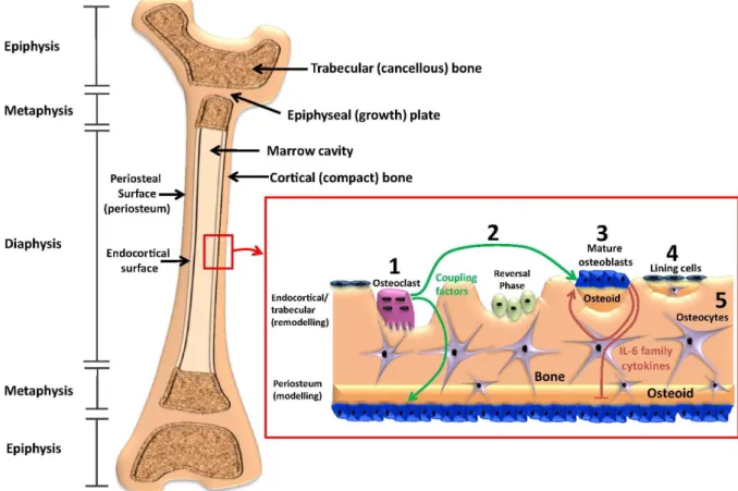
Bone adaptation as a whole is achieved by two fundamental processes: modeling and remodeling (Fig. 2.1) [7].
Fig. 2.1Structure of trabecular and cortical bone, the process of bone remodelling, and cellular signals from coupling factors and the IL-6 cytokine family that regulate bone remodelling and periosteal modelling. The proximal and distal ends of the growing murine femur (epiphyses) contain a high proportion of trabecular (cancellous) bone. Trabecular bone is also prevalent in the metaphyses, which is separated from the epiphysis by the epiphyseal (growth) plate; this is the site of longitudinal bone growth. The midshaft of the femur, diaphysis, contains a high proportion of cortical (compact) bone that surrounds the inner marrow space and trabecular regions. Bone remodelling occurs on trabecular surfaces, and on the endocortical surface (both
surfaces together are termed the endosteum). Bone modelling occurs throughout life in murine bones on the outer periosteal surface (periosteum). During bone remodelling on the endosteum, (1) osteoclasts attach to the bone surface, resorb bone and release coupling factors that stimulate osteoblast differentiation on the endosteal surface. These coupling factors also signal to periosteal osteoblasts, perhaps through the osteocyte canalicular network. After the reversal phase, which remains poorly understood in murine bones, (2) pre-osteoblasts mature, attach to the bone surface and fill the cavity created by osteoclasts with bone matrix, termed osteoid. (3) Mature osteoblasts, when their task of producing osteoid is completed, become lining cells or (4) become embedded within the osteoid as it is mineralised. These osteoblasts become osteocytes and release factors that regulate mineralisation. IL-6 family cytokines are released by the osteoblast lineage and act to stimulate osteoblast differentiation and bone matrix production on endosteal surfaces, but limit osteoblast activity on the periosteum. Reprinted with permission by Sims et al. [7] and Elsevier.
Bone modeling describes the process where bone resorption and formation occur independently at different sites to sculpt bone (i.e. during growth and/or in response to mechanical loading).
Bone remodeling, on the other hand, is the process of bone renewal throughout life during which small packets of bone are removed and subsequently replaced within so-called basic multicellular units (BMU) [6]. This coupling process is tightly controlled in space and time such that it can occur asynchronously at many different sites throughout the skeleton [8]. Both modeling and remodeling, collectively referred to as (re)modeling, are regulated by local strain distributions [6, 9] and ultimately rely on the ability of resident bone cells to sense mechanical signals (e.g. strain, fluid flow, pressure) [10-13].
Whereas experimental methods were still lacking, in silico models aiming at a better understanding of the relationship between global mechanical loads and the local stresses and strains influencing bone adaptation at the organ, tissue and cell level have been developed already years ago [14-17]. Since then, the integration of animal loading models [18] has improved the computational frameworks by providing in vivo data of bone responses to controlled loading conditions, which are crucial for the validation of simulations [19]. The distribution of the engendered strains can thus be quantified at selected surface locations and subsequently used to define the relationships between the administered loads and the structural responses that follow. Studies using mouse models have shown that mechanically-induced adaptation is controlled locally and varies between different bones of the skeleton [20] and between different sites (e.g. trabecular versus cortical bone) within the same bone [21-25]. It is to be noted though that different loading regimes have different efficiencies, which makes it challenging to determine the decisive factors that are necessary to promote bone formation [2, 26, 27]. Also, bone morphology and strain distributions vary depending on the mouse strain
[20, 28, 29], their age [30-32] and gender [33], which leads to changes of the local (re)modeling characteristics. Among these factors, aging has been intensely studied for its potential implication in age-related bone loss. Although many studies have shown anabolic effects of mechanical loading on bone mass both in young and older mice [30, 34-38], there is a lack of consensus on bone’s mechanoresponsiveness with age. While studies using a tibia-loading model have shown a reduced response of bone formation to mechanical loading both in cortical [39, 40] and trabecular [30, 36] bone with age, trabecular bone adaptation in response to loading of the caudal vertebrae was maintained with age [37]. It therefore remains unclear whether and how age-related changes in bone’s response to mechanical loading occur and whether these changes are due to morphological and material property changes alone, or due to the loss of bone’s ability to sense and/or respond to mechanical signals. Hence, it is believed that a better understanding of the cellular activities involved in bone (re)modeling will provide some answers to ensure further progress in the field of bone mechanobiology.
Studies have identified several mechanoresponsive cell types and signaling pathways involved in the mechanotransduction of extracellular stimuli into intracellular biochemical responses thereby influencing bone (re)modeling. Herein, osteocytes, known as the major mechanosensors are able to sense and integrate mechanical and chemical signals from their environment, and in turn orchestrate appropriate responses such as recruitment, differentiation and activity of effector cells [10, 13, 41]. Embedded in the matrix in a connected system of voids, called lacunae, and slender canalicular channels, collectively known as the lacuno- canalicular network (LCN), they are ideally located to sense changes in their external mechanical environment and to communicate this information between each other and other bone and marrow cells. During mechanical loading, multiple mechanical signals such as bone matrix deformation [42], changes in hydrostatic pressure [43, 44], fluid flow into and out of the LCN and shear stresses along osteocyte membranes [45-47] arise. These signals lead to force- induced conformational changes in cellular structures (tethering elements, primary cilia, cell- cell adhesions, integrin complexes), which subsequently trigger the activation of mechanoresponsive signaling pathways (e.g. Wnt-, calcium- and estrogen-signaling) resulting in altered gene production [48]. Currently, fluid flow through the LCN seems to be the primary mechanical stimulus that is sensed by cells, however the exact mechanotransduction mechanisms are still under debate and have been reviewed elsewhere [10, 11, 41, 49, 50].
Manyin vitro models have been established to investigate biological responses of individual cell types to mechanical stimulation disconnected from their native environment (Wang and Thampatty 2006, Thompson, Rubin et al. 2012, Michael Delaine-Smith, Javaheri et al. 2015).
Compared to in vivo loading models, cell-culture experiments are extremely powerful in identifying relevant mechanical cues, candidate molecules and mechanisms of mechanosensing as the cellular adaptation (e.g. strain) can be directly measured in response to controlled mechanical cues (e.g. stresses). Furthermore, by combining experimental and computational techniques to correlate these experimental outcomes (strains, stresses) with cell-specific gene expression profiles, methods are currently arising to predict and investigate the “mechanome”
of live cells [51, 52].
However, in vitro studies in bone mechanobiology have shown that strains (1-10%) that are necessary to cause signaling in two-dimensional cell cultures are much larger than strains applied to whole bones (0.04-0.3%) [53, 54]. Bacabac et al. for example showed that osteocyte- like cells are more mechanosensitive to the same mechanical stimulus when they are rounded, i.e. with a more three-dimensional morphology, than when they are stretched out on a flat surface [55]. Indeed, it has also been shownin vivothat the shape of osteocyte lacunae depends on local stress distributions and varies between different bones and different sites within the same bone [56, 57].
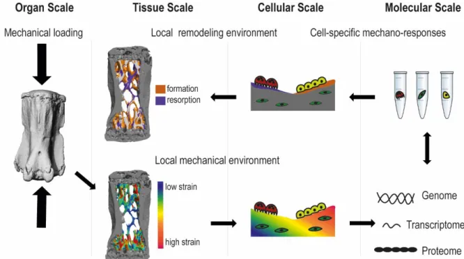
More recently, the importance of understanding bone (re)modeling in its entirety - as a result of the complex interactions among many contributing cells (osteocytes, osteoclasts, osteoblasts, marrow cells) – has been recognized [2, 7, 11, 12]. Furthermore, as strains are heterogeneously distributed throughout bone tissue, and as (re)modeling occurs at many different skeletal sites, (re)modeling activities should be assessed locally and considering signals from the local mechanical environment. Consequently, the understanding of load-induced bone (re)modeling requires a systems-level approach in which forces exerted at the organ scale can be linked to the responses at the cellular and molecular scale which lead to coordinated (re)modeling at the tissue scale (Fig. 2.2).
Fig. 2.2Multiscale process of load-induced bone remodeling. Mechanical forces exerted at the organ scale are distributed heterogeneously throughout the tissue. Cells sense these mechanical signals at the molecular level, which leads to coordinated remodeling at the tissue scale.
With increased computational power and recent experimental advances such as improved spatial and temporal resolution of imaging techniques [58, 59], dynamic bone (re)modeling can be tracked in vivo both at the tissue and the cellular level also allowing for the assessment of the micromechanical environment at each stage of development through multiscale simulation techniques [23, 60, 61].
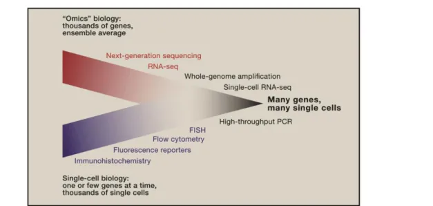
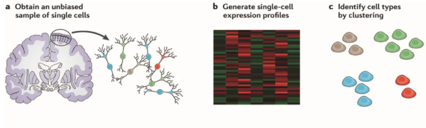
On the molecular level, advances in “omics” technologies such as genomics, transcriptomics and proteomics are more and more enabling us to comprehensively map the cellular and molecular responses to biochemical and biomechanical stimuli [62]. In fact, improvements in sequencing technology and molecular biology are now leading to the emergence of genome- wide, quantitative analysis of single cells and, hence, to the convergence of genomics and single-cell biology (Fig. 2.3) [63-65]. Single-cell biology provides unique opportunities to dissect the complex spatiotemporal dynamics of biological processes and interactions, which are averaged out in bulk assays.
Fig. 2.3 Convergence of ‘‘Omics’’ Biology and Single-Cell Biology. Technology that allows researchers to obtain genome-wide information from single cells is extending the boundaries of a field that has thus far been limited to the analyses of a select gene in eukaryotes. Reprinted with permission fromJunker et al.[63] and fromElsevier.
Combining “omics” technologies with mechanobiology, the field of mechanomics is currently emerging, which aims to deepen our understanding of cellular responses to multiple types of mechanical stimuli (e.g. shear flow, tensile stretch etc.) [51, 62].In vivo,these multiple types of mechanical stimuli are ubiquitous [10, 49] and can either act separately [66, 67] or – of higher physiological relevance - synergistically (often combined with other physical and chemical factors) on cells [68]. Furthermore, specific mechanical stimuli might cause cell type specific biological responses, which is difficult to account for when using individual cell lines [69, 70].
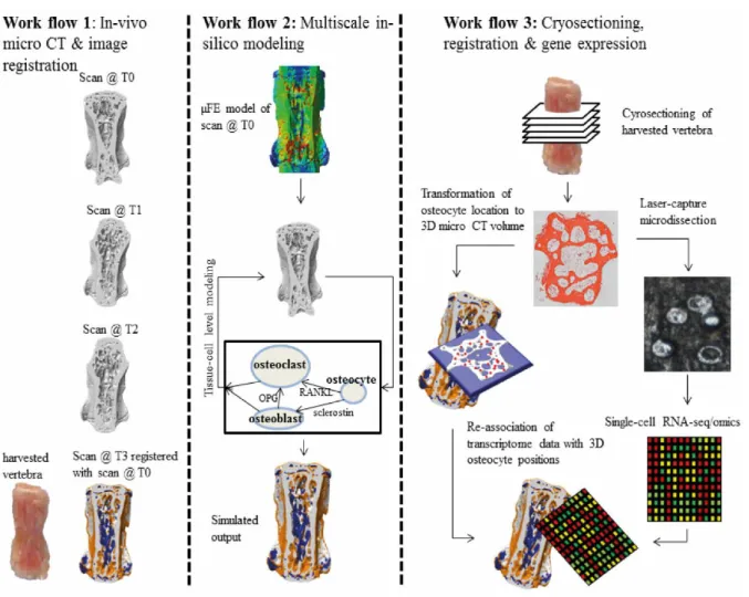
Extending the field of mechanomics to spatially resolved in vivo mechanomics in bone, we propose to combine single-cell “omics” techniques with established frameworks that locally link (re)modeling activities to mechanical environments at the tissue scale. The availability of this approach that allows to map the molecular profiles of single cells within their local 3D mechanical microenvironment will further our understanding of how bone cells interact and function as a biological system in response to mechanical loadingin vivo.
In this review, we will discuss advances in technologies and experimental methods that have enabled a better understanding of the complex interactions of mechanics and biology across different scales. More specifically, state-of-the-art technologies to quantify the mechanical environment and bone (re)modeling at tissue and cellular levels, and gene expression in mouse loading models will be discussed. Based on these methods, future, system-level strategies in the field of mechanomics are proposed, which will provide the possibility to comprehensively
map the mechanome of single cells in vivo (i.e. the responses of single cells to mechanical loadingin vivo).
2.1.2 Bone mechanobiology in mice: models & methods to study load-induced bone surface (re)modeling at different length scales
Mouse models to study load-induced bone (re)modeling (organ scale)
In contrast to other animals, mouse models present numerous advantages such as robust breeding, short generation time and the potential to examine genetic factors using transgenic technology [71]. In computational applications, they furthermore provide invaluable benefits by reducing size, complexity and computation times of the calculations. Nevertheless, the differences in the physiology of human and murine skeletons as well as among different mouse strains must be considered before translating studies of murine to human bone [72]. Unlike in humans, longitudinal bone growth does not end at sexual maturity in mice, but continues at a slow rate. Also, mice lack a well-developed Haversian system and hence intracortical remodeling only rarely occurs. However, the resorption cavities that mice use for bone remodeling during fracture healing have been shown to be very similar to the Haversian remodeling in larger animals [73, 74]. Furthermore, they do exhibit cancellous and endocortical bone remodeling as well as age-related cancellous bone loss, which have been shown to be more strongly linked to postmenopausal osteoporosis than intracortical remodeling-induced bone loss [72, 75, 76]. Hence, as long as the inherent limitations are recognized, mouse models are considered suitable to mimic human skeletal physiology [77, 78] (e.g. for osteoporosis [72]
and fracture healing [79]).
At the organ scale, a number of mouse loading models have been introduced to study load- induced bone (re)modelingin vivo. In most models, the loading is tightly controlled by directly loading a limb or bone by forces produced externally by a mechanical device. These include axial loading models of the ulna [80], fibula [81], tibia [21, 82, 83], and vertebra [84], four- point loading of the tibia [85] as well as knee loading of the tibia [86] or femur [87], and ankle loading of the tibia [25]. Alternatively, models such as treadmill running [22, 27] have been introduced which are more comparable to physiological exercise in humans. Complementarily, unloading models such as tail suspension, sciatic/tibial denervation or the injection of botulinum toxin have been used to study how bone adapts to a decrease or absence of
mechanical stimuli [18]. The effects of mechanical loading on bone tissue vary depending not only on the loading model but also on multiple parameters such as force (strain magnitude) [38, 88-92], the rate of strain (temporal change in strain magnitude within the tissue) [93-96], strain frequency (number of strain events per unit time) [97], strain gradients (spatial change in strain magnitude) [98-100], waveform, the number of loading cycles, duration of application and the inclusion of rest periods between the mechanical events [35, 101]. Moreover, the different combinations of parameters used in specific loading protocols [27, 102] together with other factors such as different animal strains [28, 29, 85], ages [21, 34] and sex [103] must also be considered when comparing results obtained by different research groups.
Methods to study load-induced bone (re)modeling (tissue scale)
The effects of loading on bone (re)modeling can be quantified at multiple scales. At the tissue scale, strain gauges and finite-element (FE) modeling can be used to characterize the strain values acting on whole bones for a specific loading model [18, 104]. Whereas strain gauges can only measure the strains at the surface to which they are attached to, FE analysis allows the computational estimation of the strain pattern throughout the entire bone.
In order to define the relationships between the administered loads and the structural responses that ensue, the outcomes of dynamic bone adaptation can be assessed. For this purpose, dynamic histomorphometry has traditionally been used, in which mice receive sequential injections of fluorescent dyes that adhere to regions of mineralizing bone, thus allowing the assessment of bone formation rates [105]. By identifying the sites of bone formation, it has been shown that responses to in vivo mechanical loading are determined locally and vary between different bones [20] and between different sites (e.g. periosteal vs. endocortical bone) within the same bone [21, 25, 92, 95]. However, the bone resorption rates cannot be assessed due to the lack of an equivalent method to label resorption over time. Furthermore, this type of analysis is limited to two-dimensional information of histological sections, which at the same time requires sample destruction. Nowadays, desktop micro-computed tomography (micro-CT) is considered a gold- standard technique to image and quantify bone as it provides a three-dimensional (3D) assessment of the net mineral response to a certain intervention or in a model of disease [58].
With information on bone mass, 3D microarchitecture and local mineralization levels, the exact amount of calcified tissue at a specific site can be measured in a standardized way, which eliminates subjectivity and inter-observer variability associated with histological methods.
Sugiyama et al. for example used both 2D fluorescent histomorphometry and micro-CT to analyze multiple bones per mouse (including loaded, adjacent and contra-lateral bones) and showed that structural changes in response to loading are not only confined to the loaded bones but also to specific sites within these bones [106]. Although, no significant differences were observed between the results obtained by histology and micro-CT, the latter is less time- and labor-intensive. Furthermore, with increasing resolution, high-resolution micro-CT scanners now allow the assessment of the 3D native environment of cells such as osteocyte lacunae and even canaliculi in mouse bones [59]. Other imaging techniques for the assessment of the osteocyte and LCN have recently been reviewed elsewhere [107]. The major limitation of both histology and ex-vivo micro-CT however, is that the measurement must be performed post- mortem, allowing only one end-point measurement per animal. In this respect, the availability ofin vivomicro-CT scanners is highly beneficial, as changes in bone in response to loading can be tracked at multiple time points within a single animal [39, 61]. For example, using a cyclic mouse tail loading model in combination with time-lapsed in vivo micro-CT and image registration techniques, Lambers et al. were able to track dynamic bone formation and resorption in trabecular mouse bone [61]. In addition, Schulte et al. were able to quantify the formation and resorption rates in 3D, similar to 2D histological parameters [108]. Birkhold et al. further developed a computational approach to identify and quantify the frequency of specific sequences of (re)modeling in response to mechanical loading of the mouse tibia [109].
By tracking spatially correlated bone formation and resorption events over time, they were not only able to show temporal differences in the (re)modeling sequences upon mechanical loading, but also to distinguish between bone modeling and remodeling events. Although they found more modeling than remodeling sites, it remains unclear if this is due to the rarer occurrence of remodeling or due to the monitoring period, which would have to be longer in order to visualize a complete remodeling cycle.
Considering the tight coordination of (re)modeling in both space and time, it seems intuitive that the local stress distributions might also play an important role in the site-specific responses of bone to mechanical loading. In this regard, the combination of time-lapsedin vivomicro-CT imaging with the above mentioned computational FE analysis has been extremely valuable to not only characterize local mechanical environments within bone, but also to link mechanical influences to local (re)modeling activities at the bone surface. By using micro-finite element models (micro-FE) derived from in vivo micro-CT images prior to loading, Schulte et al.
resolved the mechanical strains occurring throughout the trabecular micro-architecture at a
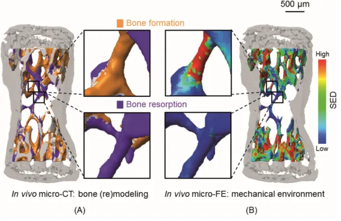
resolution of 10 µm [60]. The direct comparison of bone forming and resorbing surfaces with the micro-FE model showed that bone was formed in regions of high strain and resorbed in regions of low strain (Fig. 2.4) [60].
Fig. 2.4 Comparison of local bone formation and resorption sites with the mechanical environment. (A) Three-dimensional trabecular bone formation and resorption sites measured with in vivo micro-CT over 4 weeks. The inset shows a magnified view of formation and resorption locations in individual trabeculae. (B) Corresponding SED computed with micro-FE in the basal scan. The same regions as in (A) are enlarged. A visual comparison reveals that high SED (red) matches with sites of bone formation (yellow), while low SED (blue) is found at locations of bone resorption (violet). Reprinted with permission fromSchulte et al.[60] and PLOS ONE.
In this study, the (re)modeling activity at the bone surface was directly linked to the mechanical environment at the bone surface, defined as the strain energy density (SED). SED, which represents a scalar measure reflecting contributions from all stresses and strains acting at a given location [9, 14, 110], is often used to correlate local (re)modeling activities to the mechanical environment. However, theoretical and experimental studies in various species have linked other types of stimuli (e.g. strain magnitude [38, 88-92], strain frequency [97], strain history related parameters [111], strain rate [93-96] and strain energy gradients [98-100] to bone (re)modeling activities. Due to their relation to pressure differentials within bone, the latter parameters, namely strain rates and gradients, are thought to influence the magnitude of fluid
flow within bone, suggesting a greater physiological relevance of these parameters compared to others (e.g. SED) [95, 98, 99, 112]. In line with this hypothesis, Webster et al. [100] extended the model based on Schulte et al. further by incorporating not only the bone but also the marrow phases, which showed that mechanical signals derived from the bone marrow, namely the gradient in SED, are even more precise in predicting trabecular bone adaptation. These results suggest that many cell types - including bone marrow cells – are able to sense mechanical signals and influence bone formation and bone resorption by osteoblasts and osteoclasts respectively.
Using similar methods to Webster et al., Metzger et al. characterized the local micromechanical environment of bone marrow in trabecular compartments of porcine femora [113]. By combining micro-CT imaging, micro-scale computational fluid dynamics modeling and experimental assessments of pressure gradients during physiological loading, the authors demonstrated that deformation of whole bones induces motion and consequently shear stress in the bone marrow, which could be sensed by cells. Indeed, it has recently been shown that mechanical loading of mouse limbsin vivoinduces mechanical signaling between marrow cells [114] and increased proliferation and osteogenic gene expression in marrow stromal cells [2, 11].
Recently, Birkhold et al. also used a similar approach based on time-lapsed in vivomicro-CT and FE modeling to investigate site-specific responses of cortical bone toin vivotibial loading [23]. Compared to the periosteal surface, the response to loading was higher at the endocortical surface, which interestingly however was not linked to higher strains at that surface. Consistent with an important role of marrow cells, the authors speculate that these results may be due to the higher amount of vascularity or presence of bone marrow at the endocortical surface, which is thought to play a role in the amplification of mechanical strain.
Methods to study load-induced bone (re)modeling (cellular and molecular scale)
As is outlined above, organ- and tissue-scale information is not sufficient to validate and improve existing models aiming at understanding the underlying mechanisms of bone mechanobiology. The integration of information at the cellular and molecular level is thus required. To address this need, methods such as immunohistochemistry orin situhybridization have been used to locally determine protein or gene transcript levels within the mechanical environment of loaded bone [115-118]. For example, a reduction in expression of Sclerostin
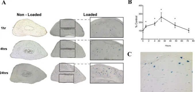
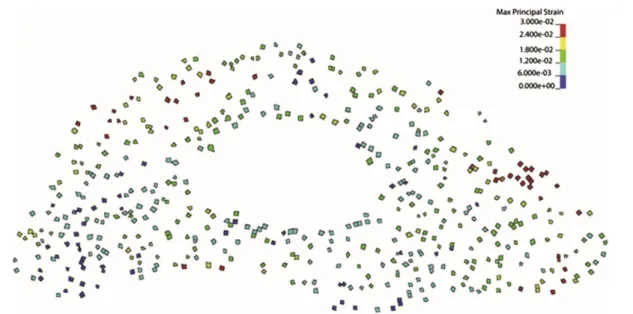
(SOST) protein has been observed both in axially loaded ulnae [115] and tibiae [118]. However, whereas Robling et al. [115] directly associated this reduction with local strain levels, Moustafa et al. [118] found that SOST expression was more closely related to sites of bone formation than to local strain levels. Possibly, these differences arise due to the different methods used for estimations of the mechanical environment within bone. Similarly, the latter approach was recently used by Lara-Castillo et al. to investigate the activation of the β-catenin pathway in osteocytes after ulna loading in mice [117]. Osteocyte gene expression patterns could not be associated to the uniform strain fields predicted by traditional FE models. Including the osteocyte lacunae (identified by histology) into the FE models increased the heterogeneity of the predicted strain field, with high strain concentrations observed around some osteocyte lacunae (Fig. 2.5 and 2.6) [117]. This strain field correlated much better with gene expression patterns.
Fig. 2.5Kinetics of β-catenin activation after a single load session. (A) Representative images and close-up of cross-sections at the midshaft region of non-loaded and loaded ulnas (B) Graph showing counts of manually counted β-catenin positive cells. Graph represents mean ± standard error of the mean (n=4, *p<0.05) (C) Increased magnification view of a cross-section at the midshaft region of a loaded ulna (24 hour time point) illustrating activated cells at the bone surface. Reprinted with permission fromLara-Castillo et al.[117] andElsevier.