Unilateral leukemic infiltration and acute angle closure as the first sign of B-cell acute lymphoblastic leukemia relapse
Abstract
Objective:Unilateral ocular leukemic infiltration with acute angle closure is an infrequent complication of B-cell acute lymphoblastic leukemia
Diana Silva
1Mafalda Mota
1(ALL-B). We present a clinical case of leukemic ocular infiltration as the
sole manifestation of ALL-B relapse.
Andreia Bilé
2Mário Ramalho
1Methods:Case description
Results: A 15-year-old female with a history of acute lymphoblastic
leukemia in remission for 2 years and pulmonary tuberculosis treated
Sara Pinto
1Graça Pires
1in the past year presented with ocular redness and decreased visual
Susana Teixeira
1acuity in the left eye (LE) with 5 days of evolution. Visual acuity was 20/20 in the right eye (RE) and absence of light perception in the left
Isabel Prieto
1eye (LE). Biomicroscopy of LE showed a small hypopion, anterior chamber cells 4+, vitreous cells 3+, and a large white mass in the vit-
1 Hospital Prof. Dr. Fernando Fonseca E.P.E.,
reous with associated vitreous hemorrhage in organization. In LE fundo- scopy, the vitreous mass occupying most of the vitreous cavity and as-
Ophthalmology Department, Amadora-Sintra, Portugal sociated hemorrhage prevented retina visualization. B-scan ultrasound
showed a multilobulated mass occupying virtually the entire vitreous
cavity with associated choroidal detachment. Forty-eight hours later, 2 Hospital Prof. Dr. Fernando Fonseca E.P.E., Pediatric she developed acute angle closure of LE with an IOP of 55 mmHg. A
Department, Amadora-Sintra, Portugal
flow cytometric analysis of the anterior chamber and vitreous showed leukemic tumor cells. The microbiologic exam and PCR forMycobacteri- um tuberculosiswere negative. No other signs of relapse of the disease were identified after investigation by the oncology department. Rescue treatment of the underlying disease was started, with symptomatic im- provement.
Conclusion:Leukemic ocular infiltration can be the only manifestation of ALL-B relapse.
Keywords:acute angle closure, B-cell acute lymphoblastic leukemia (ALL-B), ocular infiltration, relapse
Introduction
Ophthalmic manifestations of leukemia have been de- scribed previously in literature and can occur in 9 to 90%
of patients, mostly with retinal involvement [1], [2], [3].
Ophthalmic signs can be observed at the onset of disease or during follow-up [3], typically manifesting bilaterally and symmetrically [4]. However, it is uncommon that a relapsed disease presents solely with ocular manifesta- tions [1], [4], [5]. We present a clinical case of unilateral leukemic ocular infiltration with choroidal infiltration and subsequent angle closure as the sole manifestation of ALL-B relapse.
Case description
We present the case of a 15-year old girl with a history of B-cell acute lymphoblastic leukemia (t(1:19)) in clinical
remission for two years. She was under maintenance chemotherapy with dexamethasone, mercaptopurine, and melphalan. In the past year, she was also suffering from pulmonary tuberculosis having completed 9 months of therapy. She presented in our ophthalmology emer- gency room with acute visual loss of the left eye (LE) and ocular redness for 5 days. Best corrected visual acuity in her RE was 20/20 and absence of light perception in her left eye (LE). Intraocular pressure upon presentation was 12 mmHg in RE and 22 mmHg in LE. In LE biomicroscopy, she presented with a very small hypopyon, small, inferior, keratic precipitates, anterior chamber cells 4+, vitreous cells 3+, and a dense white mass was visible in the vit- reous cavity along with organized vitreous hemorrhage (Figure 1). The fundoscopy examination was made difficult by the dense vitritis. It was possible to perceive the presence of a white vitreous mass occupying most of the vitreous cavity and vitreous hemorrhage. Biomicroscopy and fundoscopy were normal in LE. B-scan ultrasound
1/3 GMS Ophthalmology Cases 2019, Vol. 9, ISSN 2193-1496
Case Report
OPEN ACCESS
Figure 1: Biomicroscopy of the left eye showed a very small hypopyon, small, inferior, keratic precipitates, anterior chamber cells 4+, vitreous cells 3+, and a dense white mass was visible in the vitreous cavity along with organized vitreous hemorrhage.
showed a multilobulated vitreous mass occupying prac- tically the entire ocular globe with associated superior choroidal detachment (Figure 2). An orbital and cranial MRI revealed an intraocular mass with gadolinium enhancement and lacrimal gland enlargement (Figure 3).
Figure 2: B-scan ultrasound of the left eye showing a multilobulated vitreous mass occupying practically the entire
ocular globe with associated superior choroidal detachment
The patient was medicated with an association of Timolol 1% and Dorzolomide 1% bid as well as topical Dexa- methasone 1% qid. Two days later, she suffered clinical worsening with severe ocular pain, headache, and naus- ea. IOP was now 55 mmHg in LE and the biomicroscopy of LE showed a shallow anterior chamber. Acute angle closure was diagnosed, and we added topical Brimonidine 0.1% bid and Acetazolamide 500 mg qid to her therapeu- tic scheme. A flow cytometry analysis of the aqueous humor and vitreous revealed ALL-B cells sharing the im- munophenotype with previously diagnosed leukemic cells (CD34–/CD10+/CD19+/CD20+ weak/CD38+). Microbio- logic analysis was negative for bacteria and fungus as well as the PCR study forMycobacterium tuberculosis.
Ocular relapse of B-cell ALL was diagnosed. The patient was re-staged but there were no signs of relapse in the bone marrow, the central nervous system or any other systemic location. Radiotherapy (30Gy) and rescue che- motherapy (INTREALL HR) were started with symptomatic improvement and better IOP control even though the pa-
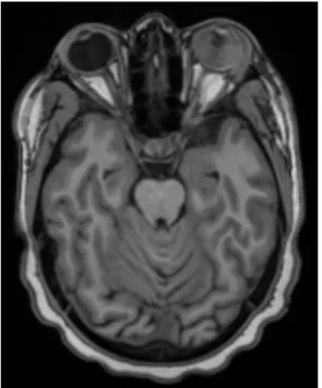
Figure 3: Orbital and cranial MRI revealed an intraocular mass with gadolinium enhancement and lacrimal gland enlargement.
No signs of CNS metastasis were found.
tient was still under topical hypotensive drugs. Final MAVC was absence of light perception and 8 months after presentation, the patient remained without any other signs of systemic relapse.
Discussion
We present a case of unilateral leukemic infiltration as the sole manifestation of relapsed ALL-B. Though, infre- quent, unilateral eye involvement as the first sign of leukemic relapse has been described previously in the literature [2], [5], [6], [7]. Our patient presented with an intraocular mass occupying the entire vitreous cavity with associated vitreous hemorrhage and choroidal detach- ment. Forty-eight hours after presenting in our emergency room, the patient evolved to acute angle closure with an IOP of 55 mmHg, secondary to the pushing effect on the iris of the very extensive vitreous mass and associated choroidal detachment. Secondary acute angle closure as
2/3 GMS Ophthalmology Cases 2019, Vol. 9, ISSN 2193-1496
Silva et al.: Unilateral leukemic infiltration and acute angle closure ...
a manifestation of leukemic infiltration is very rare, with only two other cases previously described in the literature [6], [8]. In one of the cases, the patient already had an established diagnosis of AML at the time of visual symp- toms and presented with bilateral disease [8]. In the other case, the patient was in remission for several years and presented with unilateral disease [6], making the diagnosis more challenging as in our case.
Our patient’s case also had the particularity of a challeng- ing differential diagnosis given that our patient was under maintenance chemotherapy and had history of a tubercu- losis infection in the previous year. Therefore, alternative diagnostic hypothesis such as endogenous endophthal- mitis and ocular tuberculosis had to be excluded as well, especially given that the patient was clinically in remis- sion. The microbiologic exam and PCR forMycobacterium tuberculosiswere negative and the finding of leukemic cells in the vitreous and anterior chamber was diagnostic.
Treatment of leukemic ocular infiltration comprises sys- temic chemotherapy, but if this is not an option or if the ocular disease is not responsive, ocular radiation may be combined [2], [6], typically with a total dose of 20 Gy [9].
Usually, it responds very well to systemic treatment, pre- venting the need for enucleation or evisceration [2], [5], [6], [7]. In our case, radiation therapy and chemotherapy improved the patient’s symptoms and decreased the intraocular mass. Initial visual acuity of absence of light perception at presentation suggests prolonged ocular involvement prior to the diagnosis hindering any possibility of visual recovery.
Findings of ocular leukemic relapse should prompt a new systemic work-up by the oncology team. Even though our patient still has not developed any signs of systemic leukemic relapse, in some cases where ocular findings were the only sign of leukemic relapse, bone marrow or CNS relapse ensued in the following months [5].
This case report highlights ocular leukemic infiltration as the first sign of leukemic relapse. A high suspicion index should be kept in these patients, even if in clinical remis- sion for several years. Secondary acute angle closure is a less frequent presentation that can happen especially in cases with extensive intraocular infiltration.
Notes
Competing interests
The authors declare that they have no competing in- terests.
References
1. Kincaid MC, Green WR. Ocular and orbital involvement in leukemia. Surv Ophthalmol. 1983 Jan-Feb;27(4):211-32. DOI:
10.1016/0039-6257(83)90123-6
2. Primack JD, Smith ME, Tychsen L. Retinal detachment in a child as the first sign of leukemic relapse: histopathology, MRI findings, treatment, and tumor-free follow up. J Pediatr Ophthalmol Strabismus. 1995 Jul-Aug;32(4):253-6.
3. Orhan B, Malbora B, Akça Bayar S, Avcı Z, Alioğlu B, Özbek N.
Ophthalmologic Findings in Children with Leukemia: A Single- Center Study. Turk J Ophthalmol. 2016 Apr;46(2):62-67. DOI:
10.4274/tjo.03880
4. Schachat AP, Markowitz JA, Guyer DR, Burke PJ, Karp JE, Graham ML. Ophthalmic manifestations of leukemia. Arch Ophthalmol.
1989 May;107(5):697-700. DOI:
10.1001/archopht.1989.01070010715033
5. Patel AV, Miller JB, Nath R, Shih HA, Yoon MK, Freitag SK, Papaliodis G, Chen TC, Eliott D, Kim IK. Unilateral Eye Findings:
A Rare Herald of Acute Leukemia. Ocul Oncol Pathol. 2016 Apr;2(3):166-70. DOI: 10.1159/000442951
6. Azık FM, Akıncı A, Saylı TR, Culha VK, Teberik K, Teke MY, Gürbüz F. Unilateral exudative retinal detachment as the sole presentation of relapsing acute lymphoblastic leukemia. Turk J Haematol. 2012 Jun;29(2):181-4. DOI: 10.5505/tjh.2012.72623 7. Stewart MW, Gitter KA, Cohen G. Acute leukemia presenting as a unilateral exudative retinal detachment. Retina (Philadelphia, Pa). 1989;9(2):110-4. DOI: 10.1097/00006982-198909020- 00007
8. Tumuluri K, Woo T, Crowston J, Healey PR, Gottlieb D, Maloof AJ.
Bilateral leukemic orbital infiltration presenting as proptosis and narrow-angle glaucoma. Ophthalmic Plast Reconstr Surg. 2004 May;20(3):248-50. DOI:
10.1097/01.IOP.0000129018.17256.38
9. Brady LW, Shields JA, Augsburger JJ, Day JL. Malignant intraocular tumors. Cancer. 1982 Feb 1;49(3):578-85.
Corresponding author:
Diana Silva
Praceta Papa João XXI, 1, 2º esquerdo, 2685-217 Lisboa, Portugal, Phone: +351 969743535
diana_silva1@hotmail.com
Please cite as
Silva S, Mota M, Bilé A, Ramalho M, Pinto S, Pires G, Teixeira S, Prieto I.
Unilateral leukemic infiltration and acute angle closure as the first sign of B-cell acute lymphoblastic leukemia relapse. GMS Ophthalmol Cases.
2019;9:Doc16.
DOI: 10.3205/oc000105, URN: urn:nbn:de:0183-oc0001051
This article is freely available from
http://www.egms.de/en/journals/oc/2019-9/oc000105.shtml Published:2019-04-26
Copyright
©2019 Silva et al. This is an Open Access article distributed under the terms of the Creative Commons Attribution 4.0 License. See license information at http://creativecommons.org/licenses/by/4.0/.
3/3 GMS Ophthalmology Cases 2019, Vol. 9, ISSN 2193-1496
Silva et al.: Unilateral leukemic infiltration and acute angle closure ...