Article
Comprehensive Assessment of GPR68 Expression in Normal and Neoplastic Human Tissues Using a Novel Rabbit Monoclonal Antibody
Markus Herzig
1, Pooja Dasgupta
1, Daniel Kaemmerer
2, Jörg Sänger
3, Katja Evert
4,5, Stefan Schulz
1and Amelie Lupp
1,*
1
Institute of Pharmacology and Toxicology, Jena University Hospital, D-07747 Jena, Germany;
markus.jakob.herzig@uni-jena.de (M.H.); Pooja.Dasgupta@med.uni-jena.de (P.D.);
Stefan.Schulz@med.uni-jena.de (S.S.)
2
Department of General and Visceral Surgery, Zentralklinik Bad Berka, D-99437 Bad Berka, Germany;
daniel.kaemmerer@zentralklinik.de
3
Laboratory of Pathology and Cytology Bad Berka, D-99437 Bad Berka, Germany;
pathologie.bad-berka@t-online.de
4
Department of Pathology, University of Regensburg, D-93053 Regensburg, Germany; Katja.Evert@ukr.de
5
Institute of Pathology, University Medicine of Greifswald, D-17475 Greifswald, Germany
* Correspondence: Amelie.Lupp@med.uni-jena.de; Tel.: +49-3641-9325-678; Fax: +49-3641-9325-652
Received: 10 August 2019; Accepted: 22 October 2019; Published: 23 October 2019
Abstract: GPR68 (OGR1) belongs to the proton-sensing G protein-coupled receptors that are involved in cellular adaptations to pH changes during tumour development. Although expression of GPR68 has been described in many tumour cell lines, little is known about its presence in human tumour entities. We characterised the novel rabbit monoclonal anti-human GPR68 antibody 16H23L16 using various cell lines and tissue specimens. The antibody was then applied to a large series of formalin-fixed, paraffin-embedded normal and neoplastic human tissue samples. Antibody specificity was demonstrated in a Western blot analysis of GPR68-expressing cells using specific siRNAs.
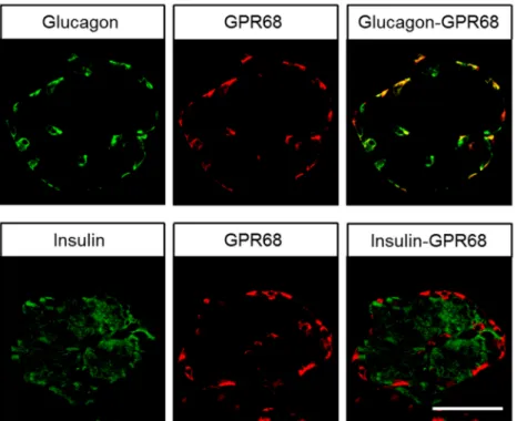
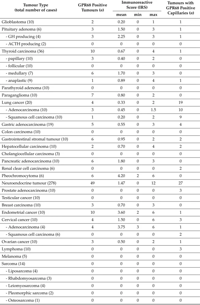
Immunocytochemical experiments revealed pH-dependent changes in subcellular localisation of the receptor and internalisation after stimulation with lorazepam. In normal tissue, GPR68 was present in glucagon-producing islet cells, neuroendocrine cells of the intestinal tract, gastric glands, granulocytes, macrophages, muscle layers of arteries and arterioles, and capillaries. GPR68 was also expressed in neuroendocrine tumours, where it may be a positive prognostic factor, in pheochromocytomas, cervical adenocarcinomas, and endometrial cancer, as well as in paragangliomas, medullary thyroid carcinomas, gastrointestinal stromal tumours, and pancreatic adenocarcinomas. Often, tumour capillaries were also strongly GPR68-positive. The novel antibody 16H23L16 will be a valuable tool for basic research and for identifying GPR68-expressing tumours during histopathological examinations.
Keywords: GPR68; OGR1; antibody; immunohistochemistry; neuroendocrine; tumours
1. Introduction
GPR68, also known as ovarian cancer G protein-coupled receptor 1 (OGR1), is a member of the proton-sensing G protein-coupled receptor (GPCR) family that contains three additional members:
GPR4, GPR65 (or T lymphocyte death-associated gene 8 protein, TDAG8), and GPR132 (or G2 accumulation protein, G2A). Unlike other receptors, the pH window of GPR68 is quite narrow.
The receptor seems to be inactive at pH 7.8 and fully active at pH 6.8 [1,2], but the opposite pattern has also been shown [3]. The pH sensing ability of GPR68 is attributed to four histidines located in its extracellular loops. It is hypothesised that at a slightly alkaline pH, GPR68 is stabilised
Int. J. Mol. Sci.2019,20, 5261; doi:10.3390/ijms20215261 www.mdpi.com/journal/ijms