Anti-lymphangiogenic compounds as novel treatment strategies in dry eye disease
Inaugural Dissertation
zur
Erlangung des Doktorgrades Dr.nat.med
der Medizinischen Fakultät und
der Mathematisch-Naturwissenschaftlichen Fakultät der Universität zu Köln
vorgelegt von
Laura Schöllhorn aus: Aachen
Köln
2016
Berichterstatter/Berichterstatterin: Prof. Dr. Thomas Langmann Prof. Dr. Ines Neundorf
Tag der letzten mündlichen Prüfung: 12. Januar 2017
“Du machst das nicht für uns…
Du machst das für dich!”
Hans Dieter und Rosi Schöllhorn
Inhaltsverzeichnis
Inhaltsverzeichnis
Inhaltsverzeichnis I
Zusammenfassung 1
Abstract 3
1. . Introduction 5
1.1. The ocular surface system 5
1.2. Anatomical components of the lacrimal functional unit 7
1.2.1. The cornea 7
1.2.2. The limbal area 8
1.2.3. The conjunctiva 8
1.2.4. The lacrimal glands 8
1.2.5. The corneal tear film 9
1.3. Ocular immune privilege 10
1.3.1. Anatomical and physical barriers of the eye 10
1.3.2. Immunoregulatory and immunosuppressive microenvironment 10
1.3.3. ACAID 11
1.3.4. Angiogenic privilege of the cornea 11
1.4. Dry eye disease 12
1.4.1. Definition, classification, therapy 12
1.4.2. Current research 15
1.4.3. Dry eye disease and corneal lymphangiogenesis 16
1.5. Aflibercept 17
1.6. Scope of the thesis 18
2. . Material 19
2.1. Reagents 19
2.2. Consumables and Equipment 20
2.3. Antibodies 22
2.4. Commercially available Kits 22
2.5. Surgical instruments 23
2.6. Animals 23
2.7. Software 23
3. . Methods 24
3.1. Animal experimental techniques 24
3.1.1. Experimental autoimmune Dry eye model 24
3.1.2. Desiccating stress model 25
3.1.3. Anesthesia 27
3.2. Clinical evaluation 28
3.2.1. Fluorescein staining score 28
3.2.2. Schirmer test 28
3.3. Organ sampling 28
3.4. Paraffin processing of organ samples 29
3.5. Immunohistochemical methods 29
3.5.1. Corneal wholemount immunostaining 29
3.5.2. Paraffin section staining with hematoxylin and eosin 30
3.6. Enzymatic methods 30
3.6.1. Lacrimal gland homogenate 30
3.6.2. Quantitation of total protein concentration 31
3.7. Flow cytometry analysis 31
3.7.1. Single cells suspension 31
3.7.1.1.Lymph nodes 31
3.7.1.2.Lacrimal glands 32
3.7.2. Cell count 32
3.7.3. Immunofluorescence labeling of cells 32
3.7.4. Flow cytometry settings 33
Inhaltsverzeichnis
3.8. Morphometrically analysis by image acquisition 33
3.8.1. Morphometric analysis of paraffin sections by light microscopy 33 3.8.2. Morphometric analysis of corneal hem- and lymphangiogenesis by fluorescence
microscopy 33
3.9. Statistical analysis 34
4. . Results 35
4.1. Development of a novel experimentally autoimmune Dry eye model similar to
Sjögren’s Syndrome dry eye 35
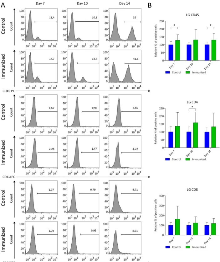
4.1.1. Short-term analysis of experimental induced autoimmune DED 36 4.1.1.1.Quantification of clinical evaluation after induction of autoimmune DED 36 4.1.1.2.Morphometric analysis of corneal neovascularization 37 4.1.1.3.Quantification of the immune response by flow cytometry analysis of corneal
draining lymph nodes 38
4.1.1.4.Quantification of the immune response by flow cytometry analysis of lacrimal
glands 40
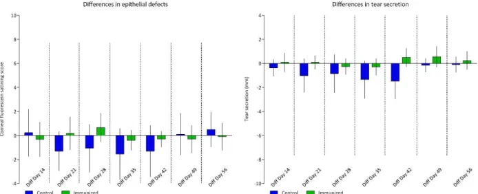
4.1.2. Long-term analysis of autoimmune induced dry eye 42
4.1.2.1.Quantification of clinical evaluation after experimentally induced autoimmune DED 42 4.1.2.2.Morphometric analysis of corneal neovascularization 43 4.1.2.3.Quantification of the immune response by flow cytometry analysis of corneal
draining lymph nodes 45
4.1.2.4.Quantification of the immune response by flow cytometry analysis of lacrimal
glands 47
4.1.2.5.Histological examination of lacrimal gland infiltrates 48 4.2. Effect of topically applied Aflibercept in a desiccating stress model reflecting non-
Sjögren’s Syndrome dry eye 50
4.2.1. Quantification of clinical evaluation regarding the effect of topically applied
Aflibercept 51
4.2.2. Morphometric analysis of neovascularization after topical application of Aflibercept 51
4.2.3. Quantification of the immune response by flow cytometry analysis of corneal draining lymph nodes after topical application of Aflibercept 53 4.3. Effect of systemically applied Aflibercept in a desiccating stress model reflecting
non-Sjögren’s Syndrome dry eye 55
4.3.1. Preventive effect of systemically applied Aflibercept on DED 56
4.3.1.1.Quantification of clinical evaluation 56
4.3.1.2.Morphometric analysis of neovascularization 57
4.3.1.3.Quantification of the immune response by flow cytometry analysis of corneal
draining lymph nodes 58
4.3.2. Therapeutic effect of systemically applied Aflibercept on DED 60
4.3.2.1.Quantification of clinical evaluation 60
4.3.2.2.Morphometric analysis of neovascularization 60
4.3.2.3.Quantification of the immune response by flow cytometry analysis of corneal
draining lymph nodes 62
5. . Discussion 64
5.1. Novel experimental autoimmune Dry eye model similar to Sjögren’s Syndrome dry
eye representing a sub-clinical model 64
5.2. Topically applied Aflibercept as novel treatment strategy for non-Sjögren’s
Syndrome dry eye 66
5.3. Systemically applied Aflibercept as novel treatment strategy for non-Sjögren’s
Syndrome dry eye 69
6. . References 72
7. . Abbreviation Index 79
8. . Danksagung 81
Erklärung 82
Zusammenfassung
Zusammenfassung
Das Trockene Auge ist eine der häufigsten Erkrankungen der Augenoberfläche, die vor allem ältere Menschen betrifft. Patienten, die an einem trockenen Auge leiden, berichten von Symptomen wie Trockenheit, Reizung, und einer verschlechterten Sehschärfe, die mit einem Verlust der Lebensqualität einhergeht. Auch wenn die Immunpathogenese in den letzten Jahren intensiv diskutiert wurde, kann auf Grund der vielen verschiedenen Auslöser und der Komplexität der Erkrankung noch immer keine kausale Therapie angeboten werden. Somit ist die Identifizierung neuer Behandlungsstrategien von wesentlicher Bedeutung.
Neuere Daten lassen vermuten, dass das antiangiogene Privileg, welches die Transparenz der Hornhaut und somit die Sehschärfe bewahrt, im Verlauf der Erkrankung gestört wird und es zu einer selektiven und spontanen kornealen Lymphangiogenese kommt. Diese pathogene Veränderung ist bereits als Risikofaktor im Bereich der Hornhauttransplantation identifiziert worden. Dabei stellen die Lymphgefäße den afferenten Arm eines Immunreflexbogens dar, indem sie den Transport von antigenem Material und Antigen-präsentierenden Zellen zu den drainierenden Lymphknoten ermöglichen.
In Bezug auf das trockene Auge gibt es erste Hinweise die korneale Lymphangiogenese als die mögliche Verbindung zur adaptiven Immunantwort angenommen, so dass Strategien, die die Lymphangiogenese modulieren, eine Chronifizierung verhindern bzw. den Krankheitsverlauf verbessern können. Eine erste Studie, in der der pro-lymhangiogene Faktor VEGF-C mit Hilfe eines prä-klinischen Medikaments inhibiert wurde, zeigte sowohl eine verminderte Entzündung als auch eine geringere Epitheliopathie.
Im Zuge dieser Arbeit wurde diese Hypothese weiter untersucht. Dazu wurde das bereits zugelassene Medikament Aflibercept hinsichtlich seiner Effizienz, die korneale Lymphangiogenese sowie die T-Zell-vermittelte Immunantwort im Mausmodell des trockenen Auges zu modulieren, getestet. Es wurden zwei verschiedene experimentelle Modelle des trockenen Auges, welche die zwei klinischen Hauptklassen widerspiegeln, verwendet: dass akut induzierbare Desiccating Stress Model, welches durch Umweltfaktoren induziert wird, und das neue, chronische Autoimmunmodell, welches durch eine autoimmune Exokrinopathie der Tränendrüsen induziert wird. Letzteres musste neu etabliert und entwickelt werden und ahmt nicht vollständig die menschliche Pathologie des Sjögren‘s Syndrom nach, spiegelt jedoch die autoimmun getriebene Entzündung und die entstehende Infiltration der Tränendrüsen wieder. Damit wurde ein neues
„subklinisches“ Modell des trockenen Auges etabliert. Aflibercept wurde dementsprechend nur im etablierten Desiccating Stress-Modell getestet. Die vorgestellten Ergebnisse lassen vermuten, dass die korneale Lymphangiogenese keine übliche phänotypische Veränderung zu sein scheint, da es in keinem der Experimente mit dem Modell des trockenen Auges induziert werden konnte. Trotz allem führte die Behandlung mit Aflibercept zu einer veränderten, z.T. abgeschwächten Immunantwort. Die vorliegende Arbeit kann die Hypothese einer wichtigen neuen Rolle der
pathologischen kornealen Lymphangiogenese in der Pathogenese des trockenen Auges nicht bestätigen. Es wird jedoch ein neues subklinisches Modell des autoimmunen trockenen Auges etabliert und die grundsätzliche Machbarkeit einer Immunmodulation beim trockenen Aue durch anti-VEGF Therapie gezeigt. Damit ergeben sich zukünftig möliche neue Therapieansätze bei Patienten mit trockenem Auge.
Abstract
Abstract
Dry eye disease (DED) is one of the most common ocular surface diseases, affecting millions of individuals. Over the last years it became a public health disorder, concerning especially the elderly.
Patients who suffer from dry eye, report symptoms like dryness, irritation and decreased visual acuity leading to a loss of life’s quality.
Even if the underlying immunopathogenesis has been verified in recent years more and more accurately, no causal treatment is available due to the various factors triggering dry eye disease and its self-intensifying viscious circle. Artificial tears and anti-inflammatory eye drops are nowadays the conventional therapy. Thus the identification of new treatment strategies is essential.
Recent data suggest that the anti-angiogenic privilege, which maintains the transparency of the cornea and preserves high visual acuity, is disturbed in DED leading to a selective and spontaneous outgrowth of lymphatic vessels. These vessels are known as risk factor for corneal graft rejection representing the afferent arm of the immune reflex arc and are shown to enable the access of antigenic material and antigen presenting cells (APCs) to the corneal draining lymph nodes. Thus, with respect to dry eye disease, corneal lymphangiogenesis is speculated to represent the potential link to the adaptive immune response and strategies modulating the lymphangiogenesis could preserve a normal phenotype or improve the disease outcome. A first study, testing blockade of pro-lymphangiogenic VEGF-C by a subclinical drug, revealed a suppressed inflammation and epitheliopathy associated with DED.
Hence, adopting this approach, we tested the hypothesis whether the already approved anti-VEGF compound Aflibercept can modulate corneal lymphangiogenesis as well as the T-cell mediated immune response occurring in dry eye disease. Therefore, two different experimental models, reflecting two major classes of clinical dry eye were contemplated to be used: the reproducible acute inducible desiccating stress model induced due to environmental stress and the novel experimental autoimmune Dry eye model induced due to a specific autoimmunological exocrinopathy of the lacrimal glands.
Regarding the latter, a new self-generated protocol had to be established to induce a “subclinical”
model of experimentally autoimmune dry eye. Since it is beyond the frame of this work to show the whole establishment, only the last attempts are shown and discussed concerning the induction of dry eye. The described protocol does not fully mimic the human pathology seen in Sjögren’s Syndrome dry eye, but reflects the autoimmunological destruction and the inflammatory infiltration of the lacrimal glands. Thus, testing Aflibercept was only performed in the desiccating stress model.
The results presented provide evidence, that corneal lymphangiogenesis does not seems to be a common phenotypical event in DED as it could not be induced in any of the experiments. However, treatment with Aflibercept leads to an altered immune response.
In summary, this work does not confirm the hypothesis of an important pathogenic role of corneal lymphangiogenesis in inflammatory dry eye disease, at least in the desiccating stress model.
Nonetheless, anti-VEGFs strategies allow for modulation of the immune response in dry eye disease thus opening new treatment avenues for future therapy of dry eye patients.
Introduction
1. Introduction
Dry eye disease (DED) is one of the most common ocular surface diseases, affecting millions of individuals. With a prevalence of about 5-35% and an incidence of 21.6% [1], dry eye increasingly becomes a public health disorder with a high financial burden [2], concerning especially the elderly [3] [4] [5].
Dry eye disease is defined as a disorder of the lacrimal functional unit (LFU) [6] [7]; a more specialized subunit of the ocular surface system [8] [9]. It can be triggered by several multifactorial factors and manifests itself with several symptoms and severity levels. Thereby the underlying immune reaction is amplified by a self-intensifying vicious circle [9]. Beside symptoms of dryness, irritation and decreased visual acuity, patients suffering from dry eye undergo a loss of life’s quality.
Despite the increased understanding of the underlying pathogenesis, no causal treatment is available. Artificial tears are the conventional and first-line treatment nowadays. Therefore, the identification of new approaches and potential therapeutics is essential.
In the following sections of the introduction, the anatomical structures and the ocular immune privilege of the ocular surface are discussed. Furthermore, the definition, the classification, the current treatment strategies and the state of the research regarding dry eye are summarized.
Furthermore, the used therapeutic Aflibercept is described.
1.1. The ocular surface system
The ocular surface system is described as an integrated system, including the “wet-surfaced and glandular epithelia of the cornea, conjunctiva, lacrimal gland, accessory lacrimal glands, nasolacrimal duct and meibomian gland, and their apical and basal matrices, linked as a functional system by both continuity of epithelia, by innervation, and the endocrine and immune systems “[8]
[9].
Based on the direct system’s exposition to the environment, including desiccation, injury and pathogens, several protective mechanisms are provided by the ocular surface system to maintain its integrity. As the smooth wet surface is responsible for most of the refractive power, the protection and maintenance of its components is the primary function of the ocular surface system.
Thus its name is linked to its primary function [9].
Within the ocular surface system, a more specialized unit can be defined: the lacrimal functional unit “comprising lacrimal glands, ocular surface (cornea, conjunctiva and meibomian glands) and lids, and the sensory and motor nerves that connect them” [6] [7]. Its overall function is to maintain the clarity of the ocular surface, which in turn depends on surface homeostasis and the integrity of the tear film which provides lubrication and a proper environment for epithelial cells [10]. Defined by Stern et al. the lacrimal functional unit “controls the major components of the tear film in a
regulated fashion and responds to environmental, endocrinological, and cortical influences.” [6] [7]
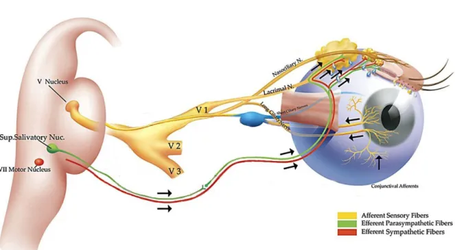
[11]. Stimulation of the corneal nerve endings of the sensory afferent nerves triggers impulses which integrate in the central nervous system through the ophthalmic branch of the trigeminal nerve; in turn generating efferent impulses terminating the optimal tear quantity and composition (see Fig. 1).
Fig. 1: Schematic illustration of the lacrimal functional unit. Stimulation of the corneal nerve endings triggers afferent impulses which integrate through the ophthalmic branch of the trigeminal nerve (V1, V2, V3) into the central nervous system. This in turn generates efferent impulses that stimulate the secretion of a healthy tear film, terminating the optimal tear quantity and composition. Illustration adapted from Beuermann et al. The lacrimal functional unit in Dry eye and Ocular Surface Disorders [7] [11].
Damage to any components of the lacrimal functional unit results in an unstable and unrefreshed tear film leading to e.g. tear film break-up causing optical aberrations, reduced tear volume, elevated tear osmolarity, and a reduced clearance of proinflammatory mediators and proteases.
Despite disturbance of the tear film, dysfunctional corneal and conjunctival cells are the most common reasons for ocular surface disorders and their pathological changes [12].
Introduction
1.2. Anatomical components of the lacrimal functional unit
1.2.1. The cornea
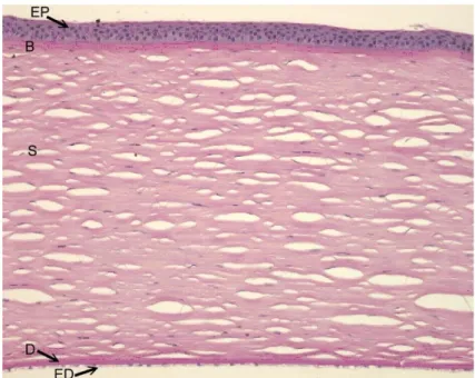
The cornea consists of five different layers which are arranged anterior to posterior as follows: the squamous non-keratinized epithelium, the anterior limiting lamina (Bowman’s layer), the corneal stroma (substantia propria), the posterior limiting lamina (Descement’s membrane) and the corneal endothelium (see Fig. 2). The outer mucous layer of the corneal epithelium thereby acts as a border to the external environment, whereas the corneal endothelium acts as an internal border to the anterior chamber.
To enable the light to proceed through the eye onto the retina the cornea has to be transparent, thus it is devoid of blood and lymphatic vessels. This is referred as “angiogenic privilege” of the eye [13] and will be discussed in a separated chapter (see 1.3.4)
Fig. 2: Histological section of a healthy cornea. It consists of five different layers: the corneal epithelium (EP), the Bowman’s layer (B), the stroma (S), the Descement’s membrane (D) and the corneal endothelium (ED).
The corneal epithelium is composed of five to six cell layers, 50 - 60 µm thick in humans and the superficial cells are flattened and enucleated [14] [15] [16]. In healthy eyes it is devoid of melanocytes and immunocompetent cells are only located at the outer edge. Desmosomes hold the adjacent cells together whereas the cells of the underlying basal lamina are held together by hemidesmosomes and anchoring filaments [16]. The anterior surface, exposed to the environment, has a specialized structural framework of microvilli and microplicae whose layer of secreted mucus (glycocalyx) support the interaction with several factors like immunoglobulins [17]. Further on, it
enables the aqueous phase of the tear film to attach to the hydrophobic non-wettable corneal epithelium [18].
As maintaining the integrity of the ocular surface is essential for vision, cell replication on the cornea is managed rapidly by mitotic activity. New cells from the limbal basal layer are recruited by the amoeboid movements of the cells in the wounded area, whereby the regenerative response is correlated to severity of damage. [19] [20].
1.2.2. The limbal area
The intervening transition area between the bulbar conjunctiva/sclera and the cornea is called corneal limbus or limbal area. It is the anatomical structure wherein the corneal epithelium becomes continuous with the conjunctival epithelium and the corneal stroma becomes continuous with the sclera (corneoscleral junction). Descement´s membrane and Bowman´s layer of the cornea end at this region.
Loops and arcades of the conjunctival blood and lymphatic capillaries appear throughout this tissue, leading to the involvement in multiple processes like nourishment of the peripheral cornea, immunosurveillance, and hypersensitivity responses. Furthermore, the corneoscleral region accommodates ocular surface progenitor cells (stem cells) in its basal epithelium [21] [22].
1.2.3. The conjunctiva
The conjunctiva is a thin translucent mucous membrane fusing the epithelium of the eyelids at their margin with the corneal epithelium at the limbal area. Until the limbal area it covers up the sclera where the transition towards the corneal epithelium begins.
Goblet cells, integrated in the conjunctival epithelium, are responsible for the production of gel-forming mucins [18] providing the adherence of the tear film to the corneal and conjunctival epithelium.
Intraepithelial dendritic (Langerhans) cells function as sentinels, whereas the subepithelial vascularized tissue contains immunocompetent cell e.g. mast cells, lymphocytes, eosinophils and plasma cells.
1.2.4. The lacrimal glands
The lacrimal gland is a branched tubuloacinar exocrine gland. It secrets electrolytes, proteins, mucins and water into the tear film whereby its right composition and amount is essential for a
Introduction
healthy ocular surface. In humans it is located in the bony orbit of the eye. It is divided into two lobes, a small palpebral and the large orbital portion. The orbital portion has fine interlobular ducts which unite to form three to five main excretory ducts, transversing the palpebral lobe. It consists of acinar, ductal and myoepithelial cells and the lobules are separated by interlobular fibrovascular tissue [23]. Due to external environmental influences and the adaptive needs of the surface epithelia, the lacrimal gland must be able to quickly adjust the composition of the tear film. This is accomplished by afferent sensory nerves which transmit the stimuli of the cornea and the conjunctiva to the central nervous system from where it is forwarded via the efferent parasympathetic and sympathetic nerves to the lacrimal glands [24].
1.2.5. The corneal tear film
In a simplified way, the corneal tear film is composed of three layers secreted by lacrimal and meibomian glands as well as the corneal and conjunctival epithelia (see Fig. 3).
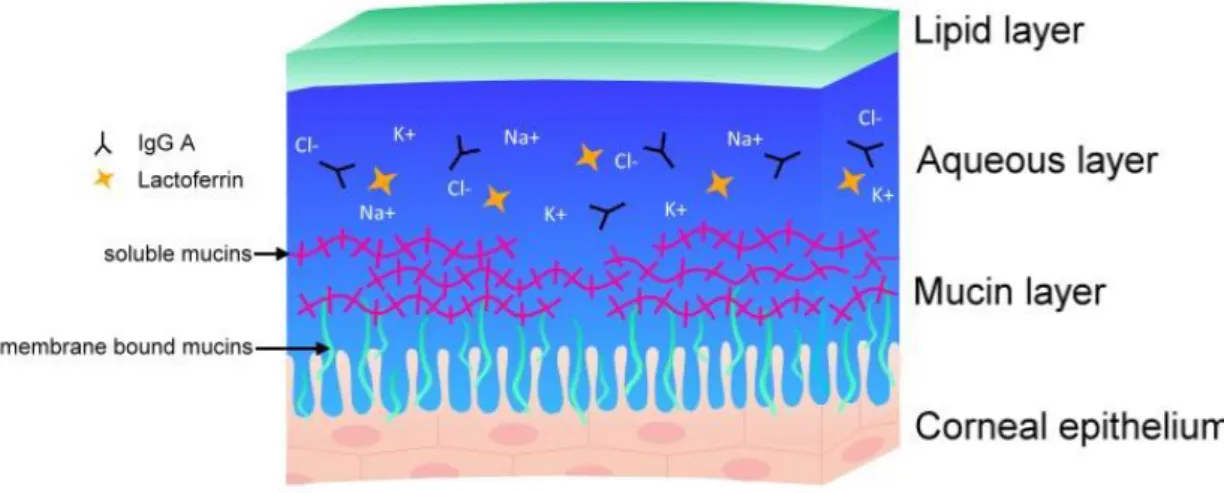
The superficial lipid layer is secreted by the meibomian glands. It consists of nonpolar lipids covering amphipathic polar lipids [25]. The former provides a barrier function at the air interface whereas the latter are in contact with the intermediate aqueous layer of the tear film providing structural stability. This layer is secreted by the lacrimal glands and contains electrolytes, ions, water and several antimicrobial proteins (further described in the following section). The deep hydrophilic mucin layer of the tear film is secreted by the goblet cells and also the corneal and conjunctival epithelial cells [26]. This layer is not wiped away by blinking. As mentioned before, membrane spanning mucins interact with the glycocalyx of the corneal epithelium enabling the tear film to stick on the hydrophobic non-wettable cornea [8] [16].
Fig. 3: Scheme of the corneal tear film, composed of three layers: the superficial lipid layer, the intermediate aqueous layer and the deep hydrophilic mucin layer.
1.3. Ocular immune privilege
The eye including all its tissues and compartments of the visual axis has to be transparent to enable light’s passage on the retina where visual response is initiated. Thus, challenges from external environment have to be recognized and processed in a way that transparency is not affected.
To balance the amount of inflammation to clear pathogens and the amount of tissue destruction the eye possesses a unique ocular immune privilege [27] which is maintained by anatomical and physical barriers, an immunoregulatory and immunosuppressive microenvironment, the anterior chamber associated immune deviation (ACAID) and the angiogenic privilege.
Thereby site- and tissue-specific mechanisms regulate both, the induction and the expression of innate and adaptive immune response.
1.3.1. Anatomical and physical barriers of the eye
The anterior segment of the eye which is highly vulnerable to pathogens is protected by a multilayer barrier system including the corneal epithelium and the tear film. Numerous antimicrobial proteins (lysozyme, lactoferrin, defensins, secretory IgG and complement factors C3 and C4) in the tear film provide a passive innate response while Toll-like receptors (TLR’s) on corneal and retinal epithelial cells provide an active innate immune response.
Furthermore, the blood: retina barrier and the absence of lymphatic vessels in the cornea prevents the fast migration of immunopathogenic cells into and from the eye, regulating the adaptive immune response.
1.3.2. Immunoregulatory and immunosuppressive microenvironment
The immune-privileged microenvironment of the eye consists of several constitutive expressed soluble and cell-bound immunoregulatory and immunosuppressive factors that mediates both, innate and adaptive immunity.
Soluble factors secreted into the aqueous humor (AqH) directly inhibit the activation of innate immunity: Transforming growth factor β (TGF-β) [28] and soluble Fas ligand (FasL) are shown to inhibit neutrophil activation; macrophage migration inhibitory factor (MIF) is shown to inhibit natural killer cells (NK cells) activity [29] [30]; calcitonin gene-related peptide is demonstrated to inhibit macrophages [31] and complement regulatory factors (CD46, CD55, CD59) are shown to inhibit complement activation [32].
With regard to the adaptive immunity it could be demonstrated that AqH inhibits T cell activation and differentiation in vivo without affecting lysis by fully functional cytotoxic T cells [33]. In addition,
Introduction
it is demonstrated that immunopathogenic T cells passing through the cornea, the iris pigment epithelium (IPE) or the retinal pigment epithelium (RPE) are neutralized by apoptosis due to direct cell contact with membrane bound FasL [34] [35] or converted in their immunological function to regulatory T cells by constitutively expressed TGF-β or CD86 [36] [37].
1.3.3. ACAID
Despite the local features mentioned above, the eye possesses an actively regulated deviant systemic immune response against eye-derived antigens; the so called anterior chamber associated immune deviation (ACAID). This specialized immune response is characterized by the suppression of CD4+ T helper 1 (TH1) and TH2 cells as well as a suppressed generation of B cells which would secrete complement fixing antibodies. Elimination of pathogens is achieved by primed CD8+ cytotoxic T cells and B cells producing non-complement fixing antibodies in the absence of inflammation while antigen-specific regulatory T cells (Tregs) inhibit the induction of T cell-mediated immunity.
Based on animal studies ACAID is shown to arise because indigenous intraocular APCs (e.g.
macrophages and dendritic cells distributed in the stroma and the anterior chamber surrounding structures (iris and ciliary body)) [38] [39] capture eye-derived antigens and migrate through the blood stream to the thymus and the marginal zone of the spleen [40] [41] [42]. Within the thymus they evoke the induction of natural killer T cells (NKT cells) which also migrate to the spleen. At this site, ocular APCs as well as NKT cells [43] [44], marginal zone B cells [45] [46] and naive antigen specific CD4+ and CD8+ T cells congregate. Together, they create a microenvironment that is rich in the cytokines TSP1 [47], TGF-β [48] and interleukin 10 (IL10) [49].
Within these cell clusters two distinct populations of Tregs emerge [50] [51]: CD4+ Tregs, which inhibit the initial induction of naive T cells into TH1 cells and CD8+ Tregs, which suppress the TH1-mediate immunity, such as delayed-type hypersensitivity (DTH). Thus, CD4+ Tregs act at the afferent arm of the ACAID in the secondary lymphoid compartment, whereas CD8+ Tregs act at the efferent arm of the ACAID in the periphery, including the eye.
Accordingly, ACAID results in a prolonged acceptance of corneal allografts [52] and solid tissue in the anterior chamber as well as the induction of a systemic tolerance to eye-derived antigens.
1.3.4. Angiogenic privilege of the cornea
To assure visual acuity the healthy cornea is transparent and devoid of blood and lymphatic vessels.
This phenomenon is highly conserved in all vertebrates and actively maintained by the so called ‚ angiogenic privilege [13] [53] [54] [55] [56].
It was demonstrated that corneal epithelium cells express soluble forms of the three major vascular endothelial growth factor (VEGF) receptors (sVEGFR-1, sVEGFR-2, sVEGfR-3) acting as a decoy receptor for the hem- and lymphangiogenic factors VEGF-A, VEGF-C, and VEGF-D [57] [58] [59] [60].
In addition endothelial cells of the cornea serve as pumps which remove fluid from the corneal stroma to the aqueous humor to keep the cornea dehydrated [61]. Fluid influx and storage in the stroma would cause irregulations in the tightly packed collagen lamellae and keratocyte network leading to increased light scatter [62] and gaps where vessels could grow in-between the lamellae [63] [64].
Further on several endogenous antiangiogenic factors like endostatin and thrombospondin, located at the epithelial basement membrane, as well as plasminogen derived angiostatin and serine protease inhibitor pigment epithelium derived factor (PEDF) maintain the corneal angiogenic privilege [65] [66] [67] [68] [69] [70] [71] [72] [73]. Endostatin causes endothelial cell cycle arrest in G1, blocks vascular endothelial growth factor (VEGF) induced mitogenic and motogenic activities in endothelial cells [65] [66], and was recently demonstrated to have an effect on lymphangiogenesis [74]. Thrombospondin exerts a strong anti-angiogenic effect via several mechanism [67] [68] and is also important for corneal alymphaticity [75] [76]. Angiostatin was shown to inhibit and regress corneal neovascularization [70]. Furthermore, it is involved in corneal avascular wound healing and downregulates endothelial cell proliferation and migration [69] [71]. PEDF is responsible for excluding vessels from invading the cornea, the vitreous and the retina [73].
Moreover the generated avascularity acts as an anatomical barrier suppressing both arms of the immune reflex arc [77] [78] maintaining the ocular immune privilege. Blood vessels providing a route of entry for immune effector cells (efferent arm) as well as lymphatic vessels enabling the effective access of antigens and APCs to the regional lymph nodes (afferent arm) are physically separated from the cornea, ending at the limbal area (see 1.2.2) [79].
1.4. Dry eye disease
1.4.1. Definition, classification, therapy
2007, on a follow-up consensus meeting of the International Dry Eye Workshop (DEWS 2007), the original definition of dry eye by Lemp [80] was updated to a more broaden definition reflecting not only the newest research but also the multifaceted aspects of the disease:
“Dry eye is a multifactorial disease of the tears and the ocular surface that results in symptoms of discomfort, visual disturbance, and tear film instability with potential damage to the ocular surface.
It is accompanied by increased osmolarity of the tear film and inflammation of the ocular surface.”
[9].
Introduction
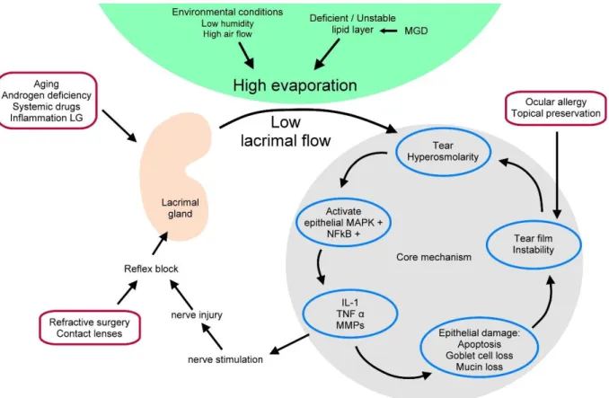
In accordance to that dry eye is recognized as an disturbance of the lacrimal functional unit (LFU), with tear hyperosmolarity and tear film instability as the main driving forces in the self-intensifying vicious circle of DED [9]. Tear hyperosmolarity leads to the activation of inflammation on the ocular surface, followed by the release of inflammatory molecules into the tears [81] [82] [83]. As a consequence, the ocular surface epithelium is damaged, characterized by cell death due to apoptosis [84], loss of goblet cell density [85], and disturbance of mucin expression [86]. The following tear film instability exacerbates the ocular surface hyperosmolarity and completes the vicious circle (see Fig. 4).
Fig. 4: Schematic mechanism of DED. Tear hyperosmolarity and tear film instability are the main forces in the self-intensifying vicious circle of dry eye. The core mechanisms are shown on the right. Tear hyperosmolarity leads to the activation of inflammation and the subsequent release of inflammatory molecules into the tears. The following corneal epithelium damage is characterized by cell death due to apoptosis, loss of goblet cell density, and disturbance of mucin expression. This in turn leads to tear film instability, which exacerbates ocular surface hyperosmolarity and completes the vicious circle. Possible risk factors are represented in the outer red circles. Adapted from DEWS (2007) [9].
Risk factors for the development of dry eye have a wide variety, resulting either in tear hyperosmolarity or in tear film instability or both. Aging, androgen deficiency, systemic drugs and local inflammatory reactions could have a direct influence on the lacrimal flow rate whereas due to
refractive surgery or wearing contact lenses a neurosensory blockade of the lacrimal flow could be induced. Environmental conditions like low humidity and high air flow as well as a deficient or unstable lipid layer due to meibomian gland dysfunction (MGD) leads to a high evaporation of the tear film. All these conditions lead to an increased tear osmolarity. In addition, several etiologies, like ocular allergy or topical preservative use, could initiate tear film instability without the prior occurrence of a tear film hyperosmolarity.
Classification of dry eye disease can either be performed by severity, since different severities need different treatment regime or by etiopathological factors, reflecting the secretory elements involved in disease induction. Using the latter, dry eye disease can be classified in two main groups:
aqueous tear-deficient dry eye (ADDE) and evaporative dry eye (EDE) [9].
ADDE arises through a failure of lacrimal gland function, resulting in a reduced tear secretion and volume. This in turn leads to a tear hyperosmolarity, inducing inflammatory responses of the epithelial cells. Due to the underlying immune response, ADDE is divided in two subclasses:
Sjögren’s syndrome dry eye (SSDE) and non-Sjögren’s syndrome dry eye (NSSDE). SSDE is caused by an exocrinopathy in which lacrimal and salivary glands are targeted by an autoimmune reaction.
Autoreactive T and B cells infiltrate the lacrimal and salivary glands, inducing cell death of acinar and ductual cells. As a result of the local inflammation, autoantigens are expressed by epithelial cells [87] and tissue specific T cells are retained [88]. NSSDE proceeds without any autoimmune features. Lacrimal gland deficiency mostly occurs due to age-related conditions, like decreased tear volume and flow, increased osmolarity [89], decreased tear film stability [90] or an alteration in the lipid layer [91].
In contrast, EDE is characterized by a normal lacrimal secretory function with a coupled excessive evaporation rate. Thereby intrinsic causes like meibomian gland dysfunction or extrinsic causes like contact lenses are described to induce EDE.
Patient who suffer from DED are likely to report symptoms of dryness, irritation, decreased visual acuity, contrast sensitivity and a loss of quality of life. Further on, compensatory responses like increased reflex tearing, blinking and meibomian gland secretion are reported.
Patients suffering aqueous tear deficiency have decreased tear film stability and tear volume. These individuals have several therapeutic options in form of artificial tears. They are applied topically to the ocular surface and viscosity, retention time and adhesion to the ocular surface of the artificial tears are determined by the polymers they based on. Common polymers are hyaluronic acid, cellulose esters, polyvinyl alcohol, povidone and carbomers. As chronic forms of DED remain due to an immune-based inflammation, topically applied corticosteroids are prescribed as effective short- term therapeutics. They block inflammatory pathways, including proinflammatory cytokine and chemokine secretion, synthesis of matrix metalloproteinases and prostaglandins as well as cell adhesion molecule expression. Cyclosporine-A (Restasis®, Allergan; Ikervis®, Santen) are the only
Introduction
FDA approved therapeutics used for DED. Cyclosporine is a fungal-derived peptide, which inhibits nuclear translocation of cytoplasmic transcription factors and the production of inflammatory cytokines. But, however, so far no causal therapies are available to cure DED.
1.4.2. Current research
Based on the current knowledge, non-Sjögren’s Syndrome dry eye is understood as a chronic inflammatory disorder, wherein the innate as well as adaptive immunity of the ocular surface are activated.
As mentioned above, increased evaporation or tear hyperosmolarity causes ocular surface inflammation. Due to the osmotic stress to the ocular surface epithelium, early innate effectors are activated, representing the afferent arm of the immune response.
Natural killer cells are shown to accumulate by 1 day of desiccating stress, representing an early source of Interferon γ (IFN-γ), IL-6, IL-23 and IL-17 [92] [93]. IFN-γ upregulates the expression of the intracellular adhesion molecule 1 (ICAM-1) on epithelial and endothelial cells and together with IL-17 it contributes to corneal barrier disruption [94] [95] [96].
Due to elevated epithelial apoptosis Toll-like receptor (TLR) activation is induced, and mitogen- activated protein kinase (MAPK) pathways and NFκB signaling are activated in epithelial cells [81]
[97] [98] [99]. MAPK activation stimulates these cells to produce several proinflammatory cytokines; e.g. IL1-β, TNF-α and IL-8, as well as several MMPs (MMP-1, -3, -9, -10 and -13) [81] [100]
[98].
IL1-β and TNF-α are demonstrated to amplify the innate immune response by upregulating the expression of costimulatory molecules like CD80/86, MHC class II antigens and CC chemokine receptor 7 (CCR7) on resident APCs, like dendritic cells (DCs) and macrophages [101]. In addition, both promote the expression of several chemokines (e.g., CCL3, CCL4, CCL5, CXCL9, CXCL10) [102]
[103] [104] [105] as well as of ICAM-1 on epihelial cells [106] [107].
Activated antigen-bearing APCs represent the link between innate and adaptive immune response, since they migrate toward the draining lymph nodes where they stimulate cognate naive T cells, reflecting the efferent arm of the immune response. APC trafficking is dependent on CCR7 signaling [108] and seems to be enhanced by isolated corneal lymphangiogenesis demonstrated to occur in experimental induced non-Sjögren’s Syndrome dry eye [109] [110] [111] as well as in Sjögren’s Syndrome dry eye [76].
CD4+ TH1, CD4+ TH17 T cells as well as CD8+ T cells are nowadays understood as the primarily effector cells of the immunopathogenesis [112] [113], which additionally is associated with an inefficient function of regulatory T cells (Tregs) [114]. While Treg homeostasis is not affected, their potential to suppress effector T cells is reduced and restricted to the TH1 T cell subpopulation [114].
Migration out of the lymphoid compartment of activated T cells toward the site of inflammation is
shown to be primarily mediated by CCR5 as well as CXCR3 [113]. T cell infiltration is facilitated by elevated expression of lymphocyte function-associated antigen 1 (LFA-1) on these T cells coupled to the increased expression of its binding partner ICAM-1 on epi- and endothelial cells [96] [106] [107].
At the ocular surface, these cells secret IFN-γ (TH1 T cells) and IL-17 (TH17 T cells). Increased expression of both derivates has been reported on human and murine ocular surface and are demonstrated to correlate with epithelial cell apoptosis, increased corneal permeability and squamous metaplasia of the corneal surface [94] [95] [112] [115] [116].
In addition, dry eye could manifest itself as an inflammatory autoimmune disorder, known as Sjögren’s Syndrome dry eye [9]. In this case it is described as an autoimmune epithelitis, wherein the exocrine glands (salivary and lacrimal glands) are infiltrated by lymphocytic and plasma cells as well as monocytic inflammatory cells [117] [118], leading to a glandular destruction of the tissue.
Thereby, the underlying exocrinopathy can be encountered alone, like in the primary Sjögren’s Syndrome dry eye or in association with other autoimmune disorders, like rheumatoid arthritis (secondary Sjögren’s Syndrome dry eye) [9].
Regarding possible autoantigens, itself little is known. Putative autoantigens have been identified in sera of patients suffering from Sjögren’s Syndrome dry eye including type 3 muscarinic acetylcholine receptor, ribonucleoprotein Ro52 and 60 (anti-Sjögren’s Syndrome antigen A, (SS-A/Ro)), La 48 (anti-Sjögren’s Syndrome antigen B, (SS-B/LA)) and αFodrin [119] [120] [121]
[122]. Nonetheless, a specific initiating autoantigen is still not identified.
1.4.3. Dry eye disease and corneal lymphangiogenesis
Only recently, experimental studies in a desiccating stress model of non-Sjögren’s Syndrome dry eye as well as in an autoimmune model reflecting Sjögren’s Syndrome dry eye revealed the isolated ingrowth of lymphatic vessels, but not blood vessels, into the physiologically avascular cornea [109]
[110] [111] [118] [76].
It was demonstrated that the lymphatic area was increased 14 days after desiccating stress while lymphangiogenic VEGF-D and VEGFR-3 levels were increased earliest on day 6. In addition, an increased homing of mature CD11b positive APCs to the corneal draining lymph nodes as well as an increased recruitment of CD11b positive monocytic cells to the cornea was detectable [109].
Furthermore, using the mouse cornea micropocket assay, IL17, one of the predominantly secreted inflammatory cytokines in DED, was demonstrated to directly induce corneal lymphangiogenesis [123]. Thus, the corneal angiogenic privilege [13] which maintains the transparency of the cornea and preserves high visual acuity is disturbed in DED.
As lymphatics were conduits that provide the access of APCs and antigenic material from the cornea to the draining lymph nodes [124] [125] it is assumed that strategies modulating lymphangiogenesis could preserve a normal phenotype or improve the disease outcome.
Introduction
First studies that have adopted this approach, revealed a significantly reduced corneal lymphangiogenesis, a weakened immune response and disease progression. Thus in vivo blockade of IL17 in the desiccating stress model demonstrated a significantly decreased corneal lymphangiogenesis and epitheliopathy [123]. Same results were reached by systemically blockade of VEGF-C, leading to a significant reduction in lymphatic vessels, epitheliopathy and CD11b positive monocytic cells in the cornea [110].
Despite these findings, corneal lymphangiogenesis is shown to be the primary risk factor for corneal allograft rejection [126] and inhibiting lymphangiogenesis before and after keratoplasty significantly increases the outcome of corneal transplantation [127] [128] [129]. Coincident, cervical lymphadenectomy (the surgical excision of draining lymph nodes) results in 90% graft survival after high-risk keratoplasty [130].
Hence, anti-lymphangiogenic therapies may be a novel effective therapeutic tool to diminish severity of DED.
1.5. Aflibercept
Aflibercept is a recombinant fusion protein with a molecular weight of approximately 115 kilo Daltons. It is composed of the Fc portion of human IgG1 fused to portions from the human extracellular VEGFR 1 and 2 domains [131]. It serves as a soluble decoy receptor binding VEGF-A, -B, and placenta growth factor (PIGF) [132]. As an ophthalmic agent, it is FDA-approved for neovascular age-related macular degeneration, macular edema and diabetic retinopathy. Human safety profile and efficacy studies demonstrated a good tolerance without any drug-related ocular or systemic adverse events and due to its high affinity blocking properties (between 0.36 and 39 pM) it allows an extended dosing interval up to 8-week interval. Further on, Aflibercept is shown to bind murine VEGF-A with a KD of 0.5 pM [132], making it a suitable compound used in several murine experimental models.
Thus, Aflibercept is already shown to suppress choroidal and subretinal neovascularization by subconjunctival and intravitreal administration [133] [134]. Furthermore, it is demonstrated to inhibit both, corneal hem- and lymphangiogenesis by systemically application in the murine inflammatory suture-induced neovascularization model as well as after keratoplasty [127] [128]
[129] [135] [126]. Furthermore, a sufficient penetration through the cornea of topical administered Aflibercept was demonstrated in the model of corneal neovascularization induced by chemical burn in rats [136].
1.6. Scope of the thesis
Recent data indicate that the anti-angiogenic privilege of the cornea is disturbed in DED, leading to a selective and spontaneous lymphangiogenesis. Lymphatic vessels were already shown to be the primarily risk factor for corneal graft rejection in animal models of corneal transplantation [126], representing the afferent arm of the immune reflex arc enabling the access of antigenic material and APCs to the corneal draining lymph nodes.
Our hypothesis was that anti-lymphangiogenic compounds could interfere in the vicious circle of dry eye disease by modulating the lymphangiogenesis as well as the T cell mediated immune response, thereby preserving a normal phenotype or improving the disease outcome.
To test this, 2 different models reflecting the two major classes of dry eye were contemplated to be used:
1) the novel, herein developed experimental autoimmune Dry eye model induced due to a specific autoimmunological exocrinopathy of the lacrimal glands reflecting Sjögren’s Syndrome dry eye.
2) the established acute desiccating stress model induced due to environmental stress reflecting non-Sjögren’s Syndrome dry eye.
In both models the efficiency of Aflibercept to modulate dry eye related lymphangiogenesis was contemplated to be tested by topical as well as systemic application. Therefore, clinical evaluation of epitheliopathy and tear secretion, immunohistochemical analysis of corneal neovascularization as well as flow cytometry analysis of corneal lymph nodes were performed.
Material
2. Material
2.1. Reagents
Tab. 1
Reagents Manufacturer
Acetone Carl Roth GmbH & CO KG
Albumin bovine fraction V BDH chemicals
Bovine serum albumin, 2% (w/v) Carl Roth GmbH & Co KG Collagenase from Clostridium histolyticum, Type IA Sigma-Aldrich Chemie GmbH DAKO® fluorescent mounting medium DAKO Diagnostic
Eosin G 1% (v/v) Carl Roth GmbH & CO KG
Ethanol 70%(v/v) Otto Fischar GmbH und Co. KG
Formaldehyde, 5% (v/v) Otto Fischar GmbH und Co. KG
Fluorescein Alcon® 10% (w/v) Alcon Pharma GmbH
Freund’s Adjuvant, Complete Sigma-Aldrich Chemie GmbH
Freund’s Adjuvant, Incomplete Sigma-Aldrich Chemie GmbH
Hematoxylin Morphisto
Hanks‘Balanced Salt solution gibco® by life technologies™
Hepes Buffer, BioWhittaker®, 1M Lonza
Medium, DMEM (1X) + GlutaMAX™ gibco® by life technologies™
Mycobacterium Tuberculosis H37 Ra, Desiccated BD Difco™
Neo-Mount® mounting medium, anhydrous Merck Millipore
Pertussis toxin from Bordetella pertussis Sigma-Aldrich Chemie GmbH Phosphate buffer saline (PBS)
Red blood cell lysis buffer Sigma-Aldrich Chemie GmbH
Sodium chloride (NaCl), 0.9% (w/v) B. Braun Melsungen AG (−)-Scopolamine hydrobromide trihydrate Sigma-Aldrich Chemie GmbH
Xylene Carl Roth GmbH & Co KG
2.2. Consumables and Equipment
Tab. 2
Consumables and Equipment Manufacturer
Autoclave, Labklav 25 SHP Steriltechnik AG
Anesthesia machine, UNO UNO BV
Precision balance, Sartorius M-pact AX6202 Sartorius AG Analytical balance, Sartorius, Competence CP64 Sartorius AG
Culture plates, CELLSTAR®, 6-well Greiner Bio One International GmbH
Cell strainer, EASYstrainer™, 40 µm Greiner Bio One International GmbH
Centrifuge, Refrigerated Benchtop, Sigma® 4K15C Sigma Laborzentrifugen GmbH Centrifuge, Refrigerated Benchtop, Heraeus® 16 Thermo Fisher scientific Centrifuge, Benchtop, Galaxy MiniStar VWR International GmbH
Cover slip Carl Roth GmbH + Co KG,
Counting chambers according to Neubauer
0.1 mm depth, 0.0025 mm² Karl Hecht GmbH&Co KG,
Cold light source KL 1500 LCD Schott AG
Digital camera ColorView III Olympus, Soft Imaging Solutions GmbH
Digital camera XM10 Olympus
Flow cytometer, guava easyCyte™ HT, benchtop Merck Millipore
Flow cytometer, Canto BD
Fluorescence microscope BX51 Olympus Optical Co., Hamburg,
Freezer, Forma 906, -86°C Thermo Fisher scientific
Fridge, G 521008, -20°C Liebherr International
Deutschland GmbH
Fridge, UK1720, 4°C Liebherr International
Deutschland GmbH
Glassware Schott AG
Gloves, Peha-soft® nitrile powderfree Paul Hartmann AG
Homogeniser, Precellys® 24 Bertin Technologies
Material
Instrument cleaning Fluid; Guava ICF® Merck Millipore
Illuminator Intensilight C-HGFI Nikon Instruments Europe B.V.
Magnetic stirrer, heatable, VMS-A VWR International GmbH Microscope Illumination, Stereo, KL1500 compact Schott AG
Microscope slides, Superfrost Ultra Plus® Thermo scientific
Microscope, Primovert Carl Zeiss Microscopy, LLC
Microtom, Microm HM400 Histo Serve
Needle, Eclipse ™ ,23G 1“ (0.6 x 25 mm) Becton, Dickinson and Company Needle, Sterican®, 30G 1/2“ (0.3 x 12 mm) B. Braun Melsungen AG
Ocular sticks, Pro-ophta®, 5 mm ø Lohmann & Rauscher GmbH &
Co. KG
Papertowels, Kolibri igeia
Phenol red threads, Zone Quick® Showa Yakuhin Co., LTD Pipette controller, accu-jet®pro Brand GMBH + CO KG Pipettes, Eppendorf® Research plus Eppendorf
Pipettes, Glass Pasteur Brand GMBH + CO KG
Pipettes, serological (5 ml, 10 ml, 25 ml) Sarstedt AG
Pipette tips Sarstedt AG
Precellys Ceramik-Kit 1.4 / 2.8 mm PEQLAB Biotechnologie GmbH Scalpel, disposable, No.11 FEATHER Safety Razor Co., Ltd.
Spectrophotometer, Epoch Microplate reader Bio-Tek
Stereomicroscop, SMZ168TP Motic Deutschland GmbH
Syringe, Dispomed®, fine dosing, 1 ml Dispomed Witt oHG
Tubes, Flow cytometry, 5 ml Sarstedt AG
Tubes, micro (1.5 ml, 2 ml) Sarstedt AG
Ultrasonic processor, Vibra cell™ 72434 Bioblock scientific
Vortex Mixer, analog VWR International GmbH
2.3. Antibodies
Tab. 3
Antibodies/Isotype Controls Manufacturer
rat anti mouse CD4 APC conjugated eBioscience rat anti mouse CD8b FITC conjugated Biolegend armenian hamster anti mouse CD11c FITC conjugated Biolegend rat anti mouse CD11b FITC conjugated AB Serotec rat anti mouse MHC2 PE conjugated BD Bioscience rat anti mouse CD45 PE conjugated BD Bioscience rabbit anti mouse Lyve-1 unconjugated AngioBio rat anti mouse CD31 FITC conjugated BD Bioscience
goat anti rabbit Cy3 Dianova
rat IgG2aκ APC conjugated eBioscience
rat IgG2bκ FITC conjugated eBioscience
armenian hamster IgG FITC conjugated Biolegend
rat IgG2b FITC conjugated BD Bioscience
rat IgG 2a κ PE conjugated eBioscience
7-AAD Viability staining solution Biolegend
rat anti mouse CD16/CD32 (Mouse BD Fc Block™) BD Bioscience
Aflibercept (Eylea®), 40 mg / ml Bayer AG
2.4. Commercially available Kits
Tab. 4
Kits Manufacturer
Pierce™ BCA Protein Assay Kit Thermo Fisher Scientific
Nuclear extract Kit Actif motif ®
Material
2.5. Surgical instruments
Tab. 5
Surgical instruments Manufacturer
Scissor, Metzenbaum F.S.T., Heidelberg, Germany
Spring scissor, Student Vannas, straight F.S.T., Heidelberg, Germany
Forcep, Sudent Dumont #5 F.S.T., Heidelberg, Germany
2.6. Animals
For all animal experiments, female C57BL/6NCrl mice from Charles River Laboratories, Germany aged 6-8 weeks were used. All animal protocols were approved in accordance with the Association for Research in Vision and Ophthalmology’s Statement for the Use of Animals in Ophthalmology and Vision Research.
2.7. Software
Tab. 6
Software Manufacturer
Cell Sense Olympus Soft Imaging solutions GmbH
FlowJo Version 10.0.7 FlowJo
Gen5™ Datenanalyse-Software Biotek
Guava Easy Cyte, Version 3.7.4 Merck Millipore
InStat 3, Version 3.10 GraphPad Software Inc.
Prism6, Version 6.05 GraphPad Software Inc.
3. Methods
3.1. Animal experimental techniques
The local animal care committee in accordance with the Association for Research in Vision and Ophthalmology’s Statement for the Use of Animals in Ophthalmology and Vision Research approved all animal protocols. As mice were purchased from external sources, they were housed for one or two weeks to acclimate.
3.1.1. Experimental autoimmune Dry eye model
First, Complete Freund’s adjuvant (CFA, 1 mg / ml, Sigma-Aldrich Chemie GmbH) was adjusted to a concentration of 2.4 mg / ml by adding desiccated Mycobacterium tuberculosis (BD Difco™). Next, equal volumes of lacrimal gland homogenate (240 µg / 100 µl per mouse, for preparation see 3.6.1) and CFA (240 µg / 100 µl per mouse) were sonificated on ice to form an emulsion.
After mice were deeply anesthetized (see 3.1.3) each mouse received 200 µl of the emulsion in total: 100 µl were injected subcutaneous (s.c.) on the base of the tail and 50 µl were injected s.c. in each flank. Furthermore, to increase immune onset mice received 2 µg Pertussis toxin (PTX, Sigma-Aldrich Chemie GmbH) intraperitoneally (i.p.). Control mice received an emulsion consisting of adjusted CFA mixed only with PBS. All mice were kept under standard animal housing.
On day 7 mice received a booster injection (BI). Therefore, equal volumes of lacrimal gland homogenate (240 µg / 100 µl per mouse) or PBS were mixed with Incomplete Freund’s adjuvant (IFA) and sonificated on ice to form an emulsion. Under anesthesia each mouse received 200 µl of the emulsion in total: 100 µl were injected s.c. on the base of the tail and 50 µl were injected s.c. in each flank.
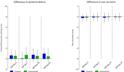
Short-term analysis (until day 14) (see Fig. 5) as well as long-term analysis (until day 56) (see Fig. 6) of immunization were performed. Short-term analysis comprise clinical evaluation of epitheliopathy and tear secretion on day 0, 3, 7, 10 and 14. Morphometric analysis of corneal neovascularization, flow cytometry analysis of corneal draining lymph nodes as well as the lacrimal glands were performed on day 7, 10 and 14. Long-term analysis comprise clinical evaluation of epitheliopathy and tear secretion on day 14, 21, 28, 35, 42, 49, and 56. Morphometric analysis of corneal neovascularization, flow cytometry analysis of corneal draining lymph nodes as well as the lacrimal glands were performed on day 14, 28, 42, and 56. Histochemical analysis of inflammatory infiltrates in lacrimal gland sections were performed on day 56.
Methods
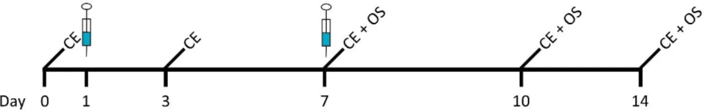
Fig. 5: Scheme of short-term immunization. Initial immunization on day 1 and booster injection on day 7 are indicated by syringes. Clinical evaluation (epitheliopathy and tear secretion; CE) was performed on day 0, 3, 7, 10 and 14. On day 7, 10 and 14 organs required for subsequent analysis were collected (organ sampling;
OS).
Fig. 6: Scheme of long-term immunization. Initial immunization on day 1 and booster injection on day 7 are indicated by syringes. Clinical evaluation (epitheliopathy and tear secretion; CE) was performed on day 14, 21, 28, 35, 42, 49, and 56. On day 14, 28, 42 and 56 organs required for subsequent analysis were collected (organ sampling; OS).
3.1.2. Desiccating stress model
Acute non-Sjögren’s Syndrome dry eye was induced in mice as described below. The used experimental setup is adapted from Dursun et al [137]. C57BL/6 mice were placed in a controlled- environment chamber (CEC) with a relative room humidity maintained at about 30% and a constant temperature of 21 to 23°C. The evaporation of the tear film leads to ocular surface lesions mimicking pathologies found in dry eye patients [112] [138]. To maximize ocular dryness, mice received 100 µl subcutaneous (s.c.) injections of the anti-cholinergic agent scopolamine hydrobromide twice a day, alternating between the left and right flanks with a concentration of 10 mg / ml. This hampers amongst mucosal secretion [137].
To determine the effect of the anti-lymphangiogenic compound Aflibercept in acute non-Sjögren’s Syndrome dry eye two experimental setups were used:
A) mice were treated topical beginning on day one for 14 consecutive days (see Fig. 7) while dessicating stress. Topical administration was performed three times a day (40 mg / ml Aflibercept, each 3 µl). Concentrations of the compound was chosen in concordance with the off label concentration used in the clinic.
Control group received an equal volume of saline solution (NaCl, 0.9% (w/v)) and naive mice serve as control for the induction of dry eye. Clinical evaluation (CE) (see 3.2) of epitheliopathy and tear secretion was performed on day 0, 3, 7, 11 and 14 and on final day mice were euthanized and organs required for subsequent analysis collected (organ sampling; OS) (see 3.3).
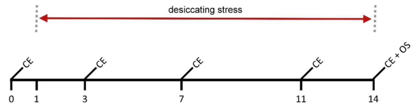
Fig. 7: Scheme of topical administration. Eye drops (3 µl) were administered three times a day (40 mg/ml Aflibercept). Clinical evaluation (epitheliopathy and tear secretion; CE) was performed on day 0, 3, 7, 11 and 14. On final day mice were euthanized and organs required for subsequent analysis collected (organ sampling; OS).
B) mice were treated systemically by two different experimental setups. In the prevention trial mice received intraperitoneally (i.p.) Aflibercept injections on day 1, 3, and 7 while mice were sitting in CEC for 14 days (see Fig. 8). In the therapy trial mice received i.p. Aflibercept injections on day 11 and 13 during dessicating stress. On day 15 mice were transferred to standard animal housing conditions till day 24, receiving i.p. injection on day 17 (see Fig. 9). Mice received 50 µl Aflibercept (25 mg/kg bodyweight) whereas the control group received an equal volume of saline solution (NaCl 0.9% (w/v)). Concentrations of the compounds were chosen in concordance with the off label concentration used in the clinic and previous corneal transplantation experiments performed with Aflibercept. Clinical evaluation (see 3.2) of epitheliopathy and tear secretion was performed on day 0, 3, 7 and 14 in the prevention trial whereas clinical evaluation in the therapy trial was performed on day 0, 7, 11, 13, 17, and 24. On final day mice were euthanized and organs required for subsequent analysis collected (organ sampling; OS) (see 3.3).
Methods
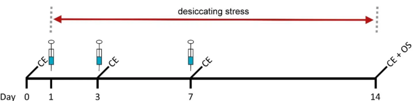
Fig. 8: Scheme of systemically administration regarding prevention. 50 µl Aflibercept (25 mg/kg bodyweight) were injected i.p. on day 1, 3, and 7 during desiccating stress (indicated by syringes). Clinical evaluation (epitheliopathy and tear secretion; CE) was performed on day 0, 3, 7 and 14. On final day mice were euthanized and organs required for subsequent analysis collected (organ sampling; OS).
Fig. 9: Scheme of systemically administration regarding therapy. 50 µl Aflibercept (25 mg / kg bodyweight) were injected i.p. on day 11 and 13 during desiccating stress and on day 17 under standard animal housing conditions (indicated by syringes). Clinical evaluation (epitheliopathy and tear secretion; CE) was performed on day 0, 7, 11, 13, 17 and 24. On final day mice were euthanized and organs required for subsequent analysis collected (organ sampling; OS).
3.1.3. Anesthesia
Prior to immunization, mice were deeply anesthetized by mean of the UNO anesthesia machine (UNO BV). First, animals were placed in the induction chamber by a flow rate of 0.7 µl / min and a concentration of 4.5% (v/v) isoflurane. After two min mice were kept out of the chamber and the desired depth of anesthesia maintained with a face mask by a flow rate of 0.25-0.3 µl / min and a concentration of 3% (v/v) isoflurane.
3.2. Clinical evaluation
To monitor disease severity, clinical evaluation by measuring corneal epitheliopathy and tear secretion was performed. Both, fluorescein staining scores and phenol red thread test were validated clinical readouts used in human diagnostics and animal experiments [139] [137]
3.2.1. Fluorescein staining score
Corneal epitheliopathy was measured by staining of epithelial surface defects with a biomicroscope (Illuminator Intensilight C-HGFI (Nikon Instruments Europe B.V)) under cobalt blue light. Two min after topical application of 1% (w/v) fluorescein to the corneal surface, the excessive fluid was removed and the spotted staining recorded with the adapted standard National Eye Institute grading system (NEI, Bethesda, MD) of 0 to 2 for each of the five areas of the cornea – central, superior, inferior, nasal and temporal. Measurements were performed for both eyes and averaged for statistical analysis.
3.2.2. Schirmer test
Measurement of the tear secretion was done by Schirmer test, using cotton phenol red threads (Zone Quick®) which were applied to the lateral canthus of the mouse eye for 15 seconds [140].
Wetting of the threads leads to a change in color and on a millimeter scale, amount of tear secretion was measured. Measurements were performed for both eyes and averaged for statistical analysis.
3.3. Organ sampling
Anatomical dissection of the required organs (cornea, lymph nodes, and lacrimal glands) was performed post mortem. Dependent on the subsequent analysis organs were placed in PBS, DMEM-medium (gibco® by life technologies™) or formaldehyde (5% (v/v), Otto Fischar GmbH und Co. KG).
For morphometric analysis of corneal neovascularization corneal wholemounts were excised by carefully cutting the cornea beneath the limbal area. Thereby the limbus is not damaged and can be used for subsequent analysis as the outermost edge. Corneal wholemounts were stored in PBS until immunohistochemical staining (see 3.5.1).
Methods
Corneal draining cervical lymph nodes used for flow cytometry analysis were dissected, connective tissue and adhering fat was completely removed and after washing in PBS lymph nodes were stored in DMEM-medium at 4°C until preparation of the single cell suspension (see 3.7.1.1). Lymph nodes were either pooled per group or analyzed per animal.
As lacrimal glands were used for both, flow cytometry analysis (see 3.7.1.2) and paraffin sections staining (see 3.5.2), they were stored in medium at 4°C or in 5% (v/v) formaldehyde. Making a skin incision below the ear, the exorbital lacrimal gland which is located posterior to the eye, is exposed.
By cutting the fascia laying around the lacrimal gland it could be dissected, washed in PBS and stored in the required solution.
3.4. Paraffin processing of organ samples
Organs preserved in formaldehyde (5% (v/v), Otto Fischar GmbH und Co. KG) were washed in tap water for 2 to 3 hours (h), replacing the water every 15 min. Afterwards organs were placed in 70%
(v/v) alcohol for 60 min, followed by a renewal of the alcohol. For further dehydration and the final infiltration with liquid paraffin, samples were putted in the Leica TP1020 tissue processor (Leica Biosystems Nussloch GmbH) according to manufacturers’ guidelines. Tissues were sectioned using the Microtome Microm HM400 (Histo Serve) and 5 to 7 µm sections were obtained. Sections were dried over night at 37°C and stored at room temperature (rT) until immunohistochemical staining (see 3.5.2).
3.5. Immunohistochemical methods
3.5.1. Corneal wholemount immunostaining
Corneal wholemounts were prepared as described previously [141]. Briefly, corneas were excised, rinsed in PBS and fixed in acetone for 30 min. After three washing steps with PBS corneas were blocked with 2% (w/v) BSA in PBS for 2 h at rT. Afterwards corneal wholemounts were stained with rabbit anti mouse LYVE-1 antibody (AngioBio) and FITC-conjugated rat anti mouse CD31 (BD Bioscience) diluted in PBS (1:200) over night at 4°C. Next day corneal tissue was washed in PBS and LYVE-1 was detected with a Cy3-conjugated secondary antibody (rabbit anti mouse; 1 : 500;
Dianova).
After a final washing step in PBS corneal wholemounts were transferred to Superfrost Ultra Plus®
microscope slides (Thermo Fisher) and covered with DAKO fluorescent mounting medium (DAKO Diagnostic). Stained wholemounts were stored at 4°C in the dark until analysis with a fluorescence microscope (see 3.8.2).
3.5.2. Paraffin section staining with hematoxylin and eosin
For paraffin section staining microsections were first deparaffinised in a descending series of alcohol (100%, 96%, 70% (v/v) alcohol; for 5 min each), starting with xylene (Carl Roth GmbH & Co KG). After washing for 5 min in aqua dest. Nuclei were stained by hematoxylin (Morphisto) for 10 min and washed in running tap water for 10 min. Afterwards connective tissue was counterstained with 1% (v/v) Eosin G (Carl Roth GmbH & Co KG) for 2 min with a following washing step in tap water. Finally, the slides were dehydrated in an ascending series of alcohol (70%, 96%, 100% (v/v); for 2 min each) ending with a 10 min washing step in xylene. Slides were covered with Neo-Mount® mounting medium (Merck Millipore) and stored at 4°C prior to analysis with a light microscope (see 3.8.1).
3.6. Enzymatic methods
3.6.1. Lacrimal gland homogenate
Lacrimal gland homogenate was prepared by means of the Precellys® 24 Homogeniser (Bertin Technologies) and the adapted Nuclear Extract Kit lysis buffer (Active Motif). Dissected and washed lacrimal glands (see 3.3) were weighted and put into an ice cold Precellys tube containing a mixture of 1.4 mm and 2.8 mm ceramic beads. After adding lysis buffer A (Tab. 7) tubes were shaken three times for 10 sec by 5500 rpm 3D speed motion. To avoid reaching of protein-denaturation heat level, tubes were always placed on ice between the shaking steps. After the last shaking step lysis buffer B (Tab. 8) was added and tubes incubated at rT for 2 h with in-between vortexing.
Total protein concentration of the homogenate was quantified by adjusted Pierce BCA Protein assay (Thermo Fisher Scientific) (see 3.6.2).
Tab. 7
Lysis buffer A µl per 1 mg tissue
10x hypotonic buffer 0.4 µl
Protease Inhibitor Cocktail 0.04 µl Phosphatase Inhibitor Cocktail 0.4 µl
dH2O 3.125 µl