Characterization of the human NLR protein
NLRC5
I n a u g u r a l – D i s s e r t a t i o n zur
Erlangung des Doktorgrades
der Mathematisch‐Naturwissenschaftlichen Fakultät der Universität zu Köln
vorgelegt von
Andreas Heinrich Neerincx
aus
Emmerich am Rhein
Köln 2013
Berichterstatter: Prof. Dr. Jonathan Howard Prof. Dr. Mats Paulsson
Tag der mündlichen Prüfung: 14.01.2013
Table of Contents
TABLE OF CONTENTS... I ABBREVIATIONS... III AMINO ACID ABBREVIATIONS... VIII
1 INTRODUCTION... 1
1.1 Innate Immunity and Pattern Recognition Receptors ... 1
1.2 Membrane‐bound Pattern Recognition Receptors ... 1
1.3 Cytosolic Pattern Recognition Receptors... 5
1.4 Nucleotide‐binding domain, leucine‐rich repeat containing proteins (NLRs) ... 7
1.4.1 The Nodosome NLRs... 9
1.4.2 The Inflammasome NLRs ... 11
1.4.3 The MHC class II transcriptional activator CIITA: a function apart from pattern recognition ... 15
1.5 Aim of the study... 22
2 MATERIAL AND METHODS ... 23
2.1 Material... 23
2.1.1 Chemicals and enzymes... 23
2.1.2 Kits ... 23
2.1.3 Cell lines and Bacteria... 23
2.1.4 Plasmids ... 25
2.1.5 Oligonucleotides ... 29
2.1.6 siRNA... 33
2.1.7 Antibodies... 34
2.1.8 Instruments... 36
2.1.9 Software... 37
2.2 Methods ... 38
2.2.1 Cell Biological Methods ... 38
2.2.2 Molecular Biological Methods ... 42
2.2.3 Biochemical Methods ... 46
3 RESULTS ... 50
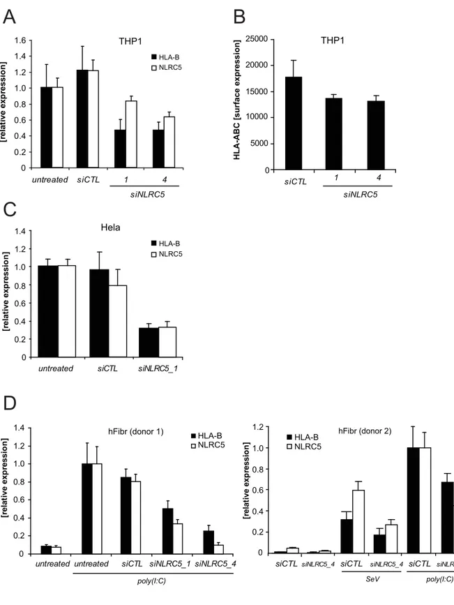
3.1 A Role for the human nucleotide‐binding domain, leucine‐rich repeat‐containing family member NLRC5 in antiviral responses ... 50
3.1.1 Structure of NLRC5 ... 50
3.1.2 Expression and Induction of NLRC5... 50
3.1.3 Innate Immune Signalling of NLRC5 ... 55
3.2 NLRC5 controls basal MHC class I gene expression in an MHC enhanceosome‐dependent
manner... 63
3.2.1 NLRC5 contributes to MHC class I gene expression ... 63
3.2.2 MHC class I gene expression correlates with NLRC5 expression levels... 67
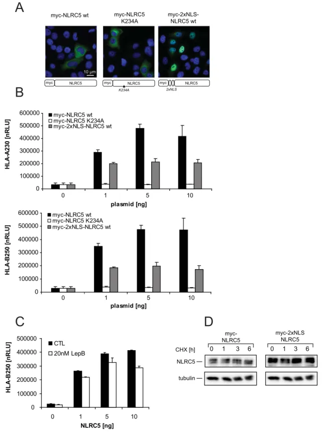
3.2.3 NLRC5‐mediated MHC class I expression is dependent on nuclear shuttling and the integral protein fold of NLRC5 ... 70
3.2.4 Nuclear–cytoplasmic shuttling of NLRC5 enhances MHC class I promoter activation... 74
3.2.5 NLRC5‐mediated MHC class I promoter activation is mediated by the MHC enhanceosome ... 76
3.3 Domain Mapping of NLRC5 and CIITA... 79
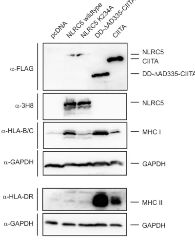
3.3.1 NLRC5/CIITA chimeric proteins are able to activate both MHC class I and II transcription 79 3.3.2 Generation of LRR domain chimeric NLR proteins ... 86
4 DISCUSSION ... 90
4.1 A Role for the human nucleotide‐binding domain, leucine‐rich repeat‐containing family member NLRC5 in antiviral responses ... 90
4.2 NLRC5 controls basal MHC class I gene expression in an MHC enhanceosome‐dependent manner... 93
4.3 Detailed function of NLRC5 in MHC transcriptional regulation ... 99
5 CONCLUSION...102
6 REFERENCES...104
7 ABSTRACT ...119
8 ZUSAMMENFASSUNG ...120
DANKSAGUNG ...122
ERKLÄRUNG...123
Abbreviations
°C Degree Celsius
anti
µg microgramme
µl microlitre
A desoxyadenosine
A20 TNFAIP3, tumor necrosis factor, alpha‐induced protein 3
aa amino acid
AAA+ ATPases associated with various cellular activities AAMP angio‐associated migratory cell protein
AD transcription activation domain ADP adenosine diphosphate
AIM2 absent in melanoma 2
APAF1 apoptotic protease‐activating factor 1
APC Allophycocyanin
APS ammonium persulfate
ASC apoptosis‐associated speck‐like protein containing a CARD ATP adenosine triphosphate
BIR baculovirus inhibitory repeat BLS bare lymphocyte syndrome
BMDMs bone marrow‐derived macrophages
bp base pairs
BS Blau Syndrome
BSA bovine serum albumin
C desoxycytidine
CIITA MHC class II transcriptional activator CARD caspase activating and recruitment domain
CC coiled‐coil
CREB cyclic‐AMP‐responsive‐element‐binding protein
CD Crohn’s Disease
CD4 cluster of differentiation 4 CD8 cluster of differentiation 8
cDNA complementary DNA
CHX Cycloheximide
cIAP cellular inhibitor of apoptosis protein CLR C‐type lectin receptor
CMV cytomegalovirus
C‐terminus carboxyl‐terminus
DAI DNA‐dependent activator of IRFs DAMPs danger‐associated molecular pattern DAP diaminopimelic acid
DISC death‐induced signalling complex
DMSO dimethylsulfoxide DNA desoxyribonucleic acid
dNTP desoxynucleotide triphosphate
ds double stranded
DTT dithiothreitol
EDTA ethylenediaminetetraacetic acid ELISA enzyme‐linked immunosorbent assay EOS early onset sarcoidosis
Erbin Erbb2 interacting protein
ERK axtracellular‐signal‐regulated kinase et al. et alii; and others
ETI effector‐triggered immunity FBS fetal bovine serum
g gravitational force
g gramme
G desoxyguanosine
GAPDH glycerinealdehyde‐3‐phosphate‐dehydrogenase GTP guanosine triphosphate
h hour
HA hemagglutinin
HAU hemagglutinin unit
HC heavy chain
HEK human embryonic kidney
HIN200 hematopoietic interferon‐inducible nuclear antigens with 200 amino acid repeats HLA human leukocyte antigens
hNLRC5 human NLRC5
HRP horseradish peroxidise i.e. id est; that is
ie‐DAP ‐D‐glutamyl‐meso‐diaminopimelic acid
IF immunofluorescence
IFI16 interferon‐‐inducible protein 16
IFN interferon
Ig immunoglobulin
IKK IB kinase
IB NF‐B inhibitor‐alpha
IL interleukin
IP immunoprecipitation
IRF interferon regulatory factor JNK c‐Jun N‐terminal kinase
kb kilobase pairs
kDa kilodalton
LPS lipopolysaccharide LRR leucine‐rich repeat
M molar
mAB monoclonal antibody
MAMP microbe‐asscociated molecular pattern MAPK mitogen‐activated protein kinase
MAVS mitochondrial antiviral‐signalling protein MCS multiple cloning site
MDA5 melanoma differentiation associated gene 5 MDP muramyl dipeptide
MEF mouse embryonic fibroblast
mg milligramme
MHC major histocompatibility complex
min minute
ml millilitre
mM millimolar
mNLRC5 murine NLRC5
MOI multiplicity of infection
mRNA messenger RNA
MyD88 myeloid differentiation primary response gene 88 NACHT domain present in NAIP, CIITA, HET‐E and TP1 NAIP neuronal apoptosis inhibitory protein
NBD nucleotide‐binding domain NEMO NF‐B essential modulator NF‐AT nuclear factor of activated T cells
NF‐B nuclear factor ‐light‐chain‐enhancer of activated B cells NF‐Y nuclear factor binding to the y box
ng nanogram
NIK NF‐B‐inducing kinase
NLR nucleotide‐binding domain and leucine‐rich repeat containing protein
NLRC nucleotide‐binding domain and leucine‐rich repeat containing protein containing a CARD domain
NLRP nucleotide‐binding domain and leucine‐rich repeat containing protein containing a PYRIN domain
NLS nuclear localization signal
№ number
NOD Nucleotide‐binding oligomerization domain NP40 Nonidet P 40
nRLU normalized relative light unit N‐terminus amino‐terminus
OD optical density
ONPG o‐Nitrophenyl‐‐D‐galactopyranosid
P phosphate
PAGE polyacrylamid gelelectrophoresis PBS phosphate buffered solution PCR polymerase chain reaction
PFA paraformaldehyde
PGN peptidoglycan
PRR pattern recognition receptor
PYD pyrin domain
PYHIN pyrin and HIN domain‐containing protein qRT‐PCR quantitative real‐time PCR
R protein resistance protein
RD repressor domain
RFX5 regulatory factor X 5
RFX‐ANK RFX‐associated ankyrin‐containing protein RFX‐AP RFX‐associated protein
RIG‐I retinoic acid‐inducible gene I
RIP2 receptor interacting serine/threonine‐protein kinase 2 RNA ribonucleic acid
ROS reactive oxygen species rpm rounds per minute
RT room temperature
RT‐PCR reverse transcription PCR SD standard deviation SDS sodium dodecyl sulphate SEAP secreted alkaline phosphatise
sec seconds
SeV Sendai Virus
siRNA small interfering RNA
ss single stranded
STAND signal transduction ATPases with numerous domains STING stimulator of interferon genes
T desoxythymidine
TAB TAK1‐binding protein
TAF TATA‐box binding protein (TBP) associated factor TAK transforming growth factor (TGF)‐beta activated kinase
TBE TRIS‐Borat‐EDTA
TBK TANK‐binding kinase
TEMED N,N,N’,N’,‐Tetramethylethylendiamin TFs transcription factors
TIR Toll‐IL‐1 receptor TLR Toll‐like receptor
TNF(‐ tumor necrosis factor(‐alpha)
TRIF Toll/IL‐1R domain containing adaptor inducing IFN‐
TRIS Tris(hydroxymethyl)‐aminomethan
Triton X‐100 polyethylene glycol p‐(1,1,3,3‐tetramethylbutyl)‐phenyl ether Tween 20 polyoxyethylen‐(20)‐sorbitan monolaureate
u unit
Ub ubiquitin
UV ultra violet
V volt
WB western blot
wt wildtype
XIAP X‐linked inhibitor of apoptosis
Amino acid abbreviations
Amino acid Three letter code One letter code
Alanine Ala A
Arginine Arg R
Asparagine Asn N
Aspartate Asp D
Cysteine Cys C
Glutamate Glu E
Glutamine Gln Q
Glycine Gly G
Histidine His H
Isoleucine Ile I
Leucine Leu L
Lysine Lys K
Methionine Met M
Phenylalanine Phe F
Proline Pro P
Serine Ser S
Threonine Thr T
Tryptophane Trp W
Tyrosine Tyr Y
Valine Val V
1 Introduction
1.1 Innate Immunity and Pattern Recognition Receptors
Multicellular organisms are constantly exposed to a variety of different microbes. To keep their integrity and to cope with potentially harmful pathogens, vertebrates have evolved a rapidly re- sponding innate immune system as well as a less rapid responding adaptive immune system. Both immune responses are triggered by different immune receptors, which are either germline- encoded to confer innate immunity or are assembled by complex somatic gene-rearrangements to assign adaptive immunity (Iwasaki and Medzhitov, 2010). The receptors triggering innate immu- nity are called pattern recognition receptors (PRRs), since they recognise specific structures unique to microbes that are not found in the host. These invariant structures are termed microbe associated molecular patterns (MAMPs). To date, a broad variety of different PRRs have been identified in mammals. These PRRs can either be membrane-bound, like the Toll-like receptors (TLRs) or C-type lectin receptors (CLRs) but also cytosolic, like the nucleotide-binding domain, leucine-rich repeat containing receptors (NLRs) or RIG-I-like receptors (RLRs). To date, several mutations in PRRs and downstream signalling components associated with severe immune- related diseases were described, in particular in TLR and NLR signalling, uncovering their impor- tance in innate immune recognition and subsequent clearing of infecting pathogens (reviewed in Netea et al., 2012; Kufer and Sansonetti, 2011).
1.2 Membrane‐bound Pattern Recognition Receptors
Among the membrane-bound PRRs, TLRs are probably the best studied family. The first human TLR, TLR 4, was identified in 1997 (Medzhitov et al., 1997). TLRs are closely related to the inter- leukin (IL)-1 receptor and the Drosophila Toll receptor which was previously reported to mediate anti-fungal immunity in flies (Medzhitov et al., 1997; Lemaitre et al., 1996). The TLR family in humans comprises 10 different family members, whereas 12 different TLRs have been reported in mice. All TLRs are type I transmembrane proteins and consist of an extracellular leucine-rich- repeat (LRR) domain, a central transmembrane domain and a cytoplasmic Toll/interleukin-1 re- ceptor (TIR) domain (Kumar et al., 2011a). Although TLRs mediate extracellular pathogen recog- nition, they are not exclusively located in the plasma membrane. In fact, among the 10 different TLRs in humans, TLR1, TLR2, TLR4, TLR5 and TLR6 are expressed at the cell surface, whereas TLR3, TLR7, TLR8 and TLR9 are expressed in the endocytic compartment (Kumar et al., 2011a).
TLRs are able to recognize a broad variety of microbe-associated molecular patterns (MAMPs),
cleic acids, detected by TLR3 (Alexopoulou et al., 2001), TLR7, TLR8 (Heil et al., 2004) or TLR9 (Hemmi et al., 2000), as well additional bacterial derived products, including LPS, detected by TLR4 (Medzhitov et al., 1997) or flagellin, detected by TLR5 (Mizel et al., 2003; Smith et al., 2003;
Hayashi et al., 2001).
Upon activation by their ligands, dimerisation was reported for at least some TLR recep- tors, including TLR2 with TLR1 and TLR6 (Jin et al., 2007; Takeda et al., 2002), TLR3 (Wang et al., 2012) and TLR4 (Park et al., 2009) to mediate downstream signalling by recruitment of TIR- domain containing adaptor molecules, such as myeloid differentiation primary response gene 88 (MyD88), Toll/IL-1R domain containing adaptor inducing IFN- (TRIF), TIR domain contain- ing adaptor protein (TIRAP) and TRIF-related adaptor molecule (TRAM) (Kumar et al., 2011a;
Saitoh et al., 2004b; Saitoh et al., 2004a). Except TLR3, all TLRs are able to recruit MyD88, which activates nuclear factor “kappa-light-chain-enhancer” of activated B-cells (NF- B) or mitogen- activated protein kinase (MAPK) pathways. TRIF, in contrast, is recruited to TLR3 and TLR4 following activation and triggers downstream NF- B and interferon regulatory factor 3 (IRF3) signalling, resulting in type I interferon and cytokine production (Kawai and Akira, 2010). TRAM and TIRAP serve as adaptor molecules, important for the specific recruitment of MyD88 and TRIF, and provide a basis for diversity in TLR signalling (Kumar et al., 2011a). Among all TLRs, TLR4 is the only TLR which is able to bind all of the four adaptor proteins and requires addi- tional molecules for LPS-recognition, including LPS-binding protein (LBP), CD14 and myeloid differentiation protein 2 (MD-2) (Peri and Piazza, 2012; Akashi-Takamura and Miyake, 2008;
Shimazu et al., 1999). TLR signalling in general can be divided into a MyD88-dependent and a MyD88-independent (TRIF-dependent) signalling cascade.
In the MyD88-dependent pathway, MyD88 activates the insulin receptor associated kinase 4 (IRAK4). Subsequently, IRAK 1 and 2 are phosphorylated and recruited to form an ac- tive signalling complex together with TNF-receptor associated factor 6 (TRAF6). TRAF6 is an E3-Ligase which assembles lysine 63-linked polyubiquitin chains on itself together with IRAK1.
For that purpose, two additional enzymes, the ubiquitin-activating enzyme Ubc13 and the ubiq-
uitin-conjugating enzyme Uev1A are recruited and form the fully active ubiquitin-transfer com-
plex together with TRAF6 (refer to figure 1.1; MyD88-dependent TLR signalling). Subsequently,
ubiquitinated TRAF6 acts as a signalling mediator in TLR signalling and interacts with TAK1, a
member of the mitogen-activated protein kinase kinase kinase (MAPKKK) family (Yamaguchi et
al., 1995). Two proteins called TAB1 and TAB2 have been identified to play crucial roles as scaf-
folding proteins for TAK1 and bind to ubiquitinated TRAF6 (Akira and Takeda, 2004). The acti-
complex (IKK) consists of IKK- , IKK- and NF- B essential modulator (NEMO or IKK-γ) and is responsible for phosphorylation of the inhibitor of NF- B (I B). Phosphorylation of I B leads to its degradation and to the release of bound p65/p50, which initiates transcription of early-phase NF- B responsive genes (Karin and Ben-Neriah, 2000). A schematic overview of the processes starting from TLR dimerization upon ligand binding and ending up in NF- B activa- tion is depicted in figure 1.1.
The MyD88-independent or TRIF-dependent pathway mainly plays a role in nucleic acid recognizing TLRs, such as TLR3, as well as partly in TLR4 signalling, resulting in IRF3- dependent type I interferon activation and late phase NF- B activation (Akira and Takeda, 2004).
It has been shown that noncanonical IKKs, for example the kinase IKK-ε, as well as the TANK- binding kinase-1 (TBK1) mediate activation of IRF3 downstream of TRIF (Fitzgerald et al., 2003b; Fitzgerald et al., 2003a; Sharma et al., 2003a; Yamamoto et al., 2002). Phosphorylation in its C-terminal regulatory domain leads to dimerisation and nuclear translocation of IRF3 where it acts as a transcription factor in conjunction with co-activators such as p300 and CREB-binding protein for early phase IFN- release. Furthermore, IRF3 also induces and cooperates with an- other IRF called IRF7, which is involved in later phase IFN- and IFN- expression (Akira and Takeda, 2004; Sato et al., 2000). Taken together, TRIF is involved in type I IFN production, but can also trigger late phase NF- B activation via at least two different MyD88-independent path- ways (Akira and Takeda, 2004).
The physiological importance of TLR signalling is highlighted by hereditary cases of se- vere diseases caused by pathogen infection (reviewed in Casanova et al., 2012). Well studied ex- amples are mutations in MyD88 and IRAK4, abolishing functional protein production. The ab- sence of these proteins result in impaired cytokine production, followed by increased bacterial infection diseases (reviewed in Netea et al., 2012). Interestingly, MyD88-IRAK4 deficiency is most problematic in early childhood, but less in the adulthood, indicating the development of adaptive immune responses over time (Netea et al., 2012; Bousfiha et al., 2010). Another example affects the MyD88-independent/TRIF-dependent signalling pathway downstream of TLR3, in- cluding mutations in TLR3 itself, in TRIF and in additional downstream molecules. These pa- tients are highly susceptible to herpesvirus encephalitis, but not to diseases caused by other in- fecting pathogens, most likely due to altered type I IFN release (Netea et al., 2012; Bousfiha et al., 2010; Zhang et al., 2007).
A second group of transmembrane receptors are Dectin-1 and Dectin-2, two members of
the C-type lectin superfamily. Although, this superfamily consists of 17 subgroups with more
than 1000 proteins, in mammals only Dectin-1 and Dectin-2 seem to function as PRRs to detect
gion, a central transmembrane domain and a cytoplasmic signalling region (reviewed in Sancho and Reis e Sousa, 2012).
Ubc13 Uev1A
TRAF6
TAK1
TAB2 TAB3NEMO IKK- α IKK- β
NF κ B IκBα
NF κB
K63Ub Ub Ub Ub Ub Ub
Ub Ub Ub Ub Ub Ub PO4
PO4 PO4
TBK1 IKK-ε
PO4 PO4
IL-8
Proinflammatory Cytokines
type I IFNs
MyD88 TRIF
IRAK1
IRAK4
MyD88-dependent MyD88-independent
p38 JNK
L x x Y
Y x x L Y x x L
SYK
PO4
PO4
PO4
MAPK NFAT
NF- κ B
ROS Dectin-1
Dectin-2
FcRγ
TLR Signalling
MyD88 MyD88
C-type Lectin Receptor Signalling
PO4
CARD9
PO4
IRF3 IRF3 IRF3
TLR2/1 TLR2/6 TLR5 TLR4/MD2
TLR3 TLR7,8,9
Nucleus Cytoplasm
Figure 1.1 Membrane‐bound PRR signalling
Detection of extracellular MAMPs is mediated by membrane‐bound Toll‐like receptors (TLRs) or
membrane‐bound C‐type lectin receptors (CLR). TLR signalling can be divided into MyD88‐dependent
(left part) and MyD88‐independent/TRIF‐dependent (middle part) signalling. Recruitment of MyD88
results in canonical NF‐B activation, whereas TRIF recruitment results in type I IFN activation via the
Although this cytoplasmic region can harbour different signalling motifs with mostly unknown signalling functions, the role of Dectin-1 and Dectin-2 with an immunoreceptor tyrosine-based activation motif (ITAM) and a hemITAM motif, respectively, has been linked to innate immunity (Sancho and Reis e Sousa, 2012). Both Dectin-1 and Dectin-2 are activated by the fungal cell wall components -1-3-glycans and -mannose, respectively. Upon activation, they dimerize and re- cruit the tyrosine kinase Syk, which is able to activate a variety of signalling pathways, like NF- B via the adaptor CARD9, MAPK pathways, the transcription factor nuclear factor of activated T cells (NFAT) or reactive oxygen species (ROS) production (Sancho and Reis e Sousa, 2012). Im- portantly, Dectin-1 and Dectin-2 do not only function as PRRs to trigger innate immunity, but can also impact cell migration, phagocytosis or cargo transport in general (Sancho and Reis e Sousa, 2012).
An overview of membrane bound PRRs and the activated signalling pathways are depicted in figure 1.1.
1.3 Cytosolic Pattern Recognition Receptors
Even though membrane bound PRRs are able to cope with a variety of different pathogens, when pathogens enter the cytoplasm, these receptors become useless.
From that point on, intracellular PRRs undertake pathogen recognition and innate immunity ac- tivation. Examples are the RIG-I-like receptors (RLRs), DNA-binding proteins like the DNA- dependent activator of IFN-regulatory factors (DAI), members of the PYHIN protein family, such as absent in melanoma 2 (AIM2) and nucleotide-binding domain-leucine-rich repeat- containing proteins (NLRs) (Schattgen and Fitzgerald, 2011; Wilkins and Gale, 2010).
Most of these cytoplasmic receptors are activated by pathogen-derived molecules. RLRs and DNA receptors are responsible for viral nucleic acid recognition whereas NLR proteins are mainly, but not exclusively, activated upon stimulation by bacterial-derived products and danger molecules. In the following paragraph, the different protein families and their signalling pathways will be briefly introduced.
The RLR family consists of three different members, namely retinoic-acid inducible gene
I (RIG-I), melanoma differentiation associated gene 5 (MDA5) and laboratory of genetics and
physiology 2 (LGP2). RLRs are expressed ubiquitously in most cell types and are closely related
to each other. Moreover, they share a similar structure belonging to the DExD/H box-containing
RNA helicase family. They consist of a C-terminal regulatory domain (RD), which is important to
confer recognition specificity for 5'-ppp dsRNA bound by the central helicase domain. Further-
more, RIG-I and MDA5 have two additional N-terminal caspase recruitment and activation
was thus proposed to have inhibitory function (Komuro and Horvath, 2006; Rothenfusser et al., 2005), another study reported a defect in type I IFN response in LGP2
-/-mice upon viral infec- tion (Venkataraman et al., 2007).
To date, the molecular basis of ligand binding to RIG-I has been investigated in great de- tail. In vitro assays revealed, that RIG-I interacts with 5’-ppp-ssRNA and dsRNA in an ATP- independent manner through its C-terminal repressor domain (RD) and helicase domain (Gee et al., 2008; Hornung et al., 2006; Pichlmair et al., 2006). Subsequent ATP binding induces confor- mational changes, leading to exposure of the CARD domains (Kowalinski et al., 2011; Luo et al., 2011; O'Neill and Bowie, 2011). Due to structural similarities, a related mechanism is also plausi- ble for other RLRs (O'Neill and Bowie, 2011). In contrast to RIG-I, MDA5 is selectively acti- vated by the dsRNA mimic poly(I:C) (Yoneyama and Fujita, 2009; Kato et al., 2006). Although detailed information of the subsequent ATP-dependent activation is still missing, it has been es- tablished, that both RIG-I and MDA5 interact through homotypic CARD-CARD interactions with the signalling-adaptor molecule IPS-1 (also known as MAVS, VISA or Cardif) (Kawai et al., 2005; Meylan et al., 2005; Seth et al., 2005; Xu et al., 2005). IPS-1 itself is associated to the outer mitochondrial membrane and consists of an N-terminal CARD domain, a central proline-rich- region (PRR) as well as a C-terminal transmembrane domain which is important for its function (Seth et al., 2005). RIG-I and MDA5 activation result in type I IFN production, mediated by the adaptor protein TRAF3 (refer to figure 1.3; right part). An interaction between IPS-1 and TRAF3 was reported to be essential for type I IFN via recruitment of the kinases (TANK)-binding kinase 1 (TBK1) and IKK- with subsequent activation of the transcription factors IRF3/7 (Fitzgerald et al., 2003b; Sharma et al., 2003b). Moreover, RIG-I and MDA5 also activate NF- B, likely by re- cruitment of TRAF2 and TRAF6 (Xu et al., 2005). This signalling platform consists of several additional factors including Fas-associated death domain (FADD), receptor interacting protein 1 (RIP1), as well as of proteins involved in the TNF-receptor (TNFR)-mediated signalling com- plex, for example TNFR-associated DD (TRADD) (Yoneyama and Fujita, 2009). For complete RIG-I activation, another adaptor protein called stimulator of interferon genes (STING), also known as mediator of IRF-3 activation (MITA), interacts directly with RIG-I, thus functioning as a scaffold (Ishikawa and Barber, 2008; Zhong et al., 2008) (also refer to Figure 1.3; right part).
Another group of intracellular nucleic acid receptors mediates DNA recognition. The fact
that dsDNA occurs in the cytoplasm only during a pathogen infection makes it an efficient target
for innate immune responses. Moreover, cytoplasmic DNA is able to trigger different pathways
(reviewed in Hornung and Latz, 2010). First, DNA receptors can induce transcriptional repro-
induce IL-1 processing by activation of the AIM2 inflammasome (Burckstummer et al., 2009;
Fernandes-Alnemri et al., 2009; Hornung et al., 2009; Roberts et al., 2009).
The first cytoplasmic receptor recognized to function as a dsDNA receptor was DNA- dependent activator of IRFs (DAI; also known as ZBP1) (Takaoka et al., 2007). DAI is able to induce type I IFN responses in an IRF3-dependent manner (Takaoka et al., 2007). However, DAI knock-down cells responded normally to DNA virus infection, thus it is likely that more than one receptor has evolved to detect dsDNA (Hornung and Latz, 2010; Wang et al., 2008).
In 2009, Chiu and colleagues, as well as Ablasser and colleagues, unveiled a dsDNA- dependent type I IFN response, which was RIG-I-, MAVS- and IRF3-dependent. During the study, they uncovered a mechanism, by which AT-rich cytoplasmic DNA serves as a template for de novo synthesis of RNA by DNA-dependent RNA polymerase III. The de novo synthesized RNA then activates RIG-I and induced type I IFN responses (Ablasser et al., 2009; Chiu et al., 2009).
Both studies not only elucidated a new pathway of dsDNA recognition, but also solved a long- standing mystery why DNA-dependent RNA polymerase is present in the cytoplasm, where normally no DNA is present (Ablasser et al., 2009; Chiu et al., 2009).
Finally, dsDNA recognition is also mediated by the recently discovered PYHIN protein family with its most prominent members absent in melanoma 2 (AIM2) and interferon inducible protein (IFI16). This protein homooligomerizes upon activation and interacts with the apoptosis- associated speck like protein containing a CARD (ASC) to form multi-protein structures called inflammasomes (refer to figure 1.3).
PYHIN proteins consist of an N-terminal PYRIN domain and one or more C-terminal HIN200 domains. The C-terminal HIN200 domain is able to bind directly to dsDNA, thus, this family appears to be composed of intracellular nucleotide sensors. In human, the PYHIN family con- sists of 4 members and the first PYHIN protein discovered to form inflammasome structures was AIM2 (Burckstummer et al., 2009; Fernandes-Alnemri et al., 2009; Hornung et al., 2009; Rob- erts et al., 2009). Another PYHIN protein, IFI16, was discussed to be involved in type I IFN re- sponses against dsDNA (Unterholzner et al., 2010) and later inflammasome formation by IFI16 was observed. Interestingly, the IFI16 inflammasome is the only nuclear inflammasome reported so far (Kerur et al., 2011).
1.4 Nucleotide‐binding domain, leucine‐rich repeat containing proteins (NLRs)
NLR proteins are the largest family of cytosolic PRRs in mammals, including 22 members in hu-
mans (Ting et al., 2006). They have a common tripartite structure, which consists of an N-
terminal effector domain, a central ATPase domain present in NAIP, CIITA, the fungal protein
peat containing region (reviewed in Kufer and Sansonetti, 2011; Koonin and Aravind, 2000).
NLR proteins belong to the STAND subclass of AAA ATPases, thus they are able to bind and hydrolyse ATP (reviewed in Kufer and Sansonetti, 2011; Danot et al., 2009; Hanson and White- heart, 2005). The detailed function of ATP-binding and hydrolysis in NLR activation is not well understood, but structural data on the closely related human apoptotic protease-activating factor- 1 (Apaf-1) suggests a model in which the N-terminal effector domain is buried by the C-terminal WD40 region in an ADP-dependent manner and thus is not accessible for downstream signalling in the inactive molecule (Riedl and Salvesen, 2007; Bao et al., 2005). Upon binding of the Apaf-1 ligand cytochrome c and upon exchange of ADP for ATP, Apaf-1 forms an oligomeric complex termed apoptosome. This apoptosome mediates caspase-9 activation and triggers apoptotic cell death (Riedl et al., 2005). Inactivation of Apaf-1 then is brought about by ATP hydrolysis result- ing in the complex disassembly.
On the basis of their N-terminal effector domains, NLR proteins can be divided into three dif- ferent groups. The two main groups, the Nodosome or NLRC NLRs and the Inflammasome or NLRP NLRs harbour an N-terminal CARD or PYRIN domain, respectively (reviewed in Kufer, 2008; Fritz et al., 2006). The third group is characterized by an effector domain excluding CARD or PYRIN. The neuronal apoptosis-inhibitory protein (NAIP) for example harbours a baculovi- rus inhibitor of apoptosis domain (BIR) (Mercer et al., 2000), whereas NLRX1 possesses a mito- chondrial targeting sequence (Tattoli et al., 2008).
However, some NLRs also harbour a less characterized N-terminal domain, as for example NLRC3 or NLRC5. A schematic overview of NLR proteins is depicted in figure 1.2.
Interestingly, NLR proteins are highly conserved and similar proteins can also be found in plants. Notably, plants lack an adaptive immune system, thus immune responses completely rely on innate immune detection mediated by membrane-bound PRRs and NLR proteins, called resis- tance proteins (R-proteins) as key players. These R-proteins have a similar structure, consisting of an N-terminal effector domain, which can either be a coiled-coil (CC) or Toll/interleukin-1 (TIR) domain, a central nucleotide binding domain (NB-ARC) and C-terminal leucine rich repeats (LRRs). Analogous to animal NLR proteins, they serve as immune receptors, although plant NLRs rather sense modified host proteins (‘modified self’ model) and pathogen-derived effector proteins to provoke effector triggered immunity (ETI) (reviewed in Maekawa et al., 2012).
In the following paragraphs, the different groups of NLR proteins will be discussed in more de-
tail.
NLRC5
P/S/T
NACHT
AD CIITA
CARD NACHT LRR
NOD1 NOD2
PYD NACHT
NLRP3
NACHT CARD CARD
NACHT DD
PYD NACHT
Apaf-1
NACHT
BIR BIR BIR
NAIP
PYD NACHT FIIND CARD
NLRP1
NB-ARC R-proteins CC/TIR
LRR
LRR
LRR
LRR LRR
WD40
LRR
Schematic Structure Function
Nodosome
Inflammasome
Inflammasome ?
Unknown Transcriptional Regulation
Apoptosome NLR proteins
Homologous proteins
Plant Immune Responses
Figure 1.2 Structure of human NLR proteins
Schematic overview of the human NLR family and homologous proteins. NLR proteins are grouped by their different N‐terminal signalling domains which determine their signalling abilities. NLR proteins are highly homologous to the apoptotic mediator Apaf‐1. Furthermore, NLR proteins are not re‐
stricted to animals, but play a crucial role as resistance proteins (R‐proteins) in plant immune re‐
sponses (based on Kufer and Sansonetti, 2011; Fritz et al., 2006 ).
1.4.1 The Nodosome NLRs
The Nodosome NLR family is characterized by the presence of an N-terminal caspase activation
and recruitment domain (CARD). This domain was first identified in an alignment study from
1996 by Bucher and colleagues and was found in proteins involved in apoptotic signalling
(Bucher et al., 1996). Later, this domain was termed caspase recruitment domain (Hofmann et al.,
1997).
The so far most extensively studied members of the Nodosome NLR family are NOD1 and NOD2. Both protein possess the typical tripartite NLR structure, except that NOD2 har- bours a prolonged N-terminus consisting of two CARD domains, in contrast NOD1 possesses only one CARD domain (Ogura et al., 2001; Inohara et al., 1999). The first functional characteri- zations of NOD1 and NOD2 were published by the Nuñez group in 1999 and 2001, respec- tively, showing that both proteins activate NF- B by involvement of the receptor interacting protein kinase 2 (RIP2) (Ogura et al., 2001; Inohara et al., 2000; Inohara et al., 1999). The elicitors for NOD1 and NOD2 were identified in 2003, when several groups reported distinct substruc- tures of peptidoglycan (PGN) that are recognized by NOD1 and NOD2 (Chamaillard et al., 2003;
Girardin et al., 2003b; Girardin et al., 2003a; Inohara et al., 2003). The minimal structure that is recognized by NOD1 was identified as γ-D-glutamyl-meso-diaminopimelic acid (ie-DAP), a structure which is found in PGN of Gram-negative bacteria (Chamaillard et al., 2003; Girardin et al., 2003b). The minimal structure that elicits NOD2 is muramyl dipeptide, present in PGN of both Gram-positive and Gram-negative bacteria (Girardin et al., 2003a; Inohara et al., 2003). The LRR region is pivotal for the recognition of PGN in both NOD1 and NOD2 (Girardin et al., 2005; Tanabe et al., 2004) and there is now evidence for a direct interaction between NOD1 and TriDAP (Laroui et al., 2011) and NOD2 and MDP (Grimes et al., 2012; Mo et al., 2012). Upon recognition of PGN fragments, NOD1 and NOD2 are thought to undergo conformational changes resulting in an interaction with the receptor interacting protein kinase 2 (RIP2) (Ogura et al., 2001; Inohara et al., 2000). Subsequently, RIP2 gets ubiquitinated at lysine 63, which serves as a docking site for the TAK1/TAB2/TAB3 complex. Although the E3 ligase being responsible for this event remains somewhat unclear, it was initially proposed that TRAF2, TRAF5, TRAF6 and ITCH are responsible for this event (Tao et al., 2009; Hasegawa et al., 2008; Abbott et al., 2007). Recently it became evident that cellular inhibitor of apoptosis 1 (cIAP1) and cellular in- hibitor of apoptosis 2 (cIAP2) as well as the X-linked inhibitor of apoptosis (XIAP) play an es- sential role in RIP2 ubiquitination (Bertrand et al., 2009; Krieg et al., 2009) and a role for the linear ubiquitin assembly complex (LUBAC) in NOD2 signalling was reported (Damgaard et al., 2012).
Similar to TLR signalling, the TAB-TAK complex activates the IKK complex, consisting of IKK- , IKK- and IKK- (NEMO). Subsequently, IKK- and IKK- phosphorylate I B , leading to ubiquitination and degradation. As a result, the canonical NF- B subunits p50 and p65 can translocate to the nucleus and bind to NF- B-specific promoter sites (reviewed in Correa et al., 2012).
NOD1 and NOD2 are not only directly controlled through binding of the elicitor, but
gawa et al., 2008). AAMP that inhibit both NOD1- and NOD2-mediated NF- B activation (Bielig et al., 2009) and Erbin that exerts an inhibitory function specific for NOD2-mediated NF-
B signalling (Kufer et al., 2006; McDonald et al., 2005). To conclude, even though NOD1 and NOD2 were identified over 10 years ago, the details of their signalling pathways still remain un- clear.
Interestingly, several mutations in the NOD1-encoding gene CARD4 and the NOD2- encoding gene CARD15 are linked to severe inflammatory disorders. Polymorphisms in CARD4, for example, are linked to asthma and increased levels of serum IgE and mutations in CARD15 are associated with increased susceptibility to Crohn’s Disease (CD), Blau syndrome (BS) and early-onset sarcoidosis (EOS). The increased susceptibility to CD is caused by several amino acid substitutions in or near the LRR of NOD2, results in a loss-of-function mutation, whereas BS and EOS are linked to constitutive NF- B activation, triggered by gain-of-function NOD2 vari- ants (reviewed in Franchi et al., 2009).
1.4.2 The Inflammasome NLRs
The group of inflammasome NLRs, or NLRPs, is the largest sub-group of NLR proteins with similar PYRIN-domain containing structures. They functionally differ from the Nodosome NLRs since they are responsible for IL-1 and IL-18 processing, two cytokines that play crucial roles in immunity. Inflammasomes are high-molecular-weight structures, which assemble in the cytoplasm. They consist of NLRP proteins together with an adaptor protein called apoptosis- associated speck-like protein (ASC) and recruit pro-caspase-1. This in turn leads to activation of caspase-1 and subsequent cleavage of pro-IL-1 or pro-IL-18 into the biological active forms IL- 1 and IL-18 (reviewed in Gross et al., 2011). Although most NLRP proteins are still uncharacter- ized, inflammasome formation was reported for NLRP1, NLRP3, NLRP6 and NLRP12. Addi- tionally, inflammasome assembly was also observed for NLRC4, a CARD-containing NLR pro- tein, as well as for a recently discovered protein family, called PYHIN proteins (Rathinam et al., 2012; Schattgen and Fitzgerald, 2011). As depicted in figure 1.3, inflammasome NLR proteins form different inflammasome complexes.
The best studied inflammasome is the NLRP3 inflammasome (Martinon et al., 2002).
Upon activation and oligomerization, NLRP3 recruits the adaptor protein ASC (Mariathasan et
al., 2004). ASC binds to NLRP3 through its PYRIN domain, providing an exposed CARD do-
main that is important for caspase-1 recruitment. Caspase-1 is expressed as a zymogen and oli-
gomerization with the inflammasome activates a process known as autoproteolysis, leading to
self-cleavage and caspase-1 activation (Cohen, 1997).
NLRP3 inflammasome signalling is tightly controlled on different levels of activation. For example, it has been shown that expression of pro-IL-1 is highly inducible by LPS (Bauernfeind et al., 2009; Unlu et al., 2007). Moreover, expression of NLRP3 itself is inducible by LPS in an NF- B-dependent manner, a process that is necessary but not sufficient for NLRP3 inflam- masome activation (Bauernfeind et al., 2009). Finally, inflammasome formation also depends on an activating stimulus. For the best-studied member NLRP3 several stimuli have been reported to trigger inflammasome assembly. On the one hand, NLRP3 is activated by several pathogens (i.e. Staphylococcus aureus, Listeria monocytogenes, Klebsiella pneumoniae, Candida albicans and influenza A virus) (Rathinam et al., 2012). On the other hand, host-derived cellular molecules referred to as danger molecules activate the inflammasome. An important role in this process plays extracellular ATP, hyaluronan, amyloid- fibrils as well as uric acid crystals (Gasse et al., 2009; Yamasaki et al., 2009; Halle et al., 2008; Mariathasan et al., 2006).
Due to this broad diversity of elicitors a direct binding of the elicitors to the inflam- masome seems unlikely. Rather, three models were proposed, in which the stimuli mentioned above trigger either potassium (K
+) efflux, ROS production or phagolysosomal destabilization, all of them resulting in NLRP3 inflammasome formation (reviewed in Rathinam et al., 2012; Jin and Flavell, 2010).
Interestingly, the CARD-containing NLRC4 protein is also able to form inflammasomes, but in contrast to NLRP3, the NLRC4 inflammasome is independent of ASC, since caspase-1 is able to directly bind to the N-terminal CARD domain of NLRC4. It was reported, that the NLRC4 inflammasome assembles upon bacterial stimuli. Several groups analysed assembly upon stimulation with flagellin derived from many different bacteria but NLRC4 inflammasomes also assemble after detection of a critical type III secretion system (T3SS) protein, which is shared by many different bacteria (Gong and Shao, 2012; Miao et al., 2010). The detailed mechanism of NLRC4 activation was long time veiled with no evidence for direct binding of flagellin to NLRC4. In 2011, two groups reported a necessity of NAIP2 and NAIP5 in recognition of bacte- rial T3SS protein and flagellin, respectively. Furthermore, direct binding between NLRC4 and NAIP2 or NAIP5 was observed, which in turn bind bacterial derived products, demonstrating that a receptor-ligand-model mediated by additional proteins could be conceivable for activation of other inflammasomes as well (Kofoed and Vance, 2011; Zhao et al., 2011) (refer to figure 1.3).
NLRP6 and NLRP12 were one of the first proteins to be referred to function as inflam-
masomes after transfection and overexpression (Grenier et al., 2002; Wang et al., 2002). To date,
there has been no activator for NLRP6 identified yet, but recent work demonstrated the in-
2011). NLRP12 was reported to function as an antagonist of proinflammatory signals, induced by TLR, but its involvement in migration of dendritic cells and myeloid cells was also reported.
Nevertheless, it seems to be dispensable for caspase-1 activation upon different stimuli (reviewed in Rathinam et al., 2012).
NLRP1 is unique among NLR proteins, since the NLRP1 inflammasome can bypass the necessity of the adaptor protein ASC because of a C-terminal CARD domain in the NLRP1 pro- tein. Nevertheless, ASC is able to enhance NLRP1 inflammasome activity (Rathinam et al., 2012).
Interestingly, NLRP1 harbours an additional domain between LRR and CARD, called function- to-find (FIIND) (refer to figure 1.2). Even though its function remains to be elucidated, autopro- teolytical cleavage can be observed, resulting in a CARD-lacking NLRP3-like NLRP1 protein (D'Osualdo et al., 2011). The only known activator of human NLRP1 so far is the bacterial pepti- doglycan component muramyl dipeptide (MDP), inducing inflammasome formation in a NOD2- dependent manner in the presence of ATP (Faustin et al., 2007; Hsu et al., 2008). Confusingly, murine NLRP1 lacks the N-terminal PYRIN domain (D'Osualdo and Reed, 2012).
Finally, PYHIN family members also assemble to inflammasomes by recruitment of the
adaptor proteins ASC, similar to NLRP3 (refer to section 1.3) (reviewed in Schattgen and Fitz-
gerald, 2011).
TAK1
TAB2 TAB3NEMO IKK-α IKK-β
NF κB I κ B α
NF κB
Ub Ub
Ub Ub Ub Ub
PO4
PO4 PO4
IL-8
Proinflammatory Cytokines (pro-IL1β, pro-IL18) K63
Ub Ub Ub Ub
UbUb
XIAP
RIP2 cIAP
NOD1 or NOD2
NLRP3
ASC NLRC4
Casp1 Casp1
pro-IL-1β IL-1β
IL18 AIM2
pro- Caspase1 NAIP 2,5,6
IPS-1
TRAF3
RIG-I/MDA5
TRAF2/6
TBK1 IKK-ε
PO4 PO4
Cytoplasm
Nucleus Mitochondrion
ER
type I IFNs
PO4
p38 JNK
LUBACIL-1β IL18
STING
pro-IL18
PO4
IRF3/7
IRF3/7 IRF3/7 NLR signalling
Nodosome Inflammasome
RLR signalling PYHIN
signalling
Figure 1.3 Cytoplasmic PRR signalling
Signalling of the intracellular NLR proteins, PYHIN proteins and RLRs proteins.
Upon activation of NOD1 or NOD2 by bacterial PGN fragments, they trigger the canonical NF‐B pathway through the RIP2 kinase (left part). Inflammasome activation results in caspase‐1 activation and IL‐1 and IL‐18 processing. A similar pathway is utilized by the PYHIN domain protein AIM2 (mid‐
dle part). On the right hand side RLR signalling is outlined, which mainly results in type I IFN activa‐
tion and TRAF2/6‐dependent NF‐B activation (based on Correa et al., 2012; Gong and Shao, 2012;
Rathinam et al., 2012; Kato et al., 2011; Schattgen and Fitzgerald, 2011).
1.4.3 The MHC class II transcriptional activator CIITA: a function apart from pattern recognition
As previously introduced, most NLR proteins are involved in either NF- B signalling or IL- 1 /IL-18 processing. Nonetheless, other functions beyond pathogen recognition have been re- ported for several NLRs (reviewed in Kufer and Sansonetti, 2011). The best characterized NLR protein with a PRR-unrelated function is the MHC class II transcriptional activator (CIITA). It shares the typical NLR tripartite structure, including NACHT domain and LRR domain, but har- bours an N-terminal transcription activation domain (AD) followed by a proline/serine/threonine-rich region (P/S/T) (reviewed in Krawczyk and Reith, 2006).
CIITA was originally discovered by an expression cloning approach using MHC class II deficient R2.25 cells, derived from the Burkitt Lymphoma B cell line Raji (Steimle et al., 1993;
Hume and Lee, 1989; Long et al., 1984; Accolla, 1983). Steimle and colleagues identified a cDNA that completely restored MHC class II expression, termed MHC class II transcriptional activator (CIITA) (Steimle et al., 1993). Polymorphisms in CIITA are associated with a severe immunodefi- ciency in patients, characterized by the lack of MHC class II expression, hence called bare lym- phocyte syndrome (BLS) (Steimle et al., 1993). Interestingly, the genes encoding the MHC class II molecules are intact in these patients, thus mutations were supposed to involve genes having a regulatory function for MHC molecule transcription (Krawczyk and Reith, 2006; Reith and Mach, 2001).
MHC molecules are pivotal for antigen presentation in antigen-presenting cells (APCs), like macrophages and dendritic cells (DCs) (Vyas et al., 2008). They are responsible for the trans- port and cell-surface-presentation of small peptides to T cell receptors (TCRs) (reviewed in van der Merwe and Dushek, 2010). Depending on the source, antigen presentation is performed by two different pathways. Cytosolic antigens deriving from intracellular bacteria or viruses as well as cellular proteins are presented at the cell surface via the MHC class I pathway, whereas exoge- nous antigens present in the endocytic compartment are presented on the cell surface via the MHC class II pathways. Although the principle of antigen presentation is similar in class I and class II, the pathways differ from each other in some major aspects. A brief overview is depicted in figure 1.4.
Antigens presented by MHC class II molecules are mainly of exogenous origin. They are
taken up by phagocytosis and undergo endosomal degradation. MHC class II molecules are as-
sembled in the ER and are composed of an chain and a -chain, which are further stabilized
by an invariant chain (li). Subsequently, the complex of MHC class II molecule and li is trans-
ported to a late endosomal compartment, termed MHC class II compartment (MIIC) (refer to
dosomal fusion. Here, li is digested and only a small peptide termed class II-associated li peptide (CLIP) remains in the antigen-binding groove. Afterwards a helper molecule, HLA-DM, mediates the exchange of CLIP with an antigen, and as a consequence, the loaded MHC class II molecules are transported to the cell surface, where foreign antigens activate CD4
+T cells (reviewed in Neefjes et al., 2011). A brief summary of MHC class II loading is depicted in the right part of figure 1.4.
endoplasmic reticulum endoplasmic
reticulum
MIIC proteasome M
peptidases
endogenous antigen
exogenous antigen CD8+ T cell response
CD4+ T cell response MHC class I
antigenpresentation pathway
MHC class II antigenpresentation pathway
cytoplasm y p TAP
Tapasin aminopeptidases
Golgi
HLA-DM
li MHC I
MHC II
early endosomes
protease cellular
proteins
Figure 1.4 Antigen presentation by APCs
Endogenous antigens, either derived from endogenous pathogens or cellular proteins, are presented on the cell surface via MHC class I molecules. Loaded MHC class I molecules are recognized by CD8
+T cells. Exogenous antigens are endocytosed and cleaved by proteases in the early endosomes. Mature MHC class II molecules are transported to a special compartment, termed MHC II compartment (MIIC), where loading of MHC class II molecules with the peptides takes place. Loaded MHC class II molecules are recognized by T cell receptors (TCR) on CD4
+T cells (based on Neefjes et al., 2011;
Vyas et al., 2008).
MHC class I molecules are key players in adaptive immune responses toward cytoplasmic
pathogens, like certain bacteria and viruses. Antigens presented by MHC class I molecules are
typically cytosolic and are generated by proteasomal degradation of proteins. Nontheless, DCs
currently both passive and active transport of peptides from phagosomes into the cytoplasm are discussed (Vyas et al., 2008). Of note, many virus-infected cells, but notably also transformed cells show reduced or impaired MHC class I molecule expression, resulting in escaping of immuno- surveillance, although the underlying mechanism is still poorly understood (Reinis, 2011). How- ever, complete absence of MHC class I antigen presentation typically results in natural killer (NK) cell-mediated lysis, known as “missing-self” hypothesis (Kumar and McNerney, 2005).
For that reason, antigen presentation of host-derived peptides by MHC class I molecules is of great importance to protect the cell from natural killer (NK) cell-mediated lysis. This protection is mediated by immediate degradation of newly synthesized proteins with subsequent loading of the peptides onto MHC class I moelcules (Reits et al., 2000; Schubert et al., 2000). Degradation is mediated by the 26S proteasome, which can either be supported by additional proteasomes called immunoproteasomes, or different subunits of the 20S proteasome subunit can be displaced by other immune specific subunits (Sijts and Kloetzel, 2011). Subsequently, the peptide fragments are transported into the endoplasmic reticulum (ER) by the peptide transporter TAP, where the MHC class I molecules are present as membrane-bound proteins stabilized by a subset of chap- erones (figure 1.4; left part). In brief, the classical MHC class I molecules consist of heavy chains (encoded by the genes HLA-A, HLA-B and HLA-C) and one
2-microglobulin chain. Peptide fragments are further cleaved to the appropriate loading size by aminopeptidases. The loading of peptides is mediated by the peptide-loading complex (PLC), including TAP, Tapasin (a chaper- one) and the MHC class I molecule. Subsequently, the antigen-loaded molecules are transported to the cell surface via the Golgi-network, where antigen-bound MHC I molecules are detected by specific T-cell receptors on CD8
+T cells (reviewed in Neefjes et al., 2011). A brief summary is depicted in figure 1.4.
In order to preserve an uncontrolled immune reaction caused by APCs, such a powerful
system has to be tightly regulated. This regulation is maintained on the one hand by the furnish-
ing of antigens (for example proteasome modifications), but on the other hand, the MHC mole-
cule expression itself is tightly regulated. In general, expression of MHC molecules is induced by
IFN- , but the principle is slightly different between MHC class I and MHC class II. MHC class I
gene promoters posses an interferon stimulatory response element (ISRE), which is occupied by
IRF1 and triggers MHC class I expression upon IFN- stimulation (refer to figure 1.6) (Gobin et
al., 1999). Although the ISRE element is lacking in MHC class II gene promoters, these genes are
also highly inducible upon IFN- stimulation, similar to MHC class I molecules. Responsible for
the IFN- –dependent induction of MHC class II genes is CIITA, which itself is highly inducible
by IFN- (Muhlethaler-Mottet et al., 1997; Steimle et al., 1994). Beside the involvement of CIITA
tor x 5 (RFX-5) (Steimle and Mach, 1995). In the following years, two additional proteins of the regulatory factor x family were described being indispensable for MHC expression, termed RFX- AP and RFX-ANK (or RFX-B) (Nagarajan et al., 1999; Masternak et al., 1998; Durand et al., 1997). These proteins, termed RFX proteins, assemble to a trimeric RFX complex, called en- hanceosome, which interacts with the MHC class II promoter. The MHC class II promoter con- sists of an S, X1, X2, Y motif (refer to figure 1.5) and the enhanceosome complex was reported to bind specifically to the X2-box inside of this motif (Steimle et al., 1995). Together with addi- tional DNA-binding factors called X2-binding protein (X2BP; a complex of cyclic-AMP- responsive-element-binding protein (CREB) and activating transcription factor (ATF)) (Moreno et al., 1999; Moreno et al., 1995) and nuclear factor binding to the Y box (NF-Y), as well as with an intact S-box sequence (Muhlethaler-Mottet et al., 2004), they assemble a platform to recruit CIITA. CIITA as a non-DNA-binding co-activator is now able to recruit the general transcrip- tion machinery and elongation factors, as well as histone-modifying enzymes to activate MHC class II transcription (reviewed in Wright and Ting, 2006; Reith et al., 2005). A schematic over- view is depicted in figure 1.5.
For the function of CIITA, nuclear localization that relies on a functional NACHT do- main is a prerequisite. In contrast to other NLR proteins, CIITA has a significant homology to GTP-binding proteins of the Ras-superfamily and CIITA was shown to bind GTP, rather than ATP (Chin et al., 1997). Mutants lacking GTP-binding and hydrolysis activity loose their ability to shuttle to the nucleus (Harton et al., 1999). Moreover, the LRR region and the N-terminal domain contribute to nuclear localization. An in-depth-analysis of the LRR region was performed by the group of Viktor Steimle, which identified several amino acids in the LRR region, pivotal for nu- clear import and MHC class II transactivator activity (Camacho-Carvajal et al., 2004; Hake et al., 2000). Interestingly, a mutant that lacks the N-terminal AD and P/S/T domain (CIITA-L335) is not able to shuttle to the nucleus. When forced to the nucleus (NLS-L335), this mutant is able to dominant-negatively inhibit MHC class II expression, probably by a more efficient binding to the MHC class II promoter, than wildtype CIITA (Camacho-Carvajal et al., 2004; Masternak et al., 2000b; Bontron et al., 1997). Thus, the N-terminal domain, the GTPase domain and the LRR domain do all contribute to nuclear localization (reviewed in Krawczyk and Reith, 2006).
As previously mentioned, additional components called enhanceosome are necessary for
CIITA-dependent MHC class II expression. The enhanceosome components are expressed
ubiquitously, thus regulation of MHC class II expression is dependent on a specific regulation of
CIITA expression itself. In contrast to NLR proteins that act as PRRs, CIITA is not kept in an
pIII and pIV are conserved in mice (Muhlethaler-Mottet et al., 1997). Each promoter was shown to possess unique transcription initiation sites, resulting in four different CIITA isoforms that differ only in their N-terminal regions. The originally described CIITA (Steimle et al., 1993) is transcribed from pIII. This promoter is mainly used in B cells, activated T cells and plasmacytoid dendritic cells (pDCs) (Krawczyk and Reith, 2006; Muhlethaler-Mottet et al., 1997). In contrast to pIII, pI was found to be active only in DCs and, interestingly, it results in a transcript with a 5’
303 bp extension that encodes a CARD-domain (CIITA-FI) (refer to figure 1.5; upper part).
Nevertheless, this CARD domain is neither involved in cell death regulation, nor in NF- B acti- vation. Rather, CIITA-FI is more potent to induce MHC class II expression in vitro, than CIITA- FIII (Nickerson et al., 2001). In 2012, Zinzow-Kramer and colleagues created CIITA-pI-deficient mice, but could not link any essential function which is restricted to CIITA-FI, thus the function of the N-terminal CARD domain remains elusive (Zinzow-Kramer et al., 2012).
S X1 X2 Y
NACHT P/S/T AD CIITA
RFX -ANK RFX unknownfactor -AP
MHC class II Polymerase
TFs
Transcription enhanceosome
complex RFX5 CREB NF-Y
pI [DCs]
pIII [B,T cells, pDCs]
pIV [IFNγ]
CIITA CARD-CIITA
pII
Nucleus Cytoplasm
Figure 1.5 Structural overview of the MHC class II promoter elements
MHC class II molecule expression is tightly regulated by the MHC class II transcriptional activator
CIITA. CIITA functions as a non‐DNA‐binding co‐activator that is recruited to a DNA‐binding complex
called enhanceosome. CIITA itself is transcribed from four different promoters. It is only ubiquitously
stimulation (pIV) in a broad variety of cells (based on Krawczyk and Reith, 2006; Ting and Trowsdale, 2002).
MHC class II expression is further highly inducible by the cytokine IL-4 in B cells (Boothby et al., 1988) and by IFN- in the majority of non-APCs (reviewed in Reith and Mach, 2001). Responsible for the tremendous upregulation of MHC class II by IFN- is the induction of CIITA-FIV, which is highly inducible by IFN- (Muhlethaler-Mottet et al., 1998; Muhlethaler- Mottet et al., 1997). Nonetheless, it has been shown that CIITA-FI is also partly induced after IFN- stimulation (Pai et al., 2002). Upregulation is mediated by the classical IFN- pathways, which requires IRF1 and STAT1 (Muhlethaler-Mottet et al., 1998). CIITA-FIV is mainly identical to FIII except for a small 3 kDa N-terminal truncation. A schematic overview of MHC class II promoter activation by CIITA and additional factors is depicted in figure 1.5.
In contrast to MHC class II, MHC class I molecules are expressed in almost all nucleated cells, although differences in expression strength are detectable among different tissues (van den Elsen et al., 2004). Noteworthy to mention here, that MHC class I self-antigen-presentation pro- tects cells from ‘missing-self’-mediated lysis by NK cells (Karre, 2002). Transcription of MHC class I genes, such as the three classical human MHC class I genes HLA-A, HLA-B and HLA-C and the
2-microglobulin-gene, are under the control of different promoter elements and vary among tissues and cell types (Johnson, 2003). Different enhancer elements are responsible for basal transcription or inducible transcription of MHC class I genes. The basal transcription is dependent on the general transcription factor TAF1 (TAF
II250), whereas the inducible transcrip- tion pathway is TAF1-independent (Howcroft et al., 2003). This transcription pathway can be initiated from different promoter sites, depicted in figure 1.6.
These sites include (1) enhancer A element, (2) interferon-regulatory-response element (ISRE),
and (3) S, X1, X2, Y-motif. Both enhancer A and ISRE element were analysed in detail by two
studies from Gobin and co-workers showing that NF- B is able to strongly induce HLA-A and
moderately induce HLA-B, but not other classical MHC class I loci (Gobin et al., 1998a), whereas
ISRE mediates interferon-driven induction of classical HLA-A, HLA-B and HLA-C molecules,
rather than non-classical HLA-E, F and G (Gobin et al., 1999). This explains the induction of
MHC class I expression by TNF- (mediated by binding of NF- B to enhancer A) and IFN-
(mediated by binding of IRF1 to ISRE). Notably, the S, X1, X2, Y motif is present in both MHC
class I and MHC class II promoter regions, showing close sequence homologies (van den Elsen et
al., 2004). In MHC class II expression, this motif was connected to CIITA-enhanceosome-
S X1 X2 Y
RFX5 CREB
MHC class I Polymerase
TFs
Transcription enhanceosome
complex NF-Y
κB1 κB2
Enhancer A ISRE
unknown factor
IRF-1
RFX-ANK RFX-AP