Signal Enhancement Strategies in Classical
Electrochemiluminescence Techniques for Modern Biosensing
Dissertation zur Erlangung des Doktorgrades der Naturwissenschaften (Dr. rer. nat.)
der Fakultät Chemie und Pharmazie der Universität Regensburg
Deutschland
vorgelegt von Michael Mayer
aus Rosenheim
im Jahr 2018
Die vorgelegte Dissertation entstand in der Zeit von März 2015 bis November 2018 am Institut für Analytische Chemie, Chemo‐ und Biosensorik der Universität Regensburg.
Die Arbeit wurde angeleitet von Prof. Dr. Antje J. Baeumner.
Promotionsgesuch eingereicht am: 26.10.2018 Kolloquiumstermin: 19.12.2018
Prüfungsausschuss
Vorsitzender: Prof. Dr. Oliver Tepner
Erstgutachterin: Prof. Dr. Antje J. Baeumner
Zweitgutachter: PD Dr. Axel Duerkop
Drittprüferin: PD Dr. Miriam Breunig
Acknowledgements
Zuerst möchte ich mich bei meiner Betreuerin Prof. Dr. Antje J. Baeumner für die Möglichkeit meine Promotion in Ihrer Gruppe zu machen und für Ihr immer offenes Ohr bei Fragen und die tolle Unterstützung bedanken. Dann möchte ich mich ebenso bei PD Dr.
Axel Duerkop bedanken, der mir auch immer bei Fragen beistand und viele gute Diskussionen und Tipps für meine Arbeit beisteuerte. Weiterhin gilt mein Dank PD Dr.
Miriam Breunig und Prof. Dr. Oliver Tepner für die Übernahme der Rollen der Drittprüferin und des Vorsitzenden in meiner Promotionsprüfung.
Bei „meinem Labor“ möchte ich mich zuerst bei Andrei und anschließend bei Christian bedanken. Dazu möchte ich auch Matthias und Arne, Simone, Meike und Franziska danken.
Anschließend danke ich auch allen anderen Kollegen unserer Arbeitsgruppe und allen anderen der Nachbararbeitsgruppen. Es war immer eine angenehme Atmosphäre vorhanden und gab viele lustige Momente. Und ein besonderes Dankeschön gilt auch Nicole für die Hilfsbereitschaft. Weiterhin bedanke ich mich bei meinen Kooperationspartnern – besonders Michael für das super Projekt und Maximilian für die große Hilfe. Dann danke ich meinen Studenten für die kompetente Hilfe während Eurer Arbeiten und Praktika oder WHKs – Christine, Jiří, Florian, Simone, Marion und Thomas.
Zuletzt bedanke ich mich bei der Feinmechanischen Werkstatt unserer Fakultät für die super Ausführung der vielen Projekte. Weiterhin möchte ich mich bei meinen Freunden in Regensburg, besonders bei Peter, Lydia, Andrea, Matthias, Carola und Anton bedanken.
Danke Euch für die vielen gemeinsamen Erlebnisse. Genauso bedanke ich mich bei meinen Heimatfreunden, Markus, Stephan, Anja, Schorsch, Max und Regina – es war immer eine super Zeit an den Wochenenden.
Zuletzt möchte ich mich ganz besonders bei meiner Familie, allen voran meinen Eltern Inge und Peter bedanken – danke für Eure immerwährende Unterstützung und alles andere.
Genauso will ich mich bei meiner Oma Rosi und meinen Onkeln Rupert und Paul besonders
bedanken.
Declaration of Collaborations
Most of the experimental and theoretical results presented in this thesis was done exclusively by the author. Though, partly, results were received together with other researchers. In accordance with § 8 Abs. 1 Satz 2 Ziff. 7 of the “Ordnung zum Erwerb des akademischen Grades eines Doktors der Naturwissenschaften (Dr. rer. nat.) an der Universität Regensburg vom 18. Juni 2009“, this paragraph states these collaborations.
ABC Spotlight on Analytics 4.0 (Chapter 1)
The literature search and writing of the editorial manuscript was done by Antje J. Baeumner and the author. The author wrote the first draft of the manuscript and Antje J. Baeumner revised the manuscript. Antje J. Baeumner is corresponding author.
Trends: Sensors and Analytical Chemistry for the Internet of Things (IoT) (Chapter 2)
The literature search and writing of the review article was done by the author and Antje J.
Baeumner. Andrei Georgescu initially contributed with a literature search and strategic thoughts. The author wrote the first draft of the article and Antje J. Baeumner revised the article. Antje J. Baeumner is corresponding author.
PAMAM dendrimers: A multifunctional nanomaterial for ECL biosensors (Chapter 3)
Sudeshna Chandra, the author and Antje J. Baeumner planned the experiments. Most of the experimental work was done by Sudeshna Chandra and some experiments by the author. Sudeshna Chandra did the data evaluation and wrote the initial draft of the manuscript. The author revised the manuscript together with Antje J. Baeumner. Antje J.
Baeumner is corresponding author.
Electrochemiluminescence Bioassays with a Water‐Soluble Luminol Derivative Can Outperform Fluorescence Assay (Chapter 4)
The author did most of the experimental work (all ECL measurements, the luminol
derivative liposome syntheses, the ECL/EC/solubility/emission characterization of the
luminol derivative and luminol derivative liposomes, the surfactant and matrix effects
study, the assay optimization, the data evaluation of these experiments and part of the
luminol liposome syntheses). Shigehiko Takegami did most of the luminol liposome
syntheses and their characterization with data evaluation. Michael Neumeier did the
luminol derivative synthesis and its full characterization with NMR and IR Spectroscopy and data evaluation. Simone Rink supported the luminol derivative liposome DNA bioassays and ECL measurements of these assays. Axel Jacobi von Wangelin supported the synthesis validation. Silja Schulte did the first luminol derivative synthesis, Moritz Vollmer optimized that synthesis. Axel G. Griesbeck supported the synthesis design. Axel Duerkop and Antje J.
Baeumner did the project administration. The author wrote most of the manuscript.
Michael Neumeier wrote the luminol derivative synthesis and NMR/IR characterization part of the manuscript. The author, Michael Neumeier, Axel Jacobi von Wangelin, Axel G.
Griesbeck, Axel Duerkop and Antje J. Baeumner revised the manuscript. Antje J. Baeumner is corresponding author.
Surfactant Interactions with Luminol and m‐Carboxy Luminol Electrochemiluminescence (Chapter 5)
Michael Mayer, Axel Duerkop and Antje J. Baeumner planned most of the experiments. The author did most of the experimental work and wrote the manuscript. Florian Gerstl, Thomas Köwer and Simone Rink helped with the ECL measurements. Maximilian Hahn discussed the surfactant adsorption models on electrodes and provided expertise.
Maximilian Hahn revised the surfactant adsorption part of the manuscript. The author, Axel Duerkop and Antje J. Baeumner revised the manuscript. Antje J. Baeumner is corresponding author.
Microfabrication strategies for ECL detection (chapter 6)
The author did most of the experimental work and wrote this chapter. Andrei Georgescu
contributed initially with expertise, microfluidic and interfacing design strategies and CAD
modeling. The mechanical workshop of the faculty of chemistry built several microfluidic
parts which were used in this project. Christine Unger, Marion Vogl and Florian Gerstl
contributed with ECL measurements on ITO and LSG electrodes. Jiří Houšť helped with
bonding tests of PMMA with PET. Christian Griesche contributed with discussions to
microfluidic design strategic ideas. Michael Mayer, Andrei Georgescu, Antje J. Baeumner,
Axel Duerkop and Thomas Hirsch discussed strategic decisions. Antje J. Baeumner was
project administrator.
Contents
Summary ... 1
Zusammenfassung ... 4
Introduction and structure of the work... 7
References ... 12
Chapter 1: ABC Spotlight on Analytics 4.0 ... 15
1. ABC Spotlight on Analytics 4.0 ... 16
2. References: ... 21
Chapter 2: Trends: Sensors and Analytical Chemistry for the Internet of Things (IoT) 22 1. Introduction ... 23
2. Sensor solutions for the point‐of‐care and in vivo detection ... 26
2.1 Appliances/accessories ... 29
2.2 Bandages, patches, tattoos ... 30
2.3 Contact lenses ... 34
2.4 In vivo sensing ... 36
2.5 Sensing enhancement through microfluidic strategies ... 40
2.6 Progress needed for IoAT in wearable point‐of‐care biosensors ... 42
2.7 Safety and security considerations ... 47
3. Sensor solutions for agriculture, food and environmental sensing ... 48
3.1 Global climate and large‐scale agriculture monitoring ... 51
3.2 Local environmental and urban area monitoring ... 52
3.3 Local agriculture monitoring ... 56
3.4 Livestock monitoring ... 57
3.5 Sensing for processed and packaged food ... 58
4. Lab‐based solutions for the IoAT ... 61
4.1 Mass spectrometry for the IoAT ... 62
4.2 Next generation DNA sequencing (NGS) for the IoAT ... 65
5. Existing commercial technologies for the IoAT ... 68
5.1 Business and IT sector initiatives can drive innovations in analytical chemistry ... 69
6. Key requirements and challenges towards a successful integration of Analytical chemistry with the IoT to generate the IoAT ... 72
7. References ... 75
Chapter 3: PAMAM dendrimers: A multifunctional nanomaterial for ECL biosensors ... 103
1. Introduction ... 104
2. Assay Principles ... 105
3. Materials and Methods ... 106
4. Results and Discussion ... 107
5. Conclusion ... 112
6. References ... 113
7. Supplementary Information ... 115
Chapter 4: Electrochemiluminescence Bioassays with a Water‐Soluble Luminol Derivative Can Outperform Fluorescence Assays ... 118
1. Introduction ... 119
2. Results and Discussion ... 120
3. Conclusion ... 125
4. References ... 126
5. Supplementary Information ... 129
5.1 Abbreviations ... 129
5.2 Experimental Procedures ... 129
5.2.1 Materials ... 129
5.2.2 Buffers and reaction mixes ... 130
5.2.3 m-Carboxy luminol synthesis ... 131
5.2.4 Detailed procedures for the synthesis of m-carboxy luminol ... 131
5.2.5 Selected NMR spectra of isolated products... 136
5.2.6 Liposome synthesis ... 140
5.2.7 ECL measurements ... 140
5.2.8 Liposome characterization ... 142
5.2.9 Surfactant study ... 143
5.2.10 Sandwich hybridization assay ... 143
5.2.11 Optimization of sandwich assay protocol steps ... 145
5.2.12 Effect of matrices ... 147
5.3 Results and Discussion ... 147
5.3.1 Luminol derivatives comparison ... 147
5.3.2 Luminol liposomes ... 149
5.3.3 m-Carboxy luminol ECL ... 151
5.3.4 m-Carboxy luminol liposomes ... 153
5.3.5 Surfactants as lysis and ECL enhancing agents ... 155
5.3.6 Optimization of sandwich assay protocol steps ... 156
5.3.7 Effect of matrices ... 157
5.4 References ... 158
Chapter 5: Surfactant Interactions with Luminol and m‐Carboxy Luminol Electrochemiluminescence ... 162
1. Introduction ... 163
2. Experimental part ... 167
2.1 Materials ... 167
2.2 Buffers and reaction mixtures ... 167
2.3 ECL measurements ... 167
2.4 ITO electrode hydrophilisation ... 168
2.5 LSG electrodes ... 168
2.6 Resistivity measurements ... 169
2.7 CL measurements ... 169
2.8 Contact angle measurements ... 169
2.9 CV measurements ... 169
2.10 Emission Scans ... 170
2.11 Liposome lysis study ... 170
3. Results and Discussion ... 171
3.1 Correlation with basic surfactant parameters. ... 171
3.2 Electrochemiluminescence effects. ... 173
3.3 Surfactant effects on the underlying electrochemical reactions of ECL. ... 177
3.4 Investigation toward a correlation between surfactant effects on EC, CL and ECL. ... 180
4. Conclusion ... 181
5. References ... 182
6. Supporting Information ... 186
6.1 Luminol and surfactant molecules ... 186
6.2 Electrode area determination ... 188
6.3 ECL with chosen surfactants for luminol and m‐carboxy luminol ECL on different electrodes ... 188
6.3.1 Remarks and settings ... 188
6.3.2 Discussion. ... 189
6.3.3 Surfactant adsorption behavior on the employed electrode surfaces ... 198
6.4 Surfactant’s ECL signal effects with respect to their cmc ... 201
6.5 Luminol and m‐carboxy luminol ECL with CTAB and CTAC ... 203
6.6 HLB correlation to quenching/ enhancement with ECL signal or EC current .. 204
6.7 Luminescence emission scans ... 205
6.7.1 Luminol ECL ... 205
6.7.2 m-Carboxy luminol ECL ... 207
6.8 Cyclic voltammetry measurements ... 209
6.8.1 CV on Gold WEs ... 209
6.9 CV on ITO WEs ... 211
6.9.1 Luminol ... 211
6.9.2 m-Carboxy luminol ... 211
6.10 Total charge transfer for different surfactants on various electrodes for both luminol species and background currents ... 212
6.11 Effect of buffer presence ... 219
6.12 Electrode surface area calculation ... 220
6.13 Chemiluminescence comparative measurements ... 221
6.14 Comparison of maximum absolute ECL signals on different electrodes ... 223
6.15 Contact angle measurements ... 224
6.16 Surfactant effect on liposome lysis ... 225
6.17 References ... 227
Chapter 6: Microfabrication strategies for ECL detection ... 229
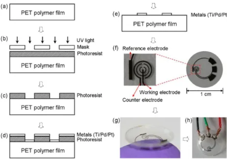
1. Introduction ... 231
1.1 Strategic route for ECL miniaturization in this project ... 232
1.2 Fluidic design strategies ... 234
2. Materials & Methods ... 238
2.1 Materials ... 238
2.2 Photolithography of ITO foils. ... 239
2.3 Silica‐mesochannels (SMC) modification of ITO electrodes ... 240
2.4 ECL and electrochemical measurements. ... 240
2.5 Hot embossing of PMMA ... 241
2.6 Thermal bonding of ITO@PET on PMMA (optimized procedure) ... 242
3. Results ... 244
3.1 Electrochemical implications towards materials choice and fabrication strategies... 244
3.2 Development of thermal bonding of PET with PMMA ... 245
3.3 Electrochemical tests with [Ru(bpy)
3]
2+‐ECL solutions on microfluidic ITO electrodes ... 247
3.4 ITO modifications ... 249
3.4.1 SMC modified ITO electrodes. ... 250
3.5 [Ru(bpy)
3]
2+‐ECL characterization and luminol on LSG electrodes. ... 252
4. Conclusions ... 256
5. References ... 258
Chapter 7: Conclusions and perspectives ... 260
References ... 266
Curriculum Vitae ... 269
Presentations ... 271
Publications ... 272
Eidesstattliche Erklärung ... 273
1
Summary
With the ascent of IT, and since Ashton has invented the term Internet of Things (IoT) in 1999, this future idea of connected machines that can do tasks and perform decision‐
control cycles without human input has become more and more attractive and is today an established future scenario. Obviously, in an IoT, “sensors for everything” are one crucial corner stone of its existence and Analytical chemistry can and must deliver them. While many challenges towards a functioning IoT remain, we are on the verge of its beginning. This can be also seen with “Analytics 4.0” in research and on the market, tending to more IT‐connected, portable, easier‐controllable and integrated solutions.
The entrance of mobility in the health sector or Point‐of‐Care (POC) diagnostics trends are alike influencing biosensing. Whether in mobile solutions or lab‐ and clinical environments, versatile, powerful and easy‐to‐adapt detection strategies like Electrochemiluminescence (ECL) are an attractive option.
Investigation and optimization of ECL strategies
The ECL molecules [Ru(bpy)
3]
2+and luminol represent the most prominent and most abundantly investigated luminophores for ECL since Bard’s accomplishment to make ECL a well‐known technique. Because both are also two of the most efficient ECL emitters that can be well‐handled in bioanalysis, and are available on the market, they are still today frequently used in research and also commercial applications. To cope with current benchmarks of sensitive detection, however a combination with a certain signal enhancement strategy is recommended. Several different routes can here be employed and one option is dendrimers. PAMAM dendrimers can function as ECL coreactant in [Ru(bpy)
3]
2+‐ECL via their amino groups and at the same time expose primary amino groups as possible bioconjugation elements. Exploring this multi‐
functionality of the dendrimers was investigated here. This was done on a model
system employing PAMAM dendrimers with [Ru(bpy)
3]
2+‐ECL together with
biotin/streptavidin as biorecognition element and analyte, respectively. The
dendrimer’s bi‐functionality was successfully proven and a joint‐role of a biorecognition
element and a possible reporter function suggests an optimum application in
homogeneous assays. A different toolset for ECL signal enhancement is offered by
2 liposomes. Numerous signaling molecules can be encapsulated inside the inner cavity of these synthetic vesicles, while they provide protection from the environment and connection‐functionality to probes via lipids and surface groups on the outside. That application was here explored, together with a newly synthesized luminol derivative obtained by a simple synthesis route from commercial starting materials and exhibiting a four times increased ECL efficiency versus standard luminol. That was necessary as a liposome enhancement was denied for the standard luminol through its poor aqueous solubility. The new m‐carboxy luminol considerably improved this feature which allowed its own encapsulation in liposomes. The superior signal generation with this dual system was proven in a model sandwich hybridization assay which yielded a 150‐
times better detection performance than the equal fluorescence‐based assay while being almost zero affected through matrices like serum, soil or river water. As such the good performance of luminol ECL together with liposomes for highly sensitive detection applications was demonstrated. A further necessary element with liposomal amplification, are surfactants to set free the signaling molecules. However, this case depicts only one example of a multitude of applications of surfactants in bioassays and biochemical methods. Hence, surfactants are commonly present solution constituents which also have to be considered in general with ECL because they can influence the ECL signals positively or negatively. This was further investigated for luminol ECL by exploring the effect of 13 different surfactants on the luminol ECL efficiency on four different electrode materials. A deeper understanding of the distinct effects was obtained by looking into ECL emission behavior, electrochemical effects, the surfaces and Chemiluminescence effects. After all, the revelation of a complicated mechanism that involves many contributing factors and as such directs signal quenching or enhancement is an important finding for assay design. In this way, the selection of a suitable surfactant is possible to exploit maximum reachable signal efficiencies.
Miniaturization of ECL assays
A combination of signal enhancement tools like a better ECL molecule derivative,
dendrimers, liposomes or surfactants has proven to boost the ECL performance
considerably. A further means of signal enhancement is offered via miniaturization,
which also makes the detection method better suited towards common application as
liquid handling and easier automation are on hand. This can be used for single ECL
assays or combinations of different ECL reagents in one system for multi‐detection.
3
Different strategies for the miniaturization of an ECL readout‐capable system were
investigated, taking requirements for [Ru(bpy)
3]
2+and luminol as ECL reporters into
account. This includes materials, electrochemical demands and simple design. Here, ITO
electrodes – while advantageous for luminol ECL could not convince with their
performance in [Ru(bpy)
3]
2+‐ECL. Alternatively, laser scribed graphene electrodes have
shown to be promising candidates for a future miniaturized system encompassing both,
luminol and [Ru(bpy)
3]
2+as ECL systems. Ultimately, the different signal amplifying
strategies, investigated in this work that can be applied standalone or combined, offer a
great toolset for state‐of‐the‐art ECL detection applications in research and also for
possible commercial applications.
4
Zusammenfassung
Mit dem Aufstieg der Internettechnologie und seit Ashton 1999 den Begriff Internet of Things (IoT) prägte, ist diese Zukunftsidee von vernetzten Maschinen, die Aufgaben und Entscheidungs‐ und Kontrollzyklen ohne menschliches Zutun ausführen können, immer attraktiver geworden und heute ein etabliertes Zukunftsszenario. Für die dienliche Existenz eines IoT sind „Sensoren für alles“ ein entscheidender Grundpfeiler, welche die analytische Chemie liefern kann und muss. Während viele Herausforderungen für ein funktionierendes IoT bestehen bleiben, stehen wir kurz vor dem Beginn. Dies zeigt sich auch in der „Analytik 4.0“ sowohl in der Forschung als auch auf dem freien Markt, die zu mehr IT‐vernetzten, portablen, einfacher steuerbaren und integrierten Lösungen neigt. Der Einstieg in die Mobilität im Gesundheitssektor oder Point‐of‐Care (POC) ‐ Diagnosetrends beeinflussen gleichermaßen die Biosensorik. Ob in mobilen Lösungen oder Labor‐ und klinischen Umgebungen ‐ vielseitige, leistungsstarke und einfach zu adaptierende Detektionsstrategien wie Elektrochemilumineszenz (ECL) sind eine attraktive Option.
Untersuchung und Optimierung von ECL‐Strategien
Die ECL‐Moleküle [Ru(bpy)
3]
2+und Luminol repräsentieren die bekanntesten und am häufigsten verwendeten Luminophore für ECL, seit der Pionierleistung von Bard ECL zu einer wohlbekannten Technik zu machen. Da beide zwei der effizientesten ECL‐Emitter sind, die in der Bioanalytik gut gehandhabt werden können und kommerziell erhältlich sind, finden sie auch heute häufig in der Forschung und in kommerziellen Anwendungen Verwendung. Um zeitgemäßen Anforderungen für sensitive Nachweise zu entsprechen, ist jedoch eine Kombination mit verschiedenen Signalverbesserungsstrategien zu empfehlen. Hierzu können mehrere verschiedene Ansätze verwendet werden, wobei Dendrimere eine der Optionen darstellen.
Polyamidoamin (PAMAM)‐Dendrimere können mittels ihrer Aminogruppen als ECL‐
Coreactant in [Ru(bpy)
3]
2+‐ECL fungieren und enthalten ebenso funktionelle primäre Aminogruppen, die eine Biokonjugation der Dendrimere ermöglichen. Für ein tieferes Verständnis dieser Multifunktionalität, wurden Dendrimere hier in ihrer Rolle als ECL‐
Coreactant parallel zu ihrer Funktion als Bioerkennungselements genutzt. Dies wurde
an einem Modellsystem, mit PAMAM‐Dendrimeren und [Ru(bpy)
3]
2+‐ECL zusammen mit
5 Biotin/Streptavidin als Bioerkennungselement bzw. Analyt untersucht. Die Nutzung der Bi‐funktionalität der Dendrimere wurde erfolgreich gezeigt und ihre universelle Rolle als Bioerkennungselement und mögliche Reporterfunktionalität suggeriert eine optimale Anwendung in homogenen Assayformaten. Liposome stellen ein weiteres Werkzeug zur ECL‐Signalverstärkung dar. Zahlreiche Signalmoleküle können dabei innerhalb des inneren Hohlraums dieser synthetischen Vesikel verkapselt werden, während diese Schutz vor der Umwelt und über Lipide, mit deren vielseitigen funktionellen Oberflächengruppen auf der Außenseite, eine einfache Kopplung zu Sonden ermöglichen. Diese Anwendung wurde hier zusammen mit einem neu synthetisierten Luminolderivat untersucht, das mittels eines einfachen Synthesewegs aus kommerziellen Ausgangsmaterialien erhalten wurde und eine vierfach höhere ECL‐
Effizienz als Standard‐Luminol zeigte. Dies wurde notwendig, da eine Liposom‐basierte
Signalverstärkung mit Standard‐Luminol aufgrund dessen schlechten Wasserlöslichkeit
verwehrt wurde. Das neue m‐Carboxy‐Luminol verbesserte dieses Merkmal
beträchtlich, was dessen eigene Verkapselung in Liposome ermöglichte. Die
herausragende Eignung zur Signalerzeugung dieses dualen Systems wurde in einem
Modell‐Sandwich‐Hybridisierungsassay nachgewiesen, der eine 150‐fach bessere
Detektionsleistung als der gleiche fluoreszenzbasierte Assay ergab, während ein
möglicher Einfluß durch Matrizen wie Serum, Boden oder Flusswasser vernachlässigbar
blieb. Anhand dieses Assays wurde die überragende Leistung von Luminol ECL
kombiniert mit Liposomen für hochempfindliche Detektionsanwendungen
offensichtlich. Ein weiteres notwendiges Element bei Liposom‐basierter Verstärkung
sind Tenside, zur kontrollierten Freisetzung der Signalmoleküle. Diese Nutzung von
Tensiden stellt jedoch nur ein Beispiel aus einer Vielzahl an Anwendungen in Bioassays
und biochemischen Verfahren dar. Daher sind Tenside üblicherweise vorhandene
Lösungsbestandteile, die im Allgemeinen auch in der ECL berücksichtigt werden
müssen, da sie die ECL‐Signalintensität positiv oder negativ beeinflussen können. Dies
wurde für Luminol‐ECL untersucht, indem die Wirkung von 13 verschiedenen Tensiden
auf die Luminol‐ECL‐Effizienz an vier verschiedenen Elektrodenmaterialien erforscht
wurde. Ein tieferes Verständnis der verschiedenen Effekte wurde durch
Untersuchungen des ECL‐Emissionsverhaltens, der elektrochemischen Effekte, der
Oberflächen und der Chemilumineszenzeffekte erworben. Dabei stellt die Entdeckung
eines komplizierten Mechanismus, zu dem viele Faktoren beitragen und welcher die
6 Signallöschung oder ‐verstärkung lenkt, einen wichtigen Befund für das Design neuer Assays dar. Auf diese Weise ist die Auswahl eines geeigneten Tensids möglich, um entweder die maximal erreichbare Signaleffizienz zu erhalten oder, falls dies nicht notwendig ist, die Wahl des bestgeeigneten Tensids auszunutzen.
Miniaturisierung von ECL Assays
Eine Kombination mit Signalverstärkungswerkzeugen wie beispielsweise einem verbesserten Derivats eines ECL Moleküls, Dendrimeren, Liposomen oder Tensiden kann die ECL‐Leistung erheblich steigern. Eine weitere Möglichkeit zur Signalverbesserung bietet hier Miniaturisierung, die die Detektionsmethode auch hinsichtlich der allgemeinen Anwendung verbessert, da eine gesteuerte Handhabung von Flüssigkeiten und eine einfachere Automatisierung zur Verfügung stehen. Dies kann in einzelnen ECL‐Assays oder bei Kombinationen von verschiedenen ECL‐Reagenzien in einem System zur Mehrfachdetektion genutzt werden. Unterschiedliche Strategien zur Miniaturisierung eines Systems, das mit ECL‐Detektion genutzt werden kann, wurden hier untersucht, wobei Anforderungen für [Ru(bpy)
3]
2+und Luminol als ECL‐Reporter berücksichtigt wurden. Dies beinhaltet Materialien, elektrochemische Anforderungen und einfaches Design. Hier konnten ITO‐Elektroden ‐ obwohl vorteilhaft für Luminol ECL
‐ mit ihrer Leistung für [Ru(bpy)
3]
2+‐ECL nicht überzeugen. Alternativ haben sich laser‐
scribed‐Graphen Elektroden als vielversprechende Kandidaten für ein zukünftiges
miniaturisiertes System erwiesen, das sowohl zur ECL Detektion von Luminol als auch
[Ru(bpy)
3]
2+geeignet ist. Letztendlich bieten die verschiedenen
Signalverstärkungsstrategien, die in dieser Arbeit untersucht wurden und die einzeln
oder kombiniert angewendet werden können, einen umfassenden Werkzeugsatz für
moderne ECL‐Detektionsanwendungen in der Forschung und auch für zukünftige
kommerzielle Anwendungen.
7
Introduction and structure of the work
The focus of this thesis lies in the advancement of Electrochemiluminescence (ECL) coupled with different signal amplification tools as sensitive detection technique towards application in modern biosensing with special considerations on highly sensitive applications and miniaturized, mobile solutions for possible implementation in growing Point‐of‐Care (POC) and Internet‐of‐Things (IoT)‐directed biosensing solutions.
More than ever, analytical chemistry and biosensing are transitioning into everyday tools for the common person, leaving an allocation as peripheries of chemistry and biochemical sciences and proceeding towards key elements and crucial toolsets of a growing and evolving IoT. Despite of today’s situation where we are still a fair step away from a functioning overall network, we are on the edge of realization and roll‐
outs of smaller and larger IoT‐shaped networks and information hives with an exponential growth. Chapter 1 in this thesis, gives a short glance in and overview of this large topic and trends while in Chapter 2 existing analytical and biosensing solutions are addressed that depict examples how analytical chemistry can be‐ and will become a helpful tool for everybody. This chapter also outlines in‐depth existing challenges (e.g.
true long‐term stability of sensors), and required advancements (e.g. joint IT‐
communication protocols), towards a successful evolution of sensing and biosensing in this setting. One large area of such an “IoT horizon” deals with evolving challenges to face existent and emerging worldwide health risks,
1how to shape the fields of modern healthcare, with developing point‐of‐care (POC) diagnostics and finally the area of highly personalized medicine. This joint venture of health risks, sensing requirements, (bio)sensing solutions and the all‐embracing framework of an upcoming IoT profoundly impacts the future direction of biosensor development.
Broken down to the actual methods and tools that are necessary and useful for such applications, the choice of a versatile detection method is one crucial decision. With respect to that, complying core requirements here are condensed under the terms of
“miniaturization& portability”, “suitability for mass production& cost reduction”,
“simple construction”, “reliability”, “automation& layman usability”, “sustainability”
and “ruggedness”. Parts or all of that are required, while the method should ideally
exhibit outstanding reachable sensitivities to make it versatile and ready to cope with
8 given needs. Also, the transition of purely scientific thoughts to show and exhaust
“what is possible”, “what is new” and “what can be realized in the lab” towards realization of true marketable products, that can cope with given requirements of POC or mobile sensing is becoming more and more important. One factor of these application‐set requirements, “miniaturization and integration” plays a key role in realizing small‐enough devices with little weight. These should either adhere to the current and future wearables realm or be embedded in a size‐restricted application setting and be true lab‐on‐chip (LOC) devices. In detail, this means that not only a single element of the systems can be small while the remaining parts are typical benchtop instruments. Here, the whole analytical process from sample introduction to results analysis must be miniaturized. Addressing that, has been disregarded often
2and is still observed in current biosensing research
3while recent works also recognize this issue and lead by example by showing true whole sensor devices that can integrate the overall analytical process.
4,5In this work, a detection method suitable for such sensing systems is investigated.
Electrochemiluminescence (ECL) as detection technique has proven over the years of its
success since A.J. Bard paved its way,
6that it is more than a niche application but a
powerful method perfectly suited for highly sensitive detection demands (See chapter
4 and e.g. Hu et al.
7). Through its intrinsic light generation at an electrode as the
analytical signal, the required instrumentation can be designed miniaturized and
simple. The optimally flat electrodes can be easily downscaled and patterened freely,
8,9are available on thin, flexible substrates (e.g. ITO on PET foil) and certain electrode
types can be simply scaled‐up to meet industrial high throughput fabrication processes
e.g. screen‐printing. The second part of the detection system simply requires to capture
the light emission which can be implemented as easily. What has been shown a lot
recently, are solutions using smartphone‐camera based readouts,
10,11off‐the‐shelf
CCD/CMOS chips, photodiodes and such systems
12that can be readily used for sensitive
ECL emission detection. At the same time, the surrounding instrumentation to power
and control the two elements of electrochemistry (potentiostat) and optical detection
can as well be broken down to very small formats.
13‐15Finally, ECL’s nature of a
luminescence process, eliminates the need of other light sources, which allows for
direct detection of the absolute amount of light present, without an urgence for filter
or monochromator systems when the system is simply enclosed from ambient light.
9 Also compared with chemiluminescence (CL) based signal generation, which omits the electrochemical side, in several applications ECL can play out different advantages which are briefly discussed. The total spatial control regarding the signal generation (in ECL) is important in flow systems, whereas reactants in CL could drift‐off and broaden the detection zone. An absolute time control over the luminescence initiation (beneficial in cases the employed assay needs lysis or incubation steps prior to detection, and luminescence termination are additionally helpful. In ECL all necessary reagents can readily be included prior to the start of luminescence and no residual emission is persistent after stopping the potential. Also, in multi detection setups (“microtiter plate‐alike design”) no need to account for residual emissive stray light from neighboring spots is advantageous. Finally, a generally more stable signal (for coreactant ECL) versus CL, the possibility to use the electrochemical signal (current or better, transferred charge) as second analytical signal in a multimodal approach (e.g.
correction for luminescence quenching or enhancement in amperometric readout
16) and the possibility to directly modify the electrode (e.g. SAM’s,
17pyrenebutyric acid,
9,18silanes
19) with probes to capture the analyte directly at the detection spot are further amenities. The necessary additional electrochemical instrumentation in ECL vs. CL doesn’t depict a major drawback as these components can all be readily miniaturized.
Also, in CL, the employed instrumentation has to cover the critical timing of luminescence initiation and simultaneous initiation of signal capturing in a reproducible way. Thus, along all and especially optical detection methods, luminescence techniques like ECL lend themselves well for sensitive and versatile ‐ but facile sensor designs. This also qualifies ECL for different routes, be it in POC applications, mobile field solutions or powerful benchtop routine assay applications.
The selection of very classical ECL luminophores (luminol, Tris(2,2’‐
bipyridyl)ruthenium(II) chloride ([Ru(bpy)
3]
2+) and their derivatives) employed within
this research project was driven by several arguments. Both ECL systems were handled,
operating them in their (oxidative‐reductive) coreactant‐ECL pathways (luminol‐H
2O
2,
[Ru(bpy)
3]
2+‐tertiary amine)
20and applying earlier findings
21,22on optimized signal
efficiencies. This route enables their application in aqueous environments while
maintaining moderate environmental conditions (pH, temperature) with their standard
forms being commercially available. Within these boundaries, both candidates are
amongst the most efficient, existent ECL emitters. The option to focus on two different
10 species which follow the same constraints, while their emission wavelengths are far separated, allows for the design of multi‐sensing solutions e.g. applying both reporters in one shared medium at the same time (concept shown in earlier master project
22) or with a two‐in‐one signal readout. Besides that, two different ECL probes offer different advantages for varying assay applications. While recent ECL research is mostly focused on different and newer types of materials, e.g. quantum dots
23or other compounds,
24[Ru(bpy)
3]
2+still represents the benchmark for commercially available ECL applications
25and luminol is commercially applied on a large scale in CL assays.
26‐28There are several, grand, unexploited prospects that the classical luminophores comprise, while the ready use in commercial ECL assay kits indicates the market relevance and possible transition towards upcoming POC and diagnostics applications.
While ECL itself is a powerful and sensitive detection technique, signal enhancement strategies beyond single‐probe approaches cannot be neglected to truly unleash the full power of detection capabilities. In this regard, different combinations of ECL reagents and signal enhancement tools can be used, which all have different advantages. The combination of [Ru(bpy)
3]
2+as ECL reagent together with PAMAM dendrimers, which is presented in chapter 3, highlights one possible route for ECL signal enhancement. Here, four different PAMAM dendrimers encompassing between 5‐16 terminal amino groups were compared for their performance in [Ru(bpy)
3]
2+‐ ECL. Additionally, the possibility to combine the function of a biorecognition element for the analyte and at the same time the role of the ECL coreactant in a single entity, i.e. the dendrimer is shown. This bi‐functionality was investigated with the model system biotin/streptavidin in a homogeneous format while it suggested that the presence of [Ru(bpy)
3]
2+in the vicinity of dendrimers at the electrode surface is beneficial towards stabilizing these assemblies and for a signal enhancement nature. A very different approach is shown in chapter 4, with the combination of a new luminol derivative with liposomes as signal enhancement means. Here, the novel synthesis of m‐carboxy luminol created a superior ECL probe. This new molecule was analyzed towards its ECL properties where its superiority compared to standard luminol, mediated by its good water solubility was shown. This also enabled its inclusion into liposomes as multiplexing probe. These were investigated for different parameters like their stability, synthesis reproducibility and their performance under changed conditions with m‐carboxy luminol as encapsulant.
The liposomes were then employed in a heterogeneous DNA sandwich hybridization
11 assay for performance comparison to the same assay employing fluorescence detection with minor optimization. Here, the superiority of this concept for highly sensitive detection demands also with respect to the presence of complex, environmental matrices (e.g. soil) was proved.
Besides the combination with dendrimers or liposomes as small‐polymer‐ or biochemical macrostructure probes for signal enhancement also the addition of surfactants can alter the ECL signal extent dramatically. This is an important, but double‐edged feature which can either strongly limit the performance of the applied system but also be used for signal enhancement. For this reason, it is important to understand the underlying mechanistical aspects which drive the signal influencing nature of the respective surfactants. In chapter 5, the influence of several different surfactants on the ECL of the luminol‐H
2O
2system was investigated for two different luminol variants and on four different electrode materials. The choice of different, charged and uncharged surfactant types as well as of gold, ITO, hydrophilized ITO and laser scribed graphene electrodes allowed to acquire a broad picture of the ambiguous surfactant effects on the employed luminol system. The underlying signal enhancing and reducing mechanisms were investigated by looking into the ECL properties, the electrochemical properties, the electrode surfaces, CL behavior and finally by comparing these with the respective surfactant properties. As such, possible influencing factors were critically assessed and suggestions for a symbiotic mechanism presented.
With the frequent presence and requirement of surfactants in different bioassays, this created library of surfactant effects on the luminol‐H
2O
2ECL system can be beneficial in intelligent assay design.
Besides signal enhancement means via combination with probes or surfactants, in
chapter 6, conceptual and assay design considerations for a possible joint‐venture of
both ECL reagents, luminol and Ru(bpy)
32+with respect to a miniaturized and versatile
ECL detection system are presented. The use of microfluidic technology can lead to
further signal enhancement effects can further prepare the ECL detection method for a
possible mobile application. Here, the capability towards a function with both ECL
systems is discussed, addressing electrode material choice, electrode performance and
limitations. A new direct thermal bonding method between the polymers PMMA and
PET is presented and the suitability of the resulting microfluidic system for luminol and
12 Ru(bpy)
32+ECL is assessed. Finally, an alternative carbon based electrode material is characterized for its general ECL capability towards microfluidic use.
References
(1) Munster, V. J.; Bausch, D. G.; de Wit, E.; Fischer, R.; Kobinger, G.; Muñoz‐Fontela, C.; Olson, S. H.; Seifert, S. N.; Sprecher, A.; Ntoumi, F.; Massaquoi, M.; Mombouli, J.‐
V. Outbreaks in a Rapidly Changing Central Africa — Lessons from Ebola. N. Engl. J.
Med. 2018. doi: 10.1056/NEJMp1807691.
(2) Mohammed, M. I.; Haswell, S.; Gibson, I. Lab‐on‐a‐chip or Chip‐in‐a‐lab: Challenges of Commercialization Lost in Translation. Procedia Technology 2015, 20, 54‐59. doi:
10.1016/j.protcy.2015.07.010.
(3) Balakrishnan, S. R.; Hashim, U.; Gopinath, S. C. B.; Poopalan, P.; Ramayya, H. R.;
Iqbal Omar, M.; Haarindraprasad, R.; Veeradasan, P. A Point‐of‐Care Immunosensor for Human Chorionic Gonadotropin in Clinical Urine Samples Using a Cuneated Polysilicon Nanogap Lab‐on‐Chip. PLoS One 2015, 10, e0137891. doi:
10.1371/journal.pone.0137891.
(4) Zór, K.; Heiskanen, A.; Caviglia, C.; Vergani, M.; Landini, E.; Shah, F.; Carminati, M.;
Martínez‐Serrano, A.; Moreno, T. R.; Kokaia, M.; Benayahu, D.; Keresztes, Z.;
Papkovsky, D.; Wollenberger, U.; Svendsen, W. E.; Dimaki, M.; Ferrari, G.; Raiteri, R.;
Sampietro, M.; Dufva, M., et al. A compact multifunctional microfluidic platform for exploring cellular dynamics in real‐time using electrochemical detection. RSC Advances 2014, 4, 63761‐63771. doi: 10.1039/c4ra12632g.
(5) Nascetti, A.; Mirasoli, M.; Marchegiani, E.; Zangheri, M.; Costantini, F.; Porchetta, A.; Iannascoli, L.; Lovecchio, N.; Caputo, D.; de Cesare, G.; Pirrotta, S.; Roda, A.
Integrated chemiluminescence‐based lab‐on‐chip for detection of life markers in extraterrestrial environments. Biosens. Bioelectron. 2018. doi:
10.1016/j.bios.2018.08.056.
(6) Tokel, N. E.; Bard, A. J. Electrogenerated chemiluminescence. IX. Electrochemistry and emission from systems containing tris(2,2'‐bipyridine)ruthenium(II) dichloride.
J. Am. Chem. Soc. 1972, 94, 2862‐2863. doi: 10.1021/ja00763a056.
(7) Hu, G. B.; Xiong, C. Y.; Liang, W. B.; Zeng, X. S.; Xu, H. L.; Yang, Y.; Yao, L. Y.; Yuan, R.;
Xiao, D. R. Highly Stable Mesoporous Luminescence‐Functionalized MOF with Excellent Electrochemiluminescence Property for Ultrasensitive Immunosensor Construction. ACS Appl. Mater. Interfaces 2018, 10, 15913‐15919. doi:
10.1021/acsami.8b05038.
(8) See chapter 6, photolithography of ITO electrodes with benchtop chemistry.
13 (9) Fenzl, C.; Nayak, P.; Hirsch, T.; Wolfbeis, O. S.; Alshareef, H. N.; Baeumner, A. J.
Laser‐Scribed Graphene Electrodes for Aptamer‐Based Biosensing. ACS Sens. 2017, 2, 616‐620. doi: 10.1021/acssensors.7b00066.
(10) Gao, W. Y.; Muzyka, K.; Ma, X. G.; Lou, B. H.; Xu, G. B. A single‐electrode electrochemical system for multiplex electrochemiluminescence analysis based on a resistance induced potential difference. Chem. Sci. 2018, 9, 3911‐3916. doi:
10.1039/c8sc00410b.
(11) Doeven, E. H.; Barbante, G. J.; Harsant, A. J.; Donnelly, P. S.; Connell, T. U.; Hogan, C.
F.; Francis, P. S. Mobile phone‐based electrochemiluminescence sensing exploiting the ‘USB On‐The‐Go’ protocol. Sens. Actuators, B 2015, 216, 608‐613. doi:
10.1016/j.snb.2015.04.087.
(12) Roda, A.; Mirasoli, M.; Michelini, E.; Di Fusco, M.; Zangheri, M.; Cevenini, L.; Roda, B.; Simoni, P. Progress in chemical luminescence‐based biosensors: A critical review.
Biosens. Bioelectron. 2016, 76, 164‐179. doi: 10.1016/j.bios.2015.06.017.
(13) Ghodsevali, E.; Morneau‐Gamache, S.; Mathault, J.; Landari, H.; Boisselier, É.;
Boukadoum, M.; Gosselin, B.; Miled, A. Miniaturized FDDA and CMOS Based Potentiostat for Bio‐Applications. Sensors (Basel) 2017, 17. doi:
10.3390/s17040810.
(14) Bezuidenhout, P.; Smith, S.; Joubert, T.‐H. A Low‐Cost Inkjet‐Printed Paper‐Based Potenaostat †. Appl. Sci. 2018, 8. doi: 10.3390/app8060968.
(15) Wojciechowski, J. R.; Shriver‐Lake, L. C.; Yamaguchi, M. Y.; Füreder, E.; Pieler, R.;
Schamesberger, M.; Winder, C.; Prall, H. J.; Sonnleitner, M.; Ligler, F. S. Organic Photodiodes for Biosensor Miniaturization. Anal. Chem. 2009, 81, 3455‐3461. doi:
10.1021/ac8027323.
(16) Qiu, H.; Yin, X.‐B.; Yan, J.; Zhao, X.; Yang, X.; Wang, E. Simultaneous electrochemical and electrochemiluminescence detection for microchip and conventional capillary electrophoresis. ELECTROPHORESIS 2005, 26, 687‐693. doi:
10.1002/elps.200410015.
(17) Kang, C. H.; Choi, Y.‐B.; Kim, H.‐H.; Choi, H. N.; Lee, W.‐Y. Electrogenerated Chemiluminescence Sensor Based on a Self‐Assembled Monolayer of Ruthenium(II)‐
bis(2,2′‐bipyridyl)(aminopropyl imidazole) on Gold Deposited Screen Printed Electrode. Electroanalysis 2011, 23, 2131‐2138. doi: 10.1002/elan.201100214.
(18) Tsuwaki, M.; Kasahara, T.; Edura, T.; Oshima, J.; Kunisawa, E.; Ishimatsu, R.;
Matsunami, S.; Imato, T.; Adachi, C.; Shoji, S.; Mizuno, J. Fabrication of a Portable Electrochemiluminescence‐induced Fluorescence Detection Chip with Microfluidic Excitation Source for Point‐of‐care Diagnostics. IEEJ Transactions on Sensors and Micromachines 2015, 135, 230‐235. doi: 10.1541/ieejsmas.135.230.
(19) Zhang, Y.; Liu, W.; Ge, S.; Yan, M.; Wang, S.; Yu, J.; Li, N.; Song, X. Multiplexed sandwich immunoassays using flow‐injection electrochemiluminescence with designed substrate spatial‐resolved technique for detection of tumor markers.
Biosens. Bioelectron. 2013, 41, 684‐690. doi: 10.1016/j.bios.2012.09.044.
14 (20) Miao, W. Electrogenerated chemiluminescence and its biorelated applications.
Chem. Rev. 2008, 108, 2506‐2553. doi: 10.1021/cr068083a.
(21) Kirschbaum‐Harriman, S.; Duerkop, A.; Baeumner, A. J. Improving ruthenium‐based ECL through nonionic surfactants and tertiary amines Analyst 2017, 142, 2648‐2653.
doi: 10.1039/C7AN00197E.
(22) Mayer, M. Luminol and ruthenium electrochemiluminescence for a dual ECL detection approach. Master thesis, Regensburg, 2015.
(23) Pan, Q. X.; Wang, J. Y.; Cheng, Y. Z.; Li, W. J.; Wang, X. D. Determination of Hydrogen Peroxide by Electrochemiluminescence Using a Chitosan‐graphene Composite Film Doped Cadmium‐Tellurium Quantum Dot Modified Glassy Carbon Electrode. Anal. Lett. 2018, 51, 1373‐1383. doi: 10.1080/00032719.2017.1374964.
(24) Carrara, S.; Aliprandi, A.; Hogan, C. F.; De Cola, L. Aggregation‐Induced Electrochemiluminescence of Platinum(II) Complexes. J. Am. Chem. Soc. 2017, 139, 14605‐14610. doi: 10.1021/jacs.7b07710.
(25) Roche Diagnostics, "Die ECL-Technologie", can be found under https://www.roche.de/diagnostics/systeme/gesamtkonzepte/elecsys‐
technologie.html#Die‐ECL‐Technologie 2018.
(26) Ortho Clinical Diagnostics, "VITROS® MicroWell Technology", can be found under https://www.orthoclinicaldiagnostics.com/en‐hk/home/products/vitros‐microwell‐
technology 2018.
(27) Elabscience, "CLIA Kits", can be found under https://www.elabscience.com/Products‐clia_kits‐108.html, 2018.
(28) Rodriguez‐Orozco, A. R.; Ruiz‐Reyes, H.; Medina‐Serriteno, N. Recent Applications
of Chemiluminescence Assays in Clinical Immunology. Mini-Rev. Med. Chem. 2010,
10, 1393‐1400. doi: 10.2174/138955710793564142.
15
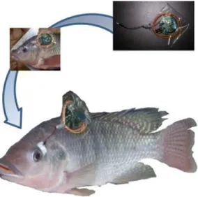
Chapter 1: ABC Spotlight on Analytics 4.0
Graphical Abstract
This chapter has been published.
Michael Mayer, Antje J. Baeumner, Anal. Bioanal. Chem., 2018, 410 (21), 5095‐5097, DOI: 10.1007/s00216‐018‐1191‐7
Author contributions:
AJB and MM did the scientific literature search. MM wrote the first draft of the manuscript. AJB revised the manuscript. AJB is corresponding author.