D i s s e r t a t i o n
B E H AV I O R A L , N E U R O N A L , A N D D E V E L O P M E N TA L C O N S E Q U E N C E S O F G E N E T I C A L LY D E C R E A S E D
T R Y P T O P H A N H Y D R O X Y L A S E 2 A C T I V I T Y
Mathematisch-Naturwissenschaftlichen Fakultät I
va l e n t i na m o s i e n k o
Präsident der Humboldt-Universität zu Berlin Prof. Dr. Jan-Hendrik Olbertz
Dekan der Mathematisch-Naturwissenschaftlichen Fakultät I Prof. Dr. Stefan Hecht
Gutachter/innen:
1. Prof. Dr. Thomas Sommer 2. Prof. Dr. Michael Bader
3. Prof. Dr. Heidrun Fink
eingereicht:16.04.2013
Tag der Mündliche Prüfung:17.09.13
Наука является основой всякого прогресса, облегчающего жизнь человечества и уменьшающего его страдания.
Склодовская-Кюри М.
Science is the basis of every progress, facilitating human living and reducing suffering Marie Sklodowska-Curie
Dedicated to my loving parents
A B S T R A C T
Serotonin (5-hydroxytryptamine, 5-HT) is a major neurotransmitter in the brain biosynthesis of which is initiated by tryptophan hydroxy- lase2(TPH2). Polymorphisms in theTPH2gene are suggested as risk factors associated with depression and anxiety in humans. Further- more, the most frequently prescribed antidepressants and anxiolytics target the serotonergic system. However, the question whether a com- plete ablation or partial reduction in brain serotonin leads to the de- velopmental, neurochemical, or psychological abnormalities remains unresolved.
In this study, I took advantage of mouse lines with various degree of decrease in TPH2activity in order to dissect the impact of5-HT loss on development, brain neurochemistry and behavior.
Using Tph2-deficient mice I showed that central serotonin is essen- tial for normal postnatal, but not prenatal development. Growth re- tardation of Tph2-/- mice was not a result of a disruption of the hypothalamo-pituitary-adrenal axis, metabolic abnormalities, or im- paired thermoregulation, but could result from reduced ultrasonic vocalization.
I tested Tph2-/- mice along with other newly generated mouse mod- els with partial TPH2 reduction, and showed that 20% reduction in central serotonin is not enough to cause changes in anxiety- and depression-like behaviors most likely due to compensatory mecha- nisms including reduced serotonin metabolism and increased5-HT1A
receptor sensitivity.
However, complete loss of central serotonin leads to a depression- like phenotype, reduced anxiety-like behavior, and exaggerated ag- gression, but no differences in activity, olfaction, memory, and adult neurogenesis.
Fluoxetine treatment ofTph2-/- mice revealed serotonin-independent action of this antidepressant on anxiety- and depression-like behavior.
Furthermore, fluoxetine drastically reduced the brain 5-HT content in mice with low TPH2 activity indicating that TPH2 activity may determine the efficiency of antidepressants targeting the serotonergic system.
Keywords: Tryptophan hydroxylase, Serotonin, Development, Be- havior, Fluoxetine
Z U S A M M E N FA S S U N G
Serotonin (5-Hydroxytryptamin,5-HT) ist ein wichtiger Neurotrans- mitter im Zentralnervensystem (ZNS). Seine Biosynthese erfolgt unter Beteiligung des Enzyms Tryptophanhydroxylase 2(TPH2). Polymor- phismen im TPH2 Gen beim Menschen sind Risikofaktoren bei der Entstehung von Depressionen und Angstverhalten. Die gängigsten Antidepressiva und Anxiolytika wirken auf das Serotonin System.
Unklar ist, ob das komplette oder teilweise Fehlen von Serotonin im Gehirn zu Entwicklungsstörungen und neurochemischen oder psy- chologischen Veränderungen führt.
In dieser Arbeit werden Mauslinien mit unterschiedlichen TPH2Akti- vitäten im ZNS verglichen und der Einfluss verringerter5-HT Konzen- trationen auf Entwicklung und Verhalten der Tiere untersucht. Zen- trales Serotonin ist nur für die postnatale Entwicklung notwendig.
Das verzögerte Wachstum vonTph2-/-Tieren ist nicht auf eine Störung der Hypothalamus-Hypophysen-Nebennieren-Achse oder auf metabo- lische Veränderungen zurückzuführen, sondern kann aus verringerter Vokalisation im Ultraschallbereich resultieren.Tph2-/-Mäuse wurden mit generierten Mausmodellen mit niedriger TPH2 Aktivität vergle- ichen. Die Ergebnisse zeigen, dass 20% weniger zentrales Serotonin nicht ausreichen, um Depression oder Angst-Verhalten herbeizuführen.
Möglicherweise greifen kompensatorische Mechanismen wie ein ver- ringerter Serotoninmetabolismus oder eine gesteigerte5-HT1A-Rezep- torsensitivität. Der komplette Verlust von Serotonin im Gehirn führt zu einem starken depressiven und weniger ängstlich Verhalten, mit erhöhter Aggression - ohne Veränderung in Aktivität, Geruchsinn, Gedächnis und adulter Neurogenese. Fluoxetine Behandlung vonTph2- defizienten Mäusen zeigte einen Serotonin-unabhängigen Effekt dieses Antodepressivums auf Angst-Verhalten und Depression. Fluoxetine reduzieren den Serotoningehalt im Gehirn von Mäusen mit geringen TPH2-Aktivität, was zeigt, dass TPH-Aktivität die Effizienz von Sero- tonin beeinflussenAntidepressiva bestimmen.
Schlagwörter: Tryptophan hydroxylase, Serotonin, Entwicklung, Ver- halten, Fluoxetine
Мы должны быть мужественней и не прекращать своей деятельности от неудач.
Константин Циолоковкий
One should be more manly, and despite multiple failures should not stop his activities Konstantin Tsiolkovsky
A C K N O W L E D G E M E N T S
First of all I very much like to thank Prof. Dr. Michael Bader and Dr. Natalia Alenina for their brilliant supervision of my PhD project.
Without these two people this work would never come out. Michael gave me the possibility to work in his laboratory, but also to perform any kind of experiment. Both of them were diligent advisors in tea- ching me which questions to ask and how to think in order to answer them. Natasha was both friend and chief during the entire time of my PhD. She had always time for answering my questions, for hel- ping me to solve technical problems arising during the experiments.
I would like to thank her for her patience, kindliness, support, all the knowledge and skills she taught me, her never-ending enthusiasm, optimism and energy.
I would like to thank Prof. Dr. Heidrun Fink and Dr. Bettina Bert for supporting our collaborative studies and giving scientific ideas that arose during this collaboration.
Many thanks to the students I worked with during all the time of my PhD, and to all the members of the group, especially to Dr. Michail Todiras, Susann Matthes, Daniel Beis, and Dr. Friederike Klempin for their help in performing some of the experiments, for very valuable scientific discussions, and the extensive support throughout my PhD.
I would like to thank Dr. Herbert Schulz from the research group of Prof. Dr. Norbert Hübner for analysing numerous gene expression data.
I would like to thank Iris Apostel-Krause for her generous help with German bureaucracy, and for the warm atmosphere she is creating in the group.
I would like to thank Sylvia Sibilak for her help regarding the issues about the legal stay in Germany.
I would like to thank Susanna da Costa Goncalves, Thorsten Riepen- hausen, Andrea Müller and Lisa Mallis for magnificent technical help in the lab, especially with valuable help in endless animal genotyp- ing, in providing necessary conditions and safety for the experimen- tal work in the laboratory. Special thanks go to Manfred Ströhmann and Sabine Grüger, who took care of the animal colonies on which
processing of multiple samples for gene expression analysis.
I would like to thank the Deutsche Akademische Austausch Dienst (DAAD) for financial support during the first 4 years of my PhD.
Special thanks go to the International Helmholtz Research School of
0Molecular Neurobiology0 without their funding I would have not been able to go to some international conferences, and its coordina- tors, Dr. Jana Dröse, Dr. Annette Schledz, and Dr. Oksana Seumenicht for their support and endless patience.
I would like to thank my family that supported first of all my deci- sion to make a PhD far away from them. Despite being not around they were and will always be my shelter and the source of positive emotions.
Last but not least, I would like to thank all my friends who were around to hear my (occasionally unreasoned) complains, who gave their advices and shared experiences, with whom I shared my fears and happy moments, and who would take me out to refresh and recharge all my mental energy.
C O N T E N T S
1 i n t r o d u c t i o n 3
1.1 Serotonin - history, metabolism, and mechanisms of action 3
1.1.1 Discovery of serotonin . . . 3
1.1.2 TPH1and TPH2: serotonin synthesising enzymes . . . . 3
1.1.3 Serotonin metabolism . . . 5
1.1.4 Formation of serotonergic neurons . . . 5
1.1.5 Serotonergic neurotransmission . . . 6
1.2 Central serotonin andTPH2in humans . . . 7
1.2.1 Mutations in TPH2 and its contribution to psychiatric states in humans . . . 7
1.2.2 Mouse as a model for human disorders . . . 9
1.3 Hyposerotonergic animal models . . . 9
1.3.1 Tph2-deficient mice . . . 9
1.3.2 Mouse models with altered formation of serotonergic neurons . . . 11
1.3.3 Mouse models with impaired serotonin storage, release and reuptake . . . 12
1.3.4 Mouse models with polymorphisms inTph2gene . . . . 13
2 a i m s o f t h e s t u d y 15 3 m at e r i a l s a n d m e t h o d s 17 3.1 Materials . . . 17
3.1.1 Reagents . . . 17
3.1.2 Kits and Markers . . . 18
3.1.3 Antibodies . . . 18
3.1.4 Equipment and expandable material . . . 19
3.1.5 Primers . . . 19
3.2 Methods . . . 20
3.2.1 Molecular biology methods . . . 20
3.2.2 Determination of plasma parameters . . . 24
3.2.3 Staining . . . 24
3.2.4 Animals . . . 26
3.2.5 Behavior assessment . . . 27
3.2.6 Telemetric measurement . . . 33
3.2.7 Neurochemical assessment . . . 33
3.2.8 Animal treatment . . . 34
4 r e s u lt s 37 4.1 Growth retardation inTph2-deficient mice . . . 37
4.1.1 Phenotype . . . 37
4.1.2 In search for explanation: hypothalamic-pituitary-adrenal (HPA) axis . . . 38
4.1.3 In search for explanation: metabolic abnormalities . . . . 40
4.1.4 In search for explanation: behavioral changes during early
development . . . 42
4.2 Behavioral and neurochemical abnormalities inTph2-deficient mice . . . 43
4.2.1 Tph2expression and serotonin levels . . . 43
4.2.2 Activity and muscle strength . . . 43
4.2.3 Olfaction . . . 44
4.2.4 Aggressive behavior . . . 47
4.2.5 Anxiety-like behavior . . . 48
4.2.6 Depresssion-like behavior . . . 49
4.2.7 Effect of fluoxetine in Tph2-deficient mice . . . 50
4.2.8 Learning and memory . . . 51
4.2.9 Neurogenesis . . . 53
4.3 Emotional and neurochemical abnormalities in mice car- ryingTph2hypomorphic and null alleles . . . 54
4.3.1 Anxiety- and depression-like behavior . . . 54
4.3.2 Serotonin content and metabolism . . . 55
4.3.3 5-HT1Areceptor function . . . 57
5 d i s c u s s i o n 59 5.1 Role of central serotonin in postnatal development . . . . 59
5.2 Tph2-deficiency and its psychological consequences . . . . 61
6 c o n c l u s i o n s 71
7 a b b r e v i at i o n s 73
b i b l i o g r a p h y 75
1
I N T R O D U C T I O N
1.1 s e r o t o n i n -h i s t o r y,m e ta b o l i s m,a n d m e c h a n i s m s o f a c t i o n
1.1.1 Discovery of serotonin
Serotonin (5-hydroxytryptamine, 5-HT) was discovered in the mid- dle of the 20th century. Scientists from the Page group were the first to isolate and identify the vasoconstrictive substance from the serum which was released while the blood was clotting [152]. Due to the source, serum, and its action, tonic, this substance encountered the name ’serotonin’ [152]. At the same time Erspamer was working on a muscle dilating substance, which he named ’enteramine’ [184]. At the end, he found out that ’serotonin’ and ’enteramine’ are the same factors. A couple of years later, in1953, Twarog and Page discovered serotonin in the central nervous system [199]. Further serotonin crea- tinine sulfate was synthesised, bursting the development of new ana- lytical methods to study serotonin metabolismin vivoandin vitro. Bet- ween 1953and 1970the processes of serotonin synthesis and degra- dation were clarified [184]. Moreover, during this time many impor- tant drugs interacting with the serotonergic system were discovered.
Among them are reserpine (further known as vesicular monoamine transporter (VMAT) inhibitor), monoamine oxidase (MAO) inhibitors (pargyline, phenelzine, tranylcypromine, isocarboxazid), lysergic acid diethylamine (LSD), aromatic amino acid decarboxylase (AADC) in- hibitor (carbidopa), tryptophan hydroxylase (TPH) inhibitor (para- chlorphenylalanine), and tricyclic antidepressants (imipramine, de- sipramine, amitriptyline, nortriptyline, protriptyline) [184].
1.1.2 TPH1and TPH2: serotonin synthesising enzymes
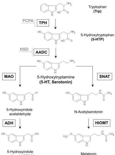
The biosynthesis of serotonin is a two-step process. On the first step the essential amino acid L-tryptophan (Trp) is metabolized to5-hydro- xytryptophan (5-HTP) by the rate-limiting enzyme TPH Figure 1. Next, 5-HTP is decarboxylated to 5-HT by the AADCFigure 1. This enzyme is involved as well in the synthesis of dopamine from l-3,4- dihydroxyphenylalanine (L-DOPA) [91], tryptamine from tyrosine [102] or tryptophan [57], and its distribution and functions are not restricted just to the serotonergic system. Thus, the whole process of serotonin synthesis in the brain is limited by two factors: availability of its sub- strate, Trp, and activity of the TPH enzyme.
Tryptophan (Trp)
5-Hydroxytryptophan (5-HTP) PCPA TPH
5-Hydroxytryptamine (5-HT, Serotonin) AADC
MAO SNAT
N-Acetylserotonin
Melatonin 5-Hydroxyindole
acetaldehyde
5-Hydroxyindole acetic acid (HIAA)
HIOMT ADH
NSD
Figure1: Serotonin synthesis and metabolism. TPH: tryptophan hy- droxylase, AADC: aromatic amino acid decarboxylase, PCPA:
parachlorophenylalanine (p-chlorophenylalanine), NSD: 3- hydroxybenzylhydrazine dihydrochloride, SNAT: serotonin N-acetyltransferase, HIOMT: hydroxy-indole-O-methyl trans- ferase, MAO: monoamine oxidase, ADH - alcohol dehydrogenase, modified from [136].
TPH belongs to the superfamily of aromatic amino acid hydroxy- lases that also includes tyrosine hydroxylase (TH) and phenylalanine hydroxylase (PAH). These are iron (Fe2+)- and tetrahydrobiopterin (BH4)-dependent monooxygenases with substantial structural simi- larities in their catalytic mechanism [65]. All aromatic amino acid hydroxylases are composed of 3 functional domains, a regulatory N-terminal domain, a catalytic domain and a C-terminal oligomeri- zation domain [208,65].
Since discovery of the serotonin synthesis pathway, it was supposed that in vertebrates only one form of TPH enzyme exists [105]. Howe- ver, in2003the existence of a second isoform,Tph2, was unravelled by genetical ablation of the only known Tph gene [214, 213]. This spec-
1.1 s e r o t o n i n - h i s t o r y, m e ta b o l i s m, a n d m e c h a n i s m s o f a c t i o n
tacular discovery gave new opportunities for studying the functions of two independent serotonin systems — central, driven byTph2, and peripheral, driven byTph1. Follow-up studies of mRNA and protein levels in rodents and human tissues confirmed TPH2to be the central isoform, which is predominantly expressed in the neurons of Raphe nuclei in the brainstem [78, 155, 213], and in peripheral myenteric neurons in the gut [147, 43], but is absent in peripheral organs like lung, heart, kidney, or liver [173, 231]. On the other hand, TPH1 is mainly expressed in the enterochromaffin cells of the gut and also in the pineal gland, where it produces 5-HT serving as a precursor for melatonin synthesis [185,213]Figure1.
1.1.3 Serotonin metabolism
In the brain, there are two main serotonin-degradation pathways. In serotonergic neurons, serotonin can be degraded to5-hydroxyindolea- cetic acid (5-HIAA) by one of the forms of MAO — MAO-AFigure1. MAO-A also metabolises norepinephrine [101]. Furthermore, another form of MAO, MAO-B, metabolises benzamidine [117]. Both enzymes are able to metabolize tyramine and dopamine [101]. Distribution of both isoforms in the brain reflects its substrate specificity [39]. In pitui- tary gland, retina and epiphysis serotonin serves as precursor in the synthesis of melatonin. First, it is degradated to N-acetylserotonin by 5-HT-N-acetyl-transferase (SNAT). Than the intermediate substrate get processed to melatonin by N-acetyl-5-HT-O-methyl-transferase (HIOMT) [41]Figure1.
1.1.4 Formation of serotonergic neurons
Neurons producing serotonin are located in a restricted region of the brainstem. They are organized in9groups of cells that form two clus- ters according to their location — B1-B5 (corresponding to the raphe pallidus, magnus, obscurus, and pontis) and B6-B9(corresponding to the dorsal and median raphe nuclei) [212,121,47]. The percentage of 5-HT to all neurons in the brain is only0.0002%. However, extensive innervations of different brain regions and spinal cord allow to regu- late a lot of essential physiological functions [121]. In mice, serotoner- gic neurons start to develop from embryonic day10to12, whereas in primates it happens during the first month of gestation [118]. Howe- ver, the full maturation of5-HT neurons in rodents occurs only after birth [121].
Several molecular factors essential for the formation of the5-HT neu- rons were identified. Among them Lmx1band Pet1 are two most es- sential transcription factors for the last stage of serotonergic neuron differentiation. Lmx1bis a LIM homeodomain-containing factor. The name of the family, LIM, was given by the initials of the three pro-
teins Linl-1, Isl-1, Mec-3, in which this homeodomain was first dis- covered. Its expression is not restricted to only serotonergic neurons, but was found as well in the subthalamic nucleus, substantia nigra, pons, and the dorsal part of the spinal cord [10]. In the mouse hind- brain, Lmx1bexpression starts at E10.5and persists during adult life [55,188].Lmx1bcontrols the expression of its downstream genePet1.
The expression of the ETS (E26 transformation-specific) domain con- taining transcription factorPet1(pheochromocytoma) was found pri- marily in neuronal tissue [70], starting at E12.75in rat [88] and at E11 in mouse [162]. In the brain,Pet1expression is restricted to the raphe nuclei and specifically to the serotonergic neurons [88]. Moreover, a conserved cis-binding site for Pet1 was found in several genes exp- ressed exclusively in serotonergic neurons such as Sert and 5-HT1A receptor [88].
Figure2: Serotonergic synaptic terminal. Abbreviations: 5-HT: serotonin, 5-HTP: 5-hydroxytryptophan, Tph2: tryptophan hydroxylase 2, AADC: aromatic amino acid decarboxylase, Sert:5-HT transporter, MAO: monoamine oxidase, 5-HIAA: 5-hydroxyindoleacetic acid, Vmat2: vesicular monoamine transporter2[62].
1.1.5 Serotonergic neurotransmission
After synthesis VMAT-mediated serotonin packaging into vesicles oc- curs Figure 2. Similar to the serotonin-synthesising enzyme, VMAT exists in two forms: central — VMAT2, and peripheral — VMAT1 [58,59]. VMAT2 is expressed in all monoaminergic neurons, thus its
1.2 c e n t r a l s e r o t o n i n a n d TPH2 i n h u m a n s
expression is not restricted only to 5-HT neurons.
Once 5-HT is packed into the vesicles it is released into the synaptic cleft via exocytosis upon cell stimulationFigure2. There it can imple- ment its action on the postsynaptic cell via specific5-HT receptors. By now,16 different serotonin receptors have been described that are di- vided into7families by homology, signal transduction pathways, and sensitivity to pharmacological modulations [16, 129]. The most inte- resting receptor for this study is the5-HT1Areceptor. It is expressed either as postsynaptic receptor in the hippocampus, cortical regions, septum, amygdala, and hypothalamus or as presynaptic receptor in dorsal and median raphe nuclei [114].5-HT1Areceptor is negatively coupled through G-proteins (αi) to adenylate cyclase. Thus, due to the negative feedback mechanisms activation of presynaptic recep- tors causes a decrease in firing of the presynaptic cell, that in term causes a decrease of serotonin release into the synaptic cleft [2,3,24].
Stimulation of postsynaptic 5-HT1A receptors causes decrease in fi- ring rates of the neurons on which they are located [24,189,193].
Once serotonin completes its action it is reuptaken back into the presynaptic cell with help of the selective serotonin transporter (SERT) Figure2. Besides the expression ofSertin5-HT neurons, it was found as well in non-serotonergic cells in different structures of the telen- cephalon, diencephalon, and brain stem [19, 117]. Pharmacological inhibition or blockage of SERT leads to the prolongation of serotonin actions, and is largely used in the treatment of psychiatric disorders [207].
1.2 c e n t r a l s e r o t o n i n a n d TPH2 i n h u m a n s
1.2.1 Mutations in TPH2and its contribution to psychiatric states in hu- mans
It has been postulated that reduction in brain serotonin leads to inc- reased depressive and aggressive behaviors. Discovery of a second, brain specific TPH isoform, TPH2, triggered genetic studies on poly- morphisms in the TPH2 gene to assess the implications of central serotonin in the development of neuropsychiatric disorders. Several single nucleotide polymorphisms (SNP) in the TPH2gene have been found to be associated with depression [82, 198, 203, 225, 229, 230], suicidal behavior [36,82,85,84, 127,169,229,232] and bipolar disor- der [36,85,84,122,127,169,203,232]. On the other hand a number of publications documented no association betweenTPH2SNPs and ma- jor depression or suicide [52, 71, 73,98,134,142]. These inconsistent results may reflect the heterogeneous nature of neuropsychiatric di- seases and populations on one side, and methodological differences, sample size and statistical power on the other.
In particular, the non-synonymous SNP (G1463A) published by Zhang
et al. (2005) seemed to be a promising step towards identifying a link betweenTPH2and depressive disorders [224]. The resulting missense mutation in the TPH2 protein (R441H) that they found in an elderly cohort of unipolar depressed patients showed an 80% decrease in 5- HT production in PC12 cells. The fact that the mutation is located within the part of the oligomerization domain that has previously been shown to be pivotal for TPH2 activity [195] together with the description of a corresponding severe pathogenic mutation in PAH (R408W) led to the expectation that the first loss of function muta- tion in human TPH2 had been identified [223]. However, this poly- morphism could not be found in any other study [22, 23, 53, 71, 74, 90,158, 202, 228]. Thus, it is probably a rare mutation within a very unique cohort with unipolar depression. Interestingly, the same SNP in TPH2 was later found as a result of RNA-editing in postmortem brain tissue of humans suffering from psychiatric disorders (drug abuse, schizophrenia, suicide) [75].
As the coding sequence ofTPH2represents less than2% of the gene, coding sequence variants are expected to be rather rare [171]. There- fore most of the so far known TPH2SNPs are located in introns and promoter regions. Although they are not likely to be of functional importance, they could affect gene expression on the transcriptional level e.g. via mRNA stability or splicing processes [25]. The T allele of theTPH2 promotor polymorphism rs4570625(-703G/T) has been shown to be involved in increased prefrontal brain activity [167], an- xiety related personality disorders [79] and amygdala reactivity [29, 33]. Therefore it might have an impact on heightened stress responsi- vity and anxiety due to alterations inTPH2expression [191]. Another intronic TPH2SNP has been reported to reduce promoter activity by reduced binding of the transcription factor POU3F2 [122, 178]. Evi- dence for an inhibitory effect of theTPH25’-UTR on gene expression has been derived from studies on common polymorphisms and hap- lotypes in this region [35]. From the so far known TPH2 missense mutations 4 have been reported in patients with clinical symptoms [137]. Nevertheless, there is still a great demand for functional and clinical data to define the role of TPH2polymorphisms in particular phenotypes of depressive disorders [81]. Furthermore, inconsistent re- sults have been published for other types of affective disorders. Some studies showed TPH2polymorphisms to be related to schizophrenia, obsessive-compulsive disorder (OCD), attention-deficit hyperactivity disorder (ADHD), and autism or panic disorder [40, 106, 141, 182, 211], while others did not observe any association [51, 141]. Con- cordant response rates from several studies with relatives suggest that antidepressant treatment response is also an inheritable trait pos- sibly influenced by TPH2 polymorphisms [67, 150]. However, apart from some studies showing a significant link between a TPH2 SNP haplotype and selective serotonin reuptake inhibitor (SSRI) response
1.3 h y p o s e r o t o n e r g i c a n i m a l m o d e l s
[159,198,200] there are again others claiming no association between this trait andTPH2SNPs in various ethnic populations [71,98,158].
1.2.2 Mouse as a model for human disorders
Studies in genetically modified laboratory animals have an impor- tant impact on our understanding of psychiatric disorders and on tes- ting new pharmacological tools that could be further used in trea- ting these diseases in humans. Especially valuable in this respect is the mouse — a species in which human mutations can be easily mi- micked by genetic modification of its genome. However, the useful- ness and validity of mouse models in evaluation of human emotional behavior is always an issue. As there are differences in brain anatomy and capacity for processing complex psychological concepts between humans and mice, it is impossible to model certain aspects of di- sease symptoms in mice. For depression, for example, it is impos- sible to show behaviors such as low self-esteem, suicidal ideation or
"fear of going crazy". On the other hand, among mammals the brains have a common structural organization, similar circuits connecting these structures, as well as conserved physiological and behavior res- ponses. Therefore, to a certain extent mouse can be used as a model for understanding human behavior and disease, but it always has to be done with caution [45].
However, mouse models with complete gene inactivation are not the only source for studying the influence of certain genes on emotional behavior. In humans a more likely source of phenotypic difference is the accumulation of certain SNPs over generations. Due to extensive inbreeding laboratory mouse strains are good models to study the ef- fect of SNPs on physiological processes and behavioral phenotypes.
1.3 h y p o s e r o t o n e r g i c a n i m a l m o d e l s 1.3.1 Tph2-deficient mice
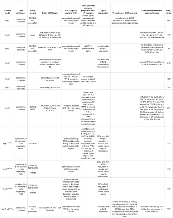
Since the discovery of Tph2 mouse models genetically depleted of brain serotonin-synthesising enzyme were created by several labora- tories [80, 78, 110, 125, 138, 196, 219]. In Table 1 the data obtained from various studies about the degree of central serotonin reduction, serotonin neuron formation and branching, and changes in other neu- rotransmitter systems in Tph2-deficient mice are summarized. Alto- gether, dramatic reduction in central 5-HT and no gross abnormali- ties in5-HT neuron formation are the main characteristics of mouse models constitutively lacking Tph2 [4, 80, 78, 110, 138, 176, 219]. Ho- wever, reports about the state of other neurotransmitter systems in mice lacking Tph2 are controversial, and indicate either no or mi-
Genetic model
Type/
deficiency Back-
ground Brain 5-HT levels
5-HT+/Tph2+
neurons (IHC)
5-HT neuronal markers/
properties of 5- HT neurons
Tph2
expression Projection of 5-HT neurons
Other neurotransmitter systems/factors
Refe- rence
Tph2-/- constitutive, complete
C57Bl/6, 6th generation
complete absence of 5-HT in the raphe
nuclei
normal SERT expression at some, dendrites and terminals of 5-
HT neurons
no difference in SERT expression in different brain regions including hippocampus
[80]
Tph2-/- constitutive, complete mixed
reduction by more than 92% in C, T, Ob, Hip, BS, Str and 68% in cerebellum
no difference in DA, DOPAC, HVA, NE, NM in C, T, Ob, Hip, BS, Str and cerebellum
[175]
Tph2-/- constitutive, complete
C57Bl/6, 6th generation
less than 1-4% is left in the brain
complete absence of 5-HT in the raphe
nuclei
mSERT is present in the
raphe
no detectable mTph2 expression
no detectable reduction in the whole brain content of NA, glutamate, GABA, DA,
DOPAC or HVA [4]
Tph2-/- constitutive, complete
near complete absence of serotonin in cerebral cortex, cerebellum, Hyp,
BS
no detectable mTph2 expression in the brainstem
around 15% increased levels of NE in the whole brain [218]
Tph2-/- constitutive, complete
complete absence of serotonin
complete absence of 5-HT or TPH2; no
TPH2 protein is detected by western
blot
unchanged protein levels of SERT and Lmx1b
[195]
Tph2-/- constitutive,
complete reduction by about 15% [125]
Tph2-/- constitutive, complete
C57Bl/6 (97%)
1,1% in RR, 3,8% in Hip, 6% in FC and
3,7% in T
complete absence of TPH2 in the raphe
nuclei
presence of SERT on the plasmatic soma membrane and projecting 5-HT fibers,in Hip;
VMAT is present in raphe neurons;
In the presence of 10
mM phenylephrine 5- HT neurons show regular firing rate
reduction in NE by 28.3% in RR, 38.6% in Hip, 43.3% in FC and 29.5% in T; DA level reduced by 71,9% in Hip and by 67,7% (relative to Tph2-/-);
reduction in NA neurons by 33.8% in arterior LC, but no difference in posterior LC; no difference in DA cell clusters
in SN, VTA and A8 [78]
Tph2Nes-cre constitutive, in
all neurons = cells expressing
nestin C57/BL6
some remaining TPH2-positive cells mainly in the dorsal part of rostral raphe
region
no difference in the expression of 5-HT1a, 5-HT1b, 5-HT2a, 5-HT2c receptors, serotonin transporter (Sert)
in F and PFC, Str, Hip, Hyp, A,
rostral and caudal raphe
region, and entorhinal cortex
80% and 90%
mTph2 reduction in rostral and caudal raphe regions, respectively
[110]
Tph2Pet1-cre constitutive, in 5-HT neurons
= cells expressing
Pet1 C57/BL6 x 129/SvEMS
x FVB/N
complete absence of 5-HT in the raphe
nuclei
90% and 95%
mTPH2 reduction in caudal and rostral raphe regions, respectively
[110]
Tph2Pet1-icre inducible, in 5- HT neurons = cells expressing
Pet1 C57/BL6 x 129/SvEMS
x FVB/N
some remaining TPH2-positive cells mainly in the dorsal part of rostral raphe region depending on the amount of tamoxifen injection
and on the age of the animals
80% mTph2 reduction in rostral and caudal raphe region
[110]
Tph2::eGFP-/- constitutive, complete
C57Bl/6, 9th generation
less than 5% in hind- and forebrain
complete absence of TPH2 in the raphe
nuclei
no detectable mTph2 expression
normal innervation of primary somatosensory, S1, pariental cortex; reduced innervation in SChN (more than 50%);
increased innervation in the NA and hippocampus (more than
50%)
increased mBDNF by 27%
in Hip, but unchanged in cortex and PVN.
[138]
Table1: Central 5-HT levels, serotonergic neurons formation and matura- tion, and other neurotransmitter systems inTph2-deficient mice RR: rostral raphe; Hip: hippocampus; FC: frontal cortex; T: thala- mus; C: cortex; Ob: olfactory bulb; BS: brain stem; Str: striatum;
Hyp: hypothalamus; A: amygdala; PVN: paraventricular nucleus;
SN: substantia nigra; DA: dopamine; NE: norepinephrine; DOPAC:
3,4 - dihydroxyphenylacetic acid; HVA: homovanillic acid; NA:
noradrenaline; NM: normetanephrine; BDNF: brain-derived neu- rotrophic factor
1.3 h y p o s e r o t o n e r g i c a n i m a l m o d e l s
nor difference in the levels of other neurotransmitters in the brain [4,80,78,138,176,219].
1.3.2 Mouse models with altered formation of serotonergic neurons Besides mice lacking the serotonin synthesising enzyme, TPH2, there are other mouse models with reduced central serotonin levels due to disrupted formation of5-HT neurons.
Lmx1b-k n o c k o u t m o u s e l i n e Lmx1b-knockout mice die in utero [55].Lmx1bis responsible for the development of not only serotoner- gic neurons but also of other neurons in the CNS, and other tissues such as kidney and limb.
Lmx1bP et1−cre m o u s e l i n e Disruption of Lmx1b gene in only Pet-1 expressing cells (Lmx1bP et1−cre) allowed to specifically dis- rupt the expression of Lmx1b in 5-HT neurons [227]. In the brains of adultLmx1bP et1−cre mice there are just residual amounts of sero- tonin. Moreover, there is almost a complete loss of 5-HT neurons in the raphe or their fibers at the target areas [227]. Interestingly, no changes in the amount of DA or NE in the brain of Lmx1bP et1−cre mice were reported.
Lmx1bP et1−icre m o u s e l i n e Inducible deletion ofLmx1bin the Pet1 expressing cells in the adulthood causes reduction of central 5- HT by 40% and a decrease in the amount of TPH2-positive neurons by85% [188].
Pet1-k n o c k o u t m o u s e l i n e Disruption of another factor im- portant for central serotonin neuron formation, Pet1, causes an 80% reduction in the number of5-HT neurons in the raphe and85-90% de- crease in central serotonin in the cortex, hippocampus, and caudate without significant changes in DA levels [89].
Pet1P et−icre m o u s e l i n e Inducible disruption of Pet1 during 6-8 weeks of adult life (Pet1P et−icre) results in reduction of Pet1 expression by 70% in rostral raphe nuclei [124]. This leads to 50% reduction in Tph2 expression and a drop of central serotonin levels to 25%. Surprisingly, Pet1 disruption induced in adulthood did not cause any changes in serotonin content or formation of5-HT neurons in B1-B3 raphe nuclei. In contrast, after tamoxifen treatment Pet-1 mRNA was decreased by more than70% in B5-B9nuclei [124].
1.3.3 Mouse models with impaired serotonin storage, release and reuptake Mice with disrupted5-HT storage, release, or reuptake represent ano- ther type of hyposerotonergic models.
Vmat2-k n o c k o u t m o u s e l i n e Mouse lacking VMAT2 are not able to store also other monoamines apart from serotonin, and there- fore, die within a few days after birth [6,66,215].
Vmat2Sert−cre m o u s e l i n e. To overcome the lack of specificity Vmat2 was ablated in SERT-expressing cells (Vmat2Sert−cre) [146].
In this mouse model a dramatic decrease by 92-96% (depending on the region) of serotonin was observed in the cortex, striatum, hip- pocampus, and brainstem. DA levels in the striatum, and NA levels in the hippocampus and cortex were unchanged. However, in the hip- pocampus of Vmat2Sert−cre mice there was a17% decrease in NA.
There was no detectable 5-HT immunoreactivity in the adult raphe, while the amount of TPH2-positive cells was unchanged [146]. Fur- thermore, mice lackingVMAT in serotonergic neurons show normal fiber formation in cerebral cortex and hippocampus [146].
Vmat2P et1−cre m o u s e l i n e As both Sert and VMAT2 are tran- siently expressed in a number of glutamatergic neurons and peri- pheral organs during development [116], it is possible that some as- pects of the phenotype observed in VMAT2Sert−cre mice could be due to the deletion of VMAT2 in these cells during development. To generate a more specific model mice with the raphe-specific inactiva- tion of the VMAT2 gene were created by using the ePet1-cre mouse line, where Cre is expressed exclusively in raphe neurons in the brain.
The brain serotonin level in VMAT2P et1−cre mice was uniformly decreased by 75% in the cortex, hippocampus, striatum, and brain- stem in prenatal E18embryos, postnatal pups as well as in adults. No changes in DA levels were found.
Sert-ov e r e x p r e s s i n g m o u s e l i n e Another mouse model with reduced central serotonin levels is aSERT-overexpressing mouse [100].
This mouse shows a 2-fold increase in the amount of SERT mRNA, and3-fold increase in the amount of SERT binding sites in the brain.
Furthermore, the level of central serotonin decreased from15to 30% depending on the region. The extracellular 5-HT levels are reduced by50-60% [100].
Interestingly, brain serotonin levels in mice lacking SERT are drama- tically decreased. Thus, one might consider it as one of the hyposero- tonergic mouse model. However, microdialysis studies revealed ele- vated extracellular5-HT levels in the brain [135].
1.3 h y p o s e r o t o n e r g i c a n i m a l m o d e l s
5-h t1Ar e c e p t o r r a p h e-ov e r e x p r e s s i n g m o u s e l i n e 5-HT1A
receptor expression in raphe nuclei is more than10times elevated in 5-HT1A receptor raphe-overexpressing mice (5-HT1ARO) [11]. These mice have a 25% decrease in 5-HIAA/5-HT ratio in the whole brain lysate when compared to controls [11]. However, in the original and follow up studies extracellular 5-HT levels in different brain regions were not studied in these mice [11,12,34].
1.3.4 Mouse models with polymorphisms in Tph2gene
It is known that certain mouse strains markedly differ in manifesta- tion of different behavior traits. Zhang et al. first surmised that the dif- ference in depression-like behavior between129X1/SvJ and BALB/cJ mice may be caused by altered TPH2 activity [224]. Indeed, sequen- cing analysis revealed a (C1473G) single-nucleotide polymorphism in the coding region of Tph2between these two strains resulting in the substitution of a highly conserved proline (Pro) residue with an argi- nine (Arg) at position 447. Consequently, it led to lowered enzyme activity in vitro [172, 224]. Moreover,5-HT content in the brain was reduced by approximately50% in the mouse strains carrying only the 1473G allele in comparison to mice homozygous for the1473C allele, which was also confirmed in an F2intercross [172,224].
However attempts to create congenic lines that differ only in the mTPH21473polymorphic allele revealed controversial results. Osipova et al. showed that transfer of the1473G allele into the C57Bl/6J genome over 3 generations (B6-1473G (G/G)) significantly decreased TPH2 activity in the brain [151]. Another study instead revealed that breed- ing the 1473G allele from DBA/2mice over eight generations to the C57Bl/6background did not result in difference in the 5-HT content in brain regions [194]. Recently in a third study it was shown that backcrossing 1473G polymorphism to C57BlB/6background over10 generations results in unchanged serotonin content either in the fore- brain, midbrain, or in the whole brain [20]. Moreover, extracellular levels of 5-HT in dorsal and medial raphe nuclei were not different between C57Bl/6mice carrying either1473C or1473G alleles [20].
At last, Beaulieu et al. generated knockin mice expressing a mutant form ofTph2. This mutation was engineered equivalent to the human R441H mutation found to be associated with major unipolar depres- sion [17]. TPH2 activity in R439H TPH2 knockin mice was reduced by 80% in the striatum, cortex and hippocampus, while the expres- sion ofSERTwas unchanged [17]. Extracellular5-HT levels in frontal cortex and hippocampus were decreased by more than half in these Tph2knockin mice [99].
2
A I M S O F T H E S T U D Y
Serotonin (5-hydroxytryptamine, 5-HT) was given a close attention since its discovery due to its involvement in the pathology of diverse psychiatric disorders and drug action. However, lack of suitable phar- macological or genetic tools to specifically influence the central but not the peripheral serotonin system did not allow drawing final conc- lusions about the processes that are directly influenced by serotonin in the brain.
In this study I took advantage of newly generated mouse models with complete or partial reduction in activity of TPH2, the enzyme responsible for central serotonin synthesis, to investigate the impact of this neurotransmitter on development, brain neurochemistry, and beha9vior.
Using these animal models I aimed at clarifying the following specific questions:
1. What is the consequence of life-long brain serotonin depletion on mouse prenatal and postnatal development?
In order to answer this question I assessed pathways involved in growth retardation of Tph2-deficient pups. In particular, functiona- lity of the hypothalamo-pituitary-adrenal axis, lipid metabolism, ther- moregulation, and the behavioral response to isolation were studied during the first postnatal weeks.
2. Does partial inactivation or complete depletion of TPH2 change central 5-HT levels and behavior?
To determine the role of brain serotonin in behavior first mice lacking Tph2were tested for alterations in general activity, olfaction, memory formation, adult neurogenesis, aggressive, anxiety-, and depression- like behaviors. To compare complete absence with partial inactiva- tion of TPH2anxiety- and depression-like behavior in mice with dec- reased TPH2 activity were evaluated. To test for potential compen- satory mechanisms I checked5-HT1Areceptor function and serotonin metabolism in the brains of hyposerotonergic animal models. In addi- tion, the ability of selective serotonin transporter inhibitor, fluoxetine, to exert its actions on anxiety- and depression-like behaviors as well as brain neurochemistry in the partial or complete absence of sero- tonin was assessed.
3
M AT E R I A L S A N D M E T H O D S
3.1 m at e r i a l s 3.1.1 Reagents
Name Company, city
5-HIAA Sigma, Steinheim
5-HT Sigma, Steinheim
5-HTP Sigma, Steinheim
Acetic acid Roth, Karlsruhe
Agarose Lonza, Switzerland
Ascorbic acid Sigma, Steinheim
Bromophenolblue Sigma, Steinheim
DAPI Sigma, Steinheim
DEPC Serva, Heidelberg
DNaseI Boehringer Mannheim, Mannheim
dNTPs Amersham Bioscience, NJ, USA
Donkey serum Sigma, Steinheim
DTT Sigma, Steinheim
EDTA Sigma, Steinheim
Ethanol Roth, Karlsruhe
Ethidiumbromid Sigma, Steinheim
First strand buffer Invitrogen, Darmstadt
Glucose Sigma, Steinheim
Glycerol Roth, Karlsruhe
HCl Roth, Karlsruhe
KCl Roth, Karlsruhe
MgCl2 Roth, Karlsruhe
M-MLV Invitrogen, Darmstadt
NaCl Roth, Karlsruhe
NaOH Roth, Karlsruhe
PCA Sigma, Steinheim
PFA Fischar, Germany
Proteinase K Invitrogen, Darmstadt
Random Hexamer Primer Boehringer, Mannheim
Rnase A Promega, Madison, WI
RNasin Promega, Madison, WI
SDS Serva, Heidelberg
Sucrose Roth, Karlsruhe
Taq polymerase Invitrogen, Darmstadt
TissueTek Sakura, Netherlands
Tris Roth, Karlsruhe
Triton X-100 Sigma, Steinheim
Trizol Invitrogen, Darmstadt
Tween 100 Sigma, Steinheim
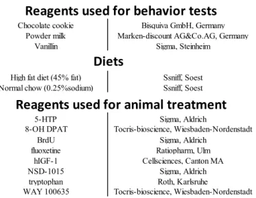
Chemicals
Chocolate cookie Bisquiva GmbH, Germany Powder milk Marken-discount AG&Co.AG, Germany
Vanillin Sigma, Steinheim
High fat diet (45% fat) Ssniff, Soest Normal chow (0.25%sodium) Ssniff, Soest
5-HTP Sigma, Aldrich
8-OH DPAT Tocris-bioscience, Wiesbaden-Nordenstadt
BrdU Sigma, Aldrich
fluoxetine Ratiopharm, Ulm
hIGF-1 Cellsciences, Canton MA
NSD-1015 Sigma, Aldrich
tryptophan Roth, Karlsruhe
WAY 100635 Tocris-bioscience, Wiesbaden-Nordenstadt
Reagents used for behavior tests
Diets
Reagents used for animal treatment
Table2: Chemicals, reagents, enzymes and animal diets used in the study
3.1.2 Kits and Markers
Name Company, locaon
100 bp Marker Biolabs, USA
ABC Kit Vector laboratories, USA
Affymetrix 1. ST Affymetrix, USA
IGF-1 ELISA Diagnostic System Laboratories, USA
Leptin ELISA R&D System, UK
RNeasy Mini Kit Qiagen, Hilden
RT2 qPCR primer assay for Tph2 Sabioscience, USA
WT expression and labelling KIT Qiagen, Hilden
Φ174 DNA/BsuRI (HaeIII) Marker, 9 Fermentas, Burlington, CDN
Table3: Kits and markers used in the study
3.1.3 Antibodies
Anbody Diluon Company, locaon
Primary
Growth hormone, rabbit 1:1000 Millipore, Germany
BrdU, rat 1:500 Biozol, Germany
Secondary
anti-rabbit AlexaFluor488 1:1000 Invitrogen, Germany anti-rat biotin-SP 1:500 Jackson ImmunoResearch, USA Table4: Primary and secondary antibodies used in the study
3.1 m at e r i a l s
3.1.4 Equipment and expandable material
Name Company, locaon
8 channel pipette Biohit, Rosbach v.d Höhe
Accu Check Roche, Manheim
AlphaImager (UV) Alpha Innotech, Germany
Automatic Pipette Witoped XP Witeg Labortechnik GmbH, Wertheim
Camera Panasonic, Japan
Centrifuge 5415C Eppendorf, Hamburg
Centrifuge Sorvall RC 5C Heraeus, Hanau
Cryostat Leica, Germany
Falcon tubes TPPR Trasadingen, Switzerland
FastPrep MP Biomedicals, France
Fine balance Kern&Sohn GmbH, Germany
Fluorescent microscope Keyence Corp., USA
Incubator Binder, USA
InfraMot TSE systems, Bad Homburg
Microwave 8020 Privileg, Fürth
Nanodrop Thermo Scientific, USA
PCR tubes Biozym Scientific GmbH, Oldendorf
pH Meter pH Level 1 WTW, Weilheim
Pipettes Gilson Disposable pipettes CellstarR 1, 2, 5, 10, 25ml Power supply for the gel chamber Appligene, France
Real time PCR machine Biosystems, USA
SasLabPro Avisoft BioAcustics, Germany
Superfrost Plus slides Menzel Gläser, Braunschweig Telemetry transmitters Data Science International (DSI), USA
Thermometer Thermo Fischer Scientific, USA
Thermomixer 5437 Eppendorf, Hamburg
USV microphone Avisoft BioAcustics, Germany
Viewer2 Biobserve, Bonn
Vortex: VibroFix Janke & Kunkel IKA, Germany
Water bath GFL, Burgwedel
Table5: Equipment and expandable materials used in the study
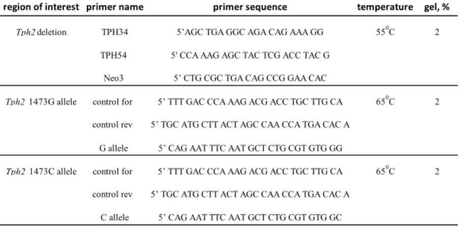
3.1.5 Primers
Primers used for qPCR
gene of interest primer name primer sequence
SERT mSERT_fw_1 GGACAAAGAGGACTGCCAAG mSERT_rev_1 GGCATAGCCAATGACAGACA TBP TBP_fw CCCTATCACTCCTGCCACACC TBP_rev CGAAGTGCAATGGTCTTTAGGTC
Primers used for animal genotyping
region of interest primer name primer sequence
annealing temperature
agarose gel, %
Tph2deletion TPH34 5’AGC TGA GGC AGA CAG AAA GG 550C 2
TPH54 5' CCA AAG AGC TAC TCG ACC TAC G Neo3 5’ CTG CGC TGA CAG CCG GAA CAC
Tph2 1473G allele control for 5’ TTT GAC CCA AAG ACG ACC TGC TTG CA 650C 2 control rev 5’ TGC ATG CTT ACT AGC CAA CCA TGA CAC A
G allele 5’ CAG AAT TTC AAT GCT CTG CGT GTG GG
Tph2 1473C allele control for 5’ TTT GAC CCA AAG ACG ACC TGC TTG CA 650C 2 control rev 5’ TGC ATG CTT ACT AGC CAA CCA TGA CAC A
C allele 5’ CAG AAT TTC AAT GCT CTG CGT GTG GC
Table6: Primers used in the study
3.2 m e t h o d s
3.2.1 Molecular biology methods
d na i s o l at i o n f r o m ta i l/e a r b i o p s i e s For genotyping of experimental animals DNA was isolated from tail or ear biopsies. The tissue was incubated overnight at 55◦C in 100 µl of the ear buffer containing 1 mg/ml Proteinase K. Next the tissues were incubated 10 min at 95◦C in order to inactivate Proteinase K. Thereafter 750 µl of TE/RNase buffer were added to the sample in order to digest RNA. After 15 min incubation at the room temperature (RT),2 µl of genomic DNA was used as a template for the PCR reaction.
d na i s o l at i o n f r o m h a i r f o l l i c l e s For some mice genoty- ping needed to be performed more than once. To minimize the stress of repeated ear or tail biopsies genotyping of animals was performed from the hair follicles. A small bundle of fur together with the follicles was collected. Hair roots were cut off and incubated55◦C overnight in 60 µl of hair buffer with 3 mg/ml proteinase K (prepared first as stock 20 mg/ml in Tris-HCl, pH=7.5). Thereafter the same steps as for DNA isolation from ear/tail biopsies were preformed.
Ear buffer
100mM Tris pH8.5 5mM EDTA
200mM NaCl 0.2% SDS
TE/RNase-buffer
20µg/ml RNase A in1x TE buffer
3.2 m e t h o d s
TE buffer
10mM Tris-HCl pH8.0 1mM EDTA
Hair buffer
10mM Tris pH8.3 50 mM KCL 0.5% Tween
r na i s o l at i o n RNA from the whole brain was extracted with Trizol reagent.100mg of the organs were diluted in1000ml of Trizol and homogenized with Fast Prep. Further steps were performed ac- cording to manufacture protocol. RNA from the hypothalamus was isolated with the RNeasy Kit according to the manufacture protocol.
Independent from the method of isolation residual genomic DNA was removed by DNase I treatment.
d e t e r m i nat i o n o f n u c l e i c a c i d c o n c e n t r at i o n DNA and RNA concentrations were determined by optical density (OD) 260 compared to OD280in a spectrophotometer (Nanodrop). When OD 260/280>1.7, the protein concentration can be neglected and the con- centration of nucleic acids was determined by the following ratio:
DNA OD260=1=50µl/ml for double stranded DNA RNA OD260=1=40µl/ml
When OD260/OD280 < 1.7 the protein concentration of the solu- tion is too high and requires additional purification steps (phenol- chlorophorm extraction or purification on a column).
s t o r a g e o f n u c l e i c a c i d s For long-term storage DNA was di- luted in DNase-free water or TE buffer and kept at -20◦C, RNA was diluted in DEPC or RNase-free water from QIAGEN, and kept at - 80◦C. Primer stocks were diluted in DNase- and RNase-free water, and kept at -20◦C. Before performing PCR the stock solutions were diluted to working concentration in ddH2O and kept at -20◦C.
DEPC water
0.1% DEPC in ddH2O
overnight mixing at37◦, than autoclaved
s e pa r at i o n o f n u c l e i c a c i d s o n a g a r o s e g e l DNA mole- cules were separated by length using 1-3% (w/v) agarose gels, con- taining 0.5 µg/ml ethidium bromide. DNA was mixed with0.1V of 10x Loading buffer, applied on agarose gel chambers and electropho- rised at 1-8 V/cm in 1x TAE buffer. DNA was visualized under 300 nm UV light. The size and the approximate concentration of the DNA
bands was determined by comparison with standardized molecular weight markers (φX174DNA/BsuRI (HaeIII) and100bp ladder).
TAE buffer
40mM Tris-Acetate pH7.4 1mM EDTA
10x DNA Loading buffer 40% Sucrose
0.02% Bromphenolblue in TE buffer
r e v e r s e t r a n s c r i p t i o n (r t) The synthesis of cDNA from to- tal RNA was done using M-MLV first strand synthesis Kit. 1 µg of total RNA was mixed with1µl of Random Hexamer primers,1µl10 mM dNTPs and 0.5 µl RNasin in 12.5 µl volume. RNA was denatu- rated for10min at60◦C and quickly chilled to4◦C. Then7.5µl of the following mix was added:
5x first strand buffer4µl 0.1M DTT2µl
RNasin (40u/ml)0.5µl M-MLV (200u/µl)1µl
The reaction was incubated for 40 min at 37◦C. Than M-MLV was inactivated at 80◦C for 10 min, afterwards the mix was kept on ice until2µl were used for PCR reaction.
m o u s e g e n o t y p i n g For the genotyping the following PCR reac- tion was set using TaqPolymerase:
genomic DNA2µl 10x PCR buffer5µl 50mM MgCl2 2.5µl 10mM dNTPs1µl 10mM primer1 1µl 10mM primer2 1µl 10mM primer3 1µl
Taq polymerase (5u/ml)0.5µl ddH20 36µl
The reaction was kept on ice before transferred to preheated PCR ma- chine. The program used for genotyping:
Denaturation5min92◦C Denaturation45sec92◦C
3.2 m e t h o d s
Annealing30sec at further specified◦C for certain primer pairs Elongation30sec72◦C
Repeat34x
Final elongation10min72◦C Forever10◦C
Genotyping of animals was performed using primer combinations specified in tableTable7. Information about the primer sequences, an- nealing temperatures, and percentage of agarose gels used for DNA separation are summarised in tableTable6.
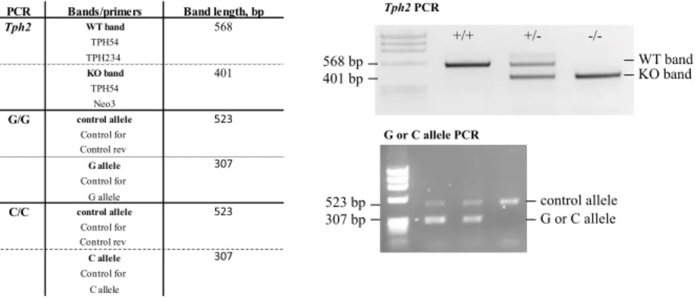
PCR Bands/primers Band length, bp
Tph2 WT band 568
TPH54 TPH234
KO band 401
TPH54 Neo3
G/G control allele 523
Control for Control rev
G allele 307
Control for G allele
C/C control allele 523
Control for Control rev
C allele 307
Control for C allele
Tph2 PCR
WT band KO band
+/+ +/- -/-
568 bp 401 bp
G or C allele PCR
523 bp 307 bp
control allele G or C allele
Table7: Primers used for genotyping of mice
q ua n t i tat i v e r e a l t i m e p c r To quantify relative gene expres- sion quantitative (real-time) polymerase chain reaction (qPCR) was performed. Real-time PCR was run in a technical triplicate using SYBR green reagent according to manufacturer protocol in a 384- well plate format. For the qPCR reaction9 µl of 2ng/µl cDNA was used. The expression of the Tph2 gene was quantified using RT2 qPCR primer assay, the expression ofSERT— using primers listed in the Table 6. Expression of gene of interest was normalized to TATA- binding protein (TBP) mRNA expression. Oligonucleotides were cus- tom made by Invitrogen. The method of Livak and Schmittgen [126] was applied to compare gene expression levels between groups, using the equation2−∆∆CT.
m i c r oa r r ay a na ly s i s For the analysis of gene expression pups at the day of birth were sacrificed by cervical dislocation. Brains were rapidly removed, hypothalamus was isolated, and then snap frozen.
Isolated and cleaned from genomic DNA RNAs were fragmented and labeled ssDNAs with WT expression and Terminal labeling kits. The labeled RNA was hybridized against the mouse gene 1.0 ST Array.
Differential expression of genes was calculated using Partek ANOVA statistic followed by FDR multiple testing corrections. Resulting p- values and FDR values indicated the probability of differential exp- ression between the different conditions.
3.2.2 Determination of plasma parameters
m e a s u r e m e n t s o f p l a s m a h o r m o n e l e v e l s IGF-1 and lep- tin levels were evaluated at postnatal day 35. Animals were anes- thetized by an intraperitoneal (i.p.) injections of ketamine (100mg/kg) and xylazine (10 mg/kg). Blood was taken transcardially. 25 µL of serum were used for IGF-1 measurement using a commercially avai- lable EIA kit. 50 µl of serum was used for the leptin measurement using a commercially available Leptin Kit.
g l u c o s e m e a s u r e m e n t s Plasma glucose levels were measured with the glucometer according to manufacturer protocol. To obtain a drop of blood1mm of the mouse tail was cut with scissors.
3.2.3 Staining
g r o w t h h o r m o n e i m m u n o h i s t o c h e m i s t r y For the analysis of pituitary glands in 2-day old Tph2-/- mice, the tissue was post- fixated in4% paraformaldehyde (PFA) in0.1M phosphate buffer for 4 hours followed by2 hours30% sucrose. After a short wash in PBS heads were embedded in Tissue-Tek O.C.T compound and cryosec- tioned in 14 µm coronal sections. Sections were dried for 2 hours at room temperature and then stored at -80◦C. For immunohistochem- istry, tissues were washed in PBS followed by PBST, and blocked with 10% normal donkey serum. Slides were incubated overnight at 4◦C with polyclonal rabbit anti-growth hormone. Anti-rabbit IgG secondary antibody conjugated with Alexa-Fluor488was applied the next day. Fluorescence images were collected using a fluorescence mi- croscope.
10x PBS for1L ddH2O 80g NaCl 2g KCl
14.4g Na2HPO2
2.4g KH2PO2 pH to7.4 PBST
o.1% Triton X-100in1xPBS
b r d u l a b e l l i n g a n d q ua n t i f i c at i o n Mice were deeply anaes- thetized with isoflurane and perfused transcardially with0.9% sodium chloride followed by 4% PFA in0.1M phosphate buffer. Brains were removed from the skulls, postfixed in 4% PFA at4◦C over night, and
3.2 m e t h o d s
transferred into30% sucrose. Sequential40µm coronal sections were cut on a microtome and cryoprotected in cryo protection solution (CPS). For BrdU staining, DNA was denatured in2N HCl for20min- utes at 37◦C. Sections were then rinsed in 0.1 M borate buffer and washed in Tris-buffered solution (TBS). Sections were stained free floating with antibodies diluted in TBS containing 3% normal don- key serum and0.1% Triton X-100. Primary anti-BrdU antibodies were applied in the concentration 1:500. Immunohistochemistry followed the peroxidase method with biotinylated secondary antibodies, ABC Elite reagent and diaminobenzidine (DAB) as chromogen. For BrdU labeling, one-in-six series of sections of each brain were DAB stained, and immunoreactive cells were counted throughout the rostra-caudal extent of the dentate gyrus. Results were multiplied by six to obtain the total number of BrdU-positive cells per dentate gyrus.
0.1M phosphate buffer for1L ddH2O
13.73g Na2HPO2
3.18g NaH2PO2 0.1M borate buffer for480ml ddH2O 3.08g Boric Acid 5N NaOH pH8.5
10x Tris buffer solution (TBS) for1710ml ddH2O
180g NaCl 264.40g Tris HCL 38.80Tris base
Cryo protection solution (CPS) 250ml25% Ethylenglycol 500ml0.1M PO4
o i l r e d o s ta i n i n g Aliquots of fresh stool samples collected prior staining were homogenised after adding water in a volume of 10µl/mg stool. After centrifugation (200g,5min)5µl of supernatant was applied to a glass slide, mixed with 5 µl of staining solution, and examined by light microscopy [83]. For preparing the stock of staining solution 0.3 g of Oil RedO was dissolved in 100ml of isop- ropanole. It was stirred overnight, filtered and stored at4◦C. Prior to staining two parts of distilled water was added to three parts of the stock solution of Oil RedO [109].