zur
Erlangung der Doktorwürde der
Naturwissenschaftlich-Mathematischen Gesamtfakultät der
Ruprecht - Karls - Universität Heidelberg
vorgelegt von
Diplom-Biologe Christoph Grundner aus Marburg
Tag der mündlichen Prüfung: 13.12.2002
Solid Phase Proteoliposomes Containing HIV-1 Envelope Glycoproteins as Antigens and Immunogens
and
HIV-1 Core Envelope Glycoproteins Deficient in T-Helper Epitopes
Gutachter: Prof. Dr. Hans-Georg Kräusslich
Richard Wyatt, Ph.D.
Acknowledgements
I am grateful to have had the opportunity to pursue my graduate work in an environment where the only limitations I experienced were my own talent.
I could not have wished for a more supportive and patient mentor than Richard Wyatt.
His guidance opened many doors for me and made these three years exciting, rewarding, and cheerful. He always gave me the freedom to stray. I have learned and drawn greatly from his insight and experience. Joseph Sodroski was always a source of inspiration and support. He set an example of scientific achievement and integrity for me. The members of the Sodroski lab past and present were great colleagues and friends for whom help, exchange, and entertaining diversions were always a matter of course. Thanks to the members of the Wyatt lab at the Vaccine Research Center and Dennis Burton's lab for my fruitful and enjoyable time at The Scripps Research Institute.
Prof. Dr. Hans-Georg Kräusslich made the connection between Heidelberg and Boston possible. I am very grateful for his support of my unconventional arrangement that turned out to be so rewarding for me. Many thanks for his kind supervision.
Alexandra Eichten was a tireless source of support. Without her companionship, these three years would not have been nearly as successful and cheerful.
My parents Hans-Georg and Brigitte always supported me and believed in me. To them this thesis is dedicated.
Abstract
The HIV-1 envelope glycoproteins gp120 and gp41 mediate binding and fusion of the virus to target cells. The envelope glycoproteins are exposed on the surface of the virus as trimeric spikes and are the major targets for neutralizing antibodies. The design of envelope glycoprotein-based subunit vaccines has been frustrated by many viral immune escape mechanisms. Trimeric envelope glycoprotein formulations hold promise to overcome limitations of monomeric envelope glycoproteins as immunogens. The generation of native, trimeric envelope glycoprotein complexes, however, remains a major challenge.
Here, solid-phase proteoliposomes containing native, trimeric HIV-1 envelope glycoprotein complexes that mimic the trimeric complex as it is found on the viral surface have been designed. In a comparative immunogenicity study, these proteoliposomes were shown to better elicit broadly neutralizing antibodies than gp120.
A second trimeric envelope glycoprotein formulation, soluble YU2 gp140-GCN4 constructs, were also shown to better elicit broadly neutralizing antibodies in rabbits, extending a previous study in mice. These data support the hypothesis that trimeric envelope glycoprotein formulations are an advance over gp120-based immunogens.
To date, only four broadly neutralizing antibodies against the HIV-1 envelope glycoproteins have been identified. Here, three novel Fab antibody fragments binding to the CD4 binding site of gp120 have been identified from phage-displayed antibody libraries with proteoliposomes. These Fab antibodies display some breadth and potency in neutralizing HIV-1. Comparison of the neutralizing activity of Fab antibodies and whole antibodies directed to the CD4 binding site suggests that these Fab antibodies may significantly gain neutralizing potency as whole antibodies.
Many HIV-1 immune escape mechanisms complicate the elicitation of broadly neutralizing antibodies. Core gp120 envelope glycoproteins derived from primary isolate viruses were found to be deficient in T-helper epitopes. This finding is suggestive of yet another HIV-1 viral immune escape mechanism, the escape from recognition by CD4+ T- helper cells.
1 Introduction ... 1
1.1 AIDS... 1
1.2 HIV... 2
1.3 The HIV-1 envelope glycoproteins: Structure and function ... 3
1.4 Antibodies in HIV-1 infection... 5
1.5 T-helper cells, B-cells, and T-helper epitopes ... 7
1.6 Immunogenicity of the envelope glycoproteins... 8
1.7 Viral immune escape mechanisms ... 10
1.8 Scope of this thesis ... 12
2 Materials and Methods ... 14
2.1 Envelope glycoprotein constructs ... 14
2.1.1 Envelope glycoprotein constructs used for the generation of PLs... 14
2.1.2 Core gp120 envelope glycoproteins... 14
2.2 Expression of envelope glycoprotein constructs ... 15
2.2.1 Expression of gp160∆CT... 15
2.2.2 Core gp120 envelope glycoprotein ... 15
2.3 Deglycosylation of envelope glycoproteins ... 16
2.4 Generation of proteoliposomes... 16
2.5 SDS-PAGE ... 18
2.5.1 gp160∆CT eluted from proteoliposomes ... 18
2.5.2 Core gp120 envelope glycoproteins... 18
2.6 Molecular exclusion chromatography ... 18
2.7 Western blot ... 19
2.8 Flow cytometric analysis ... 19
2.9 Animals, immunization, and serum preparation... 20
2.9.1 Immunization with proteoliposomes... 20
2.9.1.1 Mice ... 20
2.9.1.2 Rabbits ... 20
2.9.2 Immunization with core gp120 envelope glycoproteins ... 21
2.9.2.1 Mice ... 21
2.9.2.2 Rabbits ... 21
2.10 ELISA ... 22
2.10.1 Detection of anti-gp120 reactivity in serum ... 22
2.10.2 Detection of gp120- and gp41-binding of scFv and Fab antibodies ... 22
2.10.3 Competition ELISA with sCD4 and D-mannose... 23
2.11 Phage display ... 24
2.11.1 Panning of a naïve human phage-displayed scFv antibody library ... 24
2.11.2 Panning of biased human phage-displayed scFv and Fab antibody libraries 24 2.12 ScFv and Fab production... 26
2.13 HIV-1 single-round neutralization assay... 27
3 Results ... 29
3.1 Generation and characterization of proteoliposomes ... 29
3.1.1 Creation of gp160∆CT proteoliposomes ... 29
3.1.2 Analysis of proteoliposome protein composition ... 29
3.1.3 Characterization of the size of the gp160∆CT proteoliposome envelope glycoproteins... 31
3.1.4 Characterization of the proteoliposome membrane ... 33
3.1.5 Antigenic characterization of the gp160∆CT proteoliposomes ... 34
3.1.6 2F5 antibody binding to gp160∆CT proteoliposomes ... 36
3.1.7 CD4 induction of the 17b epitope ... 38
3.1.8 Characterization of cleavage-deficient gp160∆CT glycoproteins ... 38
3.2 Immunogenicity of proteoliposomes ... 40
3.2.1 Immunogenicity of proteoliposomes in mice ... 40
3.2.2 Immunogenicity of proteoliposomes in rabbits ... 42
3.3 Proteoliposomes as antigens ... 49
3.3.1 Identification of HIV-1 envelope glycoprotein-binding antibodies from phage-displayed antibody libraries ... 49
3.3.2 Characterization of CG5, CG9, and CG21 ... 52
3.3.3 Neutralization of in vitro HIV-1 infection by Fab CG5, CG9, and CG21.... 53
3.3.4 Neutralization of in vitro HIV-1 infection by Fabs versus IgGs ... 56
3.4 Envelope glycoproteins deficient in T-cell helper epitopes... 57
3.4.1 Anti-gp120 IgG reactivity in mouse sera raised against YU2 core gp120 and
HXBc2 core gp120 ... 58
3.4.2 Generation of fusion proteins of core gp120 and the heterologous helper epitope PADRE... 59
3.4.3 Anti-gp120 IgG reactivity in mouse sera raised against YU2 and HXBc2 core gp120 and core-PADRE gp120... 60
3.4.4 Serum reactivity against PADRE, neoepitopes, and carbohydrate moieties 62 3.4.5 Anti-gp120 IgG and IgM reactivity in rabbit serum ... 65
4 Discussion... 68
4.1 Generation of proteoliposomes... 68
4.2 Proteoliposomes as immunogens... 70
4.3 Proteoliposomes as antigens ... 76
4.4 Envelope glycoproteins deficient in T-helper epitopes... 80
5 Summary ... 85
6 References ... 86
1 Introduction
1.1 AIDS
Since the first reported cases of acquired immune deficiency syndrome (AIDS) in 1981, AIDS has become a pandemic of catastrophic proportions. Human immunodeficiency viruses (HIV) type 1 and type 2 are the etiologic agents of AIDS (10, 43). According to UNAIDS, there are an estimated 40 million people living with HIV today, predominantly of the HIV-1 type. In sub-Saharan Africa, AIDS is today the leading cause of death. The adult prevalence in many of these countries exceeds 10%.
Eastern Europe and China are presently experiencing the steepest increase in new infections and may only be at the beginning of a disastrous epidemic.
With the availability of highly active anti-retroviral therapy (HAART), the progression of HIV-infection to AIDS can be slowed. However, while 500,000 people received HAART and 25,000 people died of AIDS or related diseases in the developed world last year, in sub-Saharan Africa, only 30,000 people received HAART and 2.3 million people died (www.unaids.org). The high cost of antiviral therapies and the complexities of their administration make it unlikely that these drugs will significantly alter the course of the HIV epidemic in developing areas of the world. Only a broadly effective vaccine will eventually be able to curb the global spread of HIV.
HIV infects and depletes primarily CD4+ T-helper cells. HIV infection typically progresses through three stages: Acute, latent and AIDS (39). During the acute phase, a burst of viral replication coincides with rapid loss of CD4+ T-helper cells. The end of the acute phase, which occurs within a few weeks of infection, is characterized by a drop in viral load and a rebounding CD4+ T-helper cell count. During the subsequent latent phase, constant turnover of virus and CD4+ T-helper cells occurs, resulting in a slow and gradual decline in CD4+ T-helper cells. Eventually, the loss of CD4+ T-helper cells becomes too severe and leads to the failure of the immune system to fend off pathogens that do not generally pose a threat to immunocompetent individuals. The emergence of opportunistic infections such as Pneumocystis carinii, cytomegalovirus, or neoplasms such as Karposi’s sarcoma mark the onset of AIDS. Without treatment, AIDS patients rarely survive longer than a few years.
1.2 HIV
HIV belongs to the genus lentivirinae of the Retroviridae family. Based on sequence analysis and serologic properties, two types of HIV have been identified. HIV-1 is the prevalent strain in Africa, Asia, Europe, North- and South America. In some West African countries, HIV-2 is the dominant strain. Genetically related viruses that infect nonhuman primates, the simian immunodeficiency viruses (SIV), have been isolated and are believed to have given rise to HIV by cross-species transmission. In fact, HIV-1 exhibits more sequence homology to an SIV found in chimpanzees than it does to HIV-2.
HIV-2, in turn, is most closely related to SIV isolated from sooty mangabeys (24, 42).
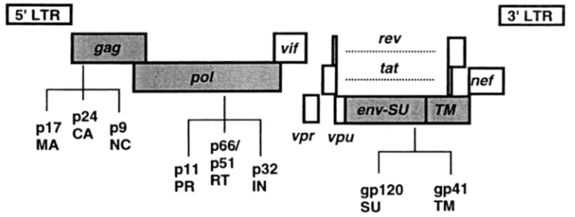
FIG. 1. Schematic diagram of the HIV-1 genome and its gene products. Virion structural genes are depicted as shaded, accessory genes as clear boxes (from Fields, Fundamental Virology). MA: Matrix protein, CA: Capsid protein, NC: Nucleocapsid protein, PR: Protease, RT: Reverse transcriptase, IN:
Integrase, SU: Surface glycoprotein (gp120), TM: Transmembrane glycoprotein (gp41).
HIV is an enveloped virus with two identical copies of a single-stranded RNA genome of about 9.2 kb. The HIV genome encodes nine genes (gag, pol, env, tat, rev, vif, vpr, vpu, and nef) in three open reading frames. It is flanked on either side by identical long terminal repeats (LTRs) that contain non-coding sequences important for viral replication (Fig. 1). After entry of the virus into the host cell, the RNA genome is reverse-transcribed by viral reverse transcriptase and the provirus is integrated into the host genome by the viral integrase. The first viral gene products to be expressed by host- cell RNA polymerases are Tat and Rev, which upregulate the subsequent expression of
structural proteins. While Gag and Gag-Pol become localized to the inner surface of the plasma membrane, Env is extensively modified in the endoplasmic reticulum. The Gag and Gag-Pol polyproteins assemble the core particle containing genomic RNA and the accessory protein Vpr. The viral particle then buds from the cell surface, acquiring in the process a membrane containing envelope glycoprotein complexes. After budding, proteolytic processing of Gag and Gag-Pol by the viral protease generates the mature, fully infectious virus (38).
1.3 The HIV-1 envelope glycoproteins: Structure and function
The HIV-1 envelope glycoproteins gp120 and gp41 facilitate binding of the virus to its target cell and mediate fusion between viral and cellular membranes (121). Initially, the envelope glycoproteins are synthesized as highly glycosylated gp160 polyprotein precursors that undergo a sequence of additional posttranslational modifications (3, 36, 85). After oligomerization in the endoplasmic reticulum, the polyprotein is cleaved into the gp120 and gp41 subunits by cellular proteases. The envelope glycoproteins remain associated through noncovalent interactions. They are extensively glycosylated and transported to the plasma membrane. From there, the envelope glycoprotein complexes are incorporated into budding virions. Many lines of evidence suggest that the mature, functional envelope glycoprotein complex on the viral surface consists of a trimer of gp120-gp41 heterodimers. The ectodomain of HIV-1 gp41 crystallizes as a trimeric coiled coil with interdigitating alpha helices to form a six-helix bundle (25, 109, 116).
The structure of the simian immunodeficiency virus (SIV) ectodomain has been solved by magnetic resonance and shown to be trimeric (22). Both the HIV-1 and SIV postfusogenic states of gp41 share close resemblance with the corresponding transmembrane proteins of viruses such as influenza (19) and Ebola virus (115). Each of these fusion determinants has been crystallized as helical bundles possessing trimeric coiled-coil motifs. The matrix proteins of HIV-1 and SIV that interact with gp41 also crystallize as trimers (48). The gp160 ectodomain of SIV (gp140) has been shown to be trimeric by biophysical analysis (26). Trimerization has also been reported for a number of gp120-gp41/gp140 ectodomain constructs (14, 122, 124).
FIG. 2. Molecular surface presentation of HIV-1 core gp120 (from Wyatt and Sodroski, 1998). A. The arrow indicates the direction of the viral membrane. The inner domain is facing towards the left, and the outer domain, which is thought to be exposed on the assembled trimer, is on the right. The red surface indicates the footprint of CD4. Green indicates the surface thought to be involved in chemokine receptor binding. The base of the V3 loop is shown in magenta. B. Conserved gp120 neutralization epitopes. C. The antigenic faces of core gp120 are shown. The non-neutralizing face (red) encompasses most of the inner domain and N- and C-terminal regions interacting with gp41. This region is thought to be largely buried on the assembled trimer complex and interacts with non-neutralizing antibodies. The silent face (yellow) consists of heavily glycosylated regions on the outer domain and is poorly immunogenic. The neutralizing face (green) extends over both domains and the bridging sheet, as well as the V2 and V3 variable loops (not shown). This surface interacts with neutralizing antibodies.
The amino acid sequence of gp120 reveals alternating variable (V1-V5) and conserved regions (C1-C5) (102). The variable regions V1-V4 are anchored at their bases with disulfide bonds and mask the CD4- and coreceptor binding sites. The atomic structure of a core gp120 deleted of variable loops V1, V2, and V3 as well as C-and N- terminal regions in complex with the receptor CD4 and the Fab fragment of the CD4- induced (CD4i) antibody 17b has been solved (54) (Fig. 2). The gp120 envelope glycoprotein is structurally organized into an inner and outer domain, linked by a bridging sheet minidomain. The inner domain is probably facing the axis of the trimeric complex and contributes most of the contacts with gp41 at the C-and N-terminal regions.
The outer domain is heavily glycosylated and is thought to face away from the trimer axis. Four antiparallel beta sheets form the bridging sheet. The CD4 binding site is situated at the interface of the three domains and is relatively well conserved among viral strains. All three domains also contribute to coreceptor binding, with the V3 loop and the base of the V1/V2 stemloop structure providing additional contacts (81, 82).
The infection process is initiated by binding of the gp120 envelope glycoprotein
A B C
to the cellular receptor CD4. This binding event presumably displaces the variable loop regions V1 and V2 on the gp120 envelope glycoprotein and exposes the coreceptor binding site, allowing for subsequent binding to the cellular coreceptor, typically the chemokine receptor CXCR4 or CCR5. These interactions trigger further conformational changes that allow for the formation of a six-helix bundle formed by two potential alpha- helical regions, N36 and C34 in the gp41 ectodomain, with three C34 helices packing into the hydrophobic grooves of a trimeric N36 coiled coil. Formation of this six-helix bundle is proposed to bring the viral and cellular membranes into close proximity and allow the insertion of the gp41 fusion peptide into the host cell membrane (25, 53, 115).
Antigenically, the surface of the gp120 core can be organized into three different faces as defined by antibody binding (121) (Fig. 2). Conserved regions that are accessible to antibodies or neutralizing ligands comprise the receptor- and coreceptor binding sites and form the neutralizing face. The heavily glycosylated outer domain is virtually invisible to the host immune system and is termed the silent face. The non-neutralizing face is oriented towards the trimer axis, is not accessible to antibodies on the functional trimer, and elicits only nonneutralizing antibodies.
1.4 Antibodies in HIV-1 infection
Vaccine-induced protection from many viral infections is based solely on the elicitation of neutralizing antibodies. It is becoming increasingly clear that for the prevention of HIV-1 infection, antibodies are also likely to be a critical component of a vaccine-elicited immune response. The level of circulating neutralizing antibodies correlated with protection from viral challenge in several animal models (12, 17). Passive immunization with neutralizing antibodies has also been demonstrated to protect from the establishment of viral infection in primates when administered to the host prior to exposure with HIV-1 (5, 58, 61). Some HIV-1-infected individuals remain asymptomatic for long periods of time and largely contain viral replication. In some cases, these long- term nonprogressors exhibit high titers of broadly neutralizing antibodies that may contribute to the favourable outcome of infection. The envelope glycoproteins are the major targets for neutralizing antibodies in vivo. However, the elicitation of antibodies that broadly neutralize HIV-1 has proven to be exceptionally difficult. The identification
of only four broadly and potently neutralizing envelope glycoprotein-directed antibodies to date from extensive screens of human and rodent antibodies illustrates the elusiveness of this class of antibodies.
During HIV-1 infection, antibodies directed against the viral envelope glycoproteins are readily elicited. In most infected individuals, two classes of neutralizing antibodies emerge, strain-restricted and broadly neutralizing antibodies. The strain- restricted antibodies are generally directed towards the second variable (V2) or third variable loop (V3) of gp120 and appear early during infection (84, 92). These antibodies neutralize only a limited number of viral isolates. Only after the initial phase of high viremiae, when the infection has already been firmly established, do more broadly neutralizing antibodies appear (103). These are directed to conformational epitopes, some of which, the CD4 binding site antibodies (CD4BS), recognize the discontinuous CD4 binding site (78, 86, 111). Another group of antibodies that neutralize T-cell line adapted (TCLA) isolates and a subset of primary isolates recognize CD4 induced (CD4i) epitopes. These antibodies bind near the gp120 bridging sheet and interfere with gp120- chemokine receptor interaction (110, 119).
One phenomenon of antibody neutralization that has misled the focus of research for years is the greater neutralization sensitivity of viral isolates that have been extensively passaged in vitro in immortalized T-cell lines compared to primary, clinical isolates (31, 44, 60, 61, 66, 71, 117, 128). Passage in cell lines selects for viruses using the CXCR4 coreceptor, such as the molecular clones HXBc2, IIIB, and MN. These TCLA viruses can be neutralized by a wide range of antibodies. Viruses directly isolated from patients, such as the molecular clones SF162, BaL, ADA, 89.6, JR-CSF, JR-FL, and YU2, preferentially use the CCR5 coreceptor or both CXCR4 and CCR5 coreceptors.
Neutralization of primary isolates generally requires much greater concentrations of neutralizing antibodies. This difference in neutralization resistance can most likely be attributed to the antibody-mediated immune selection pressure on primary isolate viruses.
It appears that primary viruses, regardless of their choice of coreceptor, are more resistant to neutralization than TCLA viruses.
To date, only four monoclonal antibodies that broadly and potently neutralize HIV-1 primary isolates have been identified. All of these antibodies have unique
properties and despite intensive study, no general scheme of neutralization of HIV-1 has emerged. The CD4 binding site antibody IgG1b12 was identified from a phage-displayed IgG Fab library obtained from the bone marrow of a long-term nonprogressor with exceptionally high titers of serum neutralizing antibodies (21, 113). It binds the CD4 binding site and neutralizes a wide range of primary isolate viruses at nanomolar concentrations. The atomic structure of this antibody reveals that it binds to the CD4 binding site similarly to CD4, extending a long, finger-like complementarity-determining region 3 (CDR3) into the recessed cavity of the CD4 binding site (95). The second broadly neutralizing gp120-directed antibody, 2G12, binds a carbohydrate moiety on the outer face of gp120 and is a rare example of a carbohydrate-reactive gp120 antibody (112). The gp41-binding antibodies 4E10 (129) and 2F5 (69) bind in close proximity on the C-terminal ectodomain of gp41. The epitope of the 2F5 antibody is thought to be linear, comprising the amino acid sequence ELDKWA, however, to date this antibody could not be elicited by peptides containing the 2F5 epitope or any other immunogen.
1.5 T-helper cells, B-cells, and T-helper epitopes
Functional activation of the cellular and the humoral arm of the immune system requires interaction with activated CD4+ T-helper cells (1, 51). At the same time, CD4+ T-helper cells are the major target for HIV-1 infection. In the course of AIDS, CD4+ T- helper cells specific for HIV-1 are preferentially infected and depleted. This loss of specific anti-HIV-1 CD4+ T-helper cells has been suggested to be a major factor in the eventual failure of the host immune system to control the virus (34, 88).
The generation of mature antibody responses requires the interaction of B-cells with activated CD4+ T-helper cells. Upon specific recognition of an antigen by the B-cell receptor, the antigen is internalized and proteolytically degraded into peptide fragments in the lysosomal compartment. Particular peptides, the T-helper epitopes, bind to the major histocompatibility complex class II (MHC class II) and are presented on the surface of the cell. The complex of MHC class II and T-helper epitope is then recognized by a specific T-cell receptor (TCR) on the surface of a CD4+ T-helper cell. This interaction and a complex series of costimulatory signals lead to the activation of the B- cell (1, 51).
Most large proteins possess one or more contiguous stretches of amino acids that can function as T-helper epitopes (90). Sequence comparison of helper epitopes naturally presented by MHC class II and structural analysis of MHC and MHC-peptide complexes suggest key anchor residues necessary for MHC class II binding and TCR engagement (16, 50, 89, 90). Although each MHC allele is characterized by a number of unique polymorphic residues along the peptide-binding cleft that limit the number of potential peptide ligands, peptides that are able to bind promiscuously to a number of different MHC alleles have been described (23, 72, 99). This observation gave rise to the concept of "universal helper-epitopes", peptides that can serve as T-helper epitopes for a number of different MHC class II alleles. One such universal T-helper epitope consists of key residues necessary for the binding to MHC class II and TCR on a polyalanine backbone.
This peptide (amino acid sequence AKFVAAWTLKAAA) was shown to bind ten out of ten tested HLA-DR molecules and was hence termed pan-DR-helper epitope (PADRE).
The PADRE epitope was also shown to bind to certain mouse MHC class II alleles, in particular I-Ab expressed by C57BL/6 mice (2).
1.6 Immunogenicity of the envelope glycoproteins
Most envelope glycoprotein-based immunogens tested so far are based on monomeric gp120. The antibody responses obtained in these studies generally did not neutralize primary HIV-1 isolates. (9, 11, 12, 30, 61, 118) The poor ability of gp120 to elicit neutralizing antibodies is also reflected in the binding characteristics of neutralizing and nonneutralizing antibodies. Neutralization has been found to correlate with binding of antibodies to the mature envelope glycoprotein complex. No such correlation could be found with monomeric gp120 (41, 83, 97).
Attempts to generate mimics of the trimeric envelope glycoprotein complex to better elicit neutralizing antibodies have been frustrated by the lability of these complexes. A number of different strategies have been pursued to stabilize the trimeric complex as soluble gp140 constructs. The deletion of the proteolytic cleavage site between gp120 and gp41 prevents shedding of noncovalently bound gp120 subunits from the complex and is common to most gp140 ectodomain constructs. The insertion of cysteine residues at specific locations on the alpha helical gp41 regions that mediate
formation of the trimeric coiled coil have been used to stabilize the trimeric organization of the envelope glycoprotein complex (37). Heterologous trimerization domains such as the bacterial transcription factor GCN4 or the bacteriophage trimerization domain fibritin have also been employed to stabilize the trimeric complex (122-124). Intersubunit disulfide bonds between gp41 and gp120 have been inserted in order to stabilize the cleaved, fully mature trimeric complex (14). The immunogenic properties of these ectodomain constructs have been disappointing, perhaps because of their heterogeneity (35). Only one of these constructs, a gp140-GCN4 construct derived from the primary isolate YU2, has been shown to elicit neutralizing antibodies better than monomeric gp120 (125). Mice immunized with these constructs generated antibodies that showed significant neutralization of the homologous primary isolate YU2, heterologous neutralization of the primary isolate ADA and the TCLA isolate HXBc2, but little neutralization of other primary isolates. This may be seen as a proof of principle that suggests better immunogenicity of trimeric constructs compared to monomeric gp120.
And yet, these molecules may not fully resemble the relevant trimeric envelope glycoprotein complex. It is conceivable that the extensive changes that need to be introduced in these molecules to stabilize the trimer may force the envelope glycoprotein in conformations they would not naturally assume. Many lines of indirect evidence suggest that the best envelope glycoprotein-based immunogen is going to be one that most closely mimics the native envelope glycoprotein complex as it is found on the viral surface. Broadly neutralizing antibodies are elicited in HIV-infected individuals, and a good correlation between neutralization and binding of these antibodies to the functional oligomeric envelope glycoprotein complex has been established (75, 83, 97). The best protection from homologous and heterologous SIV challenge that could be achieved to date was observed in a primate model after immunization with live attenuated virus (91).
Since inactivated virus in the case of HIV-1 may never be a safe immunogen (4, 6), the generation of virus-like particles (VLP) has been explored as a means of preserving the native envelope glycoprotein structure in its natural lipid environment (126, 127). These VLP, however, have been difficult to generate with envelope glycoprotein quantities large enough to elicit sufficient titers of antibodies.
1.7 Viral immune escape mechanisms
As a result of HIV-1 infection, a vigorous immune response occurs. Despite this immune response, the general slow progression to AIDS ensues. The immune system keeps the infection in check for many years until the virus eventually gains the upper hand. Both cellular and humoral effector mechanisms exert considerable immune pressure on the virus. The effect of the humoral response in particular is highlighted by the difference in neutralization sensitivity that can be observed between TCLA isolates and primary isolates (41, 65, 101). Viruses grown without antibody pressure in tissue culture become more neutralization sensitive. During chronic infection, however, HIV-1 and the host’s immune system co-evolve for many years, during which the immune system selects for ever more neutralization-resistant viruses, particularly resistant to autologous antibodies.
Many escape mechanisms from antibody-mediated neutralization have been described. Maybe most importantly, the exceptionally high genetic variability of HIV-1 allows the virus to escape from antibody recognition by mutation of antibody epitopes and necessitates the constant adaptation of the antibody response to mutated generations of progeny virus. Only antibodies elicited by structures that are limited in their ability to mutate by functional constraints, such as the receptor binding sites, can potentially neutralize different viral strains. However, large regions of the envelope glycoprotein, the exposed variable loops V1 and V2, sterically block access to the receptor binding sites while themselves eliciting mostly strain-restricted antibodies (121).
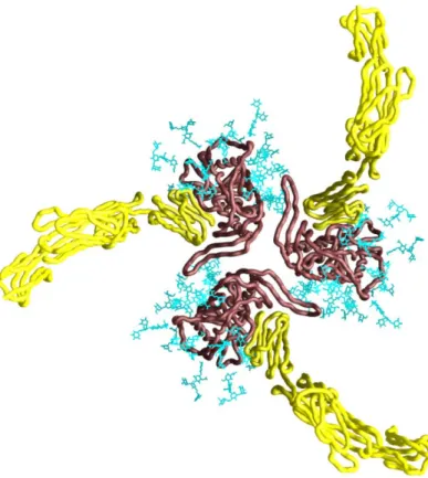
Moreover, large surfaces of gp120 are covered by carbohydrate moities, with about half of the gp120 molecular weight being contributed by carbohydrates (Fig. 3).
Since these carbohydrates are synthesized by the host glycosyltransferase system, they are considered "self" determinants by the host's immune system. Thus, the glycosylation renders the underlying foreign protein determinants of gp120 invisible to the immune system. The protective effect that even one single carbohydrate moiety can have is highlighted in the case of a gp120 mutant that deletes the glycosylation site at amino acid 301. Deletion of this single glycosylation site renders a number of primary viral isolates sensitive to neutralization by many antibodies that cannot neutralize the wild-type virus even at high concentrations (57).
FIG. 3. Model of oligomeric core gp120 (modified from Kwong et al., 2000). Depicted is a Cα worm presentation of core gp120 (brown), the two membrane-distal domains of CD4 (yellow), and the gp120 carbohydrate cores (blue) as viewed from the target cell membrane. The carbohydrate cores shown represent about half of the high-mannose and complex N-linked glycan moieties. The long, finger-like structure extending counter-clockwise at the center of the trimer represents the V1,2 stem. The variable loops V1,2 and V3 are not shown.
The complex organization of gp41 and gp120 as trimers of heterodimers itself has important implications for immune diversion. It has been suggested that in vivo, disassembled envelope glycoprotein or viral debris may be the predominant immunogen (73). The interaction of gp41 and gp120 is noncovalent, thought to be labile, and allows gp120 to shed from the functional complex. Upon disassembly of envelope glycoprotein complexes, many surfaces that are occluded on the functional spike such as the gp41/gp120 interface and areas on gp120 buried along the trimer axis are presented to the immune system (Fig. 3). Both gp120 and gp41 may also change conformation upon dissociation. As a consequence, many antibodies elicited in vivo are raised against these decoy epitopes and do not bind the functional envelope glycoprotein complex.
Thermodynamic analysis of the interaction of gp120 with its receptor CD4 revealed highly unusual thermodynamics of this binding reaction (70). The changes in enthalpy and entropy of this reaction greatly exceeds that of other protein/protein interactions. The binding of CD4 to gp120 is characterized by an extraordinarily favourable binding enthalpy of –63kcal/mol. This change reflects a large number of bonding interactions during complex formation. The compensatory high entropy of this reaction of ~52kcal/mol is suggestive of large conformational rearrangements in gp120 upon CD4 binding. Since similar results have been obtained with deglycosylated core gp120 and fully glycosylated full-length gp120, these structural rearrangements are likely to occur at the interface of the inner and outer domain and the bridging sheet. These data have been interpreted to mean that monomeric gp120 in its free state is a very flexible molecule, possibly sampling many conformations. Only upon ligand binding does gp120 assume a fixed conformation. One implication of this flexibility of free gp120 for immunogenicity is that it may present a multitude of different conformations to the immune system, most of which may be irrelevant for virus neutralization.
Mutations in viral proteins that lead to loss of recognition by CD8+
cytotoxic T lymphocytes have been documented and shown to allow for viral immune escape (15, 45, 76, 79). It is conceivable that viral proteins that lack potential T-helper lymphocyte epitopes for recognition by CD4+ T-helper cells will also be selected for in an infected individual. Such escape mutants have been documented in the case of lymphocytic choriomeningitis virus. Adoptive transfer of CD4+ T-helper cells from mice transgenic in the T-cell receptor for the immunodominant MHC class II-restricted LCMV epitope GP61-80 into C57BL/6 mice persistently infected with LCMV selected for virus mutants that failed to be recognized by CD4+ T-helper cells (27). A similar mechanism has not yet been documented for HIV-1.
1.8 Scope of this thesis
The HIV-1 envelope glycoproteins are poor immunogens. HIV-1 has evolved a plethora of immune escape mechanisms to evade host immune surveillance. Most likely, we are only at the beginning of understanding the complexities of immune escape that need to be adressed and dismantled to generate better immunogens that elicit broadly
neutralizing antibodies. It appears that the envelope glycoproteins are protected from antibody neutralization by many layers of protection at virtually every level of immune effector mechanisms.
1. The generation of better mimics of the functional native envelope glycoprotein complex as it is found on the viral surface is a promising approach to limit the number of irrelevant epitopes that are presented to the immune system by, for example, immunogens based on monomeric envelope glycoprotein. A novel envelope glycoprotein complex formulation containing native, trimeric envelope glycoprotein complexes embedded in a lipid membrane, solid-phase proteoliposomes (PLs), have been generated, characterized, and tested for immunogenicity in mice and rabbits.
2. A great wealth of information has been drawn from the four broadly neutralizing antibodies that have been identified to date. The identification of additional neutralizing antibodies would greatly aid our understanding of neutralization mechanisms and viral immune escape, and may help in the identification of novel targets for vaccine design. To this end, proteoliposomes were used to screen phage-displayed Fab and scFv libraries for novel neutralizing antibodies.
3. Escape from T-helper cell recognition has been documented in a transgenic model for LCMV as a viral immune escape mechanism (27). For HIV-1, such a mechanism has not yet been described. Based on a comparison of the immunogenicity of envelope glycoprotein core proteins derived from TCLA- and primary isolates, escape from recognition by T-helper cells is suggested as yet another immune escape mechanism of HIV-1.
2 Materials and Methods
2.1 Envelope glycoprotein constructs
2.1.1 Envelope glycoprotein constructs used for the generation of proteoliposomes The envelope glycoprotein constructs were derived from the primary R5 HIV-1 isolates YU2, JR-FL, and 89.6 and the X4, TCLA-adapted isolate HXBc2. The coding sequences for the YU2 and 89.6 envelope glycoproteins were obtained from the pSVIIIenv YU2 and pSVIIIenv 89.6 expression plasmid (106). The JR-FL envelope glycoprotein coding sequence, which contains a CD5 heterologous leader sequence in place of the normal JR-FL leader, was obtained from the AIDS repository and subcloned into the pcDNA 3.1(-) (Invitrogen) expression plasmid. The HXBc2 construct was codon-optimized for mammalian expression using overlapping primers and PCR and subcloned into pcDNA3.1(+) (Invitrogen). A sequence coding for the heterologous CD5 signal sequence was subcloned to replace the endogenous HXBc2 leader sequence. For all constructs, a cytoplasmic tail truncation was generated by introduction of a stop codon in place of the codon for amino acid 712 (704 for JR-FL) and the sequence coding for the C9-tag TETSQVAPA was added according to the QuikChange (Stratagene) protocol immediately before the stop codon. To create covalently linked gp120-gp41 glycoproteins, the proteolytic cleavage site between gp120 and gp41 was disrupted by replacing the arginines 508 and 511 with serines by QuikChange site-directed mutagensis. These modifications resulted in constructs encoding cleavage-deficient gp160∆CT envelope glycoproteins that were subsequently used to generate the proteoliposomes. Amino acid residue numbers are designated according to the prototypic HXBc2 sequence. The introduction of the desired mutations was confirmed by sequencing.
2.1.2 Core gp120 envelope glycoproteins
The envelope glycoprotein constructs were derived from the TCLA HIV-1 isolate HXBc2 and the primary isolates YU2 and JR-FL. The coding sequences of the envelope glycoproteins were derived from the pSVIIIenv HXBc2, YU2, and JR-FL expression plasmids containing deletions of the V1 and V2 variable loops as previously described
(120). Briefly, the V1/V2 coding region was deleted from amino acids 128 to 194 (HXBc2 numbering) and replaced with a Gly-Ala-Gly sequence. Subcloning of sequences encoding these V3-containing core glycoproteins into the insect expression vector pMT (SmithKline Beecham Pharmaceuticals) containing the inducible metallothionein promoter was performed as follows. The gp120 core coding regions were amplified by PCR to remove the N- and C-terminal sequences (∆82 and ∆492) and to append a 5' BamHI and a 3' XbaI site. The PCR products were digested with BamHI and XbaI. The pMT expression plasmid was digested with BglII and NheI to obtain compatible overhangs. The insert was then ligated into the pMT expression plasmid. For the generation of gp120 core constructs containing 5' and 3' PADRE sequences, the PADRE sequences (amino acid sequence: AKFVAAWTLKAAA) (2) as well as BamHI and XbaI sites were appended by PCR to the core-coding sequence in the pMT expression plasmid. The PCR products were digested with BamHI and XbaI and inserted into the pMT vector cut with BglII and NheI. The presence of the PADRE sequences was confirmed by DNA sequencing.
2.2 Expression of envelope glycoprotein constructs 2.2.1 Expression of gp160∆CT
Plasmids expressing the gp160∆CT glycoproteins (2µg DNA per 100mm dish of cells) were transfected into 293T cells using Effectene reagent (QIAGEN) following the manufacturer's protocol. For the expression of YU2 and 89.6 envelope glycoproteins, the HIV-1 Tat expressor plasmid pSVTat (0.5µg) was cotransfected with the plasmids expressing the gp160∆CT glycoproteins. Thirty-six to fourty-eight hours after transfection, cells expressing the envelope glycoproteins were harvested with PBS containing 5 mM EDTA.
2.2.2 Core gp120 envelope glycoprotein
To establish Drosophila S2 cell lines that stably express core gp120, Drosophila S2 cells were transfected by the CaPO4 method (93) using a 20:1 ratio of the gp120 construct-expressing pMT vector and the hygromycin B resistance-conferring plasmid pchygro. Following transfection, S2 cells were washed and maintained in MRD4 medium
(SmithKline Becham Pharmaceuticals) supplemented with 5% FCS and 0.1% pluronic F- 68 (Sigma) at 25oC. After 48 hours, hygromycin B (Boehringer Mannheim) was added to a final concentration of 300µg/ml. When growth was observed in the cultures 3-4 weeks after transfection, the cultures were expanded and incubated in shaking flasks at 25oC.
The cells were expanded and induced to produce recombinant envelope glycoprotein by pelleting the cells at 300xg and resuspending them in serum-free induction medium (MRD4 containing 750µM CuSO4 and 0.1% pluronic F-68). The cells were then incubated with shaking at 25oC for five to seven days. The cell supernatant was collected and filtered through a 0.45µm filter system (Corning). The supernatant was passed over an F105 antibody affinity column prepared from F105 antibody covalently linked to Protein-A Sepharose (Amersham Bioscience). The column was washed with 10 column volumes of PBS/0.5M NaCl, followed by 10 column volumes of PBS. Bound protein was eluted with 5 ml of 100mM glycine-HCl pH2.3 and the eluate was neutralized by adding 2M Tris base. The eluate was then concentrated using a Centriprep YM-30 concentrator (Millipore). The protein concentration of the preparation was determined photometrically by determining the absorption at 280nm using extinction coefficients determined for each gp120 construct according to the primary amino acid sequence.
2.3 Deglycosylation of envelope glycoproteins
For the enzymatic deglycosylation of Drosophila-produced gp120, the proteins (0.5mg/ml) were treated with 0.1 unit/ml Endoglycosidase D and 0.25 units/ml Endoglycosidase H (Roche) in 0.5M NaCl, 100mM sodium acetate buffer, pH5.7 for 10 hours at 37oC. The deglycosylation of gp120 was confirmed by SDS-PAGE as reduction in molecular weight relative to WT gp120 protein.
2.4 Generation of proteoliposomes
Paramagnetic proteoliposomes containing the HIV-1 envelope glycoprotein were created according to methods established for solubilization and membrane reconstitution of the CCR5 chemokine receptor (64). Tosyl-activated Dynabeads (Dynal) were conjugated with the 1D4 antibody (National Cell Culture Center) according to the manufacturer’s protocol. The 1D4 murine antibody recognizes the C9 epitope tag and
was used to capture the envelope glycoproteins on the Dynal beads. For the characterization of proteoliposomes, beads with a diameter of 4.5µm (M-450) were used, for immunizations and phage display, beads with a diameter of 2.8µm (M-280) were used.
Cells expressing gp160∆CT envelope glycoproteins were harvested in PBS containing 5mM EDTA, washed in PBS and lysed in 5ml solubilization buffer (100mM (NH4)2SO4, 20mM Tris-HCl (pH7.5), 1% (w/v) Cymal-5 and one tablet of Complete Protease Inhibitor Mixture [Boehringer Mannheim] per 50ml) at 4oC for 30 min on a rocking platform. For the preparation of 4x10⁸ M-450 or 2x10⁹ M-280 proteoliposomes, the amount of gp160∆CT envelope glycoprotein produced by transient transfection of six 150 mm tissue culture dishes of 293T cells was found to be sufficient for saturation of the beads. Cell debris was pelleted by centrifugation for 30min at 14,000xg. The cleared lysate was incubated with 1D4-conjugated Dynal beads for 16h at 4oC on a rocking platform. After recovery of the beads, they were extensively washed in solubilization buffer.
Lipids were obtained as chloroform solutions from Avanti Polar Lipids. The following lipids were used: 1-Palmitoyl-2-Oleoyl-sn-Glycero-3-Phosphocholine (POPC), 1-Palmitoyl-2-Oleoyl-sn-Glycero-3-Phosphoethanolamine (POPE) and Dimyristoylphosphatidic acid (DMPA) and Cholesterol at molar concentrations 45:25:20:10. The lipid mixture was dried in a 2-ml polyethylene tube under vacuum until all of the solvent was removed. PBS was added to the tube and a liposomal solution was obtained by ultrasonication for 5min in an ice bath using an Ultrasonic Processor (Heat Systems Inc.). Liposomal solutions of the head group-modified synthetic lipids 1,2- dioleyl-sn-glycero-3-phosphoethanolamine-n-(biotinyl) (Biotinyl-DOPE) and dioleoylphosphoethanolamine-lissamine rhodamine B (Rho-DOPE), at a final concentration of 1mg/ml, were prepared separately using the same protocol. For the formation of the lipid membrane, beads coated with gp160∆CT glycoprotein were incubated for 15min at RT with 1ml solubilization buffer containing 2mg of lipid mixture, and, if fluorescence labeling or biotinylation was desired, with 1% of Rho- DOPE or biotin-PE. The detergent was slowly removed by dialysis for 24h at 4°C against PBS, using a 10kDa molecular weight cutoff dialysis membrane (Slide-A-Lyzer 10 K
[Pierce]). The excess of unbound lipid and residual detergent was removed on a magnetic separator in one washing step with PBS. Proteoliposomes were stored in PBS with 0.1%
BSA/0.1% Na2N3 at 4°C for up to three months.
2.5 SDS-PAGE
2.5.1 gp160∆CT eluted from proteoliposomes
Approximately 5x10⁷ Proteoliposomes and control beads devoid of gp160∆CT were incubated at 100°C in reducing SDS sample buffer for 5min, separated on a 7.5%SDS-polyacrylamide gel and stained with Coomassie blue.
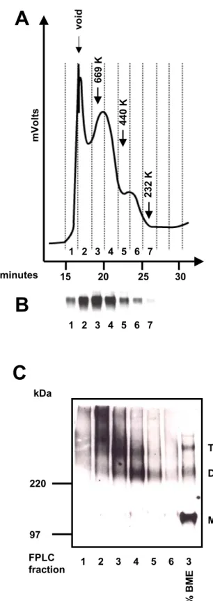
To determine that gp120 was present in the protein peaks eluted during molecular exlusion chromatography, samples from each peak were incubated for 5min at 100°C in sample buffer containing 2% BME and separated on a 7.5% SDS-polyacrylamide gel. To confirm that oligomeric forms of envelope glycoproteins could be detected in the high molecular weight peaks, the fractions were diluted in sample buffer lacking BME and separated on a 3-8% SDS-polyacrylamide gel. A sample from the peak consistent with a trimer was also analyzed in the presence of 2% BME.
2.5.2 Core gp120 envelope glycoproteins
For the detection of core gp120, 2µg of purified core gp120 was incubated at 100°C in reducing SDS sample buffer for 5min, subsequently separated on a 12% SDS- PAGE gel and stained with Coomassie blue.
2.6 Molecular exclusion chromatography
The gp160∆CT glycoproteins captured onto Dynal beads were eluted from the beads for molecular exclusion chromatography under non-denaturing conditions as follows. The beads were incubated in 0.5M MgCl2, 1% CHAPS, and 0.2mM C9 peptide (peptide sequence: TETSQVAPA) at 37oC for 30min. Approximately 5µg of eluted gp160∆CT glycoproteins were loaded onto a Superdex 200 column (Amersham Pharmacia Biotech) in a 200µl volume. The column was then eluted with PBS containing 1% CHAPS at a rate of 0.5ml/min for 40min. The eluted protein was detected by measuring the optical density at 280nm (OD280) using a Varian ProStar System (Varian
Analytical Instruments). Fractions of the flow-through were collected at 2 minute intervals using a Varian Dynamax Fraction Collector. The fractions were further analyzed by reducing and non-reducing SDS-PAGE and Western blotting using a polyclonal anti- gp120 rabbit serum for detection of HIV-1 envelope glycoproteins. A mixture of high molecular weight protein markers (Amersham Pharmacia Biotech) was eluted under identical conditions to calibrate the column.
2.7 Western blot
For the analysis of fractions eluted by molecular exclusion, Western blotting was performed after SDS-PAGE under either non-reducing or reducing conditions. Proteins were electrophoretically transferred onto a 0.45 µm Hybond-C extra membrane (Amersham). The gp160∆CT glycoproteins present in each column fraction were then detected by anti-gp120 rabbit serum at a dilution of 1:2000 and anti-rabbit IgG- horseradish peroxidase (HRP) (Sigma) at a dilution of 1:5000.
2.8 Flow cytometric analysis
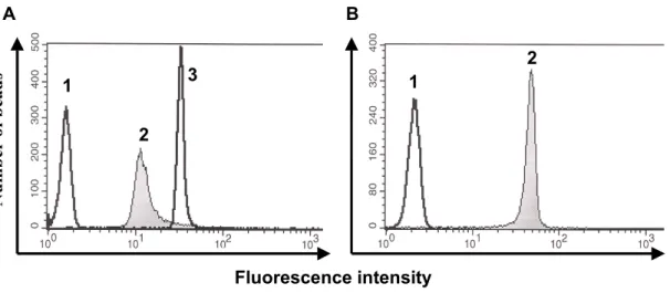
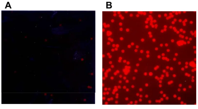
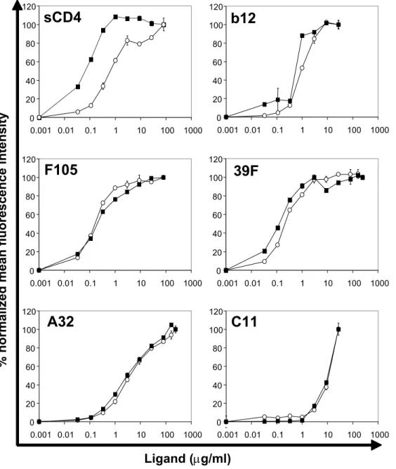
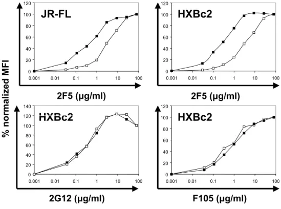
For the comparison of antibody binding to either cleavage-defective or cleavage- competent gp160∆CT envelope glycoproteins, 293T cells were transfected with plasmids expressing the two envelope glycoprotein variants. Approximately 106 cells per sample were harvested with PBS containing 5mM EDTA and washed once in FACS buffer (PBS, 2% FCS, 0.02% Na2N3). The cells were incubated for 1 hour at RT with the indicated amounts of antibodies in a volume of 100µl. After two washing steps in FACS buffer, the cells were incubated for 30min with a R-Phycoerythrin (PE)-conjugated F(ab’)2 goat anti-human antibody (Jackson Immuno Research Laboratories, Inc., West Grove, PA) at a dilution of 1:200, washed twice and analyzed with a FACScan flow cytometer and CellQuest software (Beckton Dickinson). For FACS analysis of proteoliposomes, staining was performed as described above. Staining for membrane integrity of the proteoliposomes was performed using a PE-conjugated goat anti-mouse IgG (Boehringer Mannheim) or Avidin-FITC (Sigma) at a dilution of 1:200. The following ligands were used for staining of the envelope glycoproteins: soluble CD4, the potently neutralizing CD4BS antibody IgG1b12 (kindly provided by Dennis Burton), the
F105 CD4BS antibody (kindly provided by Marshal Posner), the strain-restricted neutralizing V3 loop antibody 39F, the non-neutralizing C1/C4 antibody A32, the non- neutralizing C1/C5 antibody C11, the CD4-induced 17b antibody (all kindly provided by James Robinson) and the broadly neutralizing gp41 antibody 2F5 (kindly provided by Hermann Katinger).
2.9 Animals, immunization, and serum preparation 2.9.1 Immunization with proteoliposomes
2.9.1.1Mice
To determine the immunogenicity of M-450 Dynal beads and proteoliposomes, and to determine adjuvant requirements for immunization with proteoliposomes, groups of four BALB/c mice were inoculated as follows. For the immunization of group A, 0.5x10⁷ of M-450 conjugated with the 1D4 antibody was inoculated in 200µl MPL+TDM adjuvant (Sigma) emulsion subcutaneously. For group B, 2µg of affinity-purified YU2 gp120 was inoculated in 200µl MPL+TDM adjuvant emulsion subcutaneously.
Proteoliposomes containing gp160∆CT envelope glycoprotein (0.5x10⁷ of M-450 proteoliposomes in 200µl volume) were either resuspended in 200µl of MPL+TDM adjuvant (group D) or the adjuvant solution was administered to the site of inoculation one day prior to inoculation with proteoliposomes (group C) to limit the exposure of proteoliposomes to the detergent-containing adjuvant. The inoculation was repeated three times (two times for group D) in four week intervals. Eyebleeding was performed seven days after inoculations. The blood was collected, incubated at 4oC O/N to allow for clot formation, and serum was cleared for 10min at 14,000rpm in an Eppendorf microtube centrifuge. The cleared serum was stored at 4oC.
2.9.1.2Rabbits
Groups of five New Zealand White rabbits were inoculated intradermally with 1ml of MPL+TDM+CWS adjuvant emulsion (Sigma) containing the respective antigen.
The amounts of YU2 gp120 and YU2gp140-GCN4 envelope glycoprotein were adjusted to the same molar quantity of the gp120 moiety. Thus, each rabbit received 18.9µg of
YU2 gp120 and 25µg of YU2gp140-GCN4. Animals inoculated with proteoliposomes received 1ml MPL+TDM+CWS adjuvant emulsion containing approximately 1.8x10⁹ M- 280 proteoliposomes. Inoculations were administered 2, 6, 10, and 22 weeks after the initial inoculation. Ear bleeding was performed thirteen days after the second and seven days after the fourth inoculation and fifth inoculation (Table 1). The blood was incubated O/N at 4oC in a Vacutainer SST Gel Clot Activator (Becton Dickinson) and spun for 30min at 2000xg at 4oC. The cleared serum was collected and incubated for 30min at 55oC to inactivate complement. The sera were then stored at -20oC.
2.9.2 Immunization with core gp120 envelope glycoproteins 2.9.2.1Mice
Female, four week old mice were purchased from Taconic. Groups of six C57BL/6 and BALB/c mice were inoculated subcutaneously with 20µg of gp120 resuspended in 200µl of MPL+TDM adjuvant (Sigma). The inoculation was repeated four times in four week intervals. Eyebleeding and serum preparation was performed as described above.
2.9.2.2Rabbits
For the immunization of rabbits, 20µg of envelope glycoprotein was resuspended in 1 ml of MPL+TDM+CWS adjuvant (Sigma). Groups of five New Zealand White rabbits were inoculated intradermally with 1ml of the emulsified antigen. Inoculations were administered 4, 8, and 12 weeks after the initial inoculation. Earbleeding was performed thirteen days after each inoculation except the first. The blood samples were incubated overnight at 4oC in a Vacutainer SST Gel Clot Activator (Becton Dickinson) and cleared for 30min at 2000xg at 4oC. The cleared serum was stored at -20oC.
2.10 ELISA
2.10.1 Detection of anti-gp120 reactivity in serum
To determine the anti-gp120 reactivity in the serum raised from immunizations with proteoliposomes or core-gp120, 100ng of affinity-purified mammalian-expressed YU2 gp120 (for the sera raised against proteoliposomes) or, for the sera raised against core-gp120, affinity-purified Drosophila-expressed core or full length gp120 derived from the HXBc2 or YU2 isolate was adsorbed onto each well of a high-protein-binding microwell plate (Corning) in PBS over night at 4oC. After blocking the plates in 100µl blocking buffer (PBS/2% dry milk/5% FCS), serial serum dilutions in ELISA blocking buffer were incubated in each well for 1h at RT. After three washes with PBS/0.2%
Tween-20, mouse serum binding to gp120 was detected as follows. A 1:2000 dilution of anti-mouse-IgG-biotin antibody (Sigma) in washing buffer was added for one hour, followed after three washes by an incubation with horseradish peroxidase (HRP)- conjugated Streptavidin (Sigma) at a 1:4000 dilution for 30min. For the analysis of rabbit sera, a secondary Anti-Rabbit-IgG-HRP antibody (Sigma) was added in washing buffer at a 1:2000 dilution for one hour at RT. For the detection of IgM titers in rabbit serum, an anti-rabbit-IgM-HRP antibody (Sigma) was used at a 1:2000 dilution. Following three washes, the ELISAs were developed with 100µl TMB Peroxidase substrate (KPL). The reaction was stopped by adding 100µl 1M HCl to each well. After 5min, the optical density at 450nm was read on a microplate reader (Molecular Devices). Endpoint titers were defined as last reciprocal serum dilution at which the absorption at 450nm was greater than two-fold over the signal detected with preimmune serum.
2.10.2 Detection of gp120- and gp41-binding of scFv and Fab antibodies
For the detection of gp120-binding of Fab and scFv antibodies by ELISA, Microplate wells (Corning) were coated overnight at 4°C with 50µl of PBS containing 100ng/well of the following antigens: Affinity-purified YU2 gp120, affinity-purified IIIB gp41, and, for the detection of unspecific binding, BSA (New England Biolabs). The wells were washed twice with PBS/0.02% Tween-20 and blocked with PBS/3% BSA for 1h at 37°C. After a single wash, 20µl of Fab- or scFv-containing supernatant and 20µl
PBS/1% BSA/0.02% Tween-20 was added to the wells and allowed to incubate at 37°C for 1h. For the detection of purified scFv and Fab antibodies, the desired antibody concentration was added in 100µl PBS/0.02% Tween-20. The wells were washed four times. For Fab detection, goat anti-human IgG F(ab')2 alkaline phosphatase (Pierce) diluted 1:500 in PBS containing 1% BSA was added, and the plate was incubated for 40min at RT. The wells were washed five times and developed by adding 50µl of alkaline phosphatase substrate (one tablet of disodium p-nitrophenyl phosphate [Sigma] to 5ml of alkaline phosphatase staining buffer [pH 9.8]). After 30min, the optical density at 405nm was read on a microplate reader (Molecular Devices). For the detection of scFv, mouse anti-HIS IgG (Santa Cruz Biotechnology) was added at a 1:500 dilution in PBS/0.02%
Tween-20 and incubated for 40min at room temperature. After three washes, anti-mouse- IgG- HRP (Sigma) diluted 1:2000 in PBS/0.02% Tween-20 was added and incubated for 30min at RT. After three washes, the wells were developed and detected as described above.
2.10.3 Competition ELISA with sCD4 and D-mannose
Microplate wells (Corning) were coated overnight at 4°C with 50µl of PBS containing 100ng/well of Drosophila-expressed YU2 gp120. For sCD4 competition ELISA, the wells were incubated with sCD4 in 50µl PBS for 1h at RT. The antibodies were added to the wells at a concentration of 20µg/ml in 50µl PBS/0.02% Tween-20, thus obtaining a final antibody concentration of 10µg/ml. The wells were washed and developed as described above for Fabs.
For D-mannose competition ELISA, D-mannose (Sigma) was added to the wells in 50µl PBS/0.02% Tween-20. 50µl serum at a dilution of 1:250 in PBS/0.02% Tween-20 or 50µl antibodies at a concentration of 5µg/ml in PBS/0.02% Tween-20 were added to obtain a final serum dilution of 1:500 and a final antibody concentration of 2.5µg/ml. The wells were washed and developed as described above for rabbit serum.
2.11 Phage display
2.11.1 Panning of a naïve human phage-displayed scFv antibody library
The paramagnetic proteoliposomes were used to screen a naïve human single- chain antibody phage display library generated from a healthy uninfected donor. Before each round of selection, proteoliposomes were incubated with PBS/2% non-fat dry milk for 30min at RT. The proteoliposomes were adsorbed onto a magnet (Dynal) and incubated with the phage-displayed scFv library stock (approximately 1012 phage) for 30min at RT. After binding of the phage, the proteoliposomes were washed three times for 10min with PBS/2% non-fat dry milk after the first round of selection, four times for 10min with PBS after the second round of selection, and four times 15min after the fourth round of selection. For the first two rounds of selection, 4x10⁷ YU2 M-450 proteoliposomes were used. The following two rounds of selection were performed with 2x10⁷ proteoliposomes. The phage were eluted after each round by incubation with 100µl 0.1M HCL pH2.2 for 10min on a rocking platform. The eluted phage were neutralized with 1M Tris to pH7. After each round of panning, a counterselection step with 4x10⁷ beads conjugated with 1D4 antibody but devoid of gp160 was performed. The phage were infected into E.coli TG-1 bacteria and rescued by infection with M13 helper phage (Stratagene). After the fourth round of selection, phage produced by individual bacterial clones were tested for binding to gp120. Individual colonies were inoculated into a 96- well plate and induced to produce phage particles into the bacterial supernatant by infection with helper phage. The bacterial supernatants were tested for their ability to bind to YU2 gp120 by ELISA. Clones positive for gp120-binding were subjected to DNA sequencing to identify unique scFv antibodies.
2.11.2 Panning of biased human phage-displayed scFv and Fab antibody libraries
Six libraries generated from the bone marrow of HIV-1-infected asymptomatic donors (biased libraries) were panned with proteoliposomes as antigen. The library FDA- 2 was panned as a Fab and a scFv library, the libraries DS, DL, DA, and MT were panned as Fab libraries only. For the panning of the FDA-2 Fab library, two separate libraries were prepared initially, one containing kappa light chains and one containing lambda
light chains. Libraries of either kappa or lambda light-chain variable-region fragments were panned separately for one round with each antigen and then combined. The DS, DL, DA and MT libraries were panned separately for one round of panning and then pooled for the three subsequent pannings. All libraries were subjected to four rounds of affinity selection against the respective antigen.
For affinity selections of the biased libraries, M-280 proteoliposomes were prepared as described. Per round of panning, 30-50µl proteoliposomes were blocked with 4% non-fat dry milk for 1h at 37°C. After adsorption onto a magnet, the proteoliposomes were incubated with the phage library (1011-1012 phage diluted in 50µl of PBS/1% BSA).
The proteoliposomes were incubated with the library for 1h at 37 °C on a rocking platform. After adsorption of the proteoliposomes onto a magnet, the proteoliposomes were washed twice with PBS alone, and once in PBS/1% BSA for different times according to the stringency desired in the particular experiment. The proteoliposomes were adsorped onto a magnet and the phage were eluted using 100µl 0.1M HCL pH2.2 with 1mg/ml BSA for 10min on a rocking platform. The eluted phage were then counterscreened for 30 min at RT on a rocking platform to absorb unspecific reactivities.
The eluted phage were incubated with the following counterscreening reagents: 50µl of 1D4-coupled M-280 Dynabeads (pannings A, B, F, G, H, J, and K), 2.5µg YU2 gp120 coated onto the well of an ELISA plate (panning C), CCR5 Proteoliposomes (panning D), and YU2gp140-GCN4 complexed with sCD4 on proteoliposomes containing CCR5 (panning E). After counterscreening, the phage were infected into E.coli XL-blue and rescued with M13 helper phage.
From the enriched Fab phage pools after four rounds of selection, the pComb3 vector containing the Fab genes was obtained and engineered to secrete soluble Fab by polyclonal restriction digest of the library with NotI and NheI (New England Biolabs), thus deleting the bacteriophage coat protein geneIII. After religation of the vector, the library was transformed into E.coli XL1-blue (Stratagene). From the enriched scFv phage pools, the pComb3 vector was obtained and transformed into nonsuppressor E.coli strain TOP10 (Stratagene).
2.12 ScFv and Fab production
For small-scale production of Fab- and scFv-containing cell lysates for the screening of individual colonies for gp120- or gp41-binding, individual colonies of XL- 1blue (Fab) and TOP10 (scFv) were inoculated into 10ml superbroth medium (SB [for 1 liter, 10g of 3-(N-morpholino)propanesulfonic acid (MOPS), 30g of tryptone, and 2g of yeast extract (pH 7)] containing 20mM MgCl2, tetracycline (10µg/ml), and carbenicillin (50µg/ml) and incubated for 8h at 37°C. After adding 1mM IPTG to the cultures, they were incubated O/N at 37°C. The cells were pelleted, resuspended in 1ml PBS and lysed by three cycles of freezing in a dry-ice/ethanol bath and thawing. Cell debris was pelleted by centrifugation at 13,000xg for 10min and the cleared Fab- or scFv-containing supernatant was used for ELISA.
For large-scale preparations, a single colony of each Fab-expressing (scFv- expressing) clone of XL-1blue (TOP10) was used to inoculate an overnight culture of 10ml containing 20mM MgCl2, tetracycline (10µg/ml), and carbenicillin (50µg/ml). The overnight culture was then used to inoculate 8 liters of SB medium containing 20mM MgCl2, tetracycline (10µg/ml), and carbenicillin (50µg/ml). The cultures were incubated at 37°C with shaking at 250rpm until an optical density at 600nm of 0.8 was reached, after which time 1mM IPTG was added to each culture. The cultures were then incubated for a further 16h at 30°C. The cultures were pelleted by centrifugation at 8,000rpm for 15min in a Sorvall SLA-3000 rotor at 4°C, and the pellets were resuspended in 30ml of PBS containing 0.3 µM phenylmethylsulfonyl fluoride (PMSF). The bacteria were lysed by sonication (Misonix sonicator, 1/2-in. horn) four times at 50% cycle time (power 7 for 20s). The bacterial lysate was centrifuged at 17,000rpm in a Sorvall SS-34 rotor for 35min at 4°C. The supernatant was filtered (pore size, 0.8µm and then 0.2µm) and loaded onto a 5ml Protein G-Sepharose column for the purification of Fabs and a Ni-NTA column for the purification of scFv. The Fab was eluted with 0.2M glycine-HCl (pH2.2), neutralized with 2M Tris base (pH9). The scFv were eluted in 200mM Imidazole. Fab and scFv eluate was concentrated with a Centriprep YM-30 (Fab) and YM-10 (scFv) concentrator (Millipore) and dialyzed against PBS (pH 7).
2.13 HIV-1 single-round neutralization assay
The HIV-1 neutralizing activity of serum or antibodies was determined using a single-round virus entry assay, measuring infection by FACS detection of pseudotyped GFP reporter viruses or detection of infectious molecular clones by intracellular p24-Ag staining.
The intracellular p24-Ag neutralization assays were performed in 96-well culture plates. For each antibody or serum dilution, two wells were set up and combined to produce enough cells for precise quantitation by flow cytometry. 40µl of virusstock were incubated with the desired amount of purified antibodies in 10µl volume or 10µl of serum to obtain a serum dilution of 1:5. Approximately 20,000 TCID50 (determined by a 14-day titration assay) of HIV-1 was added to each well. After incubation for 30min at 37°C, 20µl of PBMC (1.5x10⁵ cells) was added to each well. PBMC were maintained in IL-2 culture medium containing 1µM indinavir, and the cells were fed on day 1 with 150µl of IL-2 culture medium containing indinavir. Since there were 150,000 PBMC per well, the resulting MOI was approximately 0.1.
PBMC were harvested for intracellular p24-Ag staining on day two. For intracellular p24-Ag staining, cells were transferred to V-bottomed plates and washed once in PBS containing 1% FCS. Cells were fixed and permeabilized using the Cytofix/Cytoperm Kit (BD-PharMingen). Permeabilized cells were washed twice in V- bottomed plates using the wash buffer provided by the manufacturer and resuspended for 20min at 4°C with 50 µl of a 1:160 dilution of a phycoerythrin (PE)-conjugated mouse anti-p24 Mab (KC57-RD1; Beckman Coulter, Inc.), or a mouse IgG1 isotypic control antibody. After two additional washes, HIV-1- or mock-infected PBMC were analyzed with a FACSCalibur flow cytometer (Becton Dickinson), and data analysis was performed with FlowJo software (Tree Star). For the measurement of infection with pseudotyped GFP reporter viruses, infected cells were washed once in FACS buffer and directly detected. Live cells initially gated by forward and side scatter were analyzed for intracellular expression of p24-Ag or GFP. The number of p24-Ag-positive cells or GFP- positive cells was determined using a bivariate plot of fluorescence versus forward scatter; the gate was set on mock-infected cells. To enumerate infected PBMC, cells were washed, fixed and permeabilized, and stained with the KC57 anti-p24 antibody as
described above. After forward and side scatter gating, at least 50,000 events were counted. Final quantitation of p24-Ag-positive cells was done by subtraction of background events in mock-infected PBMC (usually less than 10 positives per 50,000 events). The percent neutralization was defined as reduction in the number of p24-Ag- positive cells and GFP-positive cells, respectively, compared with the number in control wells with individual prebleed serum or no antibody, respectively.
IC50, IC80, and IC90 values were determined as follows. Antibody dose-response curves were fit with a nonlinear function and the concentration of antibody required for 50, 80, or 90% reduction (IC50, IC80, IC90, respectively) of infection was calculated by a least-squares regression analysis.
3 Results
3.1 Generation and characterization of proteoliposomes 3.1.1 Creation of gp160∆CT proteoliposomes
293T cells transiently expressing the HIV-1 YU2, JR-FL, 89.6 or HXBc2 gp160∆CT glycoproteins, which contain an alteration of the gp120-gp41 cleavage site and deletion of the gp41 cytoplasmic tail, were lysed in buffer containing Cymal-5 detergent. The gp160∆CT glycoproteins were then captured onto Dynal beads conjugated to the 1D4 antibody, which recognizes a C9 peptide tag affixed to the gp160∆CT C- terminus. Following addition of membrane lipids and dialysis of the Cymal-5 detergent, an artificial lipid bilayer is formed around the bead surface (Fig. 4). Thus, pure, properly oriented HIV-1 envelope glycoproteins, embedded in a natural membrane environment, were incorporated into an easily manipulable solid support.
3.1.2 Analysis of proteoliposome protein composition
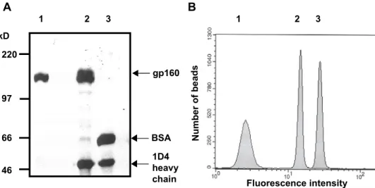
The gp160∆CT proteoliposomes were boiled in sample buffer and analyzed by reducing SDS-PAGE to determine their protein composition. As a positive control, cell
Lipid Detergent gp160∆CT
1D4 antibody
A
B
C
FIG. 4. Schematic diagram of the formation of proteoliposomes. (A) Beads conjugated with the 1D4 antibody recognizing the C9 tag were used to capture the C9 tagged gp160∆CT glycoproteins from cell lysates. The beads were then washed extensively in detergent- containing buffer to remove non C9-tagged proteins. (B) The beads were incubated with detergent- solubilized lipid and dialyzed. (C) During dialysis in PBS, the detergent is replaced by a lipid membrane which assembles around the gp160∆CT glycoproteins.
FIG. 4. Schematic diagram of the formation of proteoliposomes.
(A) Beads conjugated with the 1D4 antibody recognizing the C9 tag were used to capture the C9 tagged gp160∆CT glycoproteins from cell lysates. The beads were then washed extensively in detergent-containing buffer to remove non C9-tagged proteins.
(B) The beads were incubated with detergent-solubilized lipid and dialyzed. (C) During dialysis in PBS, the detergent is replaced by a lipid membrane which assembles around the gp160∆CT glycoproteins.