Review article:
WHY WE DO WHAT WE DO.
A BRIEF ANALYSIS OF CANCER THERAPIES
Carlos M. Galmarini, MD, PhD
Topazium Artificial Intelligence. Paseo de la Castellana 40 Pl. 8, 28046. Madrid, Spain Telephone: +34 911847846; E-mail: cmgalmarini@topazium.com
http://dx.doi.org/10.17179/excli2020-2972
This is an Open Access article distributed under the terms of the Creative Commons Attribution License (http://creativecommons.org/licenses/by/4.0/).
ABSTRACT
The goal of all medical activity is to preserve health in fit people, and to restore the sick into a state of complete physical, mental and social wellbeing. In an effort to determine whether we are achieving this last goal in oncol- ogy, herein we review the biological and clinical framework that has led to the foundations of the current anti- cancer treatment paradigm. Currently, cancer therapy is still based on the ancient axiom that states that the com- plete eradication of the tumor burden is the only way to achieve a cure. This strategy has led to a substantial im- provement in survival rates as cancer mortality rates have dropped in an unprecedented way. Despite this pro- gress, more than 9 million people still die from cancer every year, indicating that the current treatment strategy is not leading to a cancer cure, but to a cancer remission, that is “the temporary absence of manifestations of a par- ticular disease”; after months or years of remission, in most patients, cancer will inevitably recur. Our critical analysis indicates that it is time to discuss about the new key challenges and future directions in clinical oncolo- gy. We need to generate novel treatment strategies more suited to the current clinical reality.
Keywords: Neoplasm, molecular targeted therapies, immunotherapy, artificial intelligence, antiangiogenic agents
INTRODUCTION
If we ask medical students or physicians around the world why they study medicine, the answer would be almost unanimous: “to cure those who are sick and save their lives”.
But what does it really mean to cure the sick? According to the dictionary, the verb
“to cure” is defined as “to restore to health”
(Thesaurus, 2019). However, what is health?
The World Health Organization (WHO) de- fines health as “a state of complete physical, mental and social wellbeing and not merely the absence of disease or infirmity” (WHO, 2019). We can then redefine the action of curing somebody as “to restore to a state of complete physical, mental and social wellbe- ing”. In order to achieve this goal, through- out history, the medical community has de-
veloped many different therapeutic strategies that were based on the scientific paradigms available at the time. Scientific paradigms are produced by clustering information and knowledge generated over time. Indeed, a scientific paradigm is the framework con- taining the basic assumptions, approaches, and methodologies that are commonly ac- cepted by the members of a scientific com- munity. The aim of a scientific paradigm is to provide concrete solutions to specific problems. A scientific paradigm is a model to understand reality, but it is not the reality.
When scientists claim that unresolved prob- lems are often untreatable, they begin to question the prevailing paradigm and seek to replace it (Kuhn, 1970). In an effort to de- termine whether the current cancer paradigm
is achieving the goal of restoring cancer pa- tients to a state of complete physical, mental and social wellbeing, herein we review the biological and clinical framework that has led to the foundations of the current cancer treatment and prevention strategies. Our crit- ical analysis of the medical results obtained with the current paradigm indicates that it is time to replace it to generate novel treatment strategies more suited to the current clinical reality.
THE CURRENT CANCER TREAT- MENT PARADIGM: “FOR EXTREME DISEASES, EXTREME METHODS OF
CURE”
“In treating dangerous, acute diseases, when life's flame flickers at the gates of death, do not hesitate to use heroic measures:
they may avail and save your patient's breath” (Hippocrates, Aphorisms, Section 1 A6) (Scholtz, 1940a).
The current cancer treatment paradigm is based on the Hippocratic view that supports that to cure severe diseases, extreme thera- peutic approaches are the best. In cancer treatment, this paradigm has evolved along history, but always with the same ultimate goal: to cure cancer through the complete eradication of the tumor burden. In order to achieve this goal, certain combinations of surgery, radiotherapy, systemic chemothera- py, and biological and hormonal therapies are used to control the local and systemic components of the disease. Three central ax- ioms that will be explained in detail support this paradigm (Table 1).
Axiom #1: Cancer is an anatomical condi- tion: “a chance to cut is a chance to cure”
(Matmos, 2001)
The initial approach to treat cancer was purely anatomical, i.e., it was based on the eradication of the tumor mass by means of its resection or cauterization. It was not until the late 19th century, when great strides were made in general surgery (anesthesia, antisep- sis and blood transfusions), that the anatomi-
cal approach became the first potentially cu- rative anticancer treatment (Gawande, 2012).
Great masters such as Theodor Billroth (1829-1894), William Halsted (1852-1922), Harvey Cushing (1869-1939), Ernst Wertheim (1864-1920) or Allen Whipple (1881-1963), amongst others, designed and developed surgical techniques for each spe- cific cancer type in order to remove the en- tire tumor along with the lymph nodes around the tumor. Nowadays, many of these surgical procedures continue to be in use.
Radiotherapy can be compared to an invisi- ble blade and therefore, it can be considered as another anatomical approach to cure can- cer. Since 1896, when German physicist Wilhelm Roentgen (1845-1923) discovered X rays, different types of radiation have been used to treat cancer (Gianfaldoni et al., 2017). Later, in 1898, Marie (1867-1934) and Pierre Curie (1859-1906) isolated radi- um, an element that had the ability of depos- iting radiation deep inside tissues (Curie et al., 1898). This discovery led to a golden pe- riod of radiotherapy to treat patients affected by deep cancers.
Table 1: The current cancer treatment paradigm:
“For extreme diseases, extreme methods of cure”
Objective
Total eradication of the tumor mass or burden Axioms
Cancer is an anatomical disease Cancer is a genetic disease
Cancer is a microenvironmental disease Therapeutic approaches
Surgery Radiotherapy Chemotherapy Hormone therapy Targeted therapy Antiangiogenic agents Immunotherapy
However, during the last decades of the 20th century, and despite remarkable scien- tific progress, only a small fraction of pa- tients with locally restricted cancers (e.g.,
primary tumors without metastatic lesions) could be cured with these therapeutic modal- ities. Most tumors returned after surgery or radiotherapy, even if more aggressive opera- tions were performed or higher doses of ra- diation were applied. This was not surpris- ing. Ancient physicians and surgeons knew that cancer patients usually relapsed after the tumor was surgically resected. Hippocrates wisely understood that “what remains in dis- eases after the crisis is apt to produce re- lapses” (Hippocrates, Aphorisms, Section 2 A12) (Scholtz, 1940b). Moreover, interven- tions could be more harmful than no treat- ment at all. The Roman surgeon Aulus Cor- nelius Celsus (25 BC - 50 AD) in his general encyclopedia De Artibus wrote: “We reject any treatment of the latter stages, be it by caustic methods, cauterization or the scalpel.
Any aggressive measure would only irritate the process and, even if the surgeon suc- ceeded in healing the operation, the disease would inevitably recur; successful treatment would only be possible in the first stage”
(Celsus, 30; Kockerling et al., 2013). It is now clear that cancer is not an anatomical condition and therefore, anatomical thera- peutic strategies such as surgery or radio- therapy can only cure a small proportion of cancer patients; for the vast majority, these treatments are only palliative. Based on this, the role of surgery was redefined and cur- rently, surgeons have developed greater technical expertise in minimizing the amounts of healthy tissue being removed during cancer surgery. Similarly, nowadays, radiation can be aimed more precisely. Small tumors in early stage cancers can be resected without extensive amputation of healthy tis- sues, while late stage tumors cannot be erad- icated with these procedures and thus, it makes no sense for patients to undergo ag- gressive surgeries as a palliative treatment.
In summary, eradicating tumors by removing tissues or organs has not translated into a cure for most cancer patients.
Axiom #2: Cancer is a genetic disease:
“The myth of Achilles”
The seeds for establishing a relationship between genetics and cancer were planted in the beginning of the 20th century by imagina- tive scientists such as Theodor Boveri (1862- 1915) (Boveri, 2008). Boveri was ahead of his time and used experimental evidence to develop the concepts that underpin much of what is currently considered the basic tenets of cancer genetics. Boveri’s chromosome theory of genetic inheritance served as the starting point for his tumorigenesis model and the description of the key hallmarks of cancer, such as chromosomal instability, tu- mor heterogeneity, faulty tumor suppressor genes and tumor clonality (Hansford and Huntsman, 2014; Ried, 2009). The bulk of the evidence generated in the following dec- ades confirmed Boveri’s ideas. Nowadays, it is believed that cancer is a genetic disease, the result of somatic evolution, wherein a single clonal lineage acquires driver muta- tions that enables cells to circumvent con- straints on cell proliferation, and finally be- coming cancerous (Bishop, 1987; Nowell, 1976; Weinberg, 1989). A minimum of three to seven of such mutations appear to be re- quired to complete this process (Miller, 1980). Since the establishment of the first model of colorectal cancer by Bert Vogel- stein (1949- ), there was an abundance of re- search identifying and validating key genes and pathways that are dysregulated or mutat- ed in certain tumor types (Lengauer et al., 1998; Vogelstein and Kinzler, 1993). More recently, it has been accepted that the genetic variability across cancer cells is also affected by epigenetic changes, such as DNA methyl- ation. Tumors are now considered as com- plex systems composed of multiple cell sub- clones, with increasing layers of genomic complexity and heterogeneity.
The advantages conferred by the acquisi- tion of driver mutations leading to tumor- igenesis also exposed “vulnerabilities” in cancer cells that are absent in healthy cells. It was then hypothesized that these vulnerabili- ties could be considered as the “Achilles
heel” of cancer cells and exploited to target them whilst sparing healthy cells. This pos- tulate was based on the old concept of the
"poisoned arrow" developed by Paul Ehrlich (1854-1915) (Ehrlich, 1913). Indeed, Ehrlich assumed that destructive toxins developed their injurious action on parasites by binding to certain specific components that he named
"chemoreceptors”. Thus, he compared the ideal chemotherapeutic agents to a “poisoned arrow”: the fixing group of the drug, which anchored itself to the chemoreceptor of the parasite, corresponded to the arrowhead, and the warhead group was the poison smeared on the arrowhead. According to Ehrlich, the best “poisoned arrow” should include a chemical group specific to the chemorecep- tors of the parasites, with no analogue in the organs of the body, thus delivering the poi- son only to the parasite. This would allow what Ehrlich named as the “therapia steri- lisans magna”, which consisted in freeing the organism from the parasites without affect- ing body tissues.
Although originally developed for anti- biotics, the “poisoned arrow” strategy was later applied in cancer chemotherapy. This began with the clinical use of folic acid an- tagonists and nitrogen mustards in the mid- dle 40s of the 20th century (for review, see Galmarini et al., 2012). These molecules primarily targeted biochemical pathways in- volved in cell proliferation. With the discov- ery of different oncogenes, tumor suppressor genes and signaling pathways that were in- volved in carcinogenesis (“cancer chemore- ceptors”), classical chemotherapy transi- tioned to what is known as “targeted thera- pies”. Targeted therapies consist in tailor- made molecules that inhibit or modulate the very genetic mechanism underlying the neo- plasm, enabling a selective cancer treatment with minimal side effects. It was believed that this “poisoned arrow” (mutated into
“magic bullet”) strategy would lead to a can- cer cure. Several targeted therapies were de- veloped showing unparalleled activity and became the standard of care for patients hav- ing tumors with matching molecular profiles
(Dancey et al., 2012). These novel selective therapies changed completely the cancer treatment landscape. Traditionally, tumors from the same anatomical site were treated as one tumor type. However, in the last dec- ades, this notion has been replaced by the concept of targeting driver pathways in tu- mors from different anatomical sites. This is studied in basket or umbrella trials, which are designed to test the effect of one drug on a certain molecular alteration in a variety of tumor types (Redig and Janne, 2015;
Stenzinger et al., 2015).
However, despite advances in the molec- ular design of chemotherapeutic agents, most cancers are resistant to therapy at presenta- tion or become resistant after an initial re- sponse (Figure 1). Indeed, “targeted” thera- pies are facing the same drug resistance problems as conventional chemotherapeutic agents. When the problem is analyzed in depth, it appears that the causes for the fail- ure in chemotherapy are multifactorial, with many factors influencing response to treat- ment (Galmarini et al., 2012; Tredan et al., 2007). To overcome this problem, second- and third-generation selective agents have been developed for clinical use. However, unfortunately, even when treated with these novel inhibitors, tumors become resistant.
Thus, drug resistance remains a problem that limits the clinical use of “classic” and “tar- geted” drugs. Although also based on the concept that cancer is a genetic disease, the
“magic bullet” strategy targeting the “Achil- les heel” of cancer cells equally failed to achieve the goal of curing most cancer pa- tients.
Axiom #3: Cancer is a microenvironmental disease: “The forest, not the tree”
The efficacy of drugs targeting distinct cancer driver pathways varies significantly across cancer patients, and this could not on- ly be ascribed to cancer genetics. This is not surprising, as tumors are not just a cluster of mutated cells, but organ-like structures with many different components, such as non- malignant lymphoid and/or myeloid cells, as
Figure 1: The “magic bullet” strategy. Although based on the concept that cancer cells present
“vulnerabilities” that are absent in healthy cells and thus, can be exploited as therapeutic targets, the
“magic bullet” strategy failed to achieve the goal of curing most cancer patients. Indeed, tumors can be resistant to “targeted” therapies at presentation or become resistant after an initial response.
well as fibroblasts, endothelial cells, peri- cytes, and blood and lymphatic vessels, all embedded within the tumor stroma. All these components are interlinked by a vast array of cytokines, chemokines and growth factors that constitute the tumor microenvironment (TME) (Schaefer and Serrano, 2016). This concept is not new. More than a century ago, Stephen Paget (1855-1926) proposed a “seed and soil” hypothesis suggesting that the ten- dency of tumor metastases to develop in spe- cific organs was due to favorable interactions between cancer cells (the “seed”) and the or- gan microenvironment (the “soil”) (Paget, 1889). An extensive body of clinical data and experimental research has confirmed Paget’s “seed and soil” hypothesis (Fidler and Poste, 2008). Indeed, the evolution of neoplasms is determined not only by the ge- netic features of cancer cells, but also by the
selective pressure of their TME, which de- termines what changes provide adaptive benefits to the tumor (Maley et al., 2017).
The advantages conferred by the TME heter- ogeneity driving tissue-specific tumorigene- sis also exposed vulnerabilities in tumors that may be considered as an “Achilles heel”
and may be exploited as specific treatment targets, whilst sparing healthy tissues (Schneider et al., 2017). The two tumor sus- ceptibilities that could be targeted based on TME heterogeneity were tumor angiogenesis and the immune system.
Attacking tumor angiogenesis:
tumors under siege
Until the 70s, it was believed that tumor growth was primarily supported by actively recruiting blood vessels from the surround- ing tissue. On the other hand, some publica- Normal cells Cancer cells
Hypothesis
Clinical situation
Normal cells Sensitive cancer cells Resistant cancer cells Treatment
Cancer cure
Cancer relapse
Figure 1
tions claimed that the growth of solid tumors was always accompanied by neovasculariza- tion (Algire and Chalkley, 1945; Feigin et al., 1958). In this context, Judah Folkman (1933-2008) hypothesized that most solid tumors initially exist as a small cluster of cells that eventually expands to a size of ap- proximately 1-3 mm3. At this stage the cells enter into a dormant state, as simple diffu- sion of nutrients is no longer sufficient for tumors of this size (Folkman, 1971). Cancer cells then acquire the ability to release an angiogenic mediator (tumor angiogenesis factor or TAF) that stimulates the rapid for- mation of new capillaries around the tumor (Folkman et al., 1971). Only then can the tumor continue to grow. Folkman thus pro- posed to treat cancer through an “anti- angiogenic” approach: blockade of TAF ac- tivity to inhibit the formation of new blood vessels around the tumor, inducing a perma- nent non-vascularized dormant state. Thus, Folkman’s group started to avidly research for angiogenic inhibitors. The first inhibitor was found in a cartilage and suppressed tu- mor growth when it was infused into the vascular bed of murine and rabbit tumors (Brem and Folkman, 1975; Langer et al., 1980). Many other angiogenic inhibitors were subsequently discovered (Folkman and Ingber, 1992). Finally, the first clinical suc- cess in anti-angiogenic therapy came with the use of α-interferon as a treatment for he- mangioma in infants and newborns (Ezekowitz et al., 1992; White et al., 1989).
During that period, other research groups started looking for new angiogenic factors, and a dozen of them were identified in tumor and healthy tissues. The most famous angio- genic factor was discovered in 1983 by Har- old Dvorak (1937- ) et al. when they isolated what they named as the “vascular permeabil- ity factor” (VPF) (Senger et al., 1983). Late- ly, in 1989, Napoleone Ferrara (1956- ) et al.
sequenced and characterized what they named as the “vascular endothelial growth factor” (VEGF), which turned out to be VPF (Leung et al., 1989). VEGF/VPF is an endo- thelial cell mitogen regulated by hypoxia.
Later on, Ferrara et al. identified the high- affinity tyrosine kinase receptor for VEGF (de Vries et al., 1992). Ferrara’s group addi- tionally demonstrated that anti-VEGF mono- clonal antibodies neutralized human VEGF and, when injected subcutaneously to nude mice, exerted a potent inhibitory effect on cancer cell growth in several tumor cell lines (Borgstrom et al., 1998). In 2004, the first anti-angiogenic therapy, bevacizumab, was approved for cancer treatment. Nowadays, several antiangiogenic drugs targeting VEGF or other components of the angiogenic path- way are used to treat cancer (Cao et al., 2011).
However, unlike the results obtained in most preclinical tumor models, current anti- angiogenic therapies produce only modest benefits when administered as a monothera- py; clinical benefits with antiangiogenic therapies are usually achieved by combining them with existing chemotherapy (Kamrava et al., 2009; Plum et al., 2003). Moreover, a proportion of patients who initially respond to an antiangiogenic therapy subsequently relapse (Figure 2). These clinical findings demonstrate that tumors present a high de- gree of intrinsic resistance to antiangiogenic therapies or that they can acquire this re- sistance following drug treatment, as ob- served with chemotherapy. The causes of antiangiogenic resistance are not yet under- stood, but it is likely to arise from compensa- tion by other angiogenic factors and redun- dancy in angiogenic stimulators, which may allow to overcome the blockade of a particu- lar component in the angiogenic pathway (Bergers and Hanahan, 2008).
Fostering the immune system:
calling for reinforcements
The ability to circumvent local and sys- temic immune surveillance mechanisms is an essential step in tumor evolution. The TME plays an important role in the circumvention of the immune system by cancer cells. This is so because TMEs consist of an immune in- filtrate that is dominated by immunosuppres- sive cell types, such as regulatory T cells (Tregs), M2-phenotype macrophages and
Figure 2: Microenvironmental strategies. Tumors are organ-like structures and thus targeting the tumor microenvironment can show profound effects in cancer treatment. However, current antiangio- genic and modern immunotherapies produce modest benefits as a high proportion of patients who ini- tially respond subsequently relapse.
myeloid-derived suppressor cells (MDSCs) (Fridman et al., 2017). Cancer cells take ad- vantage of these particular characteristics of TMEs to avoid, subvert and circumvent the immune system (Slaney et al., 2013). Thus, restoring antitumor immunity seems to be a plausible strategy for cancer treatment.
In the late 19th century, William Coley (1862-1936) noticed that in a considerable number of patients with unresectable cancers who also suffered from accidental erysipelas (usually by Streptococcus pyogenes), tumors rapidly decreased in size or even disap- peared, with some patients remaining well for many years (Coley, 1910). Based on these clinical observations, Coley decided to administer live cultures of streptococcus of erysipelas for the treatment of sarcomas. His first inoculation was performed in a patient
suffering from an unresectable, recurrent spindle-cell sarcoma of the right tonsil; the patient also had a large metastatic tumor in the right cervical region. Coley injected 5 decigrams of a bouillon culture of strepto- coccus of erysipelas obtained from Koch’s laboratory in Germany. The patient devel- oped a severe erysipelas crisis, nearly caus- ing his death. However, after two weeks, the neck tumor incredibly disappeared, and the tonsil tumor decreased in size. Coley then treated this way ten additional patients with unresectable and advanced sarcomas and carcinomas. Again, in all cases, he observed a significant decrease in tumor size (Coley, 1910). Coley then realized that in order to achieve a therapeutic effect, it was not nec- essary to generate an erysipelas crisis, as the therapeutic activity of the erysipelas was due
to toxic bacterial products, not to the bacteria itself. These toxins produced certain changes in the blood or serum (e.g., fever and subse- quent leukocytosis) that restored the weak- ened or lost immunity (Coley, 1928). In to- tal, Coley treated more than 500 patients.
Nearly all of them exhibited a clinical im- provement, but the effect of treatment gradu- ally declined until no longer being effective, with a fatal outcome of the disease (Coley, 1928). In any case, Coley advocated the use of bacterial toxins for the treatment of all cases of unresectable sarcoma, and after all surgeries for primary sarcomas or carcino- mas, as a prophylactic measure against re- currence. The obstacles for further develop- ing Coley’s treatment were primarily the dif- ficulty in obtaining a toxin preparation with a uniform standard and the occurrence of several cases with a fatal outcome when oth- er physicians applied this treatment. Due to this, the use of Coley’s toxin approach grad- ually disappeared from the clinical setting.
Many years later, Georges Mathé (1922- 2010) resurfaced Coley’s ideas (Watts, 2010). As other physicians, Mathé believed that a cancer true remission would only be achieved after the total eradication of all cancer cells. Although surgery, radiotherapy and chemotherapy induced a substantial re- duction of the total tumor cell mass, there were always residual living cancer cells that remained and, after a certain period of time, caused the recurrence of the disease. The challenge lied in achieving a state of com- plete remission by minimizing the number of residual cancer cells without prolonging the duration of treatment, as this would increase the risk of developing a resistant cell sub- population, which would eventually lead to a relapse. Mathé thought that the only way to avoid tumor recurrence after conventional treatments was to eliminate up to the last cancer cell (Mathé, 1974). This may only be achieved by increasing the body’s ability to detect and destroy residual cancer cells by therapeutic stimulation of the immune sys- tem, a concept that he named “active immu- notherapy”. Mathé recovered Coley’s strate-
gy of using live bacteria to treat malignan- cies but, unlike Coley, Mathé had access to worldwide tested and industrially manufac- tured attenuated vaccines. In the early 60s, Mathé began to work with the Bacillus Calmette-Guérin vaccine (BCG) to achieve the unspecific activation of the immune sys- tem as a way to further treat cancer after chemotherapy (Mathé, 1968). This approach showed mixed success for different tumor types and nowadays, intravesical BCG infu- sions are still a major component of standard treatment in bladder cancer (Kamat et al., 2016; Mathé et al., 1973).
From 1980 and during the course of the following three decades, several immunolog- ical approaches were tested for the treatment of leukemia and solid tumors. In the late 80s, metastatic cancer patients have started to be treated with large doses of interferons and IL2 to enhance T-cell production. A decade later, patients with non-Hodgkin’s lympho- mas received the first monoclonal antibody, rituximab, as treatment. Currently, immuno- therapy has become one of the pillars of can- cer treatment, providing the unprecedented opportunity to, in some cases, cure several types of malignancies (Fridman, et al., 2017). Most of these groundbreaking im- munotherapies consist of monoclonal anti- bodies that block T-cell checkpoint receptors and their cognate ligands (e.g., ipilimumab, pembrolizumab, nivolumab) (Adams et al., 2015). Recently, genetically engineered au- tologous dendritic cell therapies (sipuleucel- T) or T-cell therapies (tisagenlecleucel and axicabtagene ciloleucel) have also demon- strated significant clinical responses in he- matological cancers (Miller and Sadelain, 2015). However, although modern immuno- therapies have arguably shown the most pro- found effect in cancer treatment, only about 20 % of patients has achieved true cancer cure with these therapies (Schadendorf et al., 2017) (Figure 2).
RESULTS OF THE CURRENT PARADIGM
“Those diseases which medicines do not cure, iron cures; those which iron cannot cure, fire cures; and those which fire cannot cure, are to be reckoned wholly incurable”
(Hippocrates, Aphorisms, Section 7 A87) (Scholtz, 1941).
The goal of all medical activity is to pre- vent illness and cure patients, that is, to pre- serve health in fit people, and to restore a state of complete physical, mental and social wellbeing in sick people. Our current thera- peutic strategy is based on the fact that a cancer cure can only be achieved through the complete eradication of the tumor burden.
Based on this and following the concept of the “poisoned arrow” described by Ehrlich, finding the “Achilles heel” of tumors that may be exploited as a specific target whilst sparing healthy tissues (“magic bullet” strat- egy) becomes very important. At present, these vulnerabilities include specific targets in cancer cells and tumor tissues and are be- ing targeted by local treatments (surgery and radiotherapy) and systemic treatments (chemotherapy, hormonal therapies, targeted therapies, angiogenic therapies and immuno- therapies). Indeed, the past decade has wit- nessed an explosion of combinations of these therapies. This strategy has led to a substan- tial improvement in survival rates for cancer patients and recently, the cancer mortality rate has dropped in an unprecedented way (Kort et al., 2009). Despite this progress, more than 9 million people still die from cancer every year (IARC, 2019). In addition, cancer survivors suffer chronic morbidities that impair their quality of life (Hudson et al., 2013; Jaffee et al., 2017). We must then admit that, in most patients, the “magic bul- let” strategy is not leading to a cancer cure, but to a cancer remission, that is, “the tempo- rary absence of manifestations of a particular disease” (Del Paggio et al., 2017; Sullivan et al., 2017; Thesaurus, 2019). Certainly, cur- rent treatments prolong the life of cancer pa- tients and improve their quality of life. We cannot vilify these impacts on every patient
life. Any additional time gained with the cur- rent treatments can mean a lot to a patient with the prospect of dying. However, induc- ing a remission is not the same as curing cancer. After months or years of remission, cancer will inevitably recur (Figure 3). We can continue looking for other vulnerabilities in tumors, but the problem will persist.
Therefore, the factual question remains unanswered: how can we truly cure cancer?
We can only find the answer to that question if we accept that our current cancer treatment paradigm is obsolete (Galmarini, 2020;
Galmarini and Lucius, 2020). The evaluation of the present paradigm shows many tri- umphs in basic and clinical research, but un- fortunately, continues to fail in our goal of restoring a state of complete physical, mental and social wellbeing in most cancer patients.
To cure the approximately 18 million people with cancer worldwide, we must shift from this paradigm (IARC, 2019). It is time to pause and think about the key challenges and future directions in clinical oncology.
CONCLUSIONS
There is no doubt that throughout history, the current paradigm has significantly im- proved cancer care. Today, patients with cancer live longer and with better quality of life than in the past. But the goal of curing all cancers has not been accomplished yet.
Currently, cancer therapy is still based on the thousand-year-old paradigm that states that for a cure, complete eradication of cancer cells must be achieved. According to the Hippocratic view, any treatment modality should consider the patient as a unique phys- ical, mental and social entity (Sakula, 1984).
We need to regain his wisdom. We need to recover the genius, scientific audacity and true innovation of those who, more than 100 years ago, devised and laid the foundations of modern treatments. It is necessary to inte- grate genetic, biological, clinical, psycholog- ical and social information into a new coher- ent framework or paradigm to transform it into knowledge and wisdom applied to the clinic that would lead to restoring cancer pa-
tients to their fullest physical, emotional, and social capacities. The new paradigm for can- cer treatment should be based on this holistic view (Galmarini, 2020). As the old “Masters of Medicine” said, we must treat patients, not illnesses. The real individualization of cancer treatment consists in treating each in- dividual patient following the good general practices of oncology and taking into consid- eration his/her own particular needs.
Conflict of interest
Carlos Galmarini is the founder of To- pazium Artificial Intelligence.
REFERENCES
Adams JL, Smothers J, Srinivasan R, Hoos A. Big opportunities for small molecules in immuno- oncology. Nat Rev Drug Discov. 2015;14:603-22.
Algire GH, Chalkley HW. Vascular reactions of normal and malignant tissues in vivo. I. Vascular reactions of mice to wounds and to normal and neoplastic transplants. J Natl Cancer Inst. 1945;6:73- 85.
Bergers G, Hanahan D. Modes of resistance to anti- angiogenic therapy. Nat Rev Cancer. 2008;8:592-603.
Bishop JM. The molecular genetics of cancer.
Science. 1987;235:305-11.
Borgstrom P, Bourdon MA, Hillan KJ, Sriramarao P, Ferrara N. Neutralizing anti-vascular endothelial growth factor antibody completely inhibits angio- genesis and growth of human prostate carcinoma micro tumors in vivo. Prostate. 1998;35:1-10.
Boveri T. Concerning the origin of malignant tumours by Theodor Boveri. Translated and annotated by Henry Harris. J Cell Sci. 2008;121(Suppl 1):1-84.
Brem H, Folkman J. Inhibition of tumor angiogenesis mediated by cartilage. J Exp Med. 1975;141:427-39.
Cao Y, Arbiser J, D'Amato RJ, D'Amore PA, Ingber DE, Kerbel R, et al. Forty-year journey of angio- genesis translational research. Sci Transl Med. 2011;
3:114rv3.
Coley WB. The treatment of inoperable sarcoma by bacterial toxins (the mixed toxins of the Streptococcus erysipelas and the Bacillus prodigiosus). Proc R Soc Med. 1910;3:1-48.
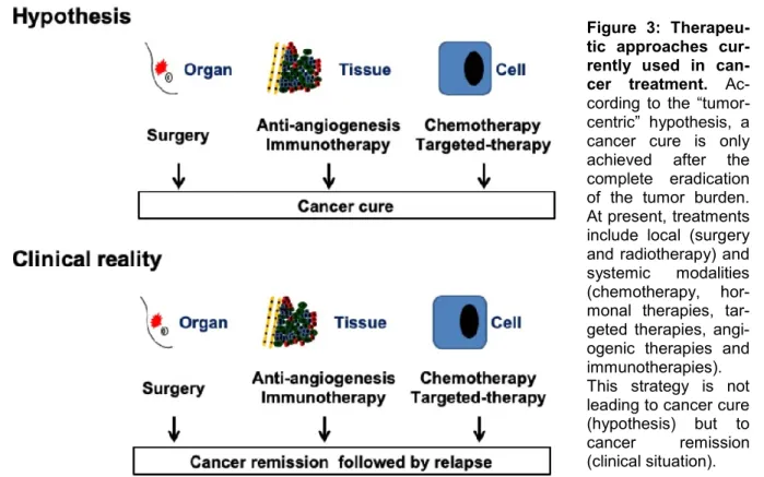
Figure 3: Therapeu- tic approaches cur- rently used in can- cer treatment. Ac- cording to the “tumor- centric” hypothesis, a cancer cure is only achieved after the complete eradication of the tumor burden.
At present, treatments include local (surgery and radiotherapy) and systemic modalities (chemotherapy, hor- monal therapies, tar- geted therapies, angi- ogenic therapies and immunotherapies).
This strategy is not leading to cancer cure (hypothesis) but to cancer remission (clinical situation).
Coley WB. End results in Hodgkin's disease and lymphosarcoma treated by the mixed toxins of erysipelas and Bacillus prodigiosus, alone or combined with radiation. Ann Surg. 1928;88:641-67.
Curie P, Curie M, Bémont G. Sur une nouvelle substance fortement radio-active, contenue dans la pechblende. C R Acad Sci Paris. 1898;127:1215-7.
Dancey JE, Bedard PL, Onetto N, Hudson TJ. The genetic basis for cancer treatment decisions. Cell.
2012;148:409-20.
de Vries C, Escobedo JA, Ueno H, Houck K, Ferrara N, Williams LT. The fms-like tyrosine kinase, a receptor for vascular endothelial growth factor.
Science. 1992;255:989-91.
Del Paggio JC, Azariah B, Sullivan R, Hopman WM, James FV, Roshni S, et al. Do contemporary randomized controlled trials meet ESMO thresholds for meaningful clinical benefit? Ann Oncol. 2017;28:
157-62.
Ehrlich P. Address in Pathology, ON CHEMIO- THERAPY: Delivered before the Seventeenth International Congress of Medicine. Br Med J. 1913;2 (2746):353-9.
Ezekowitz RA, Mulliken JB, Folkman J. Interferon alfa-2a therapy for life-threatening hemangiomas of infancy. N Engl J Med. 1992;326:1456-63.
Feigin I, Allen LB, Lipkin L, Gross SW. The endothelial hyperplasia of the cerebral blood vessels with brain tumors, and its sarcomatous transform- ation. Cancer. 1958;11:264-77.
Fidler IJ, Poste G. The "seed and soil" hypothesis revisited. Lancet Oncol. 2008;9:808.
Folkman J. Tumor angiogenesis: therapeutic implicat- ions. N Engl J Med. 1971;285:1182-6.
Folkman J, Ingber D. Inhibition of angiogenesis.
Semin Cancer Biol. 1992;3:89-96.
Folkman J, Merler E, Abernathy C, Williams G.
Isolation of a tumor factor responsible for angiogenesis. J Exp Med. 1971;133:275-88.
Fridman WH, Zitvogel L, Sautes-Fridman C, Kroemer G. The immune contexture in cancer prognosis and treatment. Nat Rev Clin Oncol. 2017;
14:717-34.
Galmarini CM. Lessons from Hippocrates: Time to change the cancer paradigm. Int J Chronic Dis. 2020;
2020:4715426.
Galmarini CM, Lucius M. Artificial intelligence: a disruptive tool for a smarter medicine Eur Rev Med Pharmacol Sci. 2020;24:7462-74.
Galmarini D, Galmarini CM, Galmarini FC. Cancer chemotherapy: a critical analysis of its 60 years of history. Crit Rev Oncol Hematol. 2012;84:181-99.
Gawande A. Two hundred years of surgery. N Engl J Med. 2012;366:1716-23.
Gianfaldoni S, Gianfaldoni R, Wollina U, Lotti J, Tchernev G, Lotti T. An overview on radiotherapy:
From its history to its current applications in dermatology. Open Access Maced J Med Sci. 2017;5:
521-5.
Hansford S, Huntsman DG. Boveri at 100: Theodor Boveri and genetic predisposition to cancer. J Pathol.
2014;234:142-5.
Hudson MM, Ness KK, Gurney JG, Mulrooney DA, Chemaitilly W, Krull KR, et al. Clinical ascertain- ment of health outcomes among adults treated for childhood cancer. JAMA. 2013;309:2371-81.
IARC. Cancer Today. Data visualization tools for exploring the global cancer burden in 2018.
http://gco.iarc.fr/today/home. Accessed 2020, September 20.
Jaffee EM, Dang CV, Agus DB, Alexander BM, Anderson KC, Ashworth A, et al. Future cancer research priorities in the USA: a Lancet Oncology Commission. Lancet Oncol. 2017;18:e653-e706.
Kamat AM, Hahn NM, Efstathiou JA, Lerner SP, Malmstrom PU, Choi W, et al. Bladder cancer.
Lancet. 2016;388:2796-810.
Kamrava M, Bernstein MB, Camphausen K, Hodge JW. Combining radiation, immunotherapy, and antiangiogenesis agents in the management of cancer:
the Three Musketeers or just another quixotic combination? Mol Biosyst. 2009;5:1262-70.
Kockerling F, Kockerling D, Lomas C. Cornelius Celsus - ancient encyclopedist, surgeon-scientist, or master of surgery? Langenbecks Arch Surg. 2013;
398:609-16.
Kort EJ, Paneth N, Vande Woude GF. The decline in U.S. cancer mortality in people born since 1925.
Cancer Res. 2009;69:6500-5.
Kuhn TS. The structure of scientific revolution. 2nd ed. Chicago, IL: The University of Chicago Press, 1970.
Langer R, Conn H, Vacanti J, Haudenschild C, Folkman J. Control of tumor growth in animals by infusion of an angiogenesis inhibitor. Proc Natl Acad Sci U S A. 1980;77:4331-5.
Lengauer C, Kinzler KW, Vogelstein B. Genetic instabilities in human cancers. Nature. 1998;396:643- 9.
Leung DW, Cachianes G, Kuang WJ, Goeddel DV, Ferrara N. Vascular endothelial growth factor is a secreted angiogenic mitogen. Science. 1989;246:
1306-9.
Maley CC, Aktipis A, Graham TA, Sottoriva A, Boddy AM, Janiszewska M, et al. Classifying the evolutionary and ecological features of neoplasms.
Nat Rev Cancer. 2017;17:605-19.
Mathé G. Active immunotherapy of L1210 leukemia given after the graft of the tumor. Rev Fr Etud Clin Biol. 1968;13:881-3.
Mathé G. La chimiothérapie des cancers. Paris:
Springer-Verlag, 1974.
Mathé G, Pannenko O, Bourut C. BCG in cancer immunotherapy. Results obtained with various BCG preparations in a screening study for systemic adjuvants. NCI Monog. 1973;39:109-14.
Matmos, 2001.
https://en.m.wikipedia.org/wiki/A_Chance_to_Cut_Is _a_Chance_to_Cure. Accessed 2020, September 20.
Miller DG. On the nature of susceptibility to cancer.
The presidential address. Cancer. 1980;46:1307-18.
Miller JF, Sadelain M. The journey from discoveries in fundamental immunology to cancer immuno- therapy. Cancer Cell. 2015;27:439-49.
Nowell PC. The clonal evolution of tumor cell populations. Science. 1976;194:23-8.
Paget S. The distribution of secondary growths in cancer of the breast. Lancet Oncol. 1889;133:571-3.
Plum SM, Hanson AD, Volker KM, Vu HA, Sim BK, Fogler WE, et al. Synergistic activity of recombinant human endostatin in combination with adriamycin:
analysis of in vitro activity on endothelial cells and in vivo tumor progression in an orthotopic murine mammary carcinoma model. Clin Cancer Res. 2003;
9:4619-26.
Redig AJ, Janne PA. Basket trials and the evolution of clinical trial design in an era of genomic medicine. J Clin Oncol. 2015;33:975-7.
Ried T. Homage to Theodor Boveri (1862-1915):
Boveri's theory of cancer as a disease of the chromo- somes, and the landscape of genomic imbalances in human carcinomas. Environ Mol Mutagen. 2009;50:
593-601.
Sakula A. In search of Hippocrates: a visit to Kos. J R Soc Med. 1984;77:682-8.
Schadendorf D, Hodi FS, Robert C, Weber JS, Margolin K, Hamid O, et al. Pooled analysis of long- term survival data from phase II and phase III trials of Ipilimumab in unresectable or metastatic melanoma. J Clin Oncol. 2017;33:1889-94.
Schaefer MH, Serrano L. Cell type-specific properties and environment shape tissue specificity of cancer genes. Sci Rep. 2016;6:20707.
Schneider G, Schmidt-Supprian M, Rad R, Saur D.
Tissue-specific tumorigenesis: context matters. Nat Rev Cancer. 2017;17:239-53.
Scholtz M. Hippocrates' aphorisms. Cal West Med.
1940a;52:126.
Scholtz M. Hippocrates' aphorisms. Cal West Med.
1940b;52:231.
Scholtz M. Hippocrates' aphorisms. Cal West Med.
1941;55:308-10.
Senger DR, Galli SJ, Dvorak AM, Perruzzi CA, Harvey VS, Dvorak HF. Tumor cells secrete a vascular permeability factor that promotes accumulation of ascites fluid. Science. 1983;219:983- 5.
Slaney CY, Rautela J, Parker BS. The emerging role of immunosurveillance in dictating metastatic spread in breast cancer. Cancer Res. 2013;73:5852-7.
Stenzinger A, Weichert W, Lennerz JK, Klauschen F.
Basket trials: Just the end of the first quarter. J Clin Oncol. 2015;33:2823-4.
Sullivan R, Pramesh CS, Booth CM. Cancer patients need better care, not just more technology. Nature.
2017;549:325-8.
Thesaurus. www.dictionary.com/browse (cure).
Accessed 2020, September 20.
Tredan O, Galmarini CM, Patel K, Tannock IF. Drug resistance and the solid tumor microenvironment. J Natl Cancer Inst. 2007;99:1441-54.
Vogelstein B, Kinzler KW. The multistep nature of cancer. Trends Genet. 1993;9:138-41.
Watts G. Georges Mathé. Lancet. 2010;376:1640.
Weinberg RA. Oncogenes, antioncogenes, and the molecular bases of multistep carcinogenesis. Cancer Res. 1989;49:3713-21.
White CW, Sondheimer HM, Crouch EC, Wilson H, Fan LL. Treatment of pulmonary hemangiomatosis with recombinant interferon alfa-2a. N Engl J Med.
1989;320:1197-200.
WHO. https://www.who.int/about/who-we- are/constitution. Accessed 2020, September 20.