Review
Hepatitis E Virus Infection: Circulation, Molecular Epidemiology, and Impact on Global Health
Srinivas Reddy Pallerla
1,2,†, Dominik Harms
3,†, Reimar Johne
4, Daniel Todt
5,6, Eike Steinmann
5, Mathias Schemmerer
7, Jürgen J. Wenzel
7, Jörg Hofmann
8, James Wai Kuo Shih
9, Heiner Wedemeyer
10,11, C.-Thomas Bock
1,3,*
,‡and Thirumalaisamy P. Velavan
1,2,12,‡1
Institute of Tropical Medicine, University of Tübingen, 72074 Tübingen, Germany;
srinivas-reddy.pallerla@uni-tuebingen.de (S.R.P.); velavan@medizin.uni-tuebingen.de (T.P.V.)
2
Vietnamese-German Center for Medical Research (VG-CARE), Hanoi 100000, Vietnam
3
Division of Viral Gastroenteritis and Hepatitis Pathogens and Enteroviruses, Department of Infectious Diseases, Robert Koch Institute, 13353 Berlin, Germany; HarmsD@rki.de
4
Unit Viruses in Food, Department Biological Safety, German Federal Institute for Risk Assessment, 10589 Berlin, Germany; Reimar.Johne@bfr.bund.de
5
Department of Molecular and Medical Virology, Ruhr University Bochum, 44801 Bochum, Germany;
daniel.todt@ruhr-uni-bochum.de (D.T.); eike.steinmann@ruhr-uni-bochum.de (E.S.)
6
European Virus Bioinformatics Center (EVBC), 07743 Jena, Germany
7
Institute of Clinical Microbiology and Hygiene, National Consultant Laboratory for HAV and HEV, University Medical Center Regensburg, 93053 Regensburg, Germany;
Mathias.Schemmerer@klinik.uni-regensburg.de (M.S.); juergen.wenzel@klinik.uni-regensburg.de (J.J.W.)
8
Institute of Virology, Charité Universitätsmedizin Berlin, Labor Berlin-Charité-Vivantes GmbH, 13353 Berlin, Germany; Joerg.Hofmann@laborberlin.com
9
Xiamen Innovax Biotech Co., Ltd., Haicang, Xiamen 361022, China; jwshih@innovax.cn
10
Department of Gastroenterology, Hepatology and Endocrinology, Hannover Medical School, 30623 Hannover, Germany; Wedemeyer.heiner@mh-hannover.de
11
German Center for Infection Research, Partner Hannover-Braunschweig, 38124 Braunschweig, Germany
12
Faculty of Medicine, Duy Tan University, Da Nang 550000, Vietnam
* Correspondence: BockC@rki.de; Tel.: +49-30-18754-2617
† S.R.P. and D.H. contributed equally to the work and thus share first authorship.
‡ C.-T.B. and T.P.V. contributed equally to the work and thus share last authorship.
Received: 22 September 2020; Accepted: 16 October 2020; Published: 20 October 2020
Abstract: Infection with hepatitis E virus (HEV) represents the most common source of viral hepatitis globally. Although infecting over 20 million people annually in endemic regions, with major outbreaks described since the 1950s, hepatitis E remains an underestimated disease. This review gives a current view of the global circulation and epidemiology of this emerging virus. The history of HEV, from the first reported enteric non-A non-B hepatitis outbreaks, to the discovery of the viral agent and the molecular characterization of the different human pathogenic genotypes, is discussed. Furthermore, the current state of research regarding the virology of HEV is critically assessed, and the challenges towards prevention and diagnosis, as well as clinical risks of the disease described. Together, these points aim to underline the significant impact of hepatitis E on global health and the need for further in-depth research to better understand the pathophysiology and its role in the complex disease manifestations of HEV infection.
Keywords: hepatitis E; infection; outbreak; epidemiology; global health
Pathogens2020,9, 856; doi:10.3390/pathogens9100856 www.mdpi.com/journal/pathogens
1. Introduction
Hepatitis E virus (HEV) is a quasi-enveloped, positive strand RNA virus belonging to the family Hepeviridae [1]. HEV is the causative agent of Hepatitis E, the most common cause of acute viral hepatitis both in resource poor and developed countries. Hepatitis E presents as a mostly asymptomatic or acute self-limiting disease with a mortality rate up to 3% in young adults [2]. However the mortality rate may reach 30% in pregnant women [3]. Furthermore, chronic hepatitis E infections may occur in high-risk groups such as immunocompromised individuals (e.g., transplant recipients), those with pre-existing liver disease, HIV-positive persons, and cancer patients [4–7]. A recent study estimates that 939 million people worldwide have been infected with HEV in the past and that 15–110 million people have recent or ongoing infections [8]. According to the WHO, an estimated 3.3 million symptomatic hepatitis E cases occur each year in endemic areas with 44,000 related deaths [9].
HEV belongs to the genus Orthohepevirus, containing four species, namely Orthohepevirus A, B, C, and D with A containing the genotypes HEV-1 to HEV-8 [10]. The genotypes that infect humans include HEV-1 to -4, -7, and Orthohepevirus C, casually termed rat HEV [10–13]. HEV-1 and -2 infect only humans and cause large waterborne outbreaks due to contaminated drinking water in endemic regions of South and Southeast Asia, Africa, and Mexico [14]. HEV-3 and -4 infect both humans and animals, cause sporadic cases in developed countries, and are mainly spread through consumption or close contact with contaminated animal products [14,15]. HEV-7 and rat HEV infections are rarely reported. HEV-1 is mainly transmitted via the fecal–oral route, but also by vertical transmission from mother to child, from person to person, and by blood transfusions [14,16]. HEV-2 is transmitted via the fecal–oral route and human-to-human [14]. In contrast, HEV-3 and -4 are transmitted by transfusion of contaminated blood products, consuming contaminated shellfish, contact with infected animals, environmental contamination by animal manure run-off, and consumption of raw or undercooked meat [14,16]. Hepatitis E is not a sole health burden of the developing world, however, with numbers of reported sporadic cases increasing in industrialized nations, where the virus is spread primarily through zoonotic transmission.
Although usually a self-limiting disease in immunocompetent persons, hepatitis E can cause serious complications in at-risk populations such as pregnant women [17] and organ transplant recipients [5,18,19]. Treatment options remain limited, and only one vaccine has been developed so far with its use limited to China [20].
This review focuses on the history and discovery of the hepatitis E virus and discusses reported outbreaks in order to share new aspects and insights into its epidemiology and global circulation.
Furthermore, it explores studies on the virology of HEV to portray a detailed picture of the known stages of the viral life cycle. Finally, it briefly summarizes the clinical aspects to underline the significant need for further research into the viral pathophysiology contributing to this underrated emerging infectious disease.
2. Discovery and History
An epidemic of HEV was reported in 1955 in Delhi, India, with about 29,000 cases of icteric
hepatitis [21]. After this epidemic, several waterborne outbreaks were reported throughout India,
and most of these cases were non-A and non-B, leading the disease to be described as enteric non-A
non-B hepatitis (ENANBH) [21]. In addition, a major water-related epidemic outbreak in the Kashmir
Valley was reported at the end of 1978, with 52,000 cases and 1700 deaths [22,23]. The symptoms of
these cases were similar to hepatitis A but were negative for both hepatitis A and hepatitis B and
were therefore confirmed as ENANBH [22]. In 1981, hepatitis occurred in a Soviet military camp in
Afghanistan. To investigate this situation, a doctor in the Russian army, Mikhail Balayan, voluntarily
ingested a pooled filtrate of stool samples from the infected soldiers, and he subsequently developed
acute hepatitis [24]. The serum of Dr. Balayan was negative for hepatitis A virus (HAV) and hepatitis
B virus (HBV), suggesting that a new pathogen was responsible for this infection. Immunoelectron
microscopy identified 20–30 nm non-enveloped virus-like particles in stool, confirming a novel
ENANBH virus [24]. In 1990, this novel ENANBH was partially cloned and sequenced and was henceforth called the hepatitis E virus (HEV) [25,26]. It was initially indicated that hepatitis E infection spreads via contaminated water and is limited to resource poor countries. Later, however, increasing reports of sporadic cases emerged in non-endemic industrialized countries with high seroprevalence in a few areas of the United States [27]. The reason for this high prevalence was not understood, and it was speculated that undetected non-pathogenic or less pathogenic HEV strains were circulating.
In 1998, genome sequences confirmed that human HEV were similar to that of HEV in pigs, suggesting zoonotic transmission pathways [28,29]. Overall, these studies led to the identification of a broad spectrum of HEV strains that are either restricted to humans, animals, or infect both.
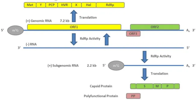
3. Virology
HEV particles have an icosahedral shape, are non-enveloped, and form virions with a diameter of about 27–34 nm [30]. The HEV genome is about 7.2 kb in size and consists of a 5
0UTR capped with 7-methylguanosine (M
7G), followed by three open reading frames (ORF1, ORF2, and ORF3).
The 3
0UTR ends with a poly(A) tail (A
n) like the eukaryotic mRNA structure. Viral replication starts with the translation of the ORF1-encoded non-structural polyprotein. An RNA-dependent RNA polymerase (RdRp) subsequently transcribes the full-length negative-sense RNA. This RNA serves as a template for the synthesis of two viral positive-sense RNAs in infected cells, a full-length genomic RNA, and a subgenomic RNA containing the capsid protein-encoding ORF2 and polyfunctional protein (PP)-encoding ORF3 [31]. The genomic arrangement and the stages of viral genome replication and protein synthesis are visualized in Figure 1.
Pathogens 2020, 9, x, 3 of 21
countries with high seroprevalence in a few areas of the United States [27]. The reason for this high prevalence was not understood, and it was speculated that undetected non-pathogenic or less pathogenic HEV strains were circulating. In 1998, genome sequences confirmed that human HEV were similar to that of HEV in pigs, suggesting zoonotic transmission pathways [28,29]. Overall, these studies led to the identification of a broad spectrum of HEV strains that are either restricted to humans, animals, or infect both.
3. Virology
HEV particles have an icosahedral shape, are non-enveloped, and form virions with a diameter of about 27–34 nm [30]. The HEV genome is about 7.2 kb in size and consists of a 5′ UTR capped with 7-methylguanosine (M
7G), followed by three open reading frames (ORF1, ORF2, and ORF3). The 3′
UTR ends with a poly(A) tail (A
n) like the eukaryotic mRNA structure. Viral replication starts with the translation of the ORF1-encoded non-structural polyprotein. An RNA-dependent RNA polymerase (RdRp) subsequently transcribes the full-length negative-sense RNA. This RNA serves as a template for the synthesis of two viral positive-sense RNAs in infected cells, a full-length genomic RNA, and a subgenomic RNA containing the capsid protein-encoding ORF2 and polyfunctional protein (PP)-encoding ORF3 [31]. The genomic arrangement and the stages of viral genome replication and protein synthesis are visualized in Figure 1.
Figure 1. Genome arrangement of hepatitis E virus (HEV) and steps of viral genome replication.
ORF1 encodes a non-structural polyprotein of varying length that consists of seven domains comprising a methyl transferase (Met), X and Y domains, a papain-like cysteine protease (PCP), a proline-rich hypervariable region (HVR), RNA helicase (Hel), and RNA-dependent RNA polymerase (RdRp) [32]. Of these seven, only the Met, Hel, and RdRp have been functionally well characterized [33–36]. The complete function of all ORF1 domains is still not fully understood. In addition, whether it functions as a multi-domain polyprotein like a “Swiss army knife” or whether it needs to be cleaved for functional activity [37] is under debate and there is still no conclusive evidence that PCP has a protease function [38]. Although ORF1 is essential for HEV replication, its HVR displays considerable sequence divergence even between isolates of the same virus genotype [39]. Size differences between different HEV genomes can be primarily attributed to the HVR region.
Analysis of different patient derived HEV isolates revealed various strains that harbor insertions from other regions of the viral genome, or from human genes, within the HVR [40].
The ORF2 encodes for the capsid protein. Its N-terminal signal peptide shuttles it to the extracellular compartment. The ORF2 protein contains three potential N-glycosylation sites [41–43],
Figure 1. Genome arrangement of hepatitis E virus (HEV) and steps of viral genome replication.
ORF1 encodes a non-structural polyprotein of varying length that consists of seven domains comprising a methyl transferase (Met), X and Y domains, a papain-like cysteine protease (PCP), a proline-rich hypervariable region (HVR), RNA helicase (Hel), and RNA-dependent RNA polymerase (RdRp) [32]. Of these seven, only the Met, Hel, and RdRp have been functionally well characterized [33–36]. The complete function of all ORF1 domains is still not fully understood.
In addition, whether it functions as a multi-domain polyprotein like a “Swiss army knife” or whether it
needs to be cleaved for functional activity [37] is under debate and there is still no conclusive evidence
that PCP has a protease function [38]. Although ORF1 is essential for HEV replication, its HVR
displays considerable sequence divergence even between isolates of the same virus genotype [39]. Size differences between different HEV genomes can be primarily attributed to the HVR region. Analysis of different patient derived HEV isolates revealed various strains that harbor insertions from other regions of the viral genome, or from human genes, within the HVR [40].
The ORF2 encodes for the capsid protein. Its N-terminal signal peptide shuttles it to the extracellular compartment. The ORF2 protein contains three potential N-glycosylation sites [41–43], and is the main immunogenic target of neutralizing antibodies [41,44]. The full-length ORF2 encodes 660aa; however, recent reports suggested that ORF2 protein is processed into at least two forms, including one or two forms of secreted glycoproteins that are not associated with infectious particles, and one unglycosylated form which is the structural component of infectious particles [45,46]. ORF2 has been well characterized for its usefulness in diagnostics and vaccines, including the vaccine Hecolin
®p239, which is currently only approved in China.
The ORF3 protein is a polyfunctional 13-kDa protein of 113 (genotype 3) or 114aa (genotypes 1, 2, and 4). Computational homology scans did not reveal any domains comparable to other known viral proteins. It has been shown to bind to microtubules and be involved in particle assembly and egress by interaction with the tumor susceptibility gene 101 protein (TSG101), a key protein involved in the endosomal sorting complexes of the ESCRT (endosomal sorting complexes required to transport) transport pathway and involved in the budding of the viruses [47–50]. Furthermore, it may play a role in infectious particle secretion via its palmitoylation and membrane association [51]. Additionally, there are also reports of its role in intracellular transduction pathways, the potential to reduce host immune responses, and protection of virus-infected cells [52–54]. A recent article reports that ORF3 is a functional ion channel required for release of infectious particles [55].
HEV exists as quasi-enveloped viral particles in blood and cell culture supernatant and as non-enveloped virions in bile and feces [56]. When they are shed into the environment, non-enveloped naked virions are enterically transmitted through contaminated water or food. So far, it is not well understood how HEV virions overcome the intestinal barrier and reach the liver. However, it is assumed that the virions first infect the enterocytes, where they multiply, and are excreted as quasi-enveloped virus particles into blood circulation, thus infecting hepatocytes [57]. On their way through the bile duct, the envelope is stripped off and naked, and more infectious virions are again released via the stool [56,58].
Naked HEV particles possibly attach to target cells via heparin sulfate proteoglycans (HSGPs) [59]
and heat shock cognate protein 70 (HSC70) [60]. Integrin α3 has been described recently [61] as a candidate receptor to mediate entry into the cells by dynamin-dependent, clathrin-mediated endocytosis, supported by the GTPases Ras-related proteins Rab5A (RAB5) and Rab7a (RAB7), which are necessary for quasi-enveloped particle internalization [59,62,63]. Quasi-enveloped particles attach less efficiently to cells and likely enter the cell in a manner similar to exosomes [63]. Not requiring HSGPs allows attachment in a non-cell-specific manner, possibly explaining HEV’s capacity to infect extrahepatic tissues [63]. Following this, lysosomal degradation of the lipid membrane (in the case of enveloped particles) and subsequent viral capsid uncoating take place, followed by release of the genomic HEV positive strand RNA into the cytoplasm [63]. The host cellular transcriptional machinery translates ORF1 polyprotein containing RdRp from HEV RNA. The polymerase transcribes the full-length negative-sense HEV RNA. From this negative strand, two RNAs are transcribed by RNA helicases and RdRp to form a full-length genomic RNA and a 2.2 kb bicistronic subgenomic RNA. These two capped and polyadenylated RNAs serve as templates for the translation of non-structural ORF1 polyproteins, ORF2 capsid proteins, and polyfunctional ORF3 proteins [64].
The subsequent steps are viral assembly and release. ORF2 and ORF3 and positive-sense genomic
HEV RNA are known to form a complex in the ER–Golgi intermediate compartment and produce
viral progeny particles [65,66]. The particles of the progeny virus bind to the TSG101 protein and are
secreted in a presumably basal fashion as enveloped particles [47,48]. When leaving hepatocytes from
the apical part, the envelope is stripped as described above [56,58]. There are many significant gaps in
the understanding of the HEV life cycle and virus–host cell interactions [67], and further studies are urgently needed. Several in vitro systems exist to study the virus. Reverse genetics models based on infectious cDNA clones have been described for several genotypes. The most commonly used are the Sar-55-related genotype 1 clone [68], the genotype 3a and 3c Kernow-C1- [69], and 47832-related [70]
clones, respectively, both of which contain insertions in the HVR, and the genotype 4 TW6196 clone [71].
A recent presentation of a novel in vitro method to produce high viral titers, allowing study of the full HEV replication cycle in cell culture, has additionally created confidence that we may overcome our limited understanding of HEV pathophysiology [72]. Although these systems are most commonly used in conventional cell culture systems, several animal infection models have been developed.
Rhesus monkeys have been shown to be susceptible to HEV-1 through -4 [28,73], while cynomolgous monkeys and chimpanzees have been used as models for HEV-1 and -2 [68,74]. As natural hosts for HEV-3 and -4, pigs can be readily infected by strains of these genotypes [75,76]. Recent reports have also described successful infection of human liver chimeric mice with HEV-1 and -3 strains [77,78].
Moreover, small animal and avian models exist for the study of animal HEV. Although animal infection models provide more physiological conditions than cell culture systems, limitations are also present.
Neither non-human primates nor mice represent natural hosts of HEV, while pigs can only be infected with HEV-3 and -4. Experiments with primates also raise ethical concerns.
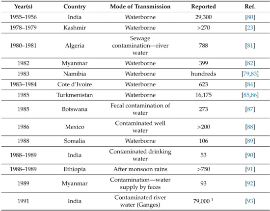
4. Outbreaks
HEV outbreaks occur mainly in resource poor countries, due to waterborne infections caused by HEV-1 and possibly HEV-2 [79], often during the monsoon season. The outbreaks are caused mainly by consumption and use of contaminated water where sanitary and hygienic conditions are poor [14].
The first laboratory confirmed HEV outbreak was reported in Delhi in 1955, and since then several outbreaks have been reported in tropical and subtropical regions of the world, especially in Asian and African countries. Reported outbreaks are summarized in Table 1.
Table 1. Reported HEV outbreaks.
Year(s) Country Mode of Transmission Reported Ref.
1955–1956 India Waterborne 29,300 [80]
1978–1979 Kashmir Waterborne
>270[23]
1980–1981 Algeria
Sewage contamination—river
water
788 [81]
1982 Myanmar Waterborne 399 [82]
1983 Namibia Waterborne hundreds [79,83]
1983–1984 Cote d’Ivoire Waterborne 623 [84]
1985 Turkmenistan Waterborne 16,175 [85,86]
1985 Botswana Fecal contamination of
water 273 [87]
1986 Mexico Contaminated well
water
>200[88]
1988 Somalia Waterborne 106 [89]
1988–1989 India Contaminated drinking
water 53 [90]
1988–1989 Ethiopia After monsoon rains
>750[91]
1989 Myanmar Contamination—water
supply by feces 93 [92]
1991 India Contaminated river
water (Ganges) 79,000
1[93]
Table 1. Cont.
Year(s) Country Mode of Transmission Reported Ref.
1991 China Waterborne 119,000 [94,95]
1991 Indonesia Waterborne 1688
1[96]
1993–1994 Pakistan Waterborne 3827 [97]
1994 Vietnam After heavy rains
>300 [98]
1994 Morocco Fecal contamination of
drinking water 73 [99]
1995–1996 Namibia Waterborne
>600[79]
1998 India Waterborne 82 [100]
1998 Indonesia Waterborne 472 [101]
1998 Pakistan
Fecal contamination—water
system
104 [102]
2002 India Contaminated water 185 [103]
2004 Central African
Republic Rainy season 213 [104]
2004 Sudan Safe water insu
fficient
>2600 [105]
2004 India Drinking untreated raw
river water 538 [106]
2004 Chad Waterborne 1442
1[107]
2005 Iraq Waterborne 268
1[108]
2005 India Contaminated drinking
water 429 [109]
2006 Sudan Waterborne 2621 [110,111]
2007–2008 India Fecal contamination of
water resources 64 [112]
2007–2008 Egypt Waterborne 28 [113]
2007–2009 Uganda Waterborne 146 [114]
2008 India Sewage contamination of
the river 23,915
1[115]
2008 Uganda Substantial
person-to-person
>10,000 [116–118]
2008–2009 Bangladesh
Sewage
contamination—municipal water
4751
1[119]
2009–2012 Uganda Contaminated water 987 [120]
2010 Bangladesh Waterborne 200 [121]
2010 India Waterborne 102 [122]
2005–2010 India Waterborne 442 [123]
2010–2011 Sudan Waterborne 39 [105]
2012 India Fecal contamination of
drinking water 180 [124]
2012 Kenya Waterborne 131 [125]
2008–2012 Central African
Republic Waterborne 745 [126]
2012–2013 South Sudan Waterborne 5080
1[127]
2013 India Sewage contamination of
drinking water 240 [128]
Table 1. Cont.
Year(s) Country Mode of Transmission Reported Ref.
2013 Cameroon Waterborne 33 [129]
2014–2015 Bangladesh Waterborne 103 [130]
2014–2016 India Waterborne 17 [131]
2016–2017 Chad Waterborne 1293 [132]
2017–2018 Nigeria Contamination of
drinking water 1376 [133]
2014–2017 Bangladesh Waterborne 661 [134]
2018 Central African
Republic Waterborne 149 [135]
2018 South Sudan Waterborne 161 [135]
2019 Pakistan Waterborne 300 [136]
2017–2020 Namibia Waterborne 7247 [137]
1Numbers based on reported estimates.
The largest outbreaks involved 79,000 ENANBH cases in Kanpur, India, between 1990 and 1991, and 119,000 cases in China between 1986 and 1988 [93–95]. Sporadic outbreaks have also occurred in recent years in Asian and African countries, but with a low number of reported cases [133–137]. Using modelling approaches, a recent study describes the ecologically most suitable hotspots for HEV viruses:
the Ganges Valley in India and Pakistan [138]. Important factors contributing to water-related outbreaks of HEV are population density, socio-economic conditions, the level of sanitation, and access to drinking water [14,138]. There are also frequent outbreaks in refugee camps, military camps, and emergency shelters in conflict- and catastrophe-affected regions [127,139–142]. In summary, HEV outbreaks can be prevented by improving sanitary conditions and ensuring access to clean drinking water.
5. Epidemiology
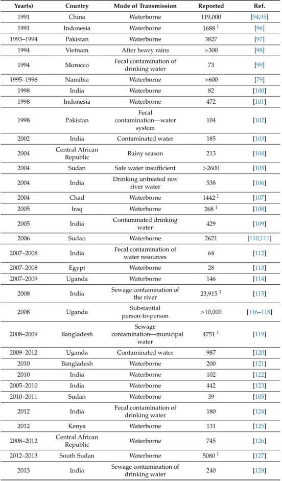
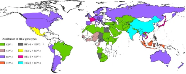
Taxonomically, the family Hepeviridae is divided into the genus Orthohepevirus with the species Orthohepevirus A, B, C, and D, and the genus Piscihepevirus. The species Orthohepevirus A contains eight distinct genotypes that infect humans and other mammals [10]. It is known that HEV-1 to -4 infect humans, and rare cases of human infection with other genotypes like HEV-7 have also been described [11,143]. Details on the genotypes of Orthohepevirus A including host range, transmission routes, and global distribution are summarized in Table 2.
Table 2. Details on Orthohepevirus A genotypes.
Genotype1
(Subtypes) Host Transmission Route Global Distribution1
HEV-1
(1a–1g) Human, primates Fecal–oral via contaminated drinking water
Mainly resource poor regions in India, Pakistan, Bangladesh, Myanmar, China, Mongolia, Morocco, Chad, Nigeria HEV-2
(2a, 2b) Human, primates Fecal–oral via contaminated drinking water
Mainly resource poor regions in Mexico, Nigeria
HEV-3 (3a–3m, 3ra)
Human, pig, wild boar, deer, mongoose,
rabbit (3ra), hare (3ra), rodents
Zoonotic via consumption or contact of/with contaminated foodstuffs; parenteral via contaminated blood donations
Mainly in industrialized countries such as
USA, Canada, China, Japan, South Korea, India, Singapore, UK, Germany, France, Italy, Spain,
Sweden, Switzerland,
Netherlands, Denmark, Hungary
Table 2. Cont.
Genotype1
(Subtypes) Host Transmission Route Global Distribution1
HEV-4 (4a–4i)
Human, pig, wild boar, cow, goat, yak,
Rhesus monkey
Zoonotic via consumption or contact of/with contaminated foodstuffs; parenteral via contaminated blood donations
Mainly in Asian countries such as China, Mongolia, Japan, South Korea, Taiwan, Cambodia, India HEV-5
(5a) Wild boar Unavailable Japan
HEV-6
(6a) Wild boar Unavailable Japan
HEV-7 (7a)
Human, dromedary camel
Zoonotic, likely via consumption
of camel meat UAE
HEV-8
(8a) Bactrian camel Unavailable China
1Information according to the International Committee on Taxonomy of Viruses (ICTV) as of September 2020 and Smith et al. 2020 [10].
Moreover, sporadic human infections with rat HEV (Orthohepevirus C) have been reported [12,13,144]. HEV epidemiology shows two differing patterns based on the global distribution of the Orthohepevirus A genotypes [9] (Figure 2). HEV-1 and HEV-2 genotypes are human pathogens that are mostly transmitted via the fecal–oral route in resource poor countries, mainly through contaminated drinking water, with several cases of vertical transmission having also been described [16]. In contrast, infections with HEV-3 and HEV-4 are autochthonous in developed countries and mainly zoonotically acquired via consumption of contaminated food, but parenteral transmission via blood transfusions have also been described [145]. Despite several HEV genotypes, only a single serotype has been reported, which simplifies seroprevalence studies, diagnosis, and vaccination [146–148].
Pathogens 2020, 9, x, 7 of 21
contaminated blood donations HEV-5
(5a) Wild boar Unavailable Japan
HEV-6
(6a) Wild boar Unavailable Japan
HEV-7 (7a)
Human, dromedary camel
Zoonotic, likely via consumption of
camel meat UAE
HEV-8
(8a) Bactrian camel Unavailable China
1 Information according to the International Committee on Taxonomy of Viruses (ICTV) as of September 2020 and Smith et al. 2020 [10].
Moreover, sporadic human infections with rat HEV (Orthohepevirus C) have been reported [12,13,144]. HEV epidemiology shows two differing patterns based on the global distribution of the Orthohepevirus A genotypes [9] (Figure 2). HEV-1 and HEV-2 genotypes are human pathogens that are mostly transmitted via the fecal–oral route in resource poor countries, mainly through contaminated drinking water, with several cases of vertical transmission having also been described [16]. In contrast, infections with HEV-3 and HEV-4 are autochthonous in developed countries and mainly zoonotically acquired via consumption of contaminated food, but parenteral transmission via blood transfusions have also been described [145]. Despite several HEV genotypes, only a single serotype has been reported, which simplifies seroprevalence studies, diagnosis, and vaccination [146–148].
Figure 2. Global HEV genotype distribution. Different colors on the map indicate the distribution of HEV genotypes (HEV-1 through -4) across the globe. The figure was created using SimpleMappr, an online tool to produce publication-quality point maps