Article
Combined Gastric and Colorectal Cancer Screening—A New Strategy
Michael Selgrad
1,2,†, Jan Bornschein
1,3,†, Arne Kandulski
1,2, Jochen Weigt
1, Albert Roessner
4, Thomas Wex
1,5and Peter Malfertheiner
1,*
1
Department of Gastroenterology, Hepatology and Infectious Diseases, Otto-von-Guericke-University of Magdeburg, Leipziger Str. 44, 39120 Magdeburg, Germany; michael.selgrad@ukr.de (M.S.);
jan.bornschein@ouh.nhs.uk (J.B.); arne.kandulski@ukr.de (A.K.); jochen.weigt@med.ovgu.de (J.W.);
t.wex@schenk-ansorge.de (T.W.)
2
Department of Internal Medicine I, University Hospital of Regensburg, Franz-Josef-Strauß-Allee 11, 93053 Regensburg, Germany
3
Translational Gastroenterology Unit, John Radcliffe Hospital, Oxford University Hospitals, Headley Way, Oxford OX3 9DU, UK
4
Department of Pathology, Otto-von-Guericke-University of Magdeburg, Leipziger Str. 44, 39120 Magdeburg, Germany; albert.roessner@med.ovgu.de
5
Medical Laboratory for Clinical Chemistry, Microbiology and Infectious Diseases, Department of Molecular Genetics, Schwiesaustr. 12, 39124 Magdeburg, Germany
* Correspondence: peter.malfertheiner@med.ovgu.de; Tel.: +0049-391-6713100; Fax: +0049-391-6713105
† These authors contributed equally to this work.
Received: 1 November 2018; Accepted: 29 November 2018; Published: 3 December 2018
Abstract: Background: Our aim was to evaluate the feasibility of a serological assessment of gastric cancer risk in patients undergoing colonoscopy in countries with low-to-moderate incidence rates.
Methods: Serum samples were prospectively collected from 453 patients (>50 years old) undergoing colonoscopies. Of these, 279 (61.6%) also underwent gastroscopy to correlate the results for serum pepsinogen I and II (sPG-I and sPG-II), sPG-I/II ratio, and anti-H. pylori antibodies with gastric histopathology findings (graded according to the updated Sydney classification and the Operative Link of Gastritis Assessment (OLGA) and the Operative Link for Gastric Intestinal Metaplasia assessment (OLGIM) systems). Results: H. pylori was found in 85 patients (30.5%). Chronic atrophic gastritis was diagnosed in 89 (31.9%) patients. High-risk OLGA (III–IV) stages were present in 24 patients, and high-risk OLGIM stages were present in 14 patients. There was an inverse correlation of sPG-I with the degree of atrophy and intestinal metaplasia (IM), as well as with the respective OLGA (r = − 0.425; p < 0.001) and OLGIM (r = − 0.303; p < 0.001) stages. A pathological sPG-I result was associated with a relative risk (RR) of 12.2 (95% confidence interval: 6.29–23.54; p < 0.001) for gastric preneoplastic changes. Conclusions: The assessment of serum pepsinogen allows the identification of patients at increased risk of gastric cancer. A prevention strategy of combining a screening colonoscopy with a serological screening for preneoplastic gastric changes should be considered in the general population.
Keywords: gastric cancer; Helicobacter pylori; screening; pepsinogen; screening colonoscopy
1. Introduction
Screening programs for colorectal cancer are well established and based on varying approaches for implementation in many parts of the world [1]. Screening colonoscopies have led to a significant reduction in the incidence and mortality of colorectal cancer [2–4]. Even in countries with low acceptance rates, screening has been shown to prevent colorectal cancer by 31–64% [5]. In contrast to
Int. J. Mol. Sci.2018,19, 3854; doi:10.3390/ijms19123854 www.mdpi.com/journal/ijms
the established and effective prevention strategies for colorectal cancer, a strategy for gastric cancer prevention has been introduced in only a few countries, Japan and Korea, both of which have high incidences of gastric cancer. Prevention strategies for gastric cancer were initially based on radiography with encouraging results. Today, screening in these countries is performed by endoscopy and serology and a combination of both [6,7].
There are two main reasons to consider gastric cancer screening in low-to-moderate incidence areas: First, gastric cancer remains one of the leading causes of cancer-related deaths, with more than 900,000 cases per year [8,9]. Second, the strong association between Helicobacter pylori (H. pylori) and gastric cancer offers the unique chance for the primary prevention of gastric carcinogenesis by eradication of H. pylori, because 89% of all gastric neoplasia cases are attributable to this infection [10–14].
H. pylori-induced gastric carcinogenesis is a multi-step process, which evolves in many cases from chronic gastritis to preneoplastic changes (atrophic gastritis and intestinal metaplasia) and ultimately into gastric cancer [15]. Therefore, the goal of gastric cancer prevention should be the earliest possible elimination of the infection by eradication therapy, ideally prior to the development of preneoplastic changes [16]. Patients in whom preneoplastic changes of the gastric mucosa are identified should be subject to meaningful surveillance programs, comparable to polyp surveillance for colorectal cancer prevention [17]. A subset of patients with preneoplastic changes will also benefit from eradication therapy, which has the potential to interrupt the progression towards neoplastic stages [18]. A simple blood test with the determination of serum pepsinogen I and II (sPG-I and sPG-II), including the calculation of the sPG-I/II ratio, in combination with the analysis of anti-H. pylori antibodies, allows for the identification of patients with severe atrophic gastritis either with or without H. pylori infection, who are at an increased risk for gastric cancer [19,20]. These high-risk patients should then be invited for a diagnostic gastroscopy to assess the presence of preneoplastic (or neoplastic) changes of the gastric mucosa and to determine if they are candidates for further surveillance [20,21]. While a population-based gastric cancer screening is highly effective in countries with high incidences of this tumour, it is unlikely to be cost-effective in countries with a lower gastric cancer rate [22–26]. For these countries, we propose the approach of a serological “pre-screening” that could be combined with an already established colorectal cancer screening program.
In the current study, we analysed the feasibility of using serological markers for the identification of patients with preneoplastic changes of the gastric mucosa among those who were undergoing colonoscopies.
2. Results
2.1. General Characteristics of the Study Population
Of the 453 individuals enrolled in this study, 196 (43.3%) were women and 257 (56.7%) were men with a mean age of 67.0 years (9.96 years standard deviation). The reasons for the colonoscopies in these individuals were colorectal cancer screening, including surveillance following a polypectomy (n = 187), and diagnostic indications such as abdominal pain (n = 47), altered bowels (n = 51), and per-anal bleeding (n = 40). The remaining 128 patients were referred for various other reasons.
Of all 453 individuals, 279 also received a diagnostic upper gastrointestinal endoscopy (61.6%) and were therefore included in the ‘core cohort’ for further analysis. The distribution of sex, age, and reason for colonoscopy were not different between the total study population and the core cohort.
2.2. Histopathological Results of Endoscopic Gastric Biopsies
Of the 279 patients who received an upper gastrointestinal endoscopy, 277 had complete biopsy
sets for the characterisation of gastric inflammation and atrophic changes according to the updated
Sydney classification. Preneoplastic changes were diagnosed in 89 (32.1%) of the patients. In 64 patients
(23.1%), atrophic changes of the gastric mucosa were seen, with a comparable distribution between
the antrum and the body. Of these, 24 patients were classified as high-risk Operative Link of Gastritis
Assessment (OLGA) stages III and IV (8.7%). In 59 patients (21.3%), IM was present, more often focused in the antrum than in the body (17.0% versus 8.3%). High-risk Operative Link for Gastric Intestinal Metaplasia assessment (OLGIM) stages were seen in 14 (5.1%) patients. In 35 patients (12.6%), active gastric inflammation was seen without preneoplastic changes.
2.3. Helicobacter Pylori Infection Status
A total of 85 patients (30.5%) in the core group was H. pylori-positive by serology (32.9% of the total study population). H. pylori-positive patients showed significantly more active gastric inflammation (54.7% versus 21.0% p < 0.001) and more preneoplastic changes compared with patients without the infection (41.2% versus 27.8%; p = 0.036). Although H. pylori status was not statistically associated with the presence of gastric atrophy (p = 0.437), there was a higher rate of patients with OLGA high-risk stages among the H. pylori-positive patients (15.5% versus 5.6%; p = 0.01). This could not be confirmed for the OLGIM stratification (p = 0.756).
2.4. Serum Pepsinogen I and Histopathological Alterations
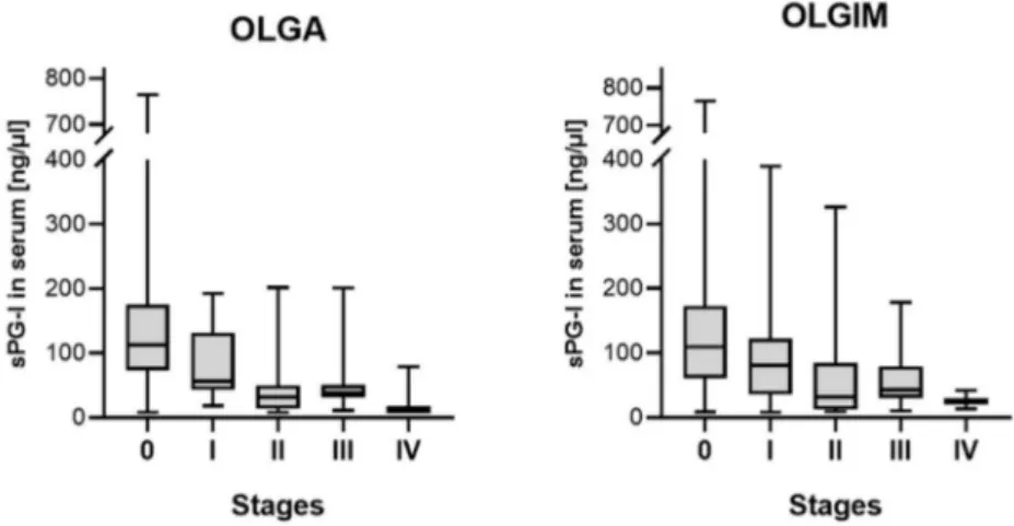
There was the expected inverse correlation of serum levels of sPG-I and the degree of atrophy and IM in the gastric antrum and body (p < 0.001). This correlation was confirmed for the respective stages according to the OLGA (r = − 0.494; p < 0.001) and OLGIM (r = − 0.325; p < 0.001) system (Figure 1).
There was furthermore a weak correlation of sPG-I with the age of the patients (r = − 0.145; p = 0.016).
Figure 1. Serum concentration of pepsinogen I according to the respective Operative Link of Gastritis Assessment (OLGA) (p < 0.001) and Operative Link for Gastric Intestinal Metaplasia Assessment (OLGIM) (p < 0.001) stages.
As mentioned above, a cut-off of sPG-I below 50 ng/µL was considered to be pathological
in accordance with the current literature. A total of 60 patients presented sPG-I levels below this
threshold. Of these, 45 (75%) showed preneoplastic changes on histology, compared with 44 of the
219 sPG-I-negative patients (20.1%; p < 0.001). There were more sPG-I-positive patients among those
with active inflammation in the gastric body (35.6% versus 19.0%; p = 0.013), which could not be
confirmed for inflammation in the antrum (p = 0.168). Of the 60 sPG-I-positive subjects, 42 (70.0%)
showed some degree of glandular atrophy (p < 0.001) independent from the location, and 28 (46.7%)
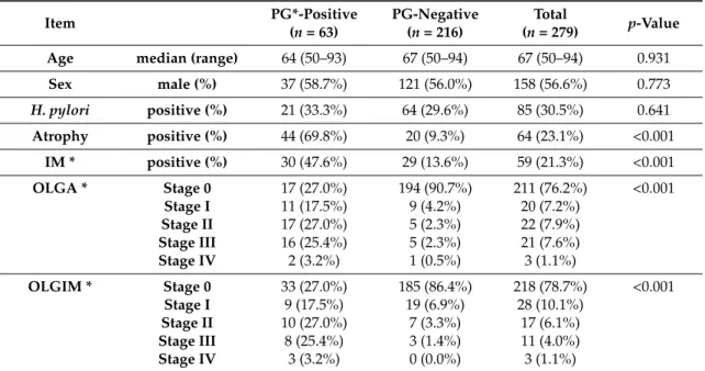
were positive for IM (p < 0.001). These results are summarised in Table 1.
Table 1. Analysis of pepsinogen status in the serum and histopathological alterations.
Item PG*-Positive
(n = 63)
PG-Negative (n = 216)
Total
(n = 279)
p-ValueAge median (range) 64 (50–93) 67 (50–94) 67 (50–94) 0.931
Sex male (%) 37 (58.7%) 121 (56.0%) 158 (56.6%) 0.773
H. pylori
positive (%) 21 (33.3%) 64 (29.6%) 85 (30.5%) 0.641 Atrophy positive (%) 44 (69.8%) 20 (9.3%) 64 (23.1%) <0.001
IM * positive (%) 30 (47.6%) 29 (13.6%) 59 (21.3%) <0.001 OLGA * Stage 0 17 (27.0%) 194 (90.7%) 211 (76.2%) <0.001
Stage I 11 (17.5%) 9 (4.2%) 20 (7.2%)
Stage II 17 (27.0%) 5 (2.3%) 22 (7.9%) Stage III 16 (25.4%) 5 (2.3%) 21 (7.6%)
Stage IV 2 (3.2%) 1 (0.5%) 3 (1.1%)
OLGIM * Stage 0 33 (27.0%) 185 (86.4%) 218 (78.7%) <0.001 Stage I 9 (17.5%) 19 (6.9%) 28 (10.1%)
Stage II 10 (27.0%) 7 (3.3%) 17 (6.1%) Stage III 8 (25.4%) 3 (1.4%) 11 (4.0%)
Stage IV 3 (3.2%) 0 (0.0%) 3 (1.1%)
PG-positive were patients with either PG1 < 50 ng/mL or a PG-ratio < 3.0. * Abbreviations: PG = pepsinogen;
IM = intestinal metaplasia; OLGA = Operative Link of Gastritis Assessment; OLGIM = Operative Link for Gastric Intestinal Metaplasia Assessment.