AUS DEM LEHRSTUHL
FÜR PSYCHIATRIE UND PSYCHOTHERAPIE PROF. DR. RAINER RUPPRECHT
DER FAKULTÄT FÜR MEDIZIN DER UNIVERSITÄT REGENSBURG
NARCOLEPSY VS. IDIOPATHIC HYPERSOMNIA:
DIFFERENTIATING GROUPS USING CLUSTER ANALYSIS
Inaugural – Dissertation zur Erlangung des Doktorgrades
der Medizin
der
Fakultät für Medizin der Universität Regensburg
vorgelegt von Emanuel Sitka
2018
Dekan: Prof. Dr. Dr. Torsten E. Reichert 1. Berichterstatter: PD Dr. Roland Popp
2. Berichterstatter: Prof. Dr. Michael Arzt Tag der mündlichen Prüfung: 22.08.2018
1
Table of contents
Zusammenfassung ... 5
1. Introduction ... 7
2. Hypersomnolence and EDS: Definition and diagnostic concepts ... 9
2.1. The ESS: A subjective measurement of EDS ... 9
2.2. Objective measurements of EDS ... 10
2.2.1. EDS and vigilance tests ... 10
2.2.2. EDS evaluation using polysomnography (PSG) ... 11
2.2.3. The MSLT ... 11
3. Narcolepsy and idiopathic hypersomnia ... 15
3.1. Narcolepsy ... 15
3.1.1. Epidemiology ... 16
3.1.2. Hypocretin, HLA DQB1-0602 and the etiology of narcolepsy ... 16
3.1.3. Clinical aspects ... 19
3.1.4. Diagnosis ... 21
3.1.5. Treatment and prognosis ... 22
3.2. Idiopathic hypersomnia ... 26
3.2.1. Epidemiology, etiology and pathophysiology ... 27
3.2.2. Clinical aspects ... 28
3.2.3. Diagnosis ... 28
3.2.4. Treatment and prognosis ... 29
3.3. Challenges in diagnosis and differential diagnosis of narcolepsy and IH ... 30
3.3.1. Clinical aspects ... 30
3.3.2. The MSLT ... 32
3.3.3. The Epworth Sleepiness Scale ... 35
2
3.3.4. PSG parameters ... 35
3.3.5. Vigilance tests ... 36
3.4. Aims of this thesis ... 37
4. The Dataset ... 39
5. Correlation and linear dependence of MSLT parameters: Sleep latencies and SOREMs ... 43
5.1. Methodical considerations ... 43
5.2. SOREMs and sleep latencies ... 46
5.2.1. Modeling assumptions ... 47
5.2.2. Results ... 48
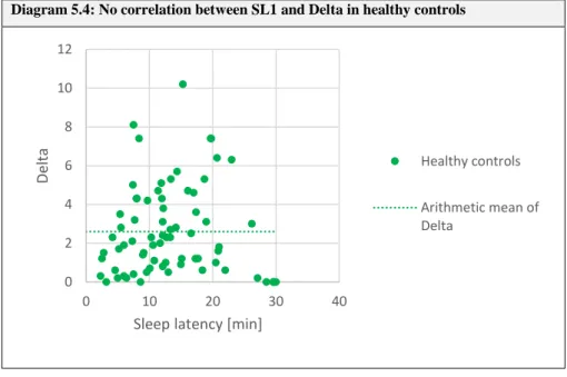
5.3. Delta and sleep latencies ... 50
5.3.1. Modeling assumptions ... 51
5.3.2. Results ... 52
5.4. Discussion ... 54
5.4.1. Sleep latencies and SOREMs ... 54
5.4.2. Delta and sleep latencies ... 57
5.5. Conclusion ... 60
6. Identifying important variables: Principal component analysis ... 62
6.1. PCA: An introduction ... 63
6.2. The PCA algorithm and the total variance criterion ... 64
6.3. Cluster variable selection ... 65
6.4. Results ... 65
6.5. Discussion ... 66
6.5.1. The selection of cluster variables ... 66
6.5.2. The selected principal components ... 67
6.6. Summary ... 74
3
7. Finding groups in the dataset: Cluster analysis ... 76
7.1. Introduction to Cluster analysis ... 76
7.1.1. Principles of cluster analysis ... 76
7.1.2. Cluster evaluation methods ... 78
7.2. K-means clustering ... 81
7.2.1. K-means clustering: iterative center calculation ... 81
7.2.2. Implementation and cluster parameters ... 82
7.2.3. Results ... 82
7.2.4. Evaluation ... 83
7.3. OPTICS: a density-based approach ... 85
7.3.1. Introduction ... 85
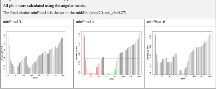
7.3.2. The angular metric and the choice of minPts ... 87
7.3.3. Cluster parameters, implementation and results ... 87
7.3.4. Evaluation ... 89
7.4. Spectral clustering ... 91
7.4.1. Spectral clustering: a graph theoretical approach ... 91
7.4.2. Implementation and cluster parameters ... 92
7.4.3. Evaluation ... 94
7.5. Discussion and cluster comparison ... 97
7.5.1. The ICSD-3 diagnoses as null hypothesis cluster solution ... 97
7.5.2. Rand indices: A first comparison of the cluster solutions ... 99
7.5.3. Cluster validation results ... 99
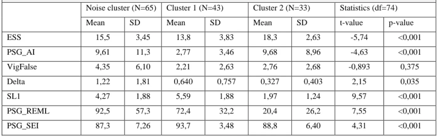
7.6. Characterization and Comparison ... 100
7.7. The narcolepsy subtypes: A critical remark based on the cluster results ... 105
7.8. Conclusion ... 107
4
8. General Discussion ... 110
8.1. Sleep latencies, SOREMs and Delta... 110
8.2. Vigilance test results ... 113
8.3. Groups in the dataset: How many are there? ... 116
8.4. IH or narcolepsy (type 2): differential diagnosis ... 121
9. Conclusion ... 127
Appendix ... 129
1. Tables ... 129
2. Diagrams ... 134
List of tables ... 141
List of diagrams ... 142
Index of abbreviations ... 144
References ... 146
5
Zusammenfassung
Das Ziel dieser Promotionsarbeit ist es, einige Beiträge zu dem Thema der Differentialdiagnose der Narkolepsie zu leisten. Gemäß der dritten Edition der ICSD (International Classification of Sleep Disorders) ist die Narkolepsie in zwei Untergruppen, Typ 1 und Typ 2, unterteilt. Während Narkolepsie Typ 1 pathophysiologisch auf den Untergang Hypocretin-freisetzender Neuronen im Hypothalamus zurückzuführen ist und klinisch häufig durch das Auftreten von Kataplexie leicht zu identifizieren ist, beruht die Diagnose der Narkolepsie Typ 2 fast ausschließlich auf den Ergebnissen des Multiplen Schlaflatenztests (MSLT). Dadurch gestaltet sich die differentialdiagnostische Abgrenzung der Narkolepsie Typ 2 von der Idiopathischen Hypersomnie als sehr schwierig.
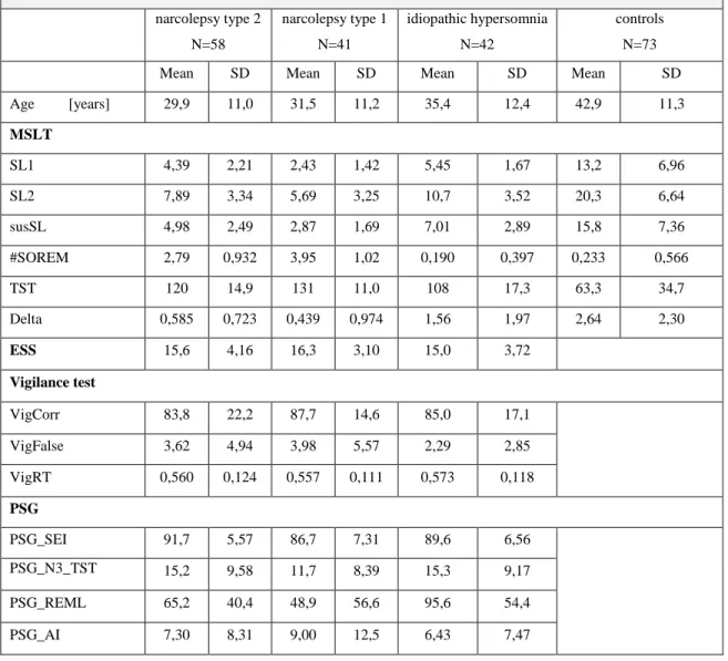
Im Rahmen dieser Doktorarbeit werden verschiedene statistische Methoden eingesetzt, um diese Probleme und ihren Zusammenhang zu den bestehenden diagnostischen Kriterien näher zu beleuchten. Für diese Fragestellung wird ein Datensatz des Schlafmedizinischen Zentrums Regensburg mit insgesamt 141 Fällen von Narkolepsie oder Idiopathischer Hypersomnie herangezogen. Ferner stehen 73 MSLT-Messungen von gesunden Kontrollpersonen zur Verfügung.
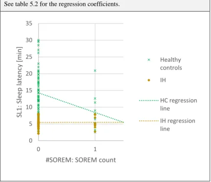
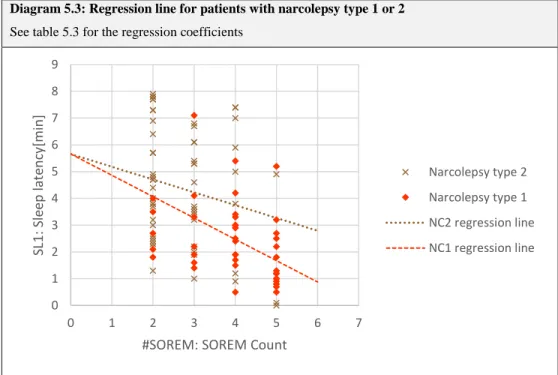
Zunächst wird im Rahmen einer linearen Regressionsanalyse die Korrelation zwischen der Häufigkeit von SOREM (sleep onset REM) – Episoden und der MSLT-Schlaflatenz genauer untersucht. Hier stellt sich ein negativer affin-linearer Zusammenhang für beide Narkolepsie- Typen und gesunde Kontrollen heraus. Für den kürzlich von Pizza et al. vorgeschlagenen Parameter Delta, der die Zeit vom ersten Einschlafen bis zum konsolidierten Einschlafen misst, ist sowohl für Narkolepsie als auch für Idiopathische Hypersomnie eine schwache positive Korrelation zur üblichen Einschlaflatenz zu verzeichnen.
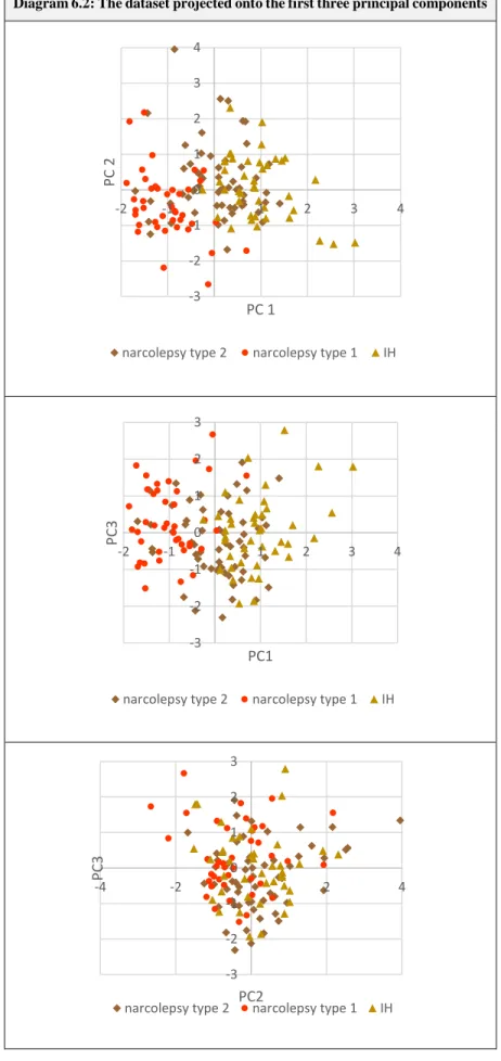
Die im Anschluss durchgeführte Hauptkomponentenanalyse erfüllt zwei wesentliche Funktionen im Rahmen der Zielsetzungen dieser Doktorarbeit. Einerseits zeigt die resultierende dominante Hauptkomponente, dass die typischen MSLT- und Polysomnographie-Parameter, die zur Beschreibung und Unterscheidung von Narkolepsie und Idiopathischer Hypersomnie angewendet werden, in der Tat eine wichtige beschreibende Achse darstellen, an der sich die größten Unterschiede im Datensatz aufschlüsseln lassen. Andererseits dienen die erhaltenen
6
Hauptkomponenten als Grundlage, um algorithmisch einen geeigneten Variablensatz für die Clusteranalysen zu gewinnen.
Für die Clusteranalysen selbst werden drei verschiedene Methoden angewandt. Entsprechend ihrer unterschiedlichen Konzepte zeigen sich deutliche Unterschiede zwischen den erhaltenen Clusterlösungen. Dennoch fallen einige Gemeinsamkeiten auf. So finden alle Clusteralgorithmen stets zwei klinisch relevante Cluster, wobei alle Methoden ähnlich zusammengesetzte Clusterpaare identifizieren. Insbesondere fällt auf, dass die Narkolepsie Typ 2-Fälle algorithmisch in weitgehend konsistenter Weise teilweise der Narkolepsie Typ 1 und teilweise der Idiopathischen Hypersomnie zugeordnet werden. Verschiedene Erklärungen für diese Beobachtung werden angeboten.
Einerseits kann auf Phänomene wie einem verzögerten Einsetzen von Kataplexie bei Narkolepsie hingewiesen werden, andererseits können methodische Schwächen des MSLT die Trennschärfe zwischen Narkolepsie Typ 2 und Idiopathischer Hypersomnie reduzieren.
Zuletzt wird ein Interpretationsansatz weiter ausgeführt, der entsprechend den Ergebnissen der Clusteranalysen zwei statt drei diagnostische Gruppen für den Datensatz vorschlägt, wobei diese beiden Gruppen entstehen, indem die Narkolepsie Typ 2-Fälle in der beobachteten Weise der Narkolepsie (ohne Subtypen) bzw. der Idiopathischen Hypersomnie zugeschrieben werden. Im direkten Vergleich zeigt sich diese alternative diagnostische Einteilung gut hinsichtlich der Clustervariablen nachvollziehbar. Diese Arbeit endet mit dem Vorschlag eines aus der dominanten Hauptkomponente abgeleiteten diagnostischen Scores, der die Differentialdiagnose insbesondere dieser neuen Kategorien zu erleichtern scheint.
7
Narcolepsy vs. idiopathic hypersomnia:
Differentiating groups using cluster analysis
1. Introduction
Narcolepsy is a disabling sleep disorder, which – despite its relative rarity – has always attracted the interest of many sleep medicine researchers. Much progress has been made in understanding the etiology and pathophysiology of this condition. In case of narcolepsy type 1 a specific loss of certain neurons in the hypothalamus has been found to be the morphologic correlate of this disease1. However, until today, no curative treatment option is available, but the majority of patients show significant improvement under the recommended medication.
Some cases of narcolepsy are easily diagnosed due to very suggestive MSLT (multiple sleep latency test) results or a clear-cut history of cataplexy. For a significant number of patients, however, diagnosis is a much more challenging task. One of the main issues in these situations is the distinction between patients with narcolepsy type 2, who by definition do not show cataplexy, and patients with idiopathic hypersomnia (IH). Until today, despite the recent update of the ICSD (International Classification of Sleep Disorders), the definition and diagnostic criteria of these conditions are heavily discussed. Additionally, patients subsumed under the diagnosis of IH show high clinical variance and heterogeneity2, 3. Furthermore, in terms of clinical presentation, a distinction between narcolepsy and IH is often not possible. Due to the absence of biomarkers, the diagnosis and differential diagnoses are based on the results of the MSLT. Since the MSLT is known to have several methodical weaknesses, scientists and clinicians remain in an unsatisfactory situation.
This thesis addresses some of the issues listed above. Using linear regression analysis, the correlation between different MSLT parameters is examined. Emphasis is put on two main aspects:
On the one hand, the correlation between the SOREM (sleep onset REM) count and the mean sleep latency is investigated. On the other hand, focus lies on the sustained sleep latency and its possible diagnostic purpose, for example via the parameter Delta which has recently been suggested by Pizza4.
8
The core of this thesis consists of a principal component analysis and subsequent cluster analyses.
Using these descriptive statistical tools, the diagnostic and differential diagnostic value of several sleep medical parameters is addressed. Furthermore, the cluster analysis results serve as foundation for the discussion, whether the current diagnostic entities, i.e. the narcolepsy subtypes and IH, actually form separate clusters and whether the results of this thesis justify the current classification system. Three entirely different cluster algorithms are employed, yielding three different cluster solutions. In order to illustrate and quantify both similarities and differences between these solutions, various methods for cluster validation and interpretation are presented.
In the final chapter of this thesis, all results of the different methodical approaches are summarized.
The general discussion further examines the question, whether the conventional classification into three groups as stated in the ICSD-3 should be challenged by the consistent finding of two essential groups reported by the cluster analyses. Eventually, a linear score is introduced as a suggestion on how the differential diagnostic process could be refined.
9
2. Hypersomnolence and EDS: Definition and diagnostic concepts
According to the ICSD-3, hypersomnolence is the occurrence of excessive sleepiness5. The conditions that are discussed in this thesis are usually accompanied by excessive daytime sleepiness (EDS), which is defined as “ […] the inability to stay awake and alert during the major waking episodes of the day, resulting in periods of irrepressible need for sleep or unintended lapses into drowsiness or sleep”5. On the contrary, the term hypersomnia should only be used to describe conditions that may cause hypersomnolence/excessive sleepiness.
Excessive sleepiness and EDS should be treated as multidimensional concepts6–8, which cannot be properly detected and quantified by a single diagnostic procedure. Both subjective and objective assessments of EDS are important in the clinical context. In the following, all diagnostic tools that are of further relevance for this thesis will be described.
2.1. The ESS: A subjective measurement of EDS
The Epworth Sleepiness Scale (ESS) is a self-administered questionnaire that was introduced in 1991 by Johns9. According to Johns, the MSLT merely allows an estimation of a very specific situational sleep propensity for the MSLT setting without addressing the general condition of EDS/sleep propensity10. In contrast to the other diagnostic tools discussed below, the ESS was designed to measure the general sleep propensity by explicitly exploring different situations in which sleepiness might occur.
The ESS consists of eight items, which represent different situations in everyday life (e.g.,
“watching TV”). For each item, the patient estimates the likeliness to fall asleep or to doze off in the given situation using the numbers 0 to 3. A score of 0 means that the patient never dozes off or falls asleep, whereas 3 represents a high chance of doing so. The individual item scores are added, resulting in a total score ranging between 0 and 249.
In the original publication by Johns, the scores of healthy controls ranged from 2 to 10 with an average value of 5,9 , whereas all IH and narcolepsy patients in this study showed ESS scores higher than 12, sometimes reaching values of 20 or more9. A normative study regarding the German version of the ESS found an average value of 6,6 and recommended regarding ESS scores higher than 10 as “clinically suspicious” and scores higher than 12 as “clinically relevant”11.
10
The ESS has been validated well for different language versions12–14, for which also the matter of internal consistency has been investigated12, 15. Indeed, high levels of consistency have been found, indicating that all items do address the same theoretic construct. Factor analysis showed the existence of one dominant factor which was interpreted as general sleep propensity by Johns15. Furthermore, most studies found an acceptable test-retest reliability12, 13, although a low reliability has been reported in a population being evaluated for sleep-related breathing disorders16.
Due to its simplicity, the ESS is widely established in clinical practice. However, its diagnostic and differential diagnostic value has been subject of discussion. A brief overview of this topic will be given in section 3.3.3. .
2.2. Objective measurements of EDS
2.2.1. EDS and vigilance tests
Vigilance can be defined as “more careful attention, especially in order to notice possible danger“17 and is therefore not directly linked to EDS. However, vigilance is needed in many tasks of everyday life, e.g. work or traffic. It is to be expected that patients suffering from severe EDS may also have an impaired vigilance, so it is reasonable to measure vigilance in hypersomnia patients.
In this thesis the term vigilance test will always refer to the Quatember-Maly test. This test, which is included in the test collection Wiener Testystem18, 19, is a digitalized version of the clock-test, which has initially been designed by Mackworth20.
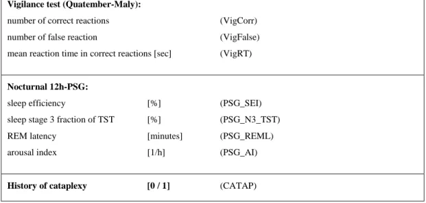
In the Quatember-Maly test the patient is instructed to follow a light source which is moving along a circle, like the second hand of a clock. In irregular intervals a wider jump of the light source occurs, which the patient has to report as quickly as possible. As test results, among other parameters, the number of correct reactions, the number of false reactions (i.e. a jump is reported that has not occurred) and the average reaction time for correct reports are obtained.
According to the test manual the number of correct reactions is the most direct measure for visual vigilance, whereas many false reactions indicate that the patient did not comprehend the test instructions or did not take the test serious21.
11
2.2.2. EDS evaluation using polysomnography (PSG)
According to the ICSD-3 criteria for IH and narcolepsy, the results of the nocturnal PSG serve two important diagnostic purposes. Mainly, the PSG is needed for the exclusion of other causes of hypersomnolence22, e.g. sleep related breathing disorders. Furthermore, if a REM sleep episode occurs in the first 15 minutes of the PSG, it is treated as an equivalent to a SOREM (sleep onset REM) episode in the MSLT, including all implications for the (differential) diagnostic process.
However, some publications indicate that there might be PSG parameters that could be of additional use for the diagnosis and differential diagnoses of IH and narcolepsy. These parameters will be discussed further in section 3.3. .
Regarding this thesis, several PSG parameters will be included in the upcoming analyses: Any SOREM episodes during the PSG are taken into account indirectly by the (PSG) REM latency. The sleep efficiency index, which is defined as the fraction of the total time in bed that is spent asleep, is also included. As a measure of sleep quality, the fraction of the total amount of sleep that is spent in sleep stage N3 is considered. Furthermore, the arousal index, which does not differentiate different causes of nocturnal arousals, is used as an estimate of sleep fragmentation.
2.2.3. The MSLT
The MSLT was designed as a tool for objective measurement of sleepiness in a standardized environment. In principle, the MSLT is based on the findings of Rechtschaffen and Kales, who were able to define EEG criteria for the human sleep architecture23. Before that, sleepiness could only be measured by observing subjects directly or by employing subjective questionnaires.
According to Arrand et al.24 in the seventies of the 20th century several studies were performed investigating the sleep behavior of subjects in a 90 minute day25–27, i.e. 60 minutes of activity and 30 minutes of rest using EEG criteria discovered by Rechtschaffen and Kales. It was found that the subjective sleepiness measured by the SSS (Stanford Sleepiness Scale) showed a strong correlation with the sleep latency of these subjects. Therefore, it was concluded that in a situation like the MSLT test environment, which does not allow any alerting stimuli, the physiological sleep tendency is unmasked and can be objectively measured using these sleep latencies28.
In 1977, the MSLT was first used to assess sleepiness in an experimental setting29. In 1979, the MSLT was used for the first time to detect REM episodes in narcolepsy patients30.
12
The guidelines formulated by Carskadon et al. in 198631 and more recently by Littner et al.32 describe in a precise manner how the MSLT should be performed. Table 2.1 summarizes these guidelines.
Table 2.1: Essential guidelines for the multiple sleep latency test (MSLT) according to Carskadon et al.31 and Littner et al.32
General considerations
- Performance of the MSLT on the day following a NPSG, no MSLT if the total sleep time was less than 6 hours - Sleep diaries 1-2 weeks preceding the MSLT
- Consideration of drug intake; withdrawal two weeks before the MSLT
o Drugs affecting sleep latency: stimulants, hypnotics, sedatives, antihistamines
o Drugs affecting REM latency: tricyclic antidepressants, MAO inhibitors, amphetamines Test settings
- Five nap opportunities in 2-hour intervals, beginning 1,5 to 3 hours after the end of nocturnal sleep - Quiet bedroom, constant and low light level, constant room temperature
- No ingestion of alcohol or caffeine during the whole day
- Between the naps, patients should be out of bed and prevented from sleeping.
Test procedure
- Recording of EEG (C3-A2, C4-A1, O1-A2, O2-A1 derivations), left and right eye electrooculograms, mental or submental EMG and ECG; bio-calibrations preceding each nap
- Each nap opportunity starts with the instruction “Please lie quietly, assume a comfortable position, keep your eyes closed and try to fall asleep.” Then, the lights are turned off.
- Each nap session is terminated after 20 minutes if no sleep occurs. In this case, a sleep latency of 20 minutes is noted.
- If the patient falls asleep within the first 20 minutes, the nap session continues for another 15 minutes starting from the first sleep epoch.
The main diagnostic results of a MSLT test are several different sleep latencies which have been calculated as the average value of the five sleep opportunities. For each sleep opportunity, the sleep latency is defined as the timespan starting from the closing of the eyes until the first episode of a prespecified sleep stage is recorded. Most of the time and in the ICSD-3 criteria for narcolepsy and IH, sleep latency refers to the timespan until the first episode of sleep stage N1 occurs.
Apart from the usual sleep latencies the concept of the sustained sleep latency will become relevant in this thesis. According to Pizza et al.4 sustained sleep latency is defined as the timespan until unequivocal sleep is reached, which is defined as at least three consecutive periods of sleep or one
13
period of a sleep stage different than N1. In the same publication the parameter Delta has also been introduced, which is defined as the difference between the sustained sleep latency and the conventional sleep latency to stage N14. Furthermore, the total sleep time in all five sleep opportunities will be considered as an important parameter later on.
Diagram 2.1: Sleep latency, sustained sleep latency and Delta
The arrows below demonstrate the sleep latency to sleep stage N1, the sustained sleep latency and Delta for a single MSLT nap opportunity. As final test results, the average values with respect to all five nap opportunities are considered.
Apart from the sleep latencies, the MSLT also allows an estimation of REM proneness using the SOREM (sleep onset REM) count. SOREM episodes are defined as REM sleep episodes occurring during the first 15 minutes after sleep onset33. For the SOREM count the number of sleep opportunities that have shown a SOREM episode are added up. Therefore, the SOREM count value can range from zero to five.
In this thesis, data obtained by a modified version of the MSLT, the MSLT30, will be evaluated.
Compared to the standard procedure, the only difference is the rule that all five nap opportunities terminate exactly after 30 minutes, regardless of the observed sleep pattern. The major advantage of this test variant is that no real-time assessment of sleep stages is necessary. Further details regarding the MSLT30 can be found in the publications of Geisler et al.34, 35.
Sleep latency to sleep stage N1 Sustained sleep latency Delta
14
As it is reflected in the current diagnostic criteria, the MSLT has become an essential tool in the diagnosis of narcolepsy and IH. Richardson et al. found in 1978 that narcolepsy patients tended to fall asleep much earlier than healthy controls36, which was an early justification for using the MSLT in the diagnostic process. Both Drake et al. and Chen et al. report an “excellent” inter- and intrarater reliability of the MSLT37, 38. There are some studies that indicate a problematic test-retest reliability of the MSLT39, 40, which will be discussed later in further detail.
Caution is needed for the interpretation of MSLT results, since it is only useful when combined with clinical findings or other diagnostic results32, 41. It is important to consider the age dependence of both the sleep latency and the SOREM frequency. Geisler et al. reported a quadratic dependence of the former from the age of the subject, reaching its minimum in middle-aged subjects35, whereas Dauvilliers et al. found an age-dependent decrease in the SOREM frequency42.
According to the review of Arand et al., the MSLT is thought to measure the physiological sleep tendency in the absence of alerting factors24. However, some concern has been raised if this is indeed the case. For example, Harrison et al. discuss a certain group of individuals showing low sleep latencies but no other signs of subjective or objective sleepiness43. This phenomenon is called
“high ‘sleepability’ without sleepiness” by the authors, raising the concern that the attribute quantified by the MSLT is merely the sleepability of the patients.
Johns, who initially introduced the ESS into clinical practice, discussed the terms “general vs situational sleep propensity”, stating that the MSLT just measures the latter in a very specific situation44. A recent critical comment of Mayer et al.45 on a study by Goldbart et al.40 discusses the question if the MSLT is indeed a suitable tool for the differential diagnosis of EDS. The authors raise the provocative question if the MSLT, often reporting unclear or inconclusive results, might be the very reason because of which the category narcolepsy without cataplexy/type 2 might have been introduced.
These findings highlight the fact that "pathological MSLT results" by themselves do not justify any diagnosis but have to be interpreted in the context of the clinical symptoms. Further issues regarding subtleties in diagnosis and differential diagnosis of narcolepsy and IH will be discussed below.
15
3. Narcolepsy and idiopathic hypersomnia
Before discussing the actual statistical methods and results of this thesis, a brief overview of the diseases addressed in this thesis will be given. After that, current issues in their diagnosis and differential diagnosis will be discussed, which will eventually lead to the motivation of the central aims of this thesis.
3.1. Narcolepsy
The term “narcolepsy” derives from the classical Greek words νάρκη (nárkē) and λῆψις (lepsis) and can be translated to “attack of numbness”. It has first been used by the French physician Jean- Baptiste-Édouard Gélineau46, who has published one of the first articles about narcolepsy in 1881.
Four years before that, Karl Friedrich Otto Westphal had published two case reports about patients showing typical symptoms of narcolepsy47, 48. More than 130 years later, having learned about SOREM episodes49, HLA genotypes50 and hypocretin51, our understanding has vastly increased.
However, until today, narcolepsy can most easily be characterized by the narcoleptic tetrad, consisting of hypnagogic hallucinations, sleep paralysis, excessive daytime sleepiness (EDS) and cataplexy33.
Hence, the typical patient suffering from narcolepsy shows a severe urge to go to sleep during daytime, which often cannot be resisted. During the transition from wakefulness to sleep, he encounters hallucinations. Furthermore, having awakened from sleep, these patients often experience an inability to move, which usually vanishes after some minutes. Finally, if these patients are exposed to certain emotional stimuli, a sudden loss of muscle tonus occurs, leading to a sudden fall. However, there are several patients suffering from most of these symptoms but cataplexy, which has led to the definition of two subgroups of narcolepsy: narcolepsy with and
Table 3.1: The narcoleptic tetrad
Symptoms Description/Comment
Hypnagogic hallucinations Hallucinations during the transition from wakefulness to sleep Sleep paralysis Paralysis for a brief time after awakening
Excessive daytime sleepiness Often leading to an irresistible urge to go to sleep
Cataplexy Sudden loss of muscle tone after exposure to emotional triggers; not present in narcolepsy type 2/without cataplexy
16
without cataplexy according to the ICSD-252, or almost equivalently narcolepsy type 1 and type 2 according to the ICSD-35.
3.1.1. Epidemiology
Narcolepsy is a rare sleep medical disease. Its prevalence varies greatly between different ethnics.
The highest prevalence of 0,16 % has been observed in Japan53, whereas in Israel, only 0,0002 % of the population are affected33, 54. In European countries narcolepsy has an intermediate prevalence value. According to Akintomide et al.33 it varies between 0,02 % and 0,05 %, which complies with a prevalence of 0,047 % reported by Ohayon et al.55. For Olmstedt County in Minnesota, USA a prevalence of 0,0563 % was reported by Silber et al56. The incidence for the latter population was 1,37/100000 per year56.
The incidence rate is highest in the second life decade, with a mean age of onset of about 24 years as reported by Dauvilliers et al.57. However, in this reference two age peaks for narcolepsy onset have been found, one around the age of 14,7 years, the second one at the age of 35. Also, narcolepsy has been found to be more common in men33. Another interesting finding is that the narcolepsy onset is strongly seasonal, reaching its maximum in April to July in Beijing in China, as reported by Han et al. .58 In this article it was also found that the incidence of narcolepsy was – with a delay of 5-7 months – correlated to the occurrence of upper airway infections and the H1N1 pandemic in China in 2009. Also, the month of birth seems to affect the individual risk for developing narcolepsy: Dauvilliers et al. found an odd ratio of 1,45 for a birth in March and 0,63 for persons born in September59.
Common comorbidities of narcolepsy are PLMS (periodic limb movement in sleep), sleep talking, REM sleep behavior disorders and depression60.
3.1.2. Hypocretin, HLA DQB1-0602 and the etiology of narcolepsy
In recent years, a convincing body of evidence has been collected suggesting that the primary cause of the narcoleptic symptoms is a hypocretin deficiency in the human brain.
Hypocretins or orexins are neuropeptides that are physiologically produced by a group of neurons in the lateral hypothalamus60. As Peyron et al. showed in an immunohistochemical study of rat brains, these neurons have very widespread projections across the brain and are involved in the regulation of food intake, blood pressure, body temperature and also the sleep-wake-cycle61. The
17
latter aspect was more highlighted by the findings of Chemelli et al. in 1999, who demonstrated that orexin knockout mice suffered from symptoms which are very similar to narcolepsy 62. Furthermore, the results of Lin et al. in 1999 indicated that the canine version of narcolepsy is caused by an altered hypocretin receptor gene 63.
Motivated by these results, Nishino et al. examined the cerebrospinal fluid of nine patients with narcolepsy type 1. In seven out of nine patients, no hypocretin could be detected, whereas all control patients had hypocretin levels above 250 pg/ml in their cerebrospinal fluid (CSF)51. Thannickal et al. conducted a post-mortem brain tissue study of four narcolepsy patients (three of which had shown cataplexy) and found a 85-95% reduction in the number of hypocretin releasing neurons in the hypothalamus1. Furthermore, gliosis was detected in the corresponding hypothalamic regions, indicating a degenerative process which may have caused the loss of neurons. Additionally, as Thannickal was able to demonstrate in another study, at least some cases of narcolepsy without cataplexy are associated with a more localized and less severe loss of hypocretin neurons, which did not affect neurons in the anterior hypothalamus64. Further studies confirmed the high specifity of undetectable low CSF levels for narcolepsy with cataplexy65, 66, but also pointed out that there are other causes that might explain reduced CSF hypocretin levels such as central nervous system (CNS) inflammation, trauma and Guillain-Barré-Syndrome33, 67, 68. Whereas the connection between narcolepsy type 1/with cataplexy and hypocretin deficiency is well established, empiric evidence hints at a more complex situation for narcolepsy type 2/without cataplexy. Krahn et al. reported that considering the total average, narcolepsy without cataplexy patients also show lowered CSF hypocretin levels. These levels however are significantly higher than those in narcolepsy with cataplexy. In this study, patients were also tested for the HLA DQB1*0602 allele, which revealed that only the HLA positive subgroup of narcolepsy without cataplexy had significantly lowered hypocretin levels66. Similarly, Mignot et al. found that only few patients diagnosed with narcolepsy without cataplexy had reduced hypocretin levels69. This apparent heterogeneity of narcolepsy type 2/without cataplexy was further investigated by Andlauer et al., who found lowered hypocretin levels in 24% of patients in a large collective of narcolepsy without cataplexy patients. They also observed that a delayed onset of cataplexy almost exclusively occurred in this subgroup70.
18
Even before these discoveries regarding hypocretin, a lot of evidence had been gathered showing that most narcolepsy patients had a HLA DQB1*0602 genotype. Conversely, being positive for HLA DQB1*0602 turned out to be a major risk factor for developing narcolepsy71. Again, a higher correlation was reported for narcolepsy with cataplexy33, 72. In patients with narcolepsy without cataplexy HLA DQB1*0602 can only be found in 40-60%60 and being positive for HLA DQB1*0602 also correlates with reduced CSF hypocretin levels70.
While according to Aran et al. HLA DQB1*0602 is beneficial in the situation of a septic shock due to a streptococcus infection73, there is some evidence indicating that precisely this improved protection against an acute streptococcus infection might also be involved in the pathogenesis of narcolepsy: Aran found that anti-streptococcal antibodies are elevated in DBQ1*0602 positive narcolepsy patients73, whereas Koepsell et al. published data which suggests that in patients having a history of a strep throat before the age of 21, narcolepsy is more than five times more common74. The connection to upper airway infection was also discussed by Han et al., who reported a time- delayed correlation between infection frequency and narcolepsy incidence58. Other environmental factors have been discussed, such as H1N1 vaccinations, head trauma or exposure to toxic agents33. In conclusion, the current knowledge about the etiology and pathogenesis of narcolepsy could be summarized like this: Environmental factors, which have not been certainly identified, lead to an autoimmune reaction, which especially affects hypocretin positive neurons in the hypothalamus60. Individuals who are positive for HLA DQB1*0602 are more likely to develop this reaction75, which eventually leads to the degeneration of the involved neurons. As a result, hypocretin is lacking in many areas of the brains, which causes the typical symptoms of narcolepsy60.
However, this model is far from comprehensive. There are some patients suffering from narcolepsy with cataplexy, who do not have lowered CSF hypocretin levels65. In reality more complex genetic interaction are thought to be responsible for the development of narcolepsy76.
Various other findings have been published showing other abnormal findings in narcolepsy patients. Several amino acids concentrations seem to be altered in the CSF of narcolepsy patients77. The COMT genotype of patients has an impact on the disease severity78. Histamine neurons are increased in patients having narcolepsy with cataplexy79, whereas the histamine CSF concentration is lowered80. The hippocampal volume is reduced in narcolepsy patients81. Other genetical factors have been identified, for example the T-cell receptor alpha polymorphism82. These results indicate
19
that despite some major breakthroughs there is still no comprehensive pathophysiological model which could explain all cases of narcolepsy, especially those without cataplexy.
It is important to consider that the symptoms of narcolepsy can also be caused by other CNS pathologies. For an example, brain lesions due to tumors, inflammation or ischemia may cause secondary narcolepsy60. Interestingly, lesions in the hypothalamus lead to the complete phenotype of narcolepsy, whereas pathologies in non-hypothalamic regions are more likely to cause cataplexy without the other symptoms of the narcolepsy tetrade83. Also Niemann-Pick disease of type C and muscular dystrophies can induce symptomatic narcolepsy60.
3.1.3. Clinical aspects
Before discussing the current diagnostic criteria, a brief overview of the typical symptoms of narcolepsy will be given. Both the variety at which the four core symptoms of narcolepsy may present themselves, and the fact that not in all patients the whole narcoleptic tetrad can be observed84, 85 contribute to the persisting issues in diagnosis and differential diagnosis.
3.1.3.1. Cataplexy
Cataplexy can be defined as “rapid eye movement (REM) sleep atony occurring at an inopportune moment”, as it is stated by Overeem et al.86. In principal, all striated muscles can be affected by cataplexy, with the diaphragm being the only exception60. The most typical forms of cataplexy are sagging of the jaw and trembling of knees.87 Using video recording, Rubboli et al. could identify three phases of cataplexy: The initial phase, the falling phase and the atonic phase. In particular, these findings suggest that apart from the negative atonic component of cataplexy there also seem to be positive motoric phenomena, possibly reflecting different aspects of motoric signs of REM sleep.88
Common triggers of cataplexy are joy, happiness, surprise and anger, in rare cases attacks of cataplexy are also triggered by sports, sudden noises and tickling87. Sturzenegger et al. have also noted that the presence of persons known to the patients increase the frequency of cataplectic attacks87. Cataplexy usually lasts from less than one second up to several minutes. Some patients encounter less than one episode per year, whereas others suffer from cataplexy many times each
20
day.60 Poryazova et al. have described a special variant of “rebound” cataplexy occurring most often on withdrawal from the antidepressant fluoxetine which acts anticataplectic89.
While cataplexy is thought to be specific for narcolepsy, cataplexy-like symptoms can also occur in non-narcoleptic individuals. According to Sturzenegger et al. these symptoms are more atypical regarding the affected muscle groups and less pronounced than attacks of clear cut cataplexy.
Additionally, they are more frequently observed in patients with hypersomnolence87.
Knudsen et al. report that a hypocretin deficiency leads to more pronounced cataplectic symptoms90. Combined with the findings of Heier et al.65, who reported a strong association between HLA DQB1*0602, cataplexy and low hypocretin levels, one may conclude that cataplexy is most likely and most severe in “typical narcolepsy patients” and that cataplexy – possibly with a delayed onset – is a direct consequence of the pathophysiological pathway described above.
3.1.3.2. EDS, Hypnagogic hallucinations and sleep paralysis
Compared to cataplexy, the occurrence of excessive daytime sleepiness is much less specific for narcolepsy. On the contrary, a lot of conditions can be responsible for this common symptom91. However, there are some features of EDS which help to simplify the differential diagnosis.
Typically, EDS resulting from narcolepsy leads to an almost irresistible urge to nap60, 87. The resulting naps are usually short and tend to be refreshing for the patients92. For some narcolepsy type 2 patients, spontaneous improvement of EDS has been observed93.
Hypnagogic hallucinations are defined as hallucinations occurring during sleep onset. Despite of being part of the narcolepsy tetrad, they are also not uncommon in healthy individuals94. Usually, they are of visual or auditory nature95, but can also include physical sensations60.
Sturzenegger et al. report that sleep paralysis occurs in about 50 % of narcolepsy patients87. Episodes of sleep paralysis usually last for a few seconds but can also persist for some minutes60. Often, sleep paralysis occurs together with hypnagogic hallucinations95. Sleep paralysis is also not specific for narcolepsy and may also occur isolated in an otherwise healthy population as a study by Bell et al. indicates96. Furthermore, Knudsen et al. pointed out that sleep paralysis and hypnagogic hallucinations are typical properties of both narcolepsy type 1 and type 2, suggesting neuronal pathways that are not affected by hypocretin may be involved in these symptoms90.
21 3.1.3.3. Other symptoms of narcolepsy
Another typical symptom of narcolepsy is automatic behavior during daytime, which has been hypothesized to be caused by microsleep episodes60, 97. Furthermore, despite the severe EDS of many narcolepsy patients, their night sleep is often disrupted by many awakenings60.
Akintomide et al. describe various minor symptoms of narcolepsy such as blurry vision and loss of concentration and memory33. It has also been observed that narcolepsy patients have an increased BMI on average98.
Other symptoms of narcolepsy patients may stem from the various comorbidities that have been described. Empirical evidence suggests that narcolepsy is often associated with PLMS60, REM behavior disorders99, sleep-related breathing disorders84 and depression100.
3.1.4. Diagnosis
According to the ICSD-3 narcolepsy can be diagnosed considering the following aspects:
Anamnesis should reveal some history of an irresistible urge to sleep or, consequently, episodes of unwillingly falling asleep. Then, for narcolepsy type 1 cataplexy is major diagnostic factor, whereas the diagnostic criteria for narcolepsy type 2 list cataplexy as an exclusion criterion. In order to objectively measure the EDS, the MSLT must be used for the definite diagnosis.
The third edition of the ICSD-3 includes another procedure for diagnosing narcolepsy type 1: The measurement of CSF concentration of hypocretin-15. This new criterion takes into account the nowadays widely accepted concept that narcolepsy type 1 is caused by hypocretin deficiency101. The CSF hypocretin concentration is measured by using a radioimmunoassay. A hypocretin concentration below 110 ng/ml or below one third of the average concentration in the general population is thought to be highly specific for narcolepsy5. However, as Knudsen et al. have remarked, measuring the CSF concentration of hypocretin-1 is of limited use in practice, since hypocretin deficient narcoleptics usually show a very severe phenotype, which allows an easy diagnosis even without referring to CSF hypocretin concentrations90.
22
Comparing the two tables, it becomes clear that narcolepsy type 2 is diagnosed by ruling out type 1 and other diseases which may cause similar symptoms. Furthermore, being deprived of both CSF measurement and cataplexy as positive diagnostic criteria, the diagnosis of narcolepsy type 2 heavily relies on clinical assessment. On the contrary, it is quite easy to follow the diagnostic criteria for narcolepsy type 1 and therefore to decide if a patient really has narcolepsy type 1.
The diagnostic uncertainty for narcolepsy type 2 is a major aspect of this thesis, and further complications arise if one has to consider the differential diagnosis of idiopathic hypersomnia.
3.1.5. Treatment and prognosis
Until today no curative treatment option is available for narcolepsy. Therefore, the therapy of narcolepsy aims at minimizing and controlling the main symptoms. There are several pharmacological treatment options that target different core symptoms of narcolepsy. In general, stimulants are used to treat narcoleptic EDS, whereas antidepressants show some effect on cataplexy and sodium oxybate affects both EDS and cataplexy.
Table 3.2: Diagnostic criteria for narcolepsy type 1 according to the ICSD-35 Both criteria A and B must be true.
A History of daily periods of irresistible need to sleep or actual lapses to sleep for at least three months.
B At least one statement of the following two has to be true.
B1 Occurrence of cataplexy and a mean sleep latency less than 8 minutes in the MSLT and at least two SOREM episodes in the MSLT. One SOREM episode can be replaced by one found in the preceding NPSG.
B2 The CSF concentration of hypocretin-1 is either below 110 pg/ml or less than a third of the average values obtained from control subjects using the same standardized assay.
Table 3.3: Diagnostic criteria for narcolepsy type 2 according to the ICSD-35 All criteria A to E must be met.
A History of daily periods of irresistible need to sleep or actual lapses to sleep for at least three months.
B A mean sleep latency less than 8 minutes in the MSLT and at least two SOREM episodes in the MSLT. One SOREM episode can be replaced by one found in the preceding NPSG.
C Cataplexy is absent.
D The CSF concentration of hypocretin-1 has not been measured or is either above 110 pg/ml or higher than one third of the average values obtained from control subjects using the same standardized assay.
E The hypersomnolence and MSLT results are not better explained by other causes.
23
Apart from that, several other drugs have also been used: Selegiline, a selective irreversible MAO- B inhibitor, proved some efficacy in the treatment of EDS and cataplexy102, but is only recommended as an “option” in the treatment of narcolepsy due to possible diet and drug interactions. Pemoline has been a treatment option for narcoleptic EDS103, but has fallen into disrepute since liver toxicity has been shown to be a rare but potentially lethal side effect104. Furthermore, there is class II evidence suggesting that mazindol has a beneficial effect on sleepiness of narcoleptics105. Finally, the 5HT2 antagonist ritanserin is also regarded as a treatment option104, acknowledging the results of two studies which indicate a beneficial effect on subjective sleepiness106 and sleep quality107.
Stimulants
The standard drug used for the treatment of EDS in narcolepsy is the stimulant modafinil60. While the precise mechanism of action is still unknown, it is believed that modafinil affects neuronal pathways of the neurotransmitters histamine, noradrenalin and dopamine33. The efficacy of modafinil, administered at a dose of 200-400 mg/d, has been shown in several studies. Billard et al. provide a summary of the most important results108, whereas Golicki et al. have conducted a meta-analysis including 1054 patients in total109. In the latter, significant improvements are reported regarding EDS measured by the MSLT, the MWT (maintenance of wakefulness test) and the Epworth sleepiness scale. Improvements in the quality of life in terms of the SF-36 have also been observed. There are also some positive results regarding the treatment with armodafinil, the R-enantiomer of the racemate modafinil.110 It is important to remark that modafinil does not seem to affect frequency or severity of cataplexy. The main side effects of modafinil include headaches, dryness of the mouth, insomnia and nausea33, whereas only a low potential for abuse has been observed60.
Other stimulants like amphetamines, methamphetamines and methylphenidate are valid alternatives to modafinil104, but Littner et al. point out that their benefit-risk-ratio is difficult to estimate due to the small numbers of patients that have been included in the respective clinical trials103.
Sodium oxybate
In cases where stimulants cannot be used or do not yield a significant effect, sodium oxybate, which is also referred to as GHB (gamma-hydroxy-butyrate), is a treatment option for EDS. GHB is a
24
natural neurotransmitter which interacts with the GABA-B receptor108. A crucial advantage of sodium oxybate is that it improves many symptoms of narcolepsy. As a study of Lammers et al.111 indicates, GHB reduces the frequency of hypnagogic hallucinations and daytime sleep attacks significantly as well as the subjective daytime sleepiness. Several studies also highlight the significant, dose-dependent effect of a regular intake of GHB on the frequency of cataplectic attacks112, 113. GHB also improves sleep quality by reducing night-time awakenings114.
While it has been shown to be equally effective (compared to modafinil) in the treatment of EDS (measured using the MWT) and to provide an additive effect when combined with modafinil in a multicenter study by Black et al.115, the drug remains problematic mainly due to its potential for abuse. GHB has been used by athletes for enhancing the release of growth hormone and is also frequently used as a “date rape” drug108. Possibly side effects of GHB are nausea, nocturnal enuresis, confusional arousals and headaches.108
Antidepressants
Furthermore, several classes of antidepressants (tricyclic antidepressants, SSRI, SSNRI) are also used as anticataplectic medication. Among the TCA, mainly clomipramine has been used to treat cataplexy108, 116. Although this is only supported by expert consensus, venlafaxine is often employed to reduce the frequency of cataplectic attacks105.
Regarding SSRIs, among others, fluoxetine and escitalopram have been shown to improve cataplexy117–119. The dosage for anticataplectic therapy may greatly differ from antidepressant doses120.
Tricyclic antidepressants, SSRIs and venlafaxine are also considered an optional treatment option for hypnagogic hallucinations and sleep paralysis104.
Possible future treatment options
Recently, pitolisant has been emerging as a new treatment option for narcoleptic EDS105. The effectiveness of this inverse H3 antagonist has already been proven in several clinical trials121. Other substance are being tested for their effectiveness on the symptoms of narcolepsy, such as JZP-100, which has been shown to significantly reduce EDS (measured by ESS score and MWT latency) in narcolepsy patients122.
25
The discovery of the involvement of hypocretin in the pathophysiology of narcolepsy has led to various efforts aiming to correct the hypocretin deficiency and thereby to provide a causal treatment for narcolepsy. Animal models have proven that this is indeed a valid treatment principle123, but both peripheral and intranasal application of hypocretin have failed to meet the expectations124. Regarding other ways of substituting hypocretin, hypocretin cell transplantation using pluripotent stem cells as well as hypocretin gene therapy has been discussed105.
Scheduled naps
Apart from pharmacological interventions, certain lifestyle recommendations have been made to narcoleptic patients. Since narcolepsy is often characterized by an irresistible urge to sleep, it has been suggested that scheduled naps might reduce sleep pressure and therefore contribute to less frequent and less severe sleep attacks. Rogers et al. as well as Mullington et al. provided some evidence that scheduled naps might be a therapeutic option for narcolepsy125, 126. However, Littner et al. state that in most cases, scheduling daytime naps is not sufficiently effective to be used as primary therapy103. In conclusion, scheduled naps are widely employed as an addition to pharmacological therapy and have shown to be beneficial especially for patients showing persisting severe daytime sleepiness under stimulant therapy127.
Prognosis and burden of narcolepsy
For narcolepsy patients pharmacological treatment reduces the disabling symptoms of narcolepsy and improves the health related quality of life128. Nevertheless, since no curative treatment is available yet, life-long medication is often required to keep the symptoms at bay. But even in patients receiving medication, the impact of the condition on the quality of life is severe: Daniels et al. report reduced scores in all domains of the SF-36, especially in “physical, energy/vitality, and social functioning”129. Narcolepsy with cataplexy is associated with significantly lower scores than narcolepsy without cataplexy130. Beck Depression Inventory (BDI) scores indicate that more than half of all narcolepsy patients show signs of depression129 and Kales et al. found higher levels of psychopathology as a consequence of narcolepsy131. Unfortunately, it is not unusual that narcolepsy is diagnosed years after the onset of the first symptoms84, which further worsens the psychosocial implications of the condition.
26
3.2. Idiopathic hypersomnia
In many ways idiopathic hypersomnia is associated with the same clinical difficulties as narcolepsy type 2: Until today the pathophysiology of the condition is unknown. Furthermore, due to the absence of a specific symptom like cataplexy for narcolepsy type 1, no reliable diagnosis is possible that is based merely on clinical grounds. Furthermore, no biomarker has been identified yet which could be used to confirm or reject the diagnosis of IH. Finally, as with narcolepsy type 2, diagnosis essentially relies on the MSLT and is therefore prone to several methodical weaknesses.
The recognition of IH as a distinct condition began in 1956, when - more than 70 years after the first description of narcolepsy - Roth reported cases of hypersomnolent patients suffering from sleep drunkenness while wakening up132. Roth also noticed that a significant fraction of hypersomnolent patients could be distinguished from typical narcolepsy cases by clinical observation133. Dement et al. remarked in 1966 that some patients showing hypersomnolence similar to narcolepsy did neither suffer from cataplexy nor from sleep paralysis134. Additionally, no early REM episodes usually occurred in these patients. Berti-Ceroni et al. as well as Passouant et al. reported similar findings in the following years135, 136. Many of the typical clinical features of IH like (compared to narcolepsy) longer lasting but unrefreshing naps and prolonged instead of fragmented night sleep were described by Rechtschaffen et al. in 1969137.
In the 1970s, the continuing research efforts of Roth regarding different kinds of central hypersomnias138, 139 eventually led to the introduction of the term “idiopathic hypersomnia” in 1976, when Roth published a classification of 642 cases of hypersomnolent patients140. Roth also suggested the notion of a polysymptomatic and a monosymptomatic form of IH. The latter was mainly characterized by EDS, which, however, was not as irresistible as in narcolepsy. The former additionally included a prolonged night sleep and great difficulties in awakening in the morning.
This proposed subdivision of IH remained extensively discussed. Bassetti et al., on the one hand, distinguished three different subgroups, which were called “classic”, “narcoleptic-like” and
“mixed”2. On the other hand, Billiard et al. described a complete and an incomplete phenotype of IH, essentially following the original suggestion by Roth141. In the ICSD-2, two forms of IH were included: IH with and without long sleep time52. Both subgroups could only be diagnosed if the MSLT yielded a sleep latency below 8 minutes. However, a study by Anderson et al. found no specific symptom for either subgroup of IH as described in the ICSD-2 as well as the somewhat
27
paradox result that several IH patients did not fulfill the 8 minute criterion142. Eventually, the diagnostic subgroups of IH were formally abandoned with the introduction of the ICSD-3101, when a major revision of the diagnostic criteria for the IH was done. As it will be pointed out later the IH subgroups as proposed by Roth are still implicitly acknowledged in the diagnostic criteria.
3.2.1. Epidemiology, etiology and pathophysiology
IH is a rare disease and seems to be even less frequent than narcolepsy. Roth reported a relative prevalence of 47%140, but more recently lower values were reported by Bassetti2 (16%) and Dauvilliers130(5%). Similar to narcolepsy, IH onset usually occurs in adolescents or young adults2,
143. According to some studies women show a slightly higher prevalence of IH2, 144.
Little is known about the etiology and pathophysiology of IH. About 30 % of cases have a positive family anamnesis142. Also, a correlation of IH with head traumas, viral illnesses and general anesthesia has been reported143, but it remains unknown if and how these factors might be involved in the disease onset. It has been shown that IH patients have normal hypocretin CSF levels145. It has been suggested that CSF histamine might serve as a biomarker for hypersomnias of central origin146, but according to the results of Dauvilliers et al., significant differences in CSF histamine levels exist neither between different groups of hypersomnia nor between hypersomnolent patients and controls147. Other findings indicated an involvement of GABA-related pathways148, but again these results could not be reproduced149. Hence, until today no neurochemical diagnostic procedure is available which allows the confirmation or exclusion of the diagnosis IH.
One of the most promising recent results was contributed by Lippert et al., who demonstrated that a dysregulation of the circadian clock might be involved in the pathogenesis of IH: More precisely, dermal fibroblasts of IH patients showed a reduced amplitude of the periodical expression of several circadian clock genes150.
Billiard et al. also mention possible genetical and immunological aspects in the pathogenesis of IH143, but the current state of knowledge does not allow to deduce a comprehensive pathophysiological model for IH.
28 3.2.2. Clinical aspects
IH lacks specific clinical features like cataplexy that might simplify the diagnosis. Like other hypersomnias, IH is also characterized by EDS. Typically, this EDS does not cause an urge to go to sleep so irresistible as in narcolepsy, and if naps are taken, they tend to be of a longer duration and less refreshing143.
Sleep drunkenness, also known as sleep inertia151, is another typical, but not specific symptom of IH. Roth describes this symptom as inertia in the process of waking up, which is associated by confusion, disorientation and bad motoric coordination. Consequently, sleep drunken patients show severe difficulties in reaching complete wakefulness and tend to fall asleep again for several times139. Despite the abandonment of different IH subgroups in the ICSD-3, sleep drunkenness is still used to describe the clinical heterogeneity in IH. In their review from 2016, Dauvilliers et al.
distinguish between IH with and without prolonged night sleep. The former group shows sleep drunkenness, EDS resulting in long, unrefreshing naps and more than 10 hours of night-sleep, which usually is of good quality93. IH without prolonged night sleep on the other hand, lacks sleep inertia, shows a normal amount of night-sleep and is mainly characterized by EDS, which results in short and refreshing naps93.
Also, signs of autonomic dysfunction like headaches, palpitations and digestive problems can occur in IH143. In contrast to narcolepsy type 1, spontaneous remission of the IH has been reported2, 141,
152.
3.2.3. Diagnosis
Like narcolepsy type 2, IH is essentially diagnosed by exclusion, which is reflected by the fact that four of the six ICSD-3 criteria listed below are designed to rule out other causes of hypersomnolence. Considering both the MSLT and the NPSG, not more than one SOREM episode is allowed to occur, since otherwise REM-sleep related disorders would be more likely. As it has been discussed before, the ICSD-3 does not explicitly acknowledge two subtypes of IH. Criterion D however takes the undisputable heterogeneity of IH into account: A sleep latency of less than 8 minutes is not required for the diagnosis of IH if more than 11 hours of sleep in 24 hours have been measured by actigraphy or PSG. Hence, IH patients that have been assigned to the former IH with long sleep time subgroup could be diagnosed with IH even without fulfilling the MSLT sleep latency threshold. For patients suffering from IH without long sleep type, who usually do not
29
complain about the typical sleep drunkenness, a sleep latency below 8 minutes is required as an objectively measurable correlate of the EDS.
3.2.4. Treatment and prognosis
Due to its similarity to narcolepsy almost all treatment options for narcolepsy have been
“borrowed” for the treatment of IH143. Bassetti summarizes that the majority of IH patients improve under treatment with stimulants, but some cases show a better response to antidepressants2.
For modafinil, which is the first line therapy option for narcolepsy, a large body of evidence suggests its efficacy for EDS in IH104, 142, 144, 153, 154. Furthermore, methylphenidate has been shown to significantly improve daytime sleepiness2, 144. In treatment resistant cases, amphetamines and pitolisant have been recommended93. Sodium oxybate yields an improvement in ESS similar to its effect on narcolepsy patients, and has also demonstrated some improvement in sleep inertia155, which in general is very hard to treat93. Finally, mazindol is an option if other drugs have failed to show significant improvement156.
As in most cases of IH daytime naps are not refreshing, planned naps are less effective in IH than in narcolepsy93. IH has an impairing effect on the quality of life that is comparable to the effect of narcolepsy type 2157. However, most patients respond well to treatments with stimulants142 and prognosis is further improved by the chance of spontaneous remission.
Table 3.4: Diagnostic criteria for idiopathic hypersomnia according to the ICSD-35 Criteria A-F must be true.
A History of daily periods of irresistible need to sleep or actual lapses to sleep for at least three months.
B Cataplexy is absent.
C Less than two SOREM episodes in the MSLT
- If the REM latency in the preceding NPSG was less than 15 minutes, no SOREM in the MSLT is allowed.
D At least one of the following criteria is true.
D1 The sleep latency in the MSLT is less than 8 minutes.
D2 24-hour PSG or wrist actigraphy combined with a sleep log show a 24h sleep time of more than 660 minutes.
E Insufficient sleep syndrome has been ruled out.
F The symptoms are not better explained by another sleep or medical disorder, or by the intake of drugs or medication.