Matsumoto and Fujita: Erythrocyte membrane protein in pseudohypoparathyroidism
281
J. Clin. Chem. Clin. Biochem.
Vol. 22, 1984, pp. 281-283
Abnormalities in Erythrocyte Membrane Protein in Pseudohypoparathyroidism Type l 1 )
By Akira Matsumoto and Takuo Fujita
The Third Department of Internat Medicine, Kobe University School of Medicine, Kobe, Japan
(Received August 2/October 17, 1983)
Summary: The erythrocyte membrane proteins of patients with pseudohypoparathyroidism type l and healthy volunteers were analysed by two-dimensional polyacrylamide gel electrophoresis in combination with a sensitive silver staining method. Electrophoretograms from patients were invariably different from control separations with respect to at least three protein spots. The possible relationship with the regulatory compo- nent of adenyl cyclase is discussed.
Abweichungen im Proteinspektrum der Erythrocytenmembran bei Pseudohypoparathyreoidismus Typ l Zusammenfassung: Die Proteine der Erythrocytenmembran wurden bei Patienten mit Pseudohypoparathy- reoidismus Typ l und gesunden Probanden durch zweidiinensionale Polyacrylamidgel-Elektrophorese in Verbindung mit einer empfindlichen Färbemethode mit Silber analysiert. Die Elektropherogramme von Pa- tienten unterschieden sich in jedem Falle in mindestens drei Proteinflecken von denen Gesunder. Die mögli- che Beziehung zu der regulatorischen Untereinheit von Adenylatcyclase wird erörtert.
Introduction
Recently, the erythrocytes of patients with pseudo- hypoparathyroidism type l were shown to be defi- cient in adenyl cyclase regulatory component (N- protein), a membrane protein which couples hormo- nal Stimuli to the enzymätic action of adenyl cyclase in the presence of GTP (l, 2). However, it is not knowii whether the N-protein is absent or function- ally altered in these patients. In order to detect the biochemical difference of efythrocyte membrane proteins, if any, between patients with this genetic disorder and normal Volunteers, two-dimensiönal gel electrophoresis in combiriation with a sensitive silver staining method was used.
Materials and Methods
General features of five patients and five healthy volunteers are summarized in täble l. All five patients, two of whom are brother
!) This worjc was supported in part by a Grant-in-Aid for Special Project Research, the Ministry of Education, Science and Cul- ture of Japan.
J. Clin. Chem. Clin. Biochem. / Vol. 22, 1984 / No. 4
and sister, were diagnosed äs typical and incomplicated pseudohy- poparathyroidism type l on the basis of typical physical character- islics, hypocalcaemia, poor urinary excretion of phosphate äs well äs cyclic AMP in the Elllhworth-Howard test. None of these test subjects had taken drugs for two weeks preceding the sampling.
Blood samples (4 ml) were drawn from the cubital vein into a heparinized syringe. After removing serum and buffy coat by cen- trifugation, erythrocytes were washed five times with 0.01 mol/1 phosphate buffered saline, pH 7.0; ghosts were prepared accord- ing to Dodge et al. (3) with slight modifications: lysis buffer used was 0.01 mol/1 phosphate buffer pH 7.0 with 2 mmol/1 phenylme- thylsulphonyl fluoride (Sigma, USA), a serine protease inhibitor.
Erythrocyte ghosts were thoroughly dialysed against water, lyo- philized and dissolved in the lysis buffer äs described by O'Farrell (4). Two-dimensional polyacrylamide gel electrophoresis was car- ried out according to the method of O'Farrell (4) with the follow- ing modifications: Concentrations of two Ampholines (LKB, Sweden) used in the first dimension disk gel and in the lysis buffer were 0.4% for Ampholine with pH ränge 5—7 and 1.6% for that with pH ränge 3.5—10. Those used in the sample overlay solution were 0.2% for Ampholine with pH ränge 5—7 and 0.8% for that with pH ränge 3.5—10. Polyacrylamide slab gels of l mm thick- ness and 10% concentration were used in the second dimension.
Silver staining of slab gels was performed by the method of M er r U et al. (5). Molecular weight markers used were; bovine serum al- bumin, 68000, ovalbumin, 43000 and chymotrypsinogen b, 25700. The pH gradient of the isoelectric focusjing gel was deter- mined by checking the pH of the water after incubating the l cm long disk gel slice with 500 of distilled water at 37 °C for l hour.
282
Matsumoto and Fujita: Erythrocyte membrane protein in pseudohypoparathyroidismResults
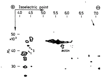
Though some individual variations of spots were ob- served on two-dimensional gel electrophoretograms, several consistent differences were elucidated be- tween five patients and five healthy volunteers (fig. l and fig. 2). The 41 kilodalton protein spot with an isoelectric point of 4.3 was completely absent in the electrophoretograms of all five patients (solid arrows l in fig. l and 2). Also a constant subtotal deficiency of the 42 kilodalton protein spot with an isoelectric point of 4.4 was found in the electrophoretogram of all five patients (solid arrows 2 in fig. l and 2). Mo- reover, the 57 kilodalton protein spot with isoelect- ric point of 6.8 was completely absent in patients (dotted arrows in figs. l and 2). The results of indi- vidual cases are also summarized in table 1.
'·· \
© Isoelectric point
40 4 5 5 0 5 5
I I I I
Θ
60
65
ι
70Tab. 1. Summary of erythrocyte membrane proteins of interest from ten test subjects.
Case No.
(Name)
1 (Kt)**
2 (Yt)**
3 ΓΠ) 4(Hw) 5 (Ski) 6 (Am) 7(Rm) 8(Ki) 9(Kn) 10 (Sku)
SexAge
625$22 616615 962 6 31929
£326 18 663
Diagnosis
PHP-1***
PHP-1 PHP-1 ΡΗΡ,Ι PHP-1 Normal Normal Normal Normal Normal
Abnormal protein (Λ/r/pI*) 41K/4.3
• r __ ****
——
—— ++
± +±
42K/4.4 57K/6.8 + —
— —- ±
± —
± —
± 4- 4- 4- + ± Φ ±
± +
* Isoelectric point.
** C se No. l and No. 2 are siblings.
*** Pseudohypoparathyroidism Type 1.
*** Relative intensity of protein Spots are presented in Symbols:
—, ±, + and Φ represents absent, barely detectable, detec- table and increased, respectively. The relative intensity of spots among each individual is determined by estimating the intensity in relation to the intensity of the actin zone n the same electrophoretogram.
50 -
actin
30 -
Fig. 1. Two-dimensional gel electrophoretogram of erythrocyte membrane proteins obtained from a healthy volunteer (case no. 6). The wide arrow indicates the position of ac- tin.
©Isoelectric point
Θ
Λ.Ο U5 5.0 5.5 6.0
ι ι ι ι ι
6.5ι 7.0ι
,10350 -
actin
30 - O
Fig. 2. Two-dimensional gel electrophoretogram of erythrocyte membrane proteins obtained from a patient with pseudo^
hypoparathyroidism type l (case no. 1). The wide arrow indicates the position of actin.
Discussion
The N-protein has recently been purified from r bbit liver plasma membranes (6, 7) s well s from turkey erythrocyte plasma membranes (8, 9). The protein complex isolated from rabbit liver plasma mem- branes consists of three iubun s with molecular weights of approximately 52, 45 and 35 kilodaltons.
Turkey erythrocyte membranes have only two sub- units with molecular weights of approximately 45 and 35 kilodaltons. The 45 kilodalton subunit has been proved to be a Substrate for ADP-ribosylation in the presence of cholera toxin. In the case of hu- man erythrocyte membrane, a 39 kilodalton protein was shown to be ADP-ribosylated in the presence of cholera toxin by Nielsen et l. (10). Kaslow et al. and
Cooper et al. reported that a 42 kilodalton proteinwas ADP-ribosylated in the presence of cholera tox- in (6) (11).
We report here a reduced concentration or absence of two proteins of this size (appr. 40 kilodaltons) in the erythrocytes of 5 patients with pseudohypopara*
thyroidism type 1. This would explain the deficient N-prptein ctivity found in the majority of patients with this disease (1). Moreover, the result that more than one protein spot is decreased or absent may be related to the observed heterogeneity of this dis- order (10, 12). When N-pfotein prepared from a murine lymphoma cell line is separated by two-di-
J. Clin. Chem. Clin. Biochem, / Vol. 22,1984 /No. 4
Matsumoto and Pujita: Erythrocyte membrane protein in pseudohypoparathyroidism
283 mensional gel electrophoresis, both 45 and 55 kilo-
dalton proteins are shown to be composed of several spots with approximate isoelectric points of 5.6 and 5.8, respectively (13). The reason for the difference of isoelectric point between the human erythrocyte and murine lymphoma cell proteins is not known, but it may be due to species and/or organ difference.
Two-dimensional polyacrylamide gel electrophoresis in combination with silver staining method accomp-
lished the highest resolution thus far obtained in ery- throcyte membrane protein analysis. Metabolie la- beling of erythrocyte membrane protein using ra- dioactive amino acids is not advisable due to the poor metabolic activity of erythrocytes. The electro- phoretic method described here, however, provides an effective strategy for research into any genetic disease abnormality which is expressed in erythro- cyte membrane protein, e.g. myotonic dystrophy and Duchenne-type muscular dystrophy (14).
References
1. Levine, M. A., Downs, R. W. Jr„ Singer, M., Marx, S. J., Aurbach, G. D. & Spiegel, A. M. (1980) Biochem. Biophys.
Res. Commun. 94, 1319-1324.
2. Farfel, Z., Brickman, A. S., Kaslow, H. R., Brothers, V. M. &
Bourne, H. R. (1980) New Engl. J. Med. 303, 237-242.
3. Dodge, J. T., Mitchell, C. & Hanahan, D. J. (1963) Arch.
Biochem. Biophys. 100, 119-130.
4. O'Farrell, P. H. (1975) J. Biol. Chem. 250, 4007-4021.
5. Merril, C. R., Goldman, D., Sedman, S. A., Ebert, M. H.
(1981) Science 277, 1437-1438.
6. Kaslow, H. R., Johnson, G. L., Brothers, V., Bourne, H. R.
(1978) Mol. Pharmacol. 75, 472-483.
7. Sternweis, P. C., Northup, J. K., Smigel, M. D., Schleifer, L.
S., Gilman, A. G. (1981) J. Biol. Chem. 256, 11517-11526.
8. Northup, J. K., Sternweis, P. C., Smigel, M. D., Schleifer, L.
S., ROSS, E. M., Gilman, A. G. (1980) Proc. Natl. Acad. Sei.
U.S.A 77, 6516-6520.
9. Sternweis, P. C., Northup, J. K., Hanski, E., Schleifer, L. S., Smigel, M. D., Gilman, A. G. (1981) Adv. Cyclic Nucleotide Res. 14, 23-36.
10. Nielsen, T. B A» Lad, P. M., Rreston, M. S., Rodbell, M.
(1980) Biochim. Biophys. Acta 629, 143-155.
11. Cooper, D. M. F., Jagus, R., Somers, R. L., Rodbell, M.
(1981) Biochem. Biophys. Res. Commun. 707, 1179-1185.
12. Rodbell, M. (1980) Nature 284, 17'-22.
13. Schleifer, L. S., Garrison, J. C., Sternweis, P. C., Northup, J.
K., Gilman, A. G. (1980) J. Biol. Chem. 255, 2641-2644.
14. Brown, H. D., Chattopadhyay, S. K., Patel, A. B. (1967) Science 757, 1577-1578.
Dr. Akira Matsumoto Dept. of Pathology Kyoto University Faculty of Medicine Joshida-Konoe Sakyo-ku
Kyoto, Japan, 606
J. Clin. Chem. Clin. Biochem. / Vol. 22, 1984 / No. 4