der Fakultät für Medizin der Universität Regensburg
The Endocannabinoid System
Exerts Anti-Inflammatory and Pro-Apoptotic Effects on Synovial Fibroblasts in Rheumatoid
Arthritis
Inaugural-Dissertation zur Erlangung des Doktorgrades
der Medizin
der
Fakultät für Medizin der Universität Regensburg
vorgelegt von Martin Apitz
2015
1. Berichterstatter: Prof. Dr. Rainer H. Straub
2. Berichterstatter: Prof. Dr. Dr. h.c. Joachim Grifka Tag der mündlichen Prüfung: 10.11.2015
1 Deutsche Zusammenfassung 1
2 Introduction 5
2.1 Rheumatoid Arthritis: Think Global, Act Local . . . 5
2.1.1 General Definition . . . 5
2.1.2 Epidemiological and Economic Relevance . . . 5
2.2 Pathogenesis of Rheumatoid Arthritis . . . 8
2.2.1 Prearthritic Phase . . . 8
2.2.2 Loss of Tolerance Phase . . . 10
2.2.3 Arthritic Phase . . . 12
2.2.4 Conclusion: Importance of Fibroblasts . . . 15
2.3 The Role of Synoviocytes in Rheumatoid Arthritis . . . . 16
2.3.1 Training the Bad Boy: Proliferation, Cell Death and Destruction . . . 16
2.3.2 The Inner Ambition: MAPK, CREB and Their Role in Survival, Invasion and Destruction . . . 20
2.3.3 Conclusion: Synoviocytes as a Therapeutic Target . 23 2.4 The Endocannabinoid System . . . 24
2.4.1 The Cannabinoid Receptors . . . 24
2.4.2 The Endogenous Ligands . . . 25
2.4.3 General Effects . . . 26
2.4.4 Conclusion: Using Complexity . . . 27
2.5 Conclusion: Synergy is Energy . . . 28
2.6 Purpose and Goal: From Pathogenesis to Treatment . . . 28
i
3 Materials and Methods 29
3.1 Patients . . . 32
3.2 Cell Culture . . . 33
3.2.1 Tissue Preparation . . . 33
3.2.2 Cryopreservation and Thawing . . . 33
3.2.3 Cell Culturing . . . 34
3.3 Detection of Intracelluar Activation of ERK, p38, c-JUN and CREB . . . 34
3.4 LDH Cytoxicity Detection . . . 35
3.5 Flow Cytometry with Annexin V and Propidium Iodide . 35 3.6 Proteome Profiler: Human Phosphokinase, Apoptosis and Human Cytokine Kit . . . 35
3.7 Statistical Analysis . . . 36
4 Results and Discussion 37 4.1 The Predominance of ERK . . . 37
4.2 The Antagonistic Effects of CB1 and CB2 on p38 . . . 38
4.3 The Antagonistic Effects of Anandamide . . . 42
4.3.1 Blocking the Recycling of AEA with JNJ Decreased MAPK Phosphorylation . . . 42
4.3.2 Repression of Downstream Proteins . . . 43
4.4 Conclusion: The Activation Pattern of AEA Resembled CB1 and CB2 . . . 45
4.5 Activation of MAPK Mainly by CB1 in Rheumatoid Arthri- tis Synovial Fibroblasts . . . 46
4.5.1 Rheumatoid Arthritis Synovial Fibroblasts Are More Prone to Stimulation . . . 46
4.5.2 The CB2 Agonist Activated ERK and CREB only in Rheumatoid Arthritis Synovial Fibroblasts . . . 46
4.5.3 The CB2 Agonist and the FAAH Inhibitor JNJ Ac- tivated ERK but Suppressed p38 . . . 46
4.5.4 AEA and the CB1 Agonist Showed Broadest Acti- vation of MAPK . . . 48
4.6 Conclusion: CB1 Agonism is Mainly Responsible for Ac-
tivation of MAPK and CREB in Synovial Fibroblasts . . 50 4.7 Consequences on Apoptosis and Cytokine Secretion . . . 50 4.7.1 CB1 Agonism Induced Apoptosis and Necrosis . . . 50 4.7.2 AEA-Attenuated Expression of Key Cytokines Af-
ter Stimulation by TNF𝛼 . . . 51 4.8 Pathogenesis: The ECS and Immune System . . . 55 4.9 Therapy: CB2Agonism and the Anti-Inflammatory Treat-
ment . . . 55
5 Summary and Outlook 57
Abbreviations 59
List of Figures 61
List of Tables 63
Bibliography 65
Die Rheumatoide Arthritis ist eine häufige Autoimmunerkrankung, die 0,5-1 % der erwachsenen Bevölkerung betrifft und als chronische Gelenkentzündung zu fortschreitender Gelenkzerstörung und systemi- schen Symptomen wie z.B. das metabolisches Syndrom und Athero- sklerose führt. In den letzten Jahrzehnten wurde das pathogenetische Verständnis der Rheumatoiden Arthritis auf Nicht-Immunzellen erwei- tert. So besetzen Fibroblasten der artikulären Synovia eine heraus- ragende Stellung in der Entwicklung der Gelenkentzündung. Im Zu- sammenspiel mit Immunzellen und Osteoklasten entwickelt sich im chronisch entzündeten Gelenk ein Gewebetumor aus tumorös verän- derten Fibroblasten, der Pannus. Diese veränderten Fibroblasten sind verantwortlich für die Synthese von Matrix-Metalloproteasen MMP, die Kapsel- und Knorpelmatrix degradieren. Die Fibroblasten hal- ten die lokale Arthritis aufrecht und koordinieren die Zerstörung an- grenzenden Knochens durch die Osteoklasten. Die Unterbindung im- munologischer Kommunikation durch sogenannte Biologika wie z.B.
TNF𝛼-Hemmer, hat das therapeutische Ergebnis deutlich verbessert, die therapeutischen Kosten jedoch auch deutlich erhöht und schwer- wiegende Nebenwirkungen der Immunsuppression eingeführt. Deshalb besteht weiterhin die Notwendigkeit, alternative und adjuvante Thera- pien zu entwickeln, die nicht nur auf das Immunsystem sondern auch auf nicht-immunologische Zellen wie die Fibroblasten abzielen. Einer der wichtigsten therapeutischen Ergebnisse muss es sein, dass diese Fi- broblasten weniger aggressiv wachsen und weniger gewebezerstörende MMP bilden. Intrazellulär wird die Synthese von MMP hauptsäch-
1
lich durch mitogen-aktivierte Proteinkinasen MAPK gesteuert. Dieses System besteht aus Proteinkinasen, die sich nach extra- oder intrazel- lulärem Signal konsekutiv und exponentiell aktivieren. Grundsätzlich lassen sich hierbei drei Kinase-Achsen unterscheiden: die Extracellular- Signal Regulated Kinase ERK, die hauptsächliche für Zellwachstum und Zelldifferenzierung verantwortlich ist, sowie die p38 MAPK und c-Jun N-Terminal Kinase JNK, die die zelluläre Antwort auf Stress ko- ordinieren. Assoziiert ist das cAMP-responsive Binding Protein CREB, das ebenso durch zellulären Stress aktiviert wird.
Somit kann ein therapeutischer Ansatz die Reduktion der Aktivität des MAPK-System in den synovialen Fibroblasten sein, um damit die Produktion von MMP zu reduzieren und die Gelenkszerstörung zu ver- langsamen.
Das endogene Cannabinoidsystem ECS wurde im Zuge der Cannabis- Forschung entdeckt und stellt ein komplexes Signalsystem aus Lipiden dar. Es besteht neben den Cannabinoid-Rezeptoren CB1 und CB2 aus den endogen produzierten Liganden Anandamid und 2-Arachidonylgly- cerol. Dieses System moduliert sowohl zentralnervös die Nahrungsauf- nahme als auch peripher das Immunsystem. Es konnte gezeigt werden, dass synthetisch hergestellte Cannabinoide in Immunzellen und Nicht- Immunzellen einiger chronischen Entzündungen, wie der Rheumatoi- de Arthritis, suppressiv auf die Aktivität der MAPK und Produktion von MMP wirken. Hingegen wurde die Rolle der endogen produzier- ten Liganden in der Rheumatoiden Arthritis bislang nur unzureichend erforscht.
Diese Arbeit verwendete humane Fibroblasten aus Patienten mit Rheu- matoider Arthritis oder degenerativer Arthrose. Im normoxischen und hypoxischen Umfeld wurden diese Zellen mit selektiven CB1- und CB2- Agonisten sowie dem endogen produzierten Anandamid vorbehandelt und zuletzt mit den pro-inflammatorischen Zytokinen TNF𝛼, Inter- leukin-1𝛽 oder dem Endotoxin Lipopolysaccharid stimuliert. Die Ak- tivität der intrazellulären MAPK ERK, p38, c-Jun und der cAMP- responsive Binding Protein CREB wurde mittels Enzyme-linked Im-
munosorbent Assay ELISA und Proteome Profiler™ gemessen. Des weiteren wurde der Anteil apoptotischer und nekrotischer Zellen mit- tels Durchflusszytometrie und Messung der Laktatdehydrogenase so- wie die Synthese einiger immunmodulatorischer Zytokine mit dem Pro- teome Profiler™ bestimmt.
Diese Arbeit zeigte, dass der CB1-Agonist die Aktivität der MAPK nach Zytokin-Stimulation signifikant erhöhte, der CB2-Agonist jedoch die Aktivität von MAPK und CREB signifikant erniedrigte. Das en- dogen produzierte Anandamid wirkte in hohen Konzentration (1 µm) wie der CB1-Agonist pro-inflammatorisch, in niedrigen Konzentration (0.1 nm) jedoch wie der CB2-Agonist anti-inflammatorisch. Ananda-
mid konnte ebenso die Produktion von pro-inflammatorischen Zytoki- nen nach Stimulation mit TNF𝛼 reduzieren. Des weiteren induzierten sowohl der CB1-Agonist als auch Anandamid Apoptose und Nekrose in den Fibroblasten.
Erstmals konnte in dieser Arbeit gezeigt werden, dass das endogen produzierte Cannabinoid Anandamid konzentrationsabhängig entwe- der pro- oder anti-inflammatorisch auf synoviale Fibroblasten wirkt.
Mögliche adjuvante Therapien mit Cannabinoiden gegen die Rheuma- toide Arthritis müssen somit die lokale Konzentration von endogenen Cannabinoiden und daher die Aktivität des ECS in der Arthritis be- rücksichtigen. Eine klare Aussage ist in Anbetracht der Komplexität des ECS sowie fehlender in-vivo-Experimente noch nicht möglich. Je- doch stellt die Modulation des ECS mit seinen vielfältigen Wirkun- gen nicht nur auf Immunzellen, sondern auch auf Nicht-Immunzellen, wie die Fibroblasten, ein mögliches zukünftiges Therapieziel gegen die Rheumatoide Arthritis dar.
2.1 Rheumatoid Arthritis: Think Global, Act Local 2.1.1 General Definition
Rheumatoid arthritis is a chronic, autoimmune-induced subset of diseases of yet-unknown origin, which is characterised by
a systemic inflammation, with antibody production as well as pul- monary or cardiovascular complications and
a local inflammation, in the joint with cartilage and bone destruction of small joints of hand and feet,
(McInnes et al., 2011) which altogether leads foremost to debilitating deformities of hands and feet, alongside a 3.17-fold risk of myocardial infarction (Maradit-Kremers et al., 2005) and stroke events, both with more lethal outcome (Davis et al., 2008). These extra-articular complications are largly responsible for higher mortality ratios ranging from 1.28 to 2.98, compared with the normal population (Gabriel et al., 2009).
2.1.2 Epidemiological and Economic Relevance
Rheumatoid arthritis affects around 0.5-1 % of the adult population in developed countries (Gabriel et al., 2009). Due to the long-term therapy required, the debilitating course of the disease and the high co- morbidity, mean healthcare expenditures per patient with RA per year in Europe and Northern America are estimated to be AC 4000-6000. In Germany, expenditures for patients with RA range from AC 2437–2981
5
annually per patient of working age, to an estimated AC 2121 per year for patients in retirement, as of 2002 (Furneri et al., 2012; Huscher et al., 2014).
Although the new biological therapeutics have overall reduced disease activity and, concomitantly, hospitalisation and work absence, the eco- nomic benefit was counterbalanced by 3- to 6-fold increases in treat- ment cost (Ziegler et al., 2010; ter Wee et al., 2012; Hagel et al., 2013).
Despite these positive trends in the therapeutic outcome, functional debilitation and thus work absence are still severe in rheumatoid arthri- tis, so that these patients still rank among patients with chronic is- chaemic heart disease and multiple sclerosis, with respect to reduction of health utility in quality of life (Huscher et al., 2006; Furneri et al., 2012).
These stastistics demonstrate an ongoing need for new, less expensive combinations of treatments to reduce climbing expenditures and eco- nomic burden, to help decreasing disease activity, and to foster quality of life and long-term survival for patients with rheumatoid arthritis.
Figure 2.1: The Multistep Pathogenesis of Rheumatoid Arthritis (McInneset al., 2011). ACPA denotes anti-citrullinated peptide antibodies and RF rheumatoid factor. Silica Dustwas added to the original picture.
2.2 Pathogenesis of Rheumatoid Arthritis
In recent years, our insight into the pathogenesis and etiology of RA has increased tremendously. With different clinical presentations and distinct genetic and environmental risk factors, RA can now be seen as a group of different diseases that share a common final pathway of inflammation and joint destruction (Scott et al., 2010; van der Helm-van Mil et al., 2008).
Although a comprehensive pathogenetic model has not yet been de- veloped, evidence is now enlarging the picture and including mes- enchymal cells like fibroblasts and osteoclasts in the inflammatory process. McInnes and Schett (McInnes et al., 2011) present three crucial phases in the development of RA (Figure 2.1).
Prearthritic Phase: Genetic predisposition, environmental exposure and combined epigenetic alteration lead to autonomous protein citrul- lination.
Loss of Tolerance Phase: Auto-antibodies are processed and secreted, especially ACPA against citrullinated proteins.
Arthritic Phase: Further biomechanical, microvascular, infectious or neuroendocrinological factors result in the clinical presentation of local and systemic inflammation.
2.2.1 Prearthritic Phase
Genetic Predisposition Twin studies revealed concordance rates of 15- 30 % in monozygotic twins against 5 % in dizygotic twins (MacGre- gor et al., 2000). Currently, the most important filial risk factor are HLA-DR𝛽 chain alleles that have the common protein sequence QKRAA, which is called theshared epitope (Gregersen et al., 1987).
The biological link to an increased susceptibility is still unknown. How- ever, the shared epitope works as a signal-transducing ligand for cal- reticulin in leukocytes, which plays a vital role in the elimination of apoptotic cells and can therefore be regarded as crucial for the devel-
Figure 2.2: The Mechanism of Epigenetics in Rheumatoid Arthritis (Grabiecet al., 2013)
opment of peripheral autoimmunity (Almeida et al., 2010). Further risk factors for rheumatoid arthritis include genetic modifications in the STAT4 signaling pathway (Remmers et al., 2007) and in T-cell activation, e.g. via PTPN22 (Begovich et al., 2004). As the low concordance rates for mono- and dizygotic twins above suggest, the exposure to diverse environmental factors mainly influence the patho- genesis and onset of RA.
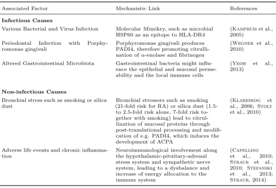
Environmental Influence In addition, a host of infectious and non- infectious causes have been marked as risk factors for developing rheuma- toid arthritis (see Table 2.1). Particularly, environmental exposure to bronchial stress such as smoking or silica dust increases the risk of developing RA in patients with susceptibility HLA alleles, such as HLA-DR1 and the shared epitope sequence or other HLA-DR4 alleles (Klareskog et al., 2006; Källberg et al., 2011). The bronchial stressors might induce citrullination of mucosal proteins through post- translational processing and modification of e.g. the peptidyl argi- nine deiminase type IV PADI4, which is required for ACPA develop- ment, the most specific auto-antibody involved in RA (McInnes et al., 2011). Also, the new field of epigenetics might provide insight into
this field of gen-environment interactions in the future.
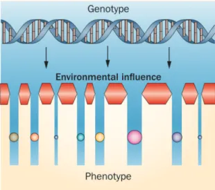
Linking together: Epigenetics The transcription is regulated mainly by the three mechanisms: DNA methylation, histone acetylation and miRNA expression. These so-called epigenetic changes are responsible for the access and binding of transcription factors and therefore create a phenotype without altering the DNA code (Grabiec et al., 2013).
They are considered crucial in two ways (Bottini et al., 2013):
Etiologically, they provide a linkage mechanism: they explain how en- vironmental exposure over time and family genetic predispositions create an individual risk for rheumatoid arthritis. The epigenetic machinery functions as a filter, which selects gene expression and therefore the cell phenotype, depending on the environmental cir- cumstances (Figure 2.2).
Pathogenically, they are involved in the transformation of leukocytes and especially synovial fibroblasts into aggressive behaviour, which is described in detail in Section 2.3.
As a consequence, external influences and internal genetic predisposi- tions in the development of RA cannot be clearly differentiated. The personal and local environment can have a long-term impact on the in- ternal genetic susceptibility to RA, therefore modulating its individual risk and variability.
2.2.2 Loss of Tolerance Phase
Citrullination 𝛼-Enolase, keratin, fibrinogen, fibronectin, collagen, and vimentin are prone to self-citrullination, against which ACPA anti- bodies are secreted. Citrullinated 𝛼-enolase is highly associated with ACPA-positive RA as well as the risk factors described in Section 2.2.1 (Mahdi et al., 2009). ACPA itself has been shown to be one of the
highest predictors of early-onset RA (van der Linden et al., 2009).
Transition to Arthritic Phase It remains unclear why the systemic loss of tolerance, alongside with the prodution of auto-antibodies such
Table 2.1: Environmental Risk Factors for Rheumatoid Arthritis
Associated Factor Mechanistic Link References
Infectious Causes
Various Bacterial and Virus Infection Molecular Mimikry, such as microbial
HSP60 as an epitope to HLA-DR4 (Kamphuiset al., 2005)
Periodontal Infection with Porphy-
romonas gingivali Porphyromonas gingivali produces
PADI4, therefore promoting citrulli- nation of𝛼-enolase and fibrinogen
(Wegner et al., 2010)
Altered Gastrointestinal Microbiota Gastrointestinal bacteria might influ- ence the epithelial and mucosal perme- ability and the local immune cells
(Yeoh et al., 2013)
Non-infectious Causes
Bronchial stress such as smoking or silica
dust Bronchial stressors such as smoking
(21-fold risk for RA) or silica dust (1.5- to 2.5-fold risk alone, 7-fold risk to- gether with smoking) lead to citrul- lination of mucosal proteins through post-translational processing and modifi- cation of e.g. PADI4, which induces the development of ACPA
(Klareskog et al., 2006; Stolt et al., 2010)
Adverse life events and chronic inflamma-
tion Neuroimmunological involvement along
the hypothalamic-pituitary-adrenal stress system and sympathetic nerve system, leading to a dysbalance and increase of energy allocation to the immune system
(Capellino et al., 2010;
Straub et al., 2010; Stefanski et al., 2013;
Straub, 2014)
as ACPA and RF, particularly affects small joints of metacarpalia and metatarsalia. Biomechanical characteristics of these joints can have an influence on the local microcirculation and protein metabolism (McInnes et al., 2011). Straub et al. (Straub, 2007, 2014) proprose a close connection between the onset and course of inflammation as well as the energy allocation. In short-term inflammatory conditions the systemic energy allocation shifts to the immune system. In chronic inflammatory conditions, the energy regulation becomes impaired by means of permanent high levels of pro-inflammatory cytokines and high activity of the sympathetic nerve system and hypothalamic-pitui- tary-adrenal axis with high cortisol level (Straub et al., 2010). The imbalance of the energy expenditure pathways contributes to common systemic features of RA such as diabetes mellitus and insulin resistance or the development of obesity and muscle wasting, since these neuro- humoral pathways meet the increased glucose demand for the immune system (Straub et al., 2010). Also, the sympathetic nerve system contributes to the local arthritic activity time-dependently and there-
fore establishes a connection between the central nervous system and the local immune system, in the joint as well as in secondary lymphatic organs (Schaible et al., 2014). It directly exerts a pro-inflammatory effect in early arthritis, but exerts an anti-inflammatory effect in later arthritic stages (Härleet al., 2005). Since sympathetic nerve density in the joint and the lymphoid organs is reduced in late arthritis, the influence of the central nervous system together with its control of energy allocation might become lost in later arthritis and replaced by alternative regulatory mechanisms such as tyrosine-hydroxylase posi- tive cells (Schaible et al., 2014; Pongratz et al., 2013).
2.2.3 Arthritic Phase
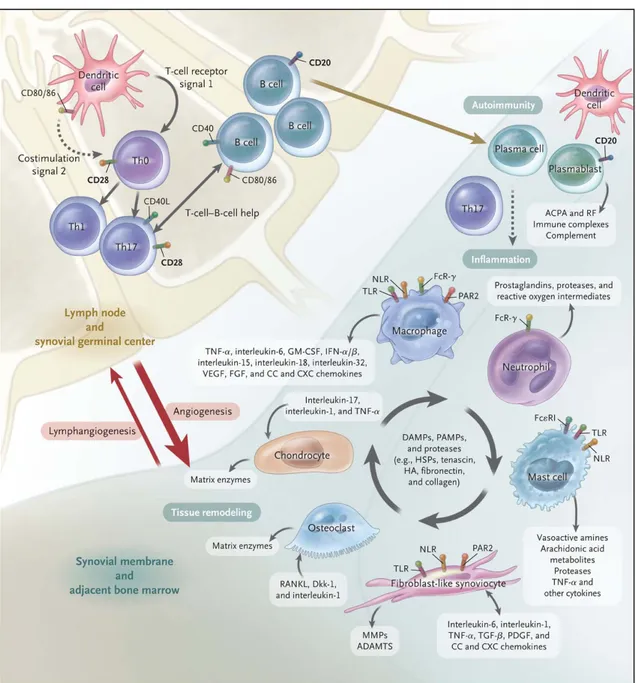
The main characteristic of RA is the highly inflamed joint containing high amounts of pro-inflammatory cytokines and tissue-degrading en- zymes (Scott et al., 2010). Three parties can be identified as main factors in the development of arthritis: leukocytes, fibroblasts and osteoclasts (Figure 2.3).
The Instigators: Leukocytes Regarding the adaptive immune system, expanding synovial myeloid and dendritic cells create a T-cell promot- ing environment and dominance of TH-17 cells, whose secreted IL-17 - together with TNF𝛼 - activates fibroblasts, chondrocytes and osteo- clasts to produce tissue-degrading enzymes, cytokines such as IL-6, chemokines and growth factors (Miossec et al., 2009; Hot et al., 2011).
Furthermore, autoreactive T-cells against citrullinated protein have been found, suggesting that the cellular immune system is directly in- volved (Cantaert et al., 2009).
B-cells are not only responsible for the production of auto-antibodies, but also for the presentation of the auto-antigen and for cytokine pro- duction itself (McInnes et al., 2011).
Macrophages help to sustain inflammation by cytokine expression and antigen presentation. They directly destroy tissue by producing reac-
Figure 2.3: Three vital players in Rheumatoid Arthritis. ADAMTS denotes a disintegrin and metalloprotease with thrombospondin-1–like domains, DAMP damage-associated molecular pattern, Dkk-1 dickkopf-1, FcR Fc receptor, Fc𝜀RI high-affinity IgE receptor, FGF fibroblast growth factor, GM-CSF granulocyte–macrophage colony-stimulating factor, HA hyaluronan, HSP heat-shock protein, IFN-𝛼/𝛽interferon-𝛼/𝛽, MMP matrix metalloproteinase, NLR nucleotide-binding oligomerization domain–like receptor, PAMP pathogen-associated molecular pattern, PAR2 protease-activated receptor 2, PDGF platelet-derived growth factor, RANKL receptor activator of nuclear factor𝜅B ligand, TGF-𝛽 transforming growth factor𝛽, Th0 type 0 helper T cell, Th1 type 1 helper T cell, Th17 type 17 helper T cell, TLR toll-like receptor, TNF-𝛼tumor necrosis factor𝛼, and VEGF vascular endothelial growth factor (McInneset al., 2011).
tive oxygen intermediates and degrading enzymes (Haringmanet al., 2005; McInnes et al., 2011).
Neutrophil Granulocytes are predominant in the synovial fluid, where they support inflammation and sustain cartilage destruction with a host of cytokines, reactive oxygen species and proteases (Eyles et al., 2006; Cornish et al., 2009).
In conclusion, leukocytes generate the highly inflammatory and hy- poxic environment that is regarded as the foundation for the develop- ment of aggressive fibroblasts.
The Culprits: Fibroblasts The joint capsule and the bone are attached to each other by the synovium, which is organised into a dense lining layer and a loose sublining stroma. Fibroblasts are of mesenchymal ori- gin and tasked with producing and modelling the extracellular matrix of the organic stroma. Thereby, they provide a scaffold for the spe- cialised cells in almost every organ, maintain extracellular homeostasis and integrate stimuli on the microenvironment such as pH reduction or oxygenic stress due to inflammation (Firestein et al., 2012).
The fibroblasts compose the synovial lining without a basement mem- brane, tight junctions or desmosomes, yet they are riveted together by cadherins such as cadherin-11, and various integrins and receptors of the Ig superfamily, e.g. ICAM and VCAM. These cellular structures play a key role not only in the attachment of the synovial lining, but also in the intracellular signaling and in the regulation of inflamma- tion (Lee et al., 2007; Chang et al., 2011).
In later arthritic stages, the synovial lining undergoes a dramatic inva- sion of T cells, dendritic cells and B-cells forming germinal centers, and particularly macrophage-like and fibroblast-like synoviocytes (Take- mura et al., 2001). All of them migrate, proliferate and increase local joint cellularity by a ten-fold (Firestein et al., 2012). Thereby, the arthritic environment is marked by pro-angiogenic factors and is extensively vascularised. But it remains hypoxic, produces reactive oxygen species and creates chronic oxidative stress on resident fibrob-
lasts (Taylor et al., 2005).
While the synovitis unfolds, Firestein (Firestein, 2003) suggest a direct interaction of macrophage-like synoviocytes and fibroblast-like synoviocytes under the regulation of primarily T-cells. This interac- tion has turned out to be more complex, since the innate immune system and adjacent bone and cartilage cells are involved as well. The initial site and onset mode of joint arthritis are still unknown.
The persistently inflamed and hypoxic environment changes the syn- oviocytes, which in turn proliferate and amass a structure called pan- nus, at which cartilage, ligaments and bone masses are progressively damaged.
The Masons: Osteoclasts Two theories were prompted by Schett and Firestein on the topic of fibroblast-osteoclast relationships (Schett et al., 2010). Depending on the first affected site, synovial inflamma- tion can spread to the bone marrow and induce osteitis. Otherwise, primary osteitis can expand to the joint space and induce synovitis.
Kleyer et al. (Kleyer et al., 2014) have coined these differing con- cepts as the hen-egg dilemma. Accordingly, further research is required to discern the prearthritic phase as local or systemic.
2.2.4 Conclusion: Importance of Fibroblasts
In conclusion, fibroblast-like synoviocytes play a vital role in the development and progression of rheumatoid arthritis and are the key players in local joint and bone destruction, alongside the immune sys- tem. Although RA is an autoimmune disease, not only should the modulation and suppression of leukocytic activity be considered for therapeutic intervention, but, eventually, decreasing the activity of fi- broblasts and osteoclasts may be key to an effective therapeutic strat- egy.
2.3 The Role of Synoviocytes in Rheumatoid Arthritis
2.3.1 Training the Bad Boy: Proliferation, Cell Death and Destruction
Fibroblast-like synoviocytes are also called synovial fibroblasts (SF) and are key factors in the maintenance of local inflammation as well as in the destruction of adjacent bone and cartilage structures.
Whereas macrophage-like synoviocytes produce a range of pro-inflam- matory cytokines, chemokines and growth factors, fibroblast-like syn- oviocytes respond with the secretion of their own host of cytokines, such as IL-6, and matrix degrading enzymes. The autocrine and paracrine circle sustains inflammation and fosters proliferation of syn- ovial tissue to a pannus at the cartilage-bone borders (Figure 2.3 and 2.4).
In the course of arthritis, fibroblast-like synoviocytes respond to the highly inflammatory environment by a changed behaviour in gene ex- pression and cell characteristics. Effectively, they are trained to be imprinted aggressors (Bottini et al., 2013).
The pannus is composed of macrophages, osteoclasts and fibroblast- like synoviocytes and shows characteristics of a local invasive tumour with ongoing destruction ofcartilage surface by fibroblast-like synovio- cytes and bone surface by osteoclasts (Figure 2.3).
The Boot Camp: Inflammatory Synovia Fibroblast-like synoviocytes play the central role in sustenance of inflammation and in bone and cartilage destruction (Leeet al., 2007). They are part of the cytokine cycle and are one of the essential sources of IL-6 in arthritis (Nishi- moto et al., 2004).
IL-6 and TNF𝛼, as well, are pleiotropic cytokines that moderate the immunological response in inflammation, haematopoesis and oncolog- ical diseases (Kishimoto, 1989; Nishimoto et al., 2000). IL-6 medi- ates between endothelial cells and SF, which, in turn, induce greater neutrophil recruitment in the synovia (Lally et al., 2005). SF are
Figure 2.4: Autocrine and Paracrine Cytokine Network in the Synovial Layer. FGF denotes fibroblast growth factor, GM-CSF granulocyte macrophage colony-stimulating factor, IFN-𝛾interferon𝛾, IL interleukin, IL-1Ra interleukin 1 receptor antagonist, M-CSF macrophage colony-stimulating factor, sTNF-R soluble tumor necrosis factor receptor, TGF-𝛽 transforming growth factor𝛽 and TNF-𝛼 tumor necrosis factor𝛼(Firestein, 2003).
responsible for the chemoattraction of monocytes by means of CCL2 (MCP-1) secretion (Villiger et al., 1992) and for T-cell recruitment
by means of CCL5 secretion (Patel et al., 2001).
These properties reflect their physiological role in the selection and recruitment of leukocytes and in the survival and differentiation of, for example, B cells, which are dependent on IL-6 stimulation by fi- broblasts in their immature stage (Firestein et al., 2012).
Fibroblasts are paramount in the prolonged survival and retention of leukocytes, as they produce Interferon-𝛼 and Interferon-𝛽 (Buckley et al., 2001), CXCL12 and its receptor CXCR4 (Burger et al., 2001;
Ohata et al., 2005), CXCL13 and its receptor CXCR5 (Amft et al., 2001) and likely other constitutive chemokines for lymphoid aggrega- tion and organisation in the synovial sublining. In essence, they are not only trainees in the inflammatory boot camp, but they also train immune cells to establish synovitis.
Lesson One: Survive and Spread Cell proliferation and cell death are crucial for developmental and homeostatic processes. In recent years, the range of possible modes of cell death has dramatically expanded.
The two most important modes are apoptosis and necrosis. Apoptosis is characterised by a regulated deconstruction of the cell, resulting in
nuclear condensation and fragmentation, followed by scattering of the cell (Mariño et al., 2014). On the other hand, necrosis is marked by cellular leakage and swelling (Vanden Berghe et al., 2014). These two forms cannot be distinctively separated, since other forms of cell death such as necroptosis, a more regulated form of necrosis, have been discovered (Vanden Berghe et al., 2014).
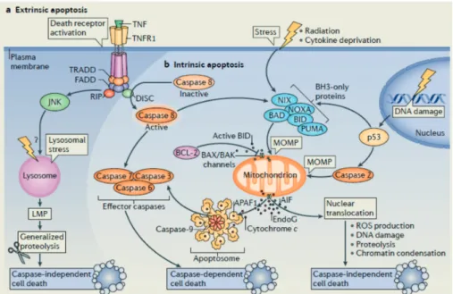
The apoptotic cell death is induced by either extrinsic or intrinsic fac- tors (Figure 2.5). The CD95 (Fas) ligand binds to its receptor, CD95, and activates several proteins, which in turn cleave the aspartat pro- tease pro-caspase 8 into its active form. In a similar mechanism TNF binds to the TNF-related apoptosis-inducing ligand (TRAIL) receptor, which also activates caspase 8. Caspase 8 triggers and orchestrates fur- ther caspases and causes the mitochondrial outer membrane permeabi- lization (MOMP). The mitochondrial leakage releases cytochrome C, which is responsible for the assembly of caspase 9 to the apoptosome.
MOMP is directly induced by cellular stress, e.g. nitroxic radicals or radiation.
Though fibroblast-like synoviocytes in RA are under inflammatory, hy- poxic and nitrogenic stress, they show a reduced sensitivity to apop- tosis. This is because chronic inflammation, in particular, changes behaviour of SF in the intimal synovial lining to a tumorous charac- ter:
Bcl-2 and mcl-2 expression increases, which serves as an anti-apoptotic signal (Matsumoto et al., 1996; Perlman et al., 2000; Liu et al., 2005).
CD95/Fas is widely expressed in RA synovia, although SF show a resistance to CD95 treatment (Imamura et al., 1998). This resis- tance continues in vitro (Meinecke et al., 2007).
P53 undergoes somatic mutation similar to many tumours (Firestein et al., 1997; Yamanishi et al., 2002).
In sum, somatic mutation of genetic content and epigenetic modu- lation of gene expression might explain certain responses by inflam-
matory signals in rheumatoid arthritis synovial fibroblasts (RASF), different from osteoarthritis synovial fibroblasts (OASF) or healthy control samples. Their resistance to apoptosis also changes the prolif- erative behaviour of SF. The morphological hallmark of this behaviour is the pannus.
Lesson Two: Invade and Infiltrate The pannus can be seen as the forefront of invasion into neighbouring structures, such as cartilage and bone. Fibroblast-like synoviocytes evade contact inhibition and substantially invade and destroy cartilage (Müller-Ladner et al., 1996), even beyond distances similar to metastasation (Lefèvre et al., 2009). The degree of fibroblast invasiveness correlates with the ra- diographic stage of joint destruction (Tolboom et al., 2005). There- fore, alteration of the invasive character of RASF is another step in the pathogenesis of RA.
Lesson Three: Destroy and Decay SF are crucial in the destruction of joint cartilage, as they produce degrading matrix metalloproteinases (MMP), of which
the secretable collagenases MMP-1, -8, -13;
the non-secretable membrane-anchored collagenases MMP-14 and -16 and
the stromelysins MMP-3 and -10
are mainly responsible for matrix degradation (Bartok et al., 2010;
McInnes et al., 2011; Firestein et al., 2012).
The secretion is triggered by cytokines including TNF𝛼, TGF𝛽 or IL-1𝛽. IL-1𝛽 is the strongest inducer of MMP secretion via activation of MAPK/AP-1, STAT and NF𝜅B pathways (Li et al., 2001; Meng- shol et al., 2000; Brauchle et al., 2000; Barchowsky et al., 2000).
Therefore, the reduction of MMP secretion by SF should be a main therapeutic target in RA. This might be achieved by a reduced se- cretion of these cytokines or by a reduced activation of intracellular signaling.
Figure 2.5: The extrinsic and intrinsic mechanism of apoptosis. AIF denotes apoptosis-inducing factor, APAF-1 apoptotic protease-activating factor1, BAK BCL-2 antagonist or killer, BAX BCL-2- associated X protein, BCL-2 b-cell lymphoma protein 2, BH3 BCL-2 homology 3, BID BH3-interacting domain death agonist, CD95 cluster of differentiation 95 or Fas receptor, DISC death-inducing signalling complex, DNA deoxyribonucleic acid, EndoG endonuclease G, FADD FAS-associated death domain, JNK JUN N-terminal kinase, MLMP lysosomal membrane permeabilization, MOMP mitochondrial outer membrane permeabilization , NIX NIP3-like protein X, PUMA and NOXA p53 upregulated modulator of apoptosis, RIP receptor-interacting protein, ROS reactive oxygen species, TNFR1 tumour necrosis factor receptor 1, TRADD TNFR1-associated death domain and TRAILR TNF-related apoptosis inducing ligand receptor (Mariñoet al., 2014).
2.3.2 The Inner Ambition: MAPK, CREB and Their Role in Survival, Invasion and Destruction
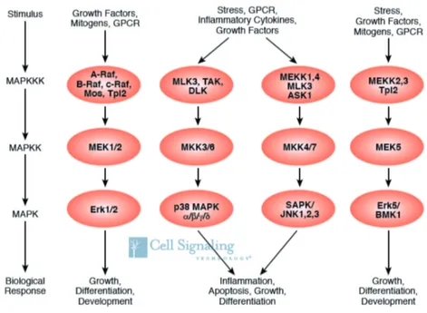
MAPK In RA fibroblasts, the intracellular system of mitogen-activated protein kinases is the most widely studied signaling pathway and vi- tal for the secretion of MMPs and inflammatory cytokines (Bottini et al., 2013). This highly conserved system consists of three distinct, but not completely separate, MAPK pathways: ERK, p38 and JNK (Figure 2.6) (Widmann et al., 1999).
After four levels of phosphorylation and activation of distinct kinases, phospholipases and cytosceletal proteins (Johnson et al., 2002), the kinases ERK, p38 and JNK enter the nucleus and activate transcrip- tion factors. By this means of shaping gene expression, ERK is mainly associated with cell proliferation and differentiation, whereas p38 and
JNK are involved in the cellular response to various forms of stress (Johnson et al., 2002). All three signaling pathways show higher ac-
tivity in RASF compared to OASF (Han et al., 1999; Schett et al., 2000), which might result from chronic inflammation and stress, but also from epigenetic changes in the genes of these proteins (Bottini et al., 2013).
ERK Signaling At least in part, ERK is activated by TNF in RASF, promotes proliferation and apoptosis (Görtz et al., 2005). Further- more, activation of ERK fosters the secretion of IL-6, IL-8 and MMP-3 (Calmon-Hamaty et al., 2011). The ERK pathway has also been shown to mediate IL-6 secretion after IL-1 stimulation of RASF (Dou et al., 2013).
JNK Signaling MMP secretion is mainly regulated by the MKK7- JNK-AP1 signaling axis (Bartok et al., 2010). Upon IL-1 stimu- lation, the kinase MKK7 is able to phosphorylate JNK, which itself activates c-JUN. Phopshorylated c-JUN binds to the FBJ murine os- teosarcoma viral oncogene homolog (FOS) and complements the AP1 transcription factor complex, which binds to specific MMP gene loci in RASF (Bottini et al., 2013). This is the reason why the secre- tion of the important MMP-1, -3 and -13 are dependent on the active JNK pathway (Han et al., 2001). JNK shows a higher activation upon stimulation with IL-1 in RASF, compared to OASF (Han et al., 1999).
p38 Signaling In addition to JNK, p38 is the pivotal regulator for fibroblast aggressiveness. Blocking the p38 pathway in RASF has revealed their involvement in MMP-1 and -3 production as well as IL- 6 and -8 secretion upon IL-1 stimulation (Westraet al., 2004;Inoue et al., 2005, 2006). The therapy of RA with p38 inhibitors did not show greater benefit compared with current regimens (Damjanov et al., 2009;Genoveseet al., 2011). This proves the complex and redundant role of the p38 signaling pathway, while heterotropic and even opposed
Figure 2.6: Overview of the MAPK system. ASK 1 denotes apoptosis signal-regulating kinase 1, BMK big MAPK kinase, DLK1 delta-like 1, ERK extracellular-signal regulated kinase, GPCR G protein-coupled receptor, JNK c-Jun N-terminal kinase, MAPK mitogen-activated protein kinase, MAPKK mitogen-activated protein kinase kinase, MAPKKK mitogen-activated protein kinase kinase kinase, MEK MAPK/ERK kinase, MEKK MAPK/ERK kinase kinase, MKK p38 MAPK kinase, MLK mixed-lineage kinase, Mos V-mos Moloney murine sarcoma viral oncogene homolog, p38 protein 38, Raf rat fibrosarcoma kinase, SAPK stress-activated protein kinase, TAK TGF𝛽-activated kinase and Tpl2 tumor progression locus 2 (Cell Signaling Technology, 2010).
function in fibroblasts and leukocytes have to be considered (McInnes et al., 2011; Bottini et al., 2013).
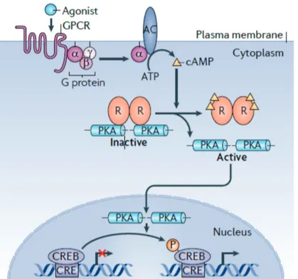
CREB pathway Another mechanism of altering gene expression by extracellular stimuli is the cAMP-responsive element binding protein (CREB). Upon stimulation by catecholamines, intracellular cyclic AMP
increases and protein kinase A is activated, which then phosphorylates CREB (Gonzalez et al., 1989). The MAPK ERK2 and p38 also are able to enhance CREB activity through the activation of mitogen- and stress-activated protein kinase MSK (Deak et al., 1998; Tan et al., 1996) and pp90(RSK) family RSK2 (Xing et al., 1996). As a result, these MAPK and CREB are able to induce IL-6 secretion after IL- 1 stimulation in RASF (Dou et al., 2013). Another mechanism of
CREB activation is the Ca2+ influx mediated by Ca2+/calmodulin- dependent kinases (Dash et al., 1991).
The active CREB binds the CREB-binding protein in the nucleus (Chrivia et al., 1993) and changes the expression of hundreds of genes affecting signaling, metabolism and proliferation (Impey et al., 2004).
Particularly in RASF, evidence of the role of the CREB pathway is rare. For example, phosphorylated CREB has been found to be higher in RASF than in OASF (Pérez-García et al., 2011) and to be func- tionally involved in the IL-6 secretion by RASF (Ishizu et al., 2010).
2.3.3 Conclusion: Synoviocytes as a Therapeutic Target The current success of biologicals in the treatment of RA proves that the modulation of the immunological process in rheumatoid arthritis is crucial for breaking the inflammatory cycle in the joint, but this might be not sufficient. Since synovial fibroblasts in RA show independent aggressive behaviour apart from immunological regulation, targeting them might be an effective adjuvant therapy. For example, Lee et al.
(Lee et al., 2007) showed the anti-inflammatory effect of a novel non- immunological antibody against cadherin-11, an important building block for the synovial layer and regulator of inflammatory responses in RASF (Chang et al., 2011). As Bottini et al. (Bottini et al., 2013) have noted, combination therapies with dual suppression both immunological cells and fibroblasts pave the way for innovative treat- ments. As one possibility, cannabinoids might offer such promising combinations.
Figure 2.7: Overview of the PKA-CREB-Pathway. AC denotes adenylyl cyclase, cAMP cyclic AMP, CRE/CREB cAMP-responsive element (CRE)-binding protein, GPCR G protein-coupled receptors, PKA protein kinase A and R regulatory subunits of PKA (Altarejoset al., 2011).
2.4 The Endocannabinoid System 2.4.1 The Cannabinoid Receptors
The Cannabinoid Receptors Cannabinoid Receptors CB1 and CB2 are G-protein coupled receptors, which, upon activation, induce the sec- ond messenger Gi/o protein to inhibit adenylate cyclase and to stimu- late MAP-Kinases. (Alexanderet al., 2011; Pertwee et al., 2010).
Cannabis research has been undertaken since the 19th century, when the psychoactive ingredients of cannabis were studied with interest (Iversen, 1999). The chemical synthesis of 𝛥9-tetrahydrocannabinol by Gaoni and Mechoulam was the gateway to serious scientific inves- tigation of the cannabinoids (Mechoulam et al., 1965).
Cannabis sativa contains over 60 substances, from which 𝛥9-tetra- hydrocannabinol are mostly responsible for psychoactivity (Mechou-
lam et al., 1970). The synthesis of 𝛥9-tetrahydrocannabinol ana- logues allowed for research in the molecular structure and physiologi- cal function of cannabinoids, e.g. in psychomotoric vigilance or body temperature. This research eventually led to the relevation of the until- then orphan receptor SKR6 to be the G-protein-coupled cannabinoid CB1 receptor (Gérard et al., 1990).
Subsequently, a second G-protein coupled cannabinoid receptor, CB2, was identified in the human promyelocytic leukaemic cell line HL 60 (Munro et al., 1993).
Non-Cannabinoid-Receptors: GPR55 and TRPV Besides the classi- cal CB-1 and -2 receptor, the endocannabinoid system also interact with the G protein-coupled receptor 55 (GPR55) and the transient receptor potential channels (TRP) such as for the vanilloid family TRPV (Alexander et al., 2011). GPR55 belongs to the GPCR su- perfamily like CB1 and CB2, but transmits its signal to Gq, G12 or G13 (Ryberg et al., 2007; Sharir et al., 2010; Alexander et al., 2011),
which generally activates downstream proteins like the GTPase Ras homolog gene family member A (RhoA) or phospholipase C. GPR55 has been reported to be involved in inflammatory pain and bone phys- iology (Sharir et al., 2010).
The TRPV receptors are divided into the two groups of thermosen- sitive non-selective cation channels TRPV1-4 and the calcium chan- nels TRPV5 and 6 (Alexander et al., 2011). Classical functions of TRPV include pain generation by heat (Starowicz et al., 2007) and regulation of the innate immune system, such as phagocytosis in macrophages (Link et al., 2010).
2.4.2 The Endogenous Ligands
The identification of receptors for tetrahydrocannabinol suggested endogenous ligands for these receptors. So far, two major endocannabi- noids have been identified: 2-arachidonylethanolamide (anandamide or AEA) and 2-arachidonylglycerol (2-AG) (Devaneet al., 1992; Me-
choulamet al., 1995). In general, these two agonists and the CB1 and CB2 receptor constitute the endogenous cannabinoid system (ECS).
2.4.3 General Effects
Since the discovery of AEA and 2-AG, the ECS has revealed to be a highly complex lipid system with distinct effects on neuronal and non-neuronal cells.
Generally, the CB1 receptor is mainly expressed in the central nervous system, whereas the CB2 receptor is widely expressed in immune cells (Pertwee et al., 2010). Changes in the activity of the endocannabi-
noid system have been shown to correlate with
neuronal conditions: pain and inflammation (Hohmann et al., 2006;
Jhaveri et al., 2007); neurological and neuropsychiatric condi- tions (Bisogno et al., 2007).
non-neuronal conditions: immunological (autoimmune and allergic) dis- orders (Oka et al., 2006); cancer (Bifulco et al., 2007); and gastrointestinal (Storr et al., 2007) and hepatic (Mallat et al., 2007) disorders.
interconnected conditions: obesity, metabolic syndrome (Matias et al., 2007) and cardiovascular disorders.
However, there have been contradictory results concerning the benefi- cial or detrimental effects of the endocannabinoids in the same tissue under a given pathological stress, which demonstrates the different interactions of the endocannabinoid system and the complexity of reg- ulation (Di Marzo et al., 2007; Pacher et al., 2006).
Considering effects on rheumatoid arthritis, evidence for a specific mode of action is rare. However, adjacent research fields of inflamma- tion and pain might provide some clue of the endocannabinoid influ- ence on RA. Thus, this introduction only presents the effect of endo- cannabinoids on inflammation and inflammatory pain in neuronal and non-neuronal systems.
Neuronal Effects: Inflammation and Pain When stressed by inflam- mation or irritants, levels of endocannabinoids in rodent skin, periph- eral nerves and spinal cord are elevated (Oka et al., 2006). Neu- ropathic pain also increases these levels (Mitrirattanakul et al., 2006; Petrosino et al., 2007), where endocannabinoids serve as anal- getic (Agarwal et al., 2007). These features lead to a recent interest in the use of cannabinoids to treat multiple sclerosis.
Though less expressed in the brain, CB2 is upregulated in microglial and glial cells in response to blood-brain barrier disruption or other inflammatory conditions (Lunn et al., 2008).
Non-Neuronal Effects: Inflammation and Cancer In gastrointestinal inflammation, the ECS is involved in visceral perception and gut motil- ity, but also in inflammation and endothelial damage, since it ame- liorates inflammation through CB1 (Di Marzo et al., 2006, 2007;
Massa et al., 2004; D’argenio et al., 2006). In the skin, the en- docannabinoid system is able to alleviate contact dermatitis in mice (Karsak et al., 2007).
Endocannabinoids exert anticarcinogenetic effects in a variety of tis- sues through both CB1 and CB2 receptors by modulating a host of different mechanisms such as mitosis, apoptosis, angiogenesis, migra- tion and metastasis (Guzmán, 2003; Bifulco et al., 2007). Yet, CB1 antagonism with rimonabant also inhibits carcinogenesis (Sarnataro et al., 2006).
2.4.4 Conclusion: Using Complexity
In conclusion, cannabinoids generally exert an anti-inflammatory ef- fect and are likely to be up-regulated in inflamed tissues. However, predictability of agonising or antagonising of the ECS in a given in- flamed tissue seems to be very limited. Also, given the difference between early and late inflammation and between murine and human physiology, beneficial effects of endocannabinoids might differ greatly.
2.5 Conclusion: Synergy is Energy
While the therapeutic outcome has significantly improved due to biological treatment, current ambitions aim to combine conventional therapeutics and to decrease cost intensity (Firestein et al., 2012;
O’Dell et al., 2013; Sokka et al., 2013). Instead of using single treatments, the usage of synergic factors in a combined therapy might be a powerful strategy to reduce dosage and adverse effects or to widen the therapeutic spectrum of established drugs, but demands appropri- ate selection of therapeutics. The different points of action of the therapeutics and their interactions with each other have to be consid- ered. Selection of the proper treatment for a particular patient will be crucial to stay ahead of the forthcoming pressure on treatment costs. There is increasing evidence that cannabinoid treatment might be useful to be integrated in such a supportive strategy.
2.6 Purpose and Goal: From Pathogenesis to Treatment Evidence of the influence of endocannabinoids on intracellular sig- naling of the fibroblast-like synoviocytes and its consequences on pro- liferation and cell death is still limited. However, this is crucial for
1. a further understanding of the pathogenic influence of the endo- cannabinoids in RASF and its role in RA, and thus
2. the development of potential new or combined therapies with es- tablished cannabinoid agonists or antagonists.
Therefore, this study investigated the influence of selective cannabi- noid agonists and the endogenous agent anandamide on two of the main mechanisms in fibroblasts, namely intracellular MAPK and CREB signaling, which coordinate proliferation, cytokine secretion and apop- tosis.
Table 3.1: Disposable Materials
Material Supplier
Culture flasks Corning, New York, USA
Multiflask BD Falcon, Plymouth, UK
Safe Lock Tubes Eppendorf, Hamburg, Germany
Table 3.2: Technical Devices
Material Supplier
Scepter™ Cell Counter Merck Millipore, Billerica, USA iMark™ Microplate Reader (for Ab-
sorbance) Bio-Rad, Hercules, USA
ChemiDoc™ XRS+ Molecular Imager
Victor Multilabel Counter Perkin Elmer, Waltham, USA Coulter EPICS XLMCL™ Beckmann Coulter, Brea, USA
29
Table 3.3: Agents and Stimulans
Material Supplier
Cannabinoids Anadamide
Tocris, Bristol, UK Am251
JTE JNJ CP47497 Hu210 Cytokines TNF𝛼
Peprotech, Rocky Hills, USA Interleukin-1𝛽
Lipopolysaccharide
Table 3.4: Cell Acquisition and Culturing
Material Supplier
RPMI
Sigma, Deisenhofen, Germany Amphotericin B 0.25 mg mL−1
Bovine Serum Albumin HEPES-Buffer 1 mol Fetal Calf Serum
Penicilline/Streptomycine Trypsin
PBS
2-Mercaptoethanol Ascorbic acid Calcium
Ammonium chloride Ammonium bicarbonate DMSO
L-glutamine 200 mmol Invitrogen, Carlsbad, USA
Dispase I Roche Diagnostics, Mannheim, Ger-
many
EDTA PAA Laboratories, Pasching, Austria
Table 3.5: Intracellular Signaling
Material Supplier
Antibodies
Phospho-p44/42 - Erk1/2 on site Thr202/Tyr204
Cell Signaling Technology, Danvers, USA
Anti-p38 (phospho T180 + Y182) Anti-
body Abcam, Cambridge, UK
Anti-CREB (phospho S133) Antibody Polyclonal Goat Anti-mouse Im- munoglobuline HRP
Dako, Glastrup, Denmark
Pierce Goat Anti-Rabbit Poly-HRP Thermo Scientific, Waltham, USA Washing, Blocking, and Develop-
ment
Triton 100X AppliChem, Darmstadt, Germany
Bovine serum albumin
Sigma, Deisendorf, Germany 3,3’,5,5’-Tetramethylbenzidine Tablet
1mg
1-Step™ Ultra TMB-ELISA Thermo Scientific, Waltham, USA Proteome Profilers
Proteome Profiler Human Phospho-
Kinase Array Kit R&D Systems, Minneapolis, USA Proteome Profiler Human Cytokine Ar-
ray Kit
Amersham ECL™ Western Blotting Reagents
GE Healthcare, Chalfont St Giles, UK
Table 3.6: Cell Viability and Cytotoxicity
Material Supplier
Proteome Profiler Apoptosis Array Kit R&D Systems, Minneapolis, USA LDH Cytotoxicity Detection Kit ClonTech, Mountain View, USA Apoptosis Detection Kit
BD Biosciences, San Jose, USA Lysis Buffer 6
3.1 Patients
Cells were acquired from a total of 24 patients with long-term rheuma- toid arthritis according to the ACR criteria (Arnett et al., 1988) and 22 patients with osteoarthritis. Every patient underwent an elective operation for knee replacement. All subjects gave informed consent to participate in this study. The study was approved by the Ethics Committee of the University of Regensburg. Relevant clinical data and anti-inflammatory drugs are listed in Table 3.7.
Table 3.7: Characteristics of Patients. CRP denotes c-reactive protein, NSAID nonsteroidal anti- inflammatory drug and DMARD disease-modifying antirheumatic drug
OA RA
Age [years] 71±9.4 61±11.3
Sex [numbers of males/females] 10/12 3/21
CRP [mg/L] 4.1±4.6 9.3±9.9
Usage of NSAID [No.] 19 22
Usage of prednisolone [No.] 1 20
Daily dosis of prednisolone [mean
±standard error in mg]
N/A 12.1±6.3
Usage of methotrexate N/A 9
Usage of DMARD N/A 13
Usage of biologicals N/A 3
3.2 Cell Culture
3.2.1 Tissue Preparation
Tissue was extracted directly after opening of the articular capsule and stored in transportation medium, which consists of RPMI-1640 with 25 mm HEPES, 5% Fetal Calf Serum, 1 % penicilline/strepto- mycin, 30 µm 2-mercaptoethanol, 0.57 mm ascorbine acid and 1.3 mm calcium.
Tissue samples were cut into 9 cm2 sections and detached from fat tis- sue or highly vascularised parts. A portion of the samples was stored at
−80∘C, and the remaining samples were used for cell culture. For ex- traction, the tissue samples were smashed and solubilised with Dispase I for 1 h at 37∘C on a rocking platform shaker. The suspension was fil- tered with 70 µm of pore size and centrifuged for 10 min at 300·g. The pellet was resuspended, incubated with erythrolysis buffer (20.7 mg ammonium chloride, 1.97 mg ammonium bicarbonate, 0.09 mg EDTA, 1 L H2O) for 5 min and re-centrifuged for 10 min at 300·g. The final pellet was resuspended with RPMI-Medium containing 10 % FCS and 1𝑥105 cells were seeded in 75 cm2 culture flasks. The culture medium was changed on the second day.
3.2.2 Cryopreservation and Thawing
For long-term storage, cells were cryo-conserved in fluid nitrogen at
−196∘C. Prior to this, cells were
1. detached with 2.5 mL EDTA/Trypsin solution for 5 min, 2. centrifuged at 300·g,
3. resuspended in a protection solution against cristallisation and cell lysis (cell culture medium with 40 % FCS and 10 % DMSO), and 4. frozen stepwise, first down to −80∘C for 48 h, then to −196∘C.
Prior to culture, frozen cells were 1. quickly thawed at 37∘C,
2. transfered to 10 mL fresh pre-warmed cell culture medium, 3. centrifuged at 300·g for 10 min,
4. resuspended in fresh pre-warmed medium and 5. finally transferred 1×10⁵ in 75 cm2 culture flask.
The culture medium was changed on the second day.
3.2.3 Cell Culturing
Cells were cultured in culture medium (RPMI-1640 with 1 % HEPES, 5 % Fetal Calf Serum, 1 % penicillin/streptomycin, 2 % L-glutamine, 0.1 % amphotericin B) at a constant temperature of 37∘C and 2 % O2/5 % CO2 in a fully saturated environment in 175 cm2 and 75 cm2 culture flasks.
Cells were split 1:2 or 1:3 with 10 % trypsin/-PBS solution every week according to their confluence. Only passages 2 to 7 were used for the experiments. This was important since macrophage-like synoviocytes are still present in the first cell culture passage and fibroblast-like synoviocytes degenerate from the 7th passage onwards (Rosengren et al., 2007).
3.3 Detection of Intracelluar Activation of ERK, p38, c-JUN and CREB
Cells were transferred into 96-well plates with 10,000 cells/well. Cells were starved for 24 h in serum-free medium to reduce basal phospho- rylation. Thereafter, they were incubated with agents described in Table 3.3. For the quantification of intracellular posphorylated ki- nases, a cell-based ELISA was performed according to the following procedure:
1. Fixation of cells by formalin (4 %)-PBS solution for 20 min
2. Washing twice with Washing Buffer (PBS, Triton 0.1 %) for 10 min 3. Incubation with Blocking Buffer (Washing Buffer, 1 % BSA) for
1 h.
4. Washing once as described in step 2.
5. Incubation of the first antibody overnight at 4∘C.
6. Washing twice as described in step 2.
7. Incubation of the second antibody for 1 h.
8. Washing twice as described in step 2.
9. Development with TMB
10. Stop reaction with 2m NaSO4.
11. Absorbance was recorded at 450 nm with an iMark™ Microplate Reader.
3.4 LDH Cytoxicity Detection
In late apoptosis and necrosis, cell membrane leakage releases in- tracellular lactate dehydrogenase. After stimulation in serum-free medium, the supernatant was transferred to a separate 96-well plate and incubated with the Cytotoxicity Detection Kit (ClonTech, USA) acoording to the retailer instructions. Measurement of absorbance was performed at 490 nm with an iMark™ Microplate Reader.
3.5 Flow Cytometry with Annexin V and Propidium Iodide Annexin V binds to extracellular phospholipid phosphatidylserine, whose translocation from the inner to the outer plasma membrane leaflet occurs during early apoptosis. In late apoptosis and necrosis, membrane stability deteriorates, so that membrane leakage leads to DNA discharge. Cytoplasmatic DNA is detected by propidium iodide (PI).
Cells were detached with Accutase™ and transferred into 1.5 mL Ep- pendorf Safe Lock Tubes. Cells were incubated with FITC and 7- AAD for 15 min at room temperature. Data acqusition was performed with the flow cytometer Coulter EPICS XLMCL™. Data output was analysed with FlowJo™ (FlowJo LLC, Oregon, USA, V.10). A sub- population of fibroblasts was set to exclude cell detritus.
3.6 Proteome Profiler: Human Phosphokinase, Apoptosis and Human Cytokine Kit
Cells were cultured in 75 cm2 culture flask and starved in serum-free medium for 48 h in normoxic or hypoxic (1 % O2) conditions. Cells were stimulated as described below, in normoxic or hypoxic environ-
ments, respectively. Cells were washed twice with Dulbecco’s PBS, solubilised with Lysis Buffer 6, and immediately stored at −25∘C.
The Human Phospho-Kinase Kit (R&D Systems, Minneapolis, USA) measures the relative level of phosphorylation of 45 different intracel- lular kinases. The Human Apoptosis Array Kit measures the expres- sion of 35 apoptosis-related proteins. Both Kits use specific antibod- ies attached to nitrocellulose slides and are performed as a sandwich enzyme-linked immunosorbent assay ELISA. Preparation and detec- tion of samples were performed according to the instructions provided by the manufacturer. Chemiluminiscence was developed using the reagents included in the kit as well as Amersham ECL™ Western Blotting Reagents. Plots were read with a BioRad ChemiDoc™ XRS+
Molecular Imager, using a defined frame of 13.5 cm x 10 cm and a bin- ning of 3x3 to increase sensitivity without any filtering. Analysis of relative dot density of the photographs was performed with Image- Lab™ (Bio-Lab Laboratories, V. 4.0.1), using the digital mean pixel intensity of the photographs for each point on the slide
3.7 Statistical Analysis
Statistical analysis and plotting were performed with SPSS (V.22, IBM™) and SigmaPlot™ software (V.13, Systat Software Inc.). Each statistical analysis was performed non-parametrically with the Mann- Whitney Rank-Sum test. A p-value <0.05 was the significance level.
4.1 The Predominance of ERK
The Proteome Profiler showed the relative phosphorylated levels of most intracellular signaling proteins and revealead ERK to be the most active MAPK in unstimulated RASF, followed by p38, pan-JNK, c-Jun and CREB in normoxia (Figure 4.1). In hypoxia, expression lev- els changed minimally, whereas pan-JNK was significantly suppressed (ibid.).
Figure 4.1: Rheumatoid arthritis synovial fibroblasts were incubated in normoxia (n=4) or hypoxia (n=5) for 24 h, lysed and incubated on Proteome Profiler™ membranes. A. Numerical expression of MAP-kinases and CREB on the Proteome Profiler™. ERK denotes extracellular-signal regulated kinase, p38 protein 38 MAPK, Pan-JNK the total amount of c-Jun N-terminal kinase, c-JUN as member of the Jun transcription factors and CREB cAMP-responsive element binding protein. B. Actual Proteome Profiler™ slides after 15 min in normoxia (left) and hypoxia (right). The numbers 1-5 refer to the set of dots left to them and denote the MAPK described in a.*= p<0.05.
37
The stimulation of RASF and OASF for 15 min showed a significantly higher phosphorylation of ERK, p38, CREB and c-JUN in normoxic and hypoxic environment (Figure 4.2). In RASF but not in OASF, the normoxic and hypoxic ERK phosphorylation was significantly higher in response to TNF (10 n mL−1) than to IL-1𝛽 (1 n mL−1), whereas the normoxic phophorylation of p38 was significantly higher after stimula- tion with IL-1𝛽. In contrast, TNF𝛼 activated c-JUN more effectively in OASF compared to RASF under hypoxic conditions (Figure 4.2).
4.2 The Antagonistic Effects of CB1 and CB2 on p38
Samples were pre-incubated with CB1 and CB2 agonists for 5 h and then stimulated with the pro-inflammatory cytokines TNF𝛼, IL-1𝛽 and Lipopolysaccharide (LPS) for 15 min. Positive control samples were stimulated only with the cytokines.
Compared to the positive control samples, CB1 and CB2 agonists showed significantly different inhibitory and stimulatory patterns on intracellular signaling in normoxia and hypoxia (Figure 4.3 and 4.4).
In normoxia, the CB2 agonist GP1a attenuated the IL-1𝛽-induced phosphorylation of the stress-related kinases p38 and CREB. In OASF, the CB2 agonist repressed TNF𝛼 stimulation of p38 and the LPS- induced phosphorylation of p38 and CREB. In hypoxia, the LPS- induced stimulation of ERK was repressed by CB2 agonism only in RASF. In OASF, GP1a more extensively repressed TNF𝛼-induced stimulation of p38 as well as the TNF𝛼- and IL-1𝛽-induced stimula- tion of CREB. The MAPK c-JUN was similarly repressed by CB1 and CB2 in both OASF and RASF after TNF𝛼, whereas only the CB2 agonist reduced c-Jun activation after IL-1𝛽 an LPS stimulation both in RASF and OASF, compared to the positive control samples.
In contrast, samples preincubated with CB1 agonist showed slightly (in OASF) and significantly (in RASF) higher p38 phosphorylation after IL-1𝛽 stimulation, compared to the positive control samples in normoxia and hypoxia (Figure 4.3 and 4.4).
Figure 4.2: Rheumatoid arthritis synovial fibroblast (RASF, n=5, n=2 for c-Jun) and Osteoarthritis synovial fibroblasts (OASF, n=5, n=2 for c-Jun) were stimulated with Tumor Necrosis Factor 𝛼 (TNF𝛼, 10 ng mL−1), Interleukin-1 (IL-1𝛽, 1 ng mL−1) or Lipopolysaccharide (LPS, 10 µg mL−1) for 15 min. The level of phosphorylation of extracellular-signal regulated kinase (ERK), p38 MAPK (p38), cAMP-responsive element binding protein (CREB) and c-Jun protein (c-JUN) was measured with ELISA. Absorbance of the samples is given in percentage of median of the control samples (NEG) on each plate. Samples were compared to the negative control samples: *= p<0.05, **= p<0.01,
***= p<0.001.