Results
3. Results
Golgi-derived sphingomyelin-enriched microdomains, GICs, were recently isolated from CHO cells (Gkantiragas et al. 2001). Compared to DRMs, GICs contain ten major proteins, i.e. caveolin-1, Flotillin-1, subunits of heterotrimeric G proteins, subunits of the v-ATPase, GAPR-1 and an unknown 45kD protein (Gkantiragas et al. 2001) (Fig. 3-3). This 45kD protein, like Flotillin-1 and GAPR- 1, exhibits a BFA-sensitive Golgi localization (Gkantiragas et al. 2001, Eberle et al.
2002). Based on these observations and other evidence, GICs are proposed to represent distinct microdomains of the Golgi complex rather than being precursors of lipid rafts at the plasma membrane. Several lines of evidence suggest that GICs may coordinate signal transduction processes at early stages of the Golgi apparatus. To further investigate the functional properties of GICs, the 45kD protein was characterized. This protein was named GREG for Golgi resident GPI-anchored protein (see below).
3.1 Isolation of GREG
On SDS-PAGE, GREG migrated as a 45kD band. By micro-sequencing using mass spectrometry, two peptides were identified from this band: one peptide contains the sequence 'ALIQEQEAQIK' (abbreviated ALI) and has been reported previously (Gkantiragas et al. 2001). The other peptide contains the sequence 'LRTAEEASITSK' (abbreviated LRT). The latter peptide sequence was also found in Band 1, 2 and 3 of GICs (Fig. 2-3). By use of antibodies against ALI, GREG was shown to localize to early Golgi compartments (Gkantiragas et al. 2001).
Database searching with the ALI peptide did not reveal any protein or EST that matched its sequence (Gkantiragas et al. 2001). Therefore, other strategies were adopted to isolate the cDNA that encodes this protein. This included the use of degenerate PCR combined with colony display.
Results
3.1.1 Cloning of the cDNA
A cDNA library constructed with mRNAs extracted from CHOwt cells, cloned into the EcoR I/Xho I sites of a Uni-ZAP vector, was used for cloning. Since the position of the peptide (ALI) in the protein was not known, two degenerate oligonucleotides were designed. Since the cDNA fragments were uni- directionally inserted into the plasmid, vector arm primers (T7 sense or M13 sense) were used for PCR in different combination with each of the two degenerate oligonucleotides (Band4s or Band4a). As shown in Fig. 3-1a, cDNA smears were amplified with the primer pairs T7 sense and Band4s or M13 sense and Band4a (see also Materials and Methods). A band of about 1.0kb was amplified with T7 sense and Band4s primer pairs. This band was reproducible and could be re-amplified after purification from the gel. By making use of the pGEM-T vector, re-amplified PCR products of this 1.0kb fragment were directly subcloned for DNA sequencing. One reading frame encoded the ALI peptide sequence and also harbored the LRT peptide, implicating that the correct fragment was cloned.
This fragment, however, did not contain a complete open-reading-frame (ORF). In an effort to amplify the remainder of the gene from the cDNA library, new primers were designed based on the sequence of the 1.0kb fragment.
However, no clear band was obtained using several different PCR conditions.
Therefore, the isolated fragment was randomly radio-labeled and used to re- screen the cDNA library by colony hybridization. Out of 1 106 clones, 40 were shown to be positive. Screened by PCR, the inserts from 4 clones containing the longest inserts (as detected by PCR) were completely sequenced. All these 4 inserts contained the same ORF which encoded a polypeptide of 203aa. The nucleotide sequence and corresponding deduced amino acid sequence from the longest insert are shown in Fig. 3-1b.
Based on the primary structure, nascent GREG has a calculated molecular weight (MW) of 22.8kD, far from the apparent MW of the native protein, 45kD.
In addition, the sequence does not contain a strong Kozak sequence (-3 to be A/G and +4 G) and an up-stream in-frame stop codon (Kozak 1977).
Results
Figure 3-1 Isolation of GREG. a, PCR screening of a CHO cDNA library. PCR was performed with a vector arm primer (T7 or M13) and degenerate primers, based on the ALI peptide (see Materials and Methods). Reactions were carried out as follows: denaturation at 94°C for 40 sec, annealing at 56°C for 45 sec, and elongation at 72°C for 1 min with a first step of 94°C for 3 min and a last step of 72°C for 5 min. A 1.0kp fragment was amplified from the library with T7 arm primer and Band4s primer after 35 cycles of PCR reactions with AmpliTaq. b, The cDNA sequence and the translated amino acid sequence of GREG. The two identified peptides are underlined. Translated GREG contains two putative N-glycosylation
Results
Northern blot analysis was therefore performed to determine the size of the transcript(s). Total RNAs were extracted from CHOwt cells and after blotting, the membrane was hybridized with the radiolabeled 1.0kb fragment. As shown in Fig. 3-2, a single transcript of about 1.8knt was detected on the blot, indicating that possibly the full length of the cDNA was not obtained. Several experiments were performed to analyze the possibility that the obtained cDNA does not encode the full length protein.
Figure 3-2 Northern blotting.
Total cellular RNAs were extracted from CHOwt cells, separated on agarose gel containing formaldehyde and blotted. After hybridization with radiolabeled PCR amplified 1.0kb fragment, the blot was exposed to X- ray films. The size of transcripts and the quality of total RNAs were determined relative to 18S (2knt) and 28S (5knt) rRNAs, whose positions are marked on the right.
Efforts were made to perform 5’-RACEs (rapid amplification of the cDNA end). The raced fragments, however, never contained sequences that overlapped with the 1.4kb fragment.
Also, different cDNA libraries were screened by PCR. Again, longer sequences were not obtained. The failure of RACE and cDNA libraries screening to obtain larger fragments could be due to 1), the low specificity of the walking primers, even with nested primers; 2), structures in the transcript, which block the reverse transcriptase to pass; 3) a too short 5’ flanking sequence beyond the
Results
recovery threshold of methods used (more than 100nt or bp); or 4) the fact that the full length fragment has been obtained.
Given the failure to amplify a longer sequence, several experiments were designed to evaluate the validity of the predicted ORF.
Figure 3-3 Sequence analysis of GREG. a, Hydrophilicity plot. The plot was calculated using the Kyte-Doolittle method of calculating hydrophilicity over a window length of 17 amino acids. The number of amino acids is shown on the X-axis. Zero on the Y- axis separates hydrophilic amino acids from hydrophobic ones. b, Prediction of coiled- coil structures. X-axis indicates the position of the amino acids in GREG and the Y-axis shows the likelihood of coiled-coil formation. c, Schematic representation of GREG. Hydrophobic regions are shown in blue; three EQ repeats are shown in green; also indicated are the positions of the two putative N-glycosylation sites.
Results
3.1.2 Characteristics of GREG
The deduced amino acid sequence of GREG shows a polypeptide of 203aa and has a calculated MW of 22.8kD. The theoretical pI of GREG is 6.01. Overlapping with the ALI peptide are three EQEAQIK (EQ in short) tandem repeats. Two N- linked glycosylation sites were predicted according the consensus sequence NXT/S (where X is any amino acid). Features of the primary structure of GREG are summarized in Fig. 3-3c.
Sequence analysis with the web-based SMART program (http://smart.embl-heidelberg.de) revealed that GREG contains a hydrophobic portion at the N-terminal end (1 – 49aa), which could act as the classical signal peptide to usher the newly synthesized polypeptide into the lumen of the ER and a C-terminal hydrophobic region spanning the last 20 amino acids (Fig. 3-3a).
Between these two hydrophobic regions, the remaining sequence was predicted to be an entire coiled-coil structure (66 – 178aa) (Fig. 3-3b). The coiled-coil motif is a heptad repeat sequence (abcdefg)n with hydrophobic residues in positions a and d, and mostly polar residues at other positions (Lupas et al. 1991). Coiled coils are the most common motifs mediating subunit-oligomerization in proteins (Burkhard et al. 2001). It has been shown that coiled-coil motif-containing proteins are involved in vesicle tethering, membrane fusion and in maintenance of Golgi structure (Shorter & Warren, 2002).
3.1.3 GREG has a luminal orientation
Analysis with the SMART program also revealed that a SNARE-like domain is present in GREG (109 – 172aa) (Fig. 3-4). SNAREs are a large family of highly conserved proteins that reside on vesicles (v-SNAREs) or target membranes (t- SNAREs). These proteins are believed to mediate membrane fusion at every step in membrane trafficking and also determine the specificity of membrane fusion (Rothman & Warren 1994, Xue & Zhang, 2002).
SNARE domains are always exposed to the cytosol for accessibility to their cognate partners. To test the topology of GREG, Golgi membranes were
Results
treated with trypsin in the presence or absence of detergent and analyzed by SDS-PAGE and western blotting (WB) using anti-GREG peptide antibodies. As Fig. 3-5 shows, peptide antibodies recognized GREG in untreated Golgi membranes as a smear from 35kD to 60kD. Trypsin digestion without detergents did not digest the protein. In contrast, the short cytosolic tail (about 1kD) of p23, a Golgi transmembrane protein (Sohn et al. 1996), was cleaved by trypsin under the same conditions, demonstrating that the Golgi membranes were sealed. After solubilization of membranes with detergent, the luminal part of p23 is accessible to trypsin (Fig. 3-5). Similarly, GREG was digested upon trypsin treatment of solubilized Golgi membranes. These results suggest that GREG localizes to the lumen of the Golgi complex. The topological orientation of GREG makes it unlikely that the protein could function as a SNARE protein.
Figure 3-4 GREG harbors a SNARE-like domain. Domain prediction was carried out using the SMART program (EMBL). The predicted SNARE-like domain (109 – 172 aa) was aligned with the SNARE domain from reported SNARE proteins. Results were graphically presented using the BoxShade program (version 3.21). Identical amino acids are marked in black and similar ones in grey.
3.1.4 GREG is a glycoprotein
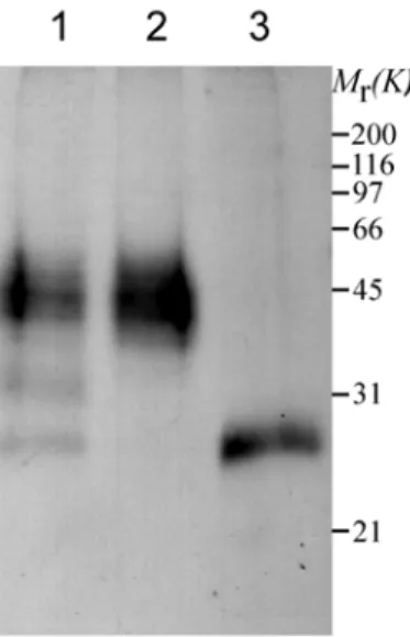
By use of anti-GREG peptide antibodies, endogenous GREG migrates as a smear from 35kD to 60kD (Fig. 3-5) as determined by SDS-PAGE and WB. This is in agreement with the identification of the LRT peptide present in Band 1, 2 and 3 of GIC proteins.
Results
Figure 3-5 Topological analysis of GREG. Golgi membranes were treated with or without trypsin in the presence or absence of TX-100 at 37°C and analyzed by immunoblot with antibodies against the identified peptide (ALI). As a control, antibodies against the luminal portion of p23, a Golgi transmembrane protein, were used to detect a difference in molecular mass (around 1kD) after cleavage of its short cytoplasmic tail. Similar results were obtained with antibodies against another peptide (SEA) (not shown). STI, soybean trypsin inhibitor.
Glycosylation is a common modification on proteins that gives rise to diffuse protein bands (smears) on SDS-PAGE. During biosynthesis in the lumen of the ER, an N-glycan core structure is added to proteins. Further processing takes place in the Golgi complex, where the diversity of N-glycan structures is generated. N-glycans added to proteins are considered to play an important role in folding, stabilization, and sorting of modified proteins (Helenius & Aebi, 2001).
The primary structure of GREG contains two Asparagine residues (66N and 93N) within the consensus sequence for N-glycosylation (NXS/T). To investigate a possible glycosylation of GREG, solubilized Golgi membranes were treated with N-glycosidase (PNGase), an enzyme that removes all N-linked sugars from asparagine. When Golgi membranes were incubated at 37°C without PNGase, GREG behaved as a smear as expected. Treatment performed in the
Results
presence of PNGase at 37°C, however, generated a single band with an apparent MW of about 28kD (Fig. 3-6 lane-3). When Golgi membranes were treated with PNGase at 4°C to reduce the enzyme activity, deglycosylation intermediates of GREG were detected (lane-1). These data show that GREG is a glycoprotein.
Figure 3-6 Glycosylation of GREG. Solubilized Golgi membranes were incubated at 37°C (lane-3) or at 4°C (lane-1) overnight with or without (lane-2) N-glycosidase PNGase and analyzed by SDS-PAGE and Western blot using antibodies against GREG. Similar results were also observed using antibodies against SEA.
3.1.5 Confirmation of the predicted ORF
As mentioned before, the deduced amino acid sequence of GREG from the cDNA sequence has a calculated molecular mass of 22.8kD. After removal of N-linked glycans from GREG, it migrates as a single band with an apparent MW of 28kD on SDS-PAGE (Fig. 3-6).
The processing of GREG from a nascent to a mature polypeptide was studied in further detail. Isolated ER fractions were analyzed by WB with anti- GREG peptide antibodies. A single band of about 35kD, together with a band of about 60kD, was detected in the ER fractions, both of which were hardly
Results
detectable in Golgi fractions (Fig. 3-7b). The 35kD band in the ER may represent the core glycosylated form of GREG, and the 60kD protein could represent a dimer of the non-glycosylated precursor (28kD) (the tendency of GREG to form dimers is discussed below).
Similar results were obtained when the complete ORF, tagged with a single myc tag at the COOH terminus, was in vitro translated in the absence or presence of microsomes. In the presence of microsomes, a 32kD band, together with a 36kD protein, was translated (Fig. 3-7a). The 32kD band might be similar to the 28kD band after deglycosylation because of the following considerations:
this construct cannot be processed for GPI-anchoring, due to the myc-tag;
therefore, it contains an additional 20 amino acids at the C-terminus as well as the amino acids representing the myc-tag. The 36kD band could arise from further modification by microsomes (successful processing of α-factor, a highly glycosylated polypeptide, demonstates that microsomes are functionally executing ER functions). These findings suggest that the obtained ORF encodes the full-length protein.
Figure 3-7 Confirmation of the predicted ORF. a, In vitro translation. The predicted ORF of GREG was fused with a single myc tag at the C-terminal end and was cloned into pcDNA3. Coupled transcription and translated was performed with 1 µg of plasmid DNA per reaction in the presence or absence of microsomes, and the translated polypeptide was labeled with [35S]-Met. 2 µL of translation mixture were analyzed on SDS-PAGE and autoradiography. b, Precursors of GREG. ER fractions, together with Golgi membranes, were analyzed by SDS-PAGE and Western blot using antibodies against the SEA peptide.
Results
3.2 The coiled-coil domain is involved in protein-protein interactions
The possibility that the apparent MW of the mature protein (45kD) is caused by an interaction of GREG with other proteins or with itself via its coiled-coil domain was investigated. The complete ORF (named ORF construct) as well as the coiled-coil domain (deletion of both the N- and C-terminal hydrophobic stretch) (named ∆-construct) was tagged with His 6 at the C-terminal end (Fig.
3-8a) and overexpressed in Pichia pastoris (these constructs could not be expressed in bacterial systems). As shown in Fig. 3-8b and c, a 28kD protein (indicated by an arrow head) was purified from lysates containing the ∆ construct, whereas a 29kD protein was purified from lysates containing the ORF construct. The MW of the purified proteins is in agreement with the deglycosylation results (Fig. 3-6 lane-3). The predicted MW of ∆ protein is about 14kD. Therefore, the 28kD band may represent a (homo)dimer. Of note, the difference in MW (about 1kD) between the purified ∆ construct and the ORF construct may originate from the additional C-terminal 20 amino acids, since the N-terminal hydrophobic stretch (49aa, around 5kD) is likely to be cleaved after serving as a signal peptide in Pichia pastoris.
Another protein of about 25kD was co-purified in stoichometric amounts on Ni-NTA beads (Fig. 3-8b, indicated by an asterisk). This protein may arise from an interaction of the coiled-coil domain with a yeast protein (25kD as a heterodimer). Alternatively, this protein may be a degradation product of the expressed construct. In this case, degradation must occur at the C-terminus since the antibody, directed against the N-terminal part (α-SEA), recognizes the degradation product (Fig. 3-8c). Thus, a homodimer may have been isolated by an interaction between a His-tagged non-degraded protein and a non-His-tagged C-terminally cleaved protein.
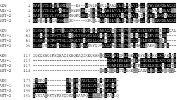
3.3 GREG is homologous to human BST-2
Blast searches performed with the deduced amino acid sequence of GREG indicated that human BST-2 (Ishikawa et al. 1995), rat DAMP-1 (unpublished
Results
data) and several mouse ESTs are similar to GREG. GREG shares 41% identity (57% similarity) with human BST-2 and 55% identity (64% similarity) with mouse and rat homologues (Fig. 3-9). The hydrophobic portions at both ends and the two N-glycosylation sites are conserved. None of these homologues, however, contains the identified peptide, ALIQEQEAQIK, and EQEAQIK tandem repeats.
This explains why no protein or EST was found in Database searches using the ALI peptide (Gkantiragas et al. 2001). No homologues were found in yeast, nematodes and worms.
Figure 3-8 Expression of GREG mutants in Pichia pastoris. a, constructs for expression in yeast cells. The ORF and ∆ mutant were cloned into pPICZ plasmid and fused with (His)6 tag. b, Expression of ORF and ∆- mutant proteins. Pichia cells were transfected with mutant proteins by electroporation and stable clones were selected in the presence of Zeocin. Protein expression was induced with methanol, and the constructs were purified by the use of Ni-NTA beads and analyzed by SDS-PAGE and Coomassie brilliant blue staining or c, by Western blot. The blot was probed with antibodies against the SEA peptide. Similar results were obtained with anti-ALI antibodies, as shown in one lane. E, elution; Nb, non- binding; W, wash.
Results
Figure 3-9 GREG is a homologue of human BST-2. Alignment of GREG with human and mouse BST-2 and rat DAMP-1 was done at EBI (http://www.ebi.ac.uk/clustalw/) and graphically presented using the program BoxShade as in Figure 3-3.
BST-2 was isolated from human bone marrow stromal cells (Ishikawa et al. 1995).
It is preferentially expressed on the surface of terminally differentiated B cells and overexpressed in multiple myeloma cells. Therefore, BST-2 could represent a potential target molecule for antibody-mediated immunotherapy for multiple myeloma (Ohtomo et al. 1999). BST-2 is predicted to be a type-II membrane protein on cell surfaces and to exist as a homodimer, similar to what has been observed for GREG. BST-2 is suggested to be engaged in pre-B cell growth, but the precise function is unknown.
3.4 GREG is a GPI-anchored protein
As predicted by the hydrophilicity plot, GREG encompasses a hydrophilic sequence, surrounded by hydrophobic sequences at each end (Fig. 3-2a). Such a characteristic is reminiscent of a precursor protein, destined for GPI linkage processing (Udenfriend & Kodukula, 1995). Indeed, sequence analysis with the
Results
web-based Big GPI prediction program revealed that GREG contains all the required signals for GPI anchorage processing and the program identified a serine residue at position 183 as a potential ω site. The presence of GREG in GICs is in agreement with the possibility of a GPI anchor modification of GREG, since GPI-anchored proteins preferentially partition into lipid rafts (Simons & Ikonen, 2000).
2.4.1 Aerolysin detects a protein in GICs similar to GREG
GPI-anchored proteins are concentrated in lipid-enriched microdomains (Brown
& Rose, 1992). These proteins are thought to be recruited to lipid-enriched microdomains at the medial Golgi en route to the plasma membrane (Brown &
Rose, 1992). Therefore, GPI-anchored proteins could be present in GICs. Several methods have been developed to detect the presence of GPI-anchored proteins.
One method is based on a pore-forming toxin, aerolysin, which is secreted by Aeromonas hydrophila. Aerolysin damages the plasma membrane of target cells by specifically binding to the glycan core of the GPI moiety of GPI-anchored proteins. Therefore, aerolysin can be used to screen for the presence of GPI- anchored proteins, irrespective of the proteinaceous domain.
GICs, prepared from CHOwt cells, were analyzed with an aerolysin overlay assay. The procedures of this assay are basically analogous to WB, except that labeled aerolysin was used in stead of antibodies (details are described in Materials and Methods). As expected, GPI-anchored proteins were detected in GICs with aerolysin. It is interesting to note that aerolysin detected smears, whose patterns are similar to that revealed by peptide antibodies (Fig. 3-10). To confirm that the protein recognized by aerolysin is indeed GREG, attempts were made to immunoprecipitate GREG either from Golgi membranes or GICs with peptide antibodies and subsequent analysis with the aerolysin overlay assay.
Immunoprecipitation of GREG was, however, not possible using peptide antibodies.
Results
Figure 3-10 GREG as a potential GPI-anchored protein. GIC proteins were resolved on SDS-PAGE and analyzed by aerolysin overlay assay. Cell lysates from BHK and CHOwt cells were used as positive controls, whereas those from a GPI-depleted CHO mutant cell line were used as a negative control. The blot was incubated with radiolabeled aerolysin and analyzed by radiography.
3.4.2 Expression of FLAG-tagged GREG in mammalian cells
The aerolysin overlay assay indicated that GREG is linked with a GPI anchor.
This knowledge was used in the design of a tagged GREG protein (GREG-Flag):
given the minor importance of the amino acids immediately N-terminal to the ω site, a single FLAG tag was inserted between the codons for 181Q and 182N by PCR (Fig. 3-11a). CHO or NRK cells were transfected with the GREG-Flag construct and stable cell clones were selected in the presence of G418 at a final concentration of 600 µg ml-1. Several clones were obtained. In this thesis, analysis of the tagged GREG was performed in the stable cell lines Cr2/3 (CHO) and Cr7 (NRK). Three hallmarks for proper expression of the tagged protein were determined: migration on SDS-PAGE, subcellular location, and recognition of the over-expressed protein by aerolysin.
Results
Figure 3-11 Expression of FLAG-tagged GREG. a, Diagram of the construct. A single FLAG tag was inserted between 181Q and 182N by PCR. After supplementing the C-terminal SP, FLAG-tagged GREG was cloned into pcDNA3 (GREG-Flag). b, Insertion of a FLAG tag has no effect on glycosylation. CHO cells were transfected with pcDNA3 containing GREG-Flag and selected with G418. Total membranes prepared from the stable cells expressing GREG-Flag were treated with or without PNGase and analyzed by immunoblot with anti-FLAG antibodies. c, Aerolysin overlay assay. Golgi membranes were prepared from CHO-derived stable cell lines expressing GREG-Flag and used for aerolysin overlay experiments as above.
As revealed by WB with peptide antibodies, endogenous GREG in Golgi membranes migrated on SDS-PAGE as a smear from 35kD to 60kD (Fig. 3-5).
Similar to the endogenous protein, Flag-tagged GREG migrated as a smear in the 45kD region. Flag-tagged GREG was sensitive to glycanase treatment and after deglycosylation, a band around 31kD was detected (Fig. 3-11b). GPI-anchorage
Results
of Flag-GREG was analyzed by preparation of Golgi membranes from Cr2/3 cells and subsequent analysis by the aerolysin overlay assay. In parallel, the same amount of Golgi membranes from CHOwt cells was analyzed. Fig. 3-11c shows that the protein pattern (smear) recognized by aerolysin is comparable to the protein pattern recognized by antibodies against the endogenous protein. These data indicate that the posttranslational processing of tagged GREG is similar to endogenous GREG.
An additional band of more than 200kD in size was observed in Golgi membranes, isolated from Cr2/3 cells (Fig. 3-11c). In light of the coiled-coil characteristic of GREG, this 200kD protein might represent oligomerizaton between individual GREG molecules or with other proteins.
3.4.3 GPI linkage of GREG
As discussed in the Introduction, PI-PLC can specifically cleave the GPI moiety from GPI-anchored proteins, thereby relieving the protein moiety from membranes. This is a widely used method to determine whether a protein is linked with a GPI moiety. To determine the presence of a GPI anchor at the C- terminus of GREG, Golgi membranes were first extracted with detergent TX-114 which undergoes phase separation at room temperature when in an aqueous solution. Under these conditions, integral membrane proteins and GPI-anchored proteins partition in the detergent phase, whereas the peripheral and soluble proteins are retained in the aqueous phase (Bordier 1981). Subsequently, an aliquot of the detergent phase is incubated in the presence or absence of PI-PLC.
After incubation, these samples are extracted again with TX-114. After PI-PLC treatment, endogenous GREG is partially present in the aqueous phase, while GREG is hardly detectable in the aqueous phase upon mock treatment (data not shown). Extension of the incubation time did not increase the amount of GREG in the aqueous phase. Similar results were obtained when changing to different incubation times and different substrate concentrations. Some GPI-anchored proteins are less sensitive to PI-PLC treatment owing to additional modifications
Results
such as palmitoylation (Fugerson, 1999). Such modifications could be involved in the partial sensitivity of GREG to PI-PLC (data not shown).
Figure 3-12 GREG is linked to a GPI moiety. Golgi membranes from stable cell lines were extracted with TX-114. The resulting detergent phase was incubated in the presence or absence of PI-PLC for 36 hrs. Samples were extracted again with TX-114 and the corresponding aqueous and the detergent phase were analyzed by Western blot with the indicated antibodies. A, aqueous phase and D, detergent phase.
To address the problems mentioned above, procedures to perform PI-PLC treatment were modified. Cr2/3 cells were first treated with β-cycloheximide for 1 hour before preparing total membranes. This eliminates a pool of newly synthesized GREG with possibly higher palmitoylation levels of the inositol ring
Results
(see Introduction). After extracted with TX-114, the detergent phase was incubated at 37°C in the presence or absence of PI-PLC. After incubation, both mock and treatment samples were extracted with TX-114. Finally, the samples were precipitated with chloroform/methanol and analyzed by WB. As shown in Fig. 3-12, a substantial amount of GREG was revealed in the aqueous phase of PI- PLC treatment and tagged GREG could not be detected anymore in the detergent phase. As a control, p23 always remained in the detergent phase.
In summery, both endogenous and tagged GREG are posttranslationally modified with a GPI anchor at the C-terminus.
3.5 GREG resides at the Golgi complex
By IF using a peptide antibody, endogenous GREG has been shown to localize exclusively to the Golgi complex (Gkantiragas et al. 2001). Confirmation of the Golgi localization of GREG is important because so far no GPI-anchored protein has been described to reside at an intracellular organelle.
3.5.1 Immunolocalization of the tagged GREG
As shown above, a FLAG tag insertion does not influence the properties of GREG protein, including GPI anchorage processing and other modifications such as glycosylation. The subcellular localization of GREG-Flag was determined in stably transfected NRK (Cr7) and CHO (Cr2/3) cells. As shown in Fig. 3-13, in both cell lines, GREG-Flag localizes to a perinuclear structure using FLAG tag antibodies. These signals co-localized with Man II, a Golgi marker. The Golgi localization of GREG is sensitive to BFA treatment. In NRK (Cr7) cells, consistent with previous observations, no specific signals of GREG were viewed at the plasma membrane (Fig. 3-13a). In CHO (Cr2/3) cells, signals at plasma membrane could be observed by IF (Fig. 3-13b). These signals were, however, also observed by IF in non-transfected CHO cells using the antibody against the FLAG-tag (data not shown), indicating a non-specific cross-reactivity of this antibody with a CHO protein.
Results
Figure 3-13 Subcellular localization of GREG-Flag. NRK- (a) or CHO (b) cells constitutively expressing GREG-Flag were treated with or without 5 µM of BFA for 30min.
Cells were fixed and labeled with antibodies against FLAG (red) and Man II (green). Images were collected with the use of a Zeiss LSM510 and adjusted using the equipped software. A representative confocal section of the cells is shown.
Results
3.5.2 Biochemical localization of the native and tagged GREG
BFA is a drug that induces redistribution of Golgi proteins to the ER by inhibition of the guanine nucleotide exchange factors (GEFs) of ARF proteins, thus leading to Golgi structure breakdown (Helms & Rothman, 1992, Donaldson et al. 1992, Mansour et al. 1999). BFA-sensitive localization has been used as evidence of Golgi location of a protein of interest. Relatively short times of incubation of cells with BFA have no effect on the localization of late Golgi proteins, such as TGN38. In this way, early Golgi proteins can be distinguished from late Golgi proteins (Steegmaier et al. 1999). However, low levels of functional GREG protein at the plasma membrane cannot be excluded from the IF data. This is important since in contrast to GREG, most GPI-anchored proteins are delivered to the plasma membrane along the biosynthetic secretory pathway (Chatterjee & Mayer 2001).
Additional evidence for the intracellular localization of GREG was obtained from experiments using density gradient centrifugation. The strategy adopted in these experiments is that upon cycloheximide treatment, the distribution pattern on a gradient will not alter if GREG has a steady-state localization to the Golgi complex. If a protein localizes to the plasma membrane at steady state, it will be detected in Golgi fractions as it passes through this organelle upon biosynthesis en route to the plasma membrane. Upon cycloheximide treatment, however, all newly synthesized proteins will move out of the Golgi complex to the plasma membrane. To biochemically determine the location of GREG, Cr2/3 cells were treated with cycloheximide to block novel protein synthesis. Post-nuclear supernatants from these cells were fractionated on a velocity gradient. 12 Fractions were collected from top to bottom and subsequently analyzed by WB. Fraction 1 is enriched in soluble proteins from the cytosol, whereas fraction 2 represents the plasma membrane (Jenne et al., 2002).
To analyze the migration of GREG on these gradients, GM-130 was used as a Golgi marker, and FolR, a GPI-anchored protein that recycles between the plasma membrane and endosomes (Mayor et al. 1998), was used as a plasma
Results
Figure 3-14 Biochemical localization of GREG. CHOwt or cells stably expressing GREG-Flag were treated with (+CH) or without (-CH) β-cycloheximide for 5 hrs. The post- nuclear cell lysates were overlayed and fractionated on a velocity Nycodenz gradient at 100,000g for 1 hr. Fractions collected from top to bottom were analyzed by SDS-PAGE and Western blot with antibodies against the indicated proteins.
Results
In the absence of cycloheximide, FolR could be detected in fractions from 3 to 12 (Fig. 3-14a). The major pool of FolR is enriched in fractions 7-9. In the presence of cycloheximide, however, FolR is enriched in fractions 2 and 3, with minor amounts in fractions 7 and 8, respectively (Fig. 3-14b). These data indicate that in the presence of cycloheximide, FolR accumulates at the plasma membrane by moving out of the Golgi complex, in agreement with its plasma membrane localization. WB revealed that GREG co-migrates with both FolR and GM-130 in untreated cells. Upon cycloheximide treatment, the distribution pattern of GREG does not undergo major changes and was found to be enriched in fraction 6-8.
GREG proteins could not be detected in fraction 2 (Fig. 3-14). This behavior of GREG in the presence of cycloheximide is in contrast to that of FolR and highlights the different localization of these two different GPI-anchored proteins.
These data suggest that GREG localizes to the Golgi complex.
A protein enriched in fraction 1 and 2 is recognized by the FLAG antibody (Fig. 3-14). Indeed, FLAG antibodies were reported to recognize a plasma membrane protein of similar MW (Schafer & Braun, 1995). It is possible that the signals at the plasma membrane of CHO cells observed in IF images (Fig. 3-13b) may originate from this protein.
The behavior of wild-type GREG was also determined on the same density gradients. The distribution pattern of wild-type GREG is similar to GREG-Flag in Cr2/3 cells (Fig. 3-14), supporting the notion that GREG-Flag mimics its wild-type counterpart.
3.6 Mapping the Golgi targeting domain of GREG
GREG has been defined as an unusual GPI-anchored protein in that it localizes to the Golgi complex. A GREG-related protein, human BST-2, localizes to the surface of the cell and ectopically expressed BST-2 in CHO cells is also found at the plasma membrane (Ohtomo et al. 1999). Compared to GREG, both human BST-2 and its mouse homologue lack the EQ tandem repeats. The SMART program suggests that the EQ repeat occupies the core region of a SNARE-like
Results
functions as a SNARE protein. Based on comparison between GREG and its homologues, the EQ tandem repeat seems the most likely candidate for targeting GREG to the Golgi complex.
3.6.1 The C-terminal domain of GREG contains a Golgi targeting signal
To investigate a possible role of the EQ tandem repeat in Golgi targeting, GREG was first truncated into two domains, constituting amino acids 1-111 (N-term construct) or 112-203 (C-term construct) (Fig. 3-15a). The N-terminal domain harbors both N-glycosylation sites, while the C-terminal domain contains the EQ tandem repeat. The N-terminal and the C-terminal domain were supplemented with the missing signal for GPI anchorage processing and signal peptide, respectively. A FLAG tag was inserted at the same position as in the full length construct of GREG (Fig. 3-11a). Both domains were cloned into a pcDNA3 expression vector and after sequence verification subsequently used for transient transfection of CHO cells.
IF revealed that the C-terminal domain co-localized with Man II (Fig. 3- 15b). Cycloheximide treatment did not change the staining pattern of its subcellular localization. In contrast, the N-terminal domain was transported to the plasma membrane and did not co-localize with Man II upon cycloheximide treatment. Instead, it was scattered over the plasma membrane as punctuate structures (Fig. 3-15b). These data demonstrate that the Golgi targeting signal localizes to the C-terminal domain of GREG, in agreement with the prediction based on by sequence comparison. Next, the EQ tandem repeat was specifically deleted from GREG (Fig. 3-15a). IF revealed that this ∆EQ protein was delivered to the plasma membrane through the secretory pathway (Fig. 15b), consistent with observations of the ectopically expressed BST-2 in CHO cells. Apparently, EQ tandem repeat is required to localize GREG to the Golgi complex.
Results
Figure 3-15 The EQ repeat is required for Golgi localization of GREG. a, Schematic representation of the constructs. All constructs were cloned into pcDNA3 and the corresponding mutant proteins are all linked with a signal peptide for targeting to the ER lumen as well as a signal peptide for GPI anchorage. b, Subcellular localization of mutant proteins. CHO cells transiently expressing indicated mutant proteins were treated with β- cycloheximide for 1 hr and fixed for indirect immunofluorescence with antibodies against FLAG (red) and Man II (green). Images shown are representative confocal sections.
Results
3.6.2 EQ repeat is not sufficient for Golgi targeting of a soluble protein
To investigate the efficiency of the EQ tandem repeat as a Golgi targeting motif, it was fused with GFP, a soluble protein that does not have any Golgi targeting sequences. A signal peptide sequence was added to the fused EQ-GFP construct to allow entry into the secretory pathway. After transfection, cells were treated with cycloheximide to exclude Golgi localization which might represent staining en route to the plasma membrane. Under these conditions, the fused GFP construct did not display a typical Golgi localization (data not shown). In addition, IF levels of the fused GFP steadily declined, indicating that the GFP- construct was secreted from the cells. These data demonstrate that the EQ tandem repeat is not sufficient to target GFP to the Golgi complex.
It should be noted that the EQ tandem repeat is only the core portion of an untypical SNARE-related domain in GREG (Fig. 3-4). Golgi targeting of EQ repeat may require either proximal amino acids of this SNARE-related domain, or sequences from other parts of the molecule. Other factors, e.g. membrane association, may influence its Golgi targeting as well.
3.6.3 Is the EQ repeat a universal Golgi targeting signal?
To perform a variety of unique functions at the Golgi complex, Golgi-resident proteins must be retained amid those sorted to other destinations. So far, only few Golgi targeting signals have been identified. Recent findings implicate a GRIP motif shared by the coiled-coil Golgin family of proteins (Kjer-Nielsen et al.
1999, Munro & Nichols 1999). The above data have characterized GREG to specifically locate at the Golgi complex and showed that an EQ tandem repeat within a coiled-coil region is essential for the subcellular localization of GREG.
An EQ tandem repeat might act as a Golgi localization motif in other Golgi resident proteins as well. By using web-based BLAST (to retrieve short nearly exact matches), several proteins were identified that contain similar tandem repeats, such as EQEGQVR (in XP_091107.5), EQEEMLR (in XP_165407.1), EQEKQMR (in XP_208786.1), EQEEKIR (in BAC05084.1) and
Results
EQEERLR (in BAC05050.1). Interestingly, these proteins are predicted to be related to Golgin-97, one member of the Golgin family of proteins. The long coiled-coil Golgin family of proteins is peripheral or integral membrane proteins locating to the cytoplasmic site of the Golgi complex. This family of proteins is believed to participate in maintenance of the Golgi architecture by stitching together distinctive cisternae, aligned in parallel. GREG is, however, confined to the Golgi lumen. How could the EQ tandem repeat function as a Golgi targeting signal amongst proteins with opposite membrane orientations?
Figure 3-16 Glycosylation of ∆EQ. Total membranes from CHO cells expressing either GREG-Flag or ∆EQ were treated with PNGase and analyzed by SDS-PAGE and immunoblot with anti-FLAG antibodies.
In view of its central position in a SNARE-related coiled-coil domain, this repeat may influence the oligomerization of GREG. After deglycosylation, the
Results
∆EQ protein migrates as two distinct bands on SDS-PAGE. One has a MW of just above 21kD, while the other migrates as a 42kD protein, possibly representing a dimer (Fig. 3-16). Therefore, deletion of the EQ tandem repeat did not affect dimerization, in agreement with observations of the human BST-2 protein. The migration of glycosylated ∆EQ protein on SDS-PAGE is, however, different from that of GREG-Flag. Further efforts should be made to investigate the possibility that the EQ tandem repeat affects oligomerization of these proteins.
3.7 GREG does not recycle between phagosomes and the Golgi complex
Subunits of heterotrimeric G proteins, Flotillin-1 and v-ATPases have been identified in mature phagosomes (Dermine et al. 2001). Flotillin-1 associates with phagosomes during their maturation, i.e. phagosomal Flotillin-1 is not recruited from the plasma membrane (Dermine et al. 2001). Recently, another GIC protein, GAPR-1, has been recently found to be recruited to phagosomes in a time- dependent fashion (Kaloyanova & Helms, unpublished data). The origin of phagosomal GAPR-1 or other GIC proteins has not been defined but they may originate from GICs at the Golgi complex. Similarly, GREG might also move out of the Golgi complex and/or recycle between the Golgi complex and other subcellular compartments such as phagosomes.
To investigate this possibility, phagosomes were prepared from phagocytic CHO cells stably expressing CD16, after being fed with Latex beads as described (Desjardins et al. 1994). Golgi membranes from phagocytotic CHO cells were extracted with TX-114. The resulting detergent pellet and corresponding supernatant, as well as phagosomes, were analyzed by WB. As shown in Fig. 3-17, GREG cannot be detected in phagosomes whilst Flotillin-1 did present in these phagosomes. The migration pattern and membrane attachment characteristics of GREG in Golgi membranes were not affected when cells were incubated with Latex beads. These data indicate that in contrast to other GIC proteins, GREG does not recycle between phagosome and the Golgi complex.
Results
Figure 3-17 GREG does not cycle between the Golgi and phagosomes. Golgi
membranes and phagosomes from CHO cells constitutively expressing CD16 were analyzed by SDS-PAGE and immunoblot with antibodies against the SEA peptide. Flotillin-1 (western analysis for Flotillin-1 was done by Kaloyanova) and p23 were used as controls.
3.8 GREG is essential in maintenance of Golgi integrity
Long coiled-coil proteins play an essential role in Golgi structures (Shorter &
Warren, 2002). As described above, GREG is a coiled-coil protein, similar to the family of Golgi matrix proteins. Despite the opposite orientation of these proteins, GREG may interact with other Golgi luminal proteins through its long coiled coil and thus to be involved in the assembly of Golgi structures.
Alternatively, it could form trans-pairs with one molecule on either side of a cisterna, thus stabilizing a flattened cisterna.
Results
2.8.1 Blockade of GREG expression vesiculates the Golgi complex
Several elegant methods are developed to characterize the function of a protein.
Among these, generating knockout animals are widely adopted. The finding of dsRNA as a tool to inhibit gene expression makes it possible to specifically reduce the levels of a protein of interest in individual cells rather than generating knockout animals (Hannon, 2002). This method was used to investigate the contribution of GREG in maintenance or generation of the Golgi structure. To this end, dsRNA was generated by in vitro transcription and subsequently transfected into Cr2/3 cells.
Figure 3-18 GREG is involved in maintenance of Golgi integrity. A plasmid containing the ∆ mutant was linearized at either end. Double-strand RNAs were generated by in vitro transcription and delivered into CHO-originated stable cells expressing GREG- Flag (Cr2/3 cell line). Cells were fixed and labeled for GREG-Flag (red), Man II (green) and nuclei (blue). Images were collected with the use of a Zeiss inverted fluorescence microscope equipped with a cooled CCD camera. The arrow indicates one cell with undetectable levels of GREG-Flag.
Results
IF revealed that levels of expressed Flag-GREG (and thus also of the endogenous protein) decreased to different extents in various cells. Likewise, the Golgi structure was affected to different extents. Strikingly, scattered and punctuate Golgi structures were visualized in cells where the level of GREG was not detectable anymore (Fig. 3-18). In cells with reduced, but detectable levels of GREG, fragmented and packaged ribbon-like Golgi structures co-existed. Cell cycle-dependent Golgi fragmentation in the affected cells could be ruled out by nuclear staining. These data support the notion that GREG is essential in maintaining the Golgi structure and that low amounts of GREG are sufficient for a structural role of GREG.
3.8.2 A dominant negative mutant of GREG
For its structural role, membrane association is expected to be an essential feature of GREG, either in trans-pair formation or in communication to cytosolic complexes involved in maintenance of the Golgi structure. In either case, release of GREG from membranes should lead to an effect similar to the knockdown expression of GREG by dsRNAi.
To affect the membrane binding properties of GREG, the C-terminal signal peptide, necessary for GPI anchorage processing, was deleted from wtGREG. A FLAG tag was added to the C-terminal end of the construct (∆GPI-GREG) (Fig. 3- 19a). As might be expected, transient expression of this mutant GREG shows that glycosylation (due to lacking the glycan core of the GPI anchor) was affected by deletion of the C-terminal signal peptide. By WB, several bands or smears were detected with the antibody against the FLAG epitope (Fig. 3-19b and compare with Fig. 3-6). From the in vitro translation data (Fig. 3-7a), the bands or smears below 40kD could represent the ER forms. Upon PNGase treatment, two bands could be identified. The apparent MW of the high MW band (60kD) indicates that it could represent a homodimer of the low MW protein (28kD).
Deglycosylation of endogenous GREG, however, generated only one band (Fig.
3-6). Dimer formation could be due to high levels of over-expression. In
Results
contain a membrane anchoring signal. However, no protein was detected in the media and it was found tightly associated with cellular membranes.
IF showed that expression of the mutant protein led to fragmentation of the Golgi architecture (Fig. 3-19c). Golgi fragmentation by this mutant supports a structural role of GREG that requires membrane attachment. It also indicates that
∆GPI-GREG acts as a dominant negative mutant. In this case, this potentially soluble mutant competes for endogenous GREG, thereby disrupting formation of e.g. trans-pairs.
The fragmentation of Golgi structures was assessed quantitatively. To this end, cells from 4 different scopes were counted. Out of 105 cells in total, 94 were shown to express the mutant protein. 51 of these cells revealed either a fragmented Golgi or no Golgi signals (54%). Under steady state, 4 to 6% of CHO cells exhibited fragmented Golgi structures, representing the cell-cycle dependent Golgi fragmentation. Under mock treatment conditions (with empty plasmid DNA), around 10% of cells revealed fragmented Golgi or no Golgi signals. The difference between mock transfected and non-treated cells may originate from harmful effects of the transfection reagent (liposomes).
3.8.3 Membrane attachment is not enough for GREG to function
The role of membrane attachment was further examined by replacement of the GPI moiety with a transmembrane domain of p23 (23TM-GREG) (Fig. 3-19a).
Again, a FLAG tag was added to trace the construct. The migration pattern on SDS-PAGE, glycosylation characterics as well as homodimer formation are similar to those of the ∆GPI-GREG mutant (Fig. 3-19b). However, the expression level is relatively low.
Similar to ∆GPI-GREG, expression of 23TM-GREG results in fragmentation of Golgi structures (Fig. 3-19c). Quantification of Golgi fragmentation revealed that 50% of cells expressing 23TM-GREG have fragmented Golgi or no Golgi signals (transfection efficiency was about 70%.).
Results
Figure 3-19 The GPI moiety of GREG is indispensable. a, Dia-
gram of the constructs.
Both mutant constructs with a FLAG tag were cloned into pcDNA3. b, Expression of mutant GREG proteins. Total membranes from CHO cells transiently express- ing either mutant protein were treated without or with PNGase and analyz- ed by SDS-PAGE and immunoblot. c, Effect of mutant protein expression on Golgi structures. CHO cells transiently transfect- ed with either mutant construct were fixed for indirect immunofluore- scence with antibodies against FLAG (red) and Man II (green). Images shown are representative sections. d, Subcellular localization of 23TM- GREG. CHO cells were transiently transfected with 23TM-GREG, fixed and labeled for 23TM- GREG (red) and the ER marker calreticulin (green). Representative confocal sections are shown.
Results
Unlike the partial Golgi location of ∆GPI-GREG, 23TM- GREG was distributed in structures with a staining pattern similar to the ER protein calreticulin, although it cannot be excluded that small amounts still located to the Golgi complex (Fig. 3-19d). Because trans-pairs could potentially still be formed with 23TM-GREG, these results indicate that either formation of trans-pairs is not important for maintenance of the Golgi structure or that recruitment into microdomains is essential for the function of GREG.
3.9 Recruitment into GICs is required for GREG to function
GPI-anchored proteins have been shown to preferentially partition into lipid- enriched microdomains and to transmit signals to the cell (Tsui-Pierchala et al.
2002). Since a transmembrane segment was not able to functionally replace the GPI anchor in GREG, a specific lipid requirement might therefore contribute to the functional activity of GREG. To test a microdomain association of the various GREG constructs, total membranes were prepared from cells expressing either tagged full length GREG or tagged mutant GREGs. Membranes were extracted with cold TX-100 and lipid-enriched microdomains were isolated as detergent- insoluble fractions. Caveolin-1 was used as a marker for detergent-insoluble fractions, whereas p23, a transmembrane protein, was used as a marker for detergent-soluble fractions. As shown in Fig. 3-20, upon TX-100 extraction, p23 was predominantly present in the supernatant. Caveolin-1, on the other hand, persisted in pellets which represent lipid-enriched microdomains. GREG-Flag preferentially remained in the detergent-insoluble pellets. Both mutant proteins partitioned in the detergent-soluble fractions and only small amounts of the mutants could be detected in the pellets. The small amounts of mutant GREG in the pellets may reflect an interaction between the mutant proteins and endogenous GREG and may explain the observed effects of the mutants on the Golgi complex. These experiments suggest that recruitment of GREG into the lipid-enriched microdomains via the GPI moiety is indispensable for GREG to function.
Results
Figure 3-20 Segregation of both GREG mutants from lipid-enriched microdomains. Total membranes were prepared from CHO cells expressing mutant GREG proteins lacking a GPI anchor (∆GPI-GREG and 23TM-GREG) or GREG-Flag.
Membranes were extracted with TX-100 in cold PEN buffer on ice for 30 min and centrifuged at 100,000Xg for 1 hr. The resulting supernatants (S) and pellets (P) were analyzed by SDS- PAGE and Western blot with antibodies against the indicated proteins.
3.10 Golgi fragmentation by mutant GREG is independent of apoptosis and microtubule integrity
As discussed in the Introduction, Golgi stacking is regulated by the Golgi matrix which contains the GRASP family of Golgi stacking proteins, the Golgin family of long coiled-coil proteins and a spectrin/ankyrin framework through protein-
Results
protein and protein-lipid interactions. Cleavage of such proteins perturbs the interactions among these proteins and thus leaves distinct cisternae separately.
The matrix proteins can be cleaved by caspases during apoptosis (Mancini et al.
2000, Lane et al. 2002, Chiu et al. 2002), resulting in scattered punctuate Golgi structures in apoptotic cells.
Is the effect of Golgi fragmentation by expression of mutant GREGs due to induction of apoptosis? A quality control system is active in eukaryotic cells to survey protein synthesis and folding in the ER. Aggregates of protein in the ER activate ER-located caspases and initiate the cell-suicide machinery (Nakagawa et al. 2000). As the above data have shown that GREG tends to cluster together, this might cause apoptosis.
An important feature of an apoptotic cell is condensation of the nucleus which can be visualized by staining DNA with dyes like DAPI. ∆GPI-GREG was expressed in CHO cells and cells that express the mutant protein were chosen according to their Golgi fragmentation as detected with Man II. As shown in Fig.
3-21a, cells that display dispersed Golgi structures show normal nuclei staining when compared to non-transfected cells. Similar results were obtained with the 23TM-GREG mutant. Condensed and fragmented nuclei could be observed in cells in which apoptosis was induced with a drug, showing that DAPI staining is sensitive enough to monitor cells undergoing apoptosis. In addition, the active form of caspase-2 could not be detected in cells expressing the mutant proteins (Fig. 3-21b). Collectively, fragmentation of the Golgi induced by expression of the mutant GREGs occurs in an apoptosis-independent fashion.
The microtubule cytoskeleton has been shown to play an important role in the organization of the Golgi complex. Its motor proteins position the Golgi complex around the centrosome (Allan et al. 2002). Once microtubules are depolymerized by treating cells with drugs like nocodazole, the Golgi ribbon structure is fragmented and redistributes to the cell periphery. IF, however, revealed no differences of microtubule staining between wild-type cells and those expressing the mutant GREG causing fragmented Golgi structures (Fig. 3-
Results
22). This indicates that disruption of the Golgi apparatus by expression of the mutant GREG occurs in a way independent of microtubule integrity.
Figure 3-21 Golgi fragmentation induced by expression of mutant GREG proteins is independent of apoptosis. a, CHO cells were transiently transfected with
∆GPI-GREG, fixed and labeled for ∆GPI-GREG (A), Man II (B) and nuclei (C). CHOwt cells were labeled for Man II (E) and nuclei (F) as an apoptosis negative control. HeLa cells were induced to undergo apoptosis with the drug apromycin for 5 hrs and labeled for GM130 (G) and nuclei (H) as a positive control. b, Absence of active forms of caspase-2 in cells expressing mutant GREG proteins. Total cell lysates were prepared from CHO cells expressing either mutant GREG proteins lacking a GPI anchor, expressing GREG-Flag, or from apoptotic cells (apoptosis induced as above) and analyzed for the presence of active forms of caspase-2 by SDS-PAGE and western-blotting.
Results
Figure 3-22 Expression of mutant GREG has no effect on the structure of
microtubules. CHO cells were transiently transfected with ∆GPI-GREG, fixed and stained with antibodies against α-tubulin (red) and Man II (green). Images showing both normal cells and cells with punctuate Golgi staining were collected with the use of LSM510 confocal microscope.
3.11 Prolonged expression of GPI anchor-deficient GREG induces apoptosis In lymphocytes, GPI-anchored proteins have been described to transfer signals across the plasma membrane to the cell interior, affecting downstream heterotrimeric G proteins in lipid rafts (Solomon et al. 1996, 1998). In GICs, both heterotrimeric G proteins (cytoplasmic orientation) and GREG (luminal orientation) are present. Activation of G proteins could lead to the observed effects with mutant GREG proteins, since heterotrimeric G proteins have been implicated in the structural organization of the Golgi apparatus (Jamora et al., 1997). Therefore, the effect of expression of mutant GREG proteins on the partitioning of heterotrimeric G proteins was investigated. In these experiments, total DRM was prepared from transfected cells as described above and analyzed by SDS-PAGE and WB with anti-Gαi3 and anti-Gβ antibodies, respectively.
Upon expression of the mutant proteins for 8h, the partitioning of heterotrimeric G proteins to lipid rafts showed no differences as compared to wt cells (Fig. 3- 23a). When the expression time was prolonged to 30h, the partitioning of heterotrimeric G proteins into DRMs was still similar in Cr2/3 and wild-type cells. Unexpectedly, however, an additional band of about 25kD as well as a smear around 33kD was detected with antibodies against subunits of
Results
heterotrimeric G proteins (Fig. 3-23b). Treatment of Golgi membranes with trypsin revealed that the 25kD band is likely derived from Gα subunits, whereas the smear originated from Gβ subunits (Fig. 3-23c). These data suggest that prolonged expression of the mutant GREGs induces degradation of heterotrimeric G proteins.
Is the degradation specific for heterotrimeric G proteins or are other proteins degraded as well? The same blot was probed with antibodies against Flotillin-1 and GAPR-1. As shown in Fig. 3-23b, both proteins are preferentially recruited into DRM, in agreement with previous observations (Gkantiragas et al.
2001, Eberle et al. 2002). The amount of both proteins was strongly reduced when cells transfected with mutant GREG were compared with Cr2/3 (GREG-Flag) and wild-type cells. The amount of DRM analyzed was, however, comparable under all conditions as shown by the analysis of the Golgi protein p23, which has been shown to segregate from GICs (Gkantiragas et al. 2001). These data demonstrate that upon prolonged expression of the mutant GREG, GIC proteins including subunits of heterotrimeric G proteins, Flotillin-1 and GAPR-1 are specifically degraded (Fig. 3-23b). It is tempting to speculate that functional inhibition of GREG by prolonged expression of the dominant negative mutant proteins specifically destroys the GIC structure.
In contrast to Flotillin-1 and GAPR-1, however, degradation of Caveolin-1 was not observed upon prolonged expression of GREG mutants. This may be explained by the fact that the majority of Caveolin-1 locates outside of the Golgi complex (Smart et al. 1999). Similar results are expected for subunits of v- ATPase, which are present in GICs (Gkantiragas et al. 2001) but the majority of which does not localize to the Golgi complex (Stevens & Forgac 1997).
Induction of apoptosis could also be observed by IF upon prolonged expression of mutant GREG proteins (Fig. 3-24). The mechanism of induction of apoptosis upon prolonged expression of mutant proteins remains to be determined.
Results
Figure 3-23 Effect of mutant GREG protein expression on GIC proteins.
a,Total DRMs were prepared from CHO wt cells or from CHO cells expressing modified GREG proteins (8 hrs of expressing mutant proteins lacking a GPI anchor). The detergent- insoluble fraction (P) and detergent-soluble fraction (S) were analyzed by SDS-PAGE and immunoblot with antibodies against subunits of heterotrimeric G proteins, caveolin-1, Flotillin-1 and GAPR-1, as indicated. b, similar to a, except that mutant protein was expressed for 30 hrs. c, Digestion of Golgi membranes with trypsin. Golgi membranes were treated with trypsin and analyzed by Western blot with antibodies against Gαi3 (left panel) and Gαi3 and Gβ2 (right panel).
Results
Figure 3-24 Golgi fragmentation induces apoptosis. CHO cells were transiently transfected with ∆GPI-GREG for 30 hrs, fixed and stained with antibodies against FLAG (red), Man II (green) and DAPI (blue).
3.12 Interaction between GREG and other GIC proteins
If GREG functions through activation of heterotrimeric G proteins on the other side of the membrane, inhibitors of heterotrimeric G proteins may abolish the observed effects of the mutant proteins. Therefore, CHO cells were transfected with ∆GPI-GREG in the presence of pertussis toxin for 24h. The Golgi structure was, however, still disrupted (data not shown). Since pertussis toxin interferes with the coupling of heterotrimeric G proteins to serpentine receptors (containing seven transmembrane spanning domains), it cannot be excluded that alternative mechanisms of activation of G proteins are still possible after ADP ribosylation of the G proteins (Helms, 1997).
Sub-complexes have been described to exist in GICs (Gkantiragas et al.
2001). However, since the identity of GREG was not known at that time, it was
Results
not examined in those experiments. Therefore, Golgi membranes from Cr2/3 cells were solubilized in PBS containing 1% NP-40 and 1% TX-100 and protease inhibitors and cleared by centrifugation at 14,000 rpm for 15 min. The supernatant was incubated with Protein-A beads pre-coupled with antibodies against the FLAG tag at 4°C for 2h. After three washes with IP buffer, the precipitates were eluted into sample buffer. Subunits of heterotrimeric G proteins, GAPR-1 and caveolin-1 were found to co-precipitate with GREG (Fig. 3- 25). Because of their topological orientations, direct protein-protein interaction between GREG and these proteins cannot occur.
Figure 3-25 Interaction of GREG with other GIC proteins. Golgi membranes prepared from CHO cells constitutively expressing GREG-Flag were solubilized in TEN buffer containing 1% of NP-40, 1% of TX-100 and protease inhibitors. Cleared supernatants were incubated with antibodies against FLAG at 4°C for 2 hrs and IPs were analyzed by SDS- PAGE and Western blot with antibodies against the indicated proteins.
Results
Pull-down of these cytosolically oriented proteins with antibodies against tagged GREG could be mediated by the lipid scaffold. Indeed, interactions between cytosolically oriented caveolin and GPI-anchored proteins have been observed. This interaction was likely to be mediated by palmitoyl moieties of caveolin, which could occur through interaction with acyl groups of the GPI anchor (Sotgia et al. 2002). Similar mechanisms may apply to the interaction of GREG with GAPR-1 and heterotrimeric G proteins, both of which are acylated (Lee et al. 2001, Eberle et al. 2002).
The presence of Flotillin-1 in the immuno-precipitates could provide an alternative explanation for pull-down of cytosolically oriented protein by GREG antibodies on account of the fact that its transmembrane domain allows a direct interaction with proteins on either side of the Golgi membrane. To discriminate between protein-protein and protein-lipid interactions, these experiments could be repeated after disruption of the lipid scaffold.