AUS DEM LEHRSTUHL für Dermatologie Prof. Dr. Dr. h.c. Michael Landthaler
DER FAKULTÄT FÜR MEDIZIN DER UNIVERSITÄT REGENSBURG
DYSPLASTIC MELANOCYTIC NEVI OF THE LOWER LEG: SEX- AND SITE-SPECIFIC HISTOPATHOLOGY
DYSPLASTISCHE MELANOZYTÄRE NÄVI DES UNTERSCHENKELS: GESCHLECHTS- UND LOKALISATIONSSPEZIFISCHE HISTOPATHOLOGIE
Inaugural – Dissertation zur Erlangung des Doktorgrades
der Medizin
der
Fakultät für Medizin der Universität Regensburg
vorgelegt von Brigitte Coras-Stepanek
2013
Dekan: Prof. Dr. Dr. Torsten E. Reichert 1. Berichterstatter: Prof. Dr. Dr. h.c. Michael Landthaler 2. Berichterstatter: PD Dr. Petra Rümmele
Tag der mündlichen Prüfung: 05.12.2013
Inhaltsverzeichnis
Publizierte Arbeit (original) 1 (599)
Deutsche Zusammenfassung 5
Einleitung
Fragestellung des Projektes
Material und Methoden, Statistische Analyse Ergebnisse
Diskussion
Publikationen 17
Danksagung 21
ORIGINAL STUDY
Dysplastic Melanocytic Nevi of the Lower Leg: Sex- and Site-Specific Histopathology
Brigitte Coras, MD, Michael Landthaler, MD, Wilhelm Stolz, MD, and Thomas Vogt, MD
Abstract:Site-specific histopathology features have been reported for acral, auricular, flexural, and genital melanocytic nevi, however, to the best of our knowledge, site- and sex-specific histology of dys- plastic nevi on the lower leg (between knee and ankle) of women (DN-LW) has not been reported. In this retrospective histopathology study, we compared DN-LW (N = 42) with appropriate control groups of (1) DN of the lower leg of men (N = 20; DN-LM), (2) DN from the back of women (N = 20), (3) common nevi of the lower leg of women (N = 40), and (4) levels 1–2 superficial spreading melanoma of the lower leg of women (N = 20). Compared with dysplastic nevi on the back, DN-LW were smaller in diameter and exhibited a significantly higher score for pagetoid spread (P,0.05). DN-LW compared with DN-LM showed sex-specific differences with (1) pagetoid spread (P, 0.05), (2) cytologic atypia (P,0.05), (3) presence of large melanocytes (P,0.05), and (4) band-like pigmentation in the dermis underlying the nevus (54% in DN-LW vs. 15% in DN-LM). As with other body sites, the dermatopathologist should be aware that dysplastic nevi occurring on the lower leg in women have site- and sex-specific features. Knowing this profile may lower the risk of misdiagnosing DN-LW and melanoma of the lower leg of women.
Key Words: dysplastic nevus, melanocytes, melanoma, female, lower leg
(Am J Dermatopathol2010;32:599–602)
INTRODUCTION
In many textbooks of dermatopathology, the criteria between early malignant melanoma and severe dysplastic melanocytic nevi show an overlap.1,2 Difficulties arise in discriminating dysplastic nevus (DN) from melanoma (MM) histologically due to the various morphologic and site-specific features which may show some overlap between the 2 entities.
Reported case series have described the specific features of melanocytic nevi of anatomic sites including acral, auricular, flexural, and genital areas.2,3However, one body site that can cause histopathologic dilemmas is the lower leg and, to the best of our knowledge, this has not been adequately studied. The
lower leg is the commonest site for women to develop superficial spreading MM,4 so for the dermatopathologist distinguishing DN from level 1 to 2 superficial spreading MM is an important daily challenge. When the dermatopathologist is unable to exclude early MM in an atypical lesion, the lesion is sometimes unnecessarily widely excised as if it were MM.
In this retrospective histopathology study, dysplastic nevus of the lower leg of women (DN-LW) was compared with (1) dysplastic nevus of the lower leg of men (DN-LM), (2) dysplastic nevus from the back of women (DN-BW), (3) common nevus of the lower leg of women (CN-LW), and (4) early superficial spreading MM of the lower leg of women (MM-LW). The aim was to assess if DN-LW showed site- and sex-specific characteristics. A secondary outcome was to define the relative significance of usually applied histologic criteria to discriminate DN and MM on the lower legs of women.
MATERIALS AND METHODS
The database of the Department of Dermatology, University of Regensburg, Germany, was scanned over a 3-year period 2004–2006 for the diagnosis ‘‘melanocytic nevus, lower leg’’ as defined between knee and ankle. Spitz nevus, blue nevus, intradermal nevus, and unclassified lesions were excluded. Lesions required negative surgical margins.
DN was defined as a nevus with gross architectural and/or cytologic disorder as established by 3 expert dermato- pathologists (B.C., T.V., M.L.), first independently from each other and then in consensus conferences.
A specific subgroup, DN of the lower leg in women (DN-LW), was identified. Comparator groups were also identi- fied and comprised (1) DN-BW, (2) CN-LW, (3) levels 1–2 superficial spreading MM-LW, and (4) DN-LM. The lower age limit to define adults was 21 years.
The analyses and final diagnoses were based on a list of criteria that were defined before the study (Table 1).5
Each investigator assigned predefined scores for each of the individual features. In the conferences, the scores were discussed and the consensus scores were established. The scoring system was calibrated by producing a set of reference lesions representing various scores. This set was then used during the expert evaluations.
The same scoring system was applied for the analysis of the control groups.
Statistical Analysis
The SPSS 13.0 software package was used to analyze the basic statistical features, mean score values, and
From the University of Regensburg, Clinic of Dermatology and Allergology, Hospital Munich Schwabing, Munich, Germany.
Reprints: Brigitte Coras, MD, University of Regensburg, Clinic of Dermatology and Allergology, Hospital Munich Schwabing, Ko¨lner Platz 1, D-80804 Munich, Germany (e-mail: brigitte.coras@klinikum-muen- chen.de).
CopyrightÓ2010 by Lippincott Williams & Wilkins
Am J DermatopatholVolume 32, Number 6, August 2010 www.amjdermatopathology.com | 599
95% confidence intervals of the means were established.
Differences of the means were subject to nonparametric testing using the Mann–WhitneyUtest. Results were considered to be significant, if aP# 0.05 was obtained.
RESULTS
The database identified 996 melanocytic nevi of the lower leg following the specific exclusions as listed above.
These were predominantly junctional benign melanocytic nevi. Site-specific features of melanocytic lesions in women were assessed by comparing 42 DN-LW with common nevi of the same site (N = 40), DN-BW (N = 20), and MM-LW (N = 20). Comparison of DN-LM (N = 20) and DN-LW (N = 42) provided information regarding sex-specific differences. The age range was 21–60 years, similar in all groups.
The histologic differences are summarized in Tables 2 and 3.
CN-LW and DN-LW were similar with regards to size, presence of elastosis, maturation, and bridging between rete.
As expected, differences in symmetry, circumscription, cytologic atypia, and presence of large melanocytes were seen, consistent with the definition of DN. Pagetoid spread was observed in approximately half of the DN-LW, and was severe in one-third. Fibroplasia was frequent in DN-LW and was usually focal rather than diffuse. A characteristic dermal pigment band was observed below approximately half of the dysplastic nevi, but not seen in common nevi.
Compared with DN-BW, DN-LW were significantly smaller (4.99 vs. 2.8 mm) and were more likely to show pagetoid spread (P,0.05).
The major characteristics of ‘‘male’’ dysplastic nevi (DN-LM) were a predominantly compound architecture and little or no pagetoid spread. By comparison, the pattern for
‘‘female’’ dysplastic nevi (DN-LW) was characterized by pre- dominantly junctional architecture, prominent pagetoid spread, and the presence of dermal pigment deposits below the nevus.
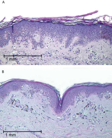
Figure 1 provides a photograph of 2 ‘‘prototypic’’ DN-LWs.
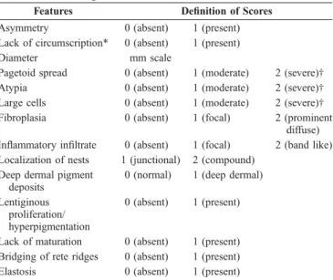
TABLE 1.Histologic Features Assessed and Scored
Features Definition of Scores
Asymmetry 0 (absent) 1 (present)
Lack of circumscription* 0 (absent) 1 (present)
Diameter mm scale
Pagetoid spread 0 (absent) 1 (moderate) 2 (severe)†
Atypia 0 (absent) 1 (moderate) 2 (severe)†
Large cells 0 (absent) 1 (moderate) 2 (severe)†
Fibroplasia 0 (absent) 1 (focal) 2 (prominent
diffuse) Inflammatory infiltrate 0 (absent) 1 (focal) 2 (band like) Localization of nests 1 (junctional) 2 (compound)
Deep dermal pigment deposits
0 (normal) 1 (deep dermal) Lentiginous
proliferation/
hyperpigmentation
0 (absent) 1 (present)
Lack of maturation 0 (absent) 1 (present) Bridging of rete ridges 0 (absent) 1 (present)
Elastosis 0 (absent) 1 (present)
*Okun definition: ‘‘a neoplasm is considered to show poorly defined lateral borders if there are significant intervals between individual tumor cells, and between groups of tumor cells, at the lateral perimeter, and/or decreasing density of aggregation of individual cells as the lateral perimeter is approached’’.5
†Moderate: average number#5, severe: average number.5 in 3 consecutive sections analyzed.
TABLE 2.Comparison of Site-Related and Sex-Related Characteristics (%)
Site Related
Sex Related DN-BW
(n = 20)
DN-LW (n = 42)
DN-LM (n = 20) Symmetry
Present 65.00 61.95 70.00
Absent 35.00 38.10 30.00
Circumscripton
Present 65.00 38.09 65.00
Absent 35.00 61.90 35.00
Diameter (mm), mean 4.99 2.8 3.06
Pagetoid spread
0 50.00 23.80 60.00
1 50.00 42.85 40.00
2 0 33.33 0
Cytologic atypia
0 65.00 42.85 95.00
1 35.00 42.85 5.00
2 0 14.28 0
Large melanocytes
0 10.00 21.42 35.00
1 90.00 30.95 65.00
2 0 47.60 0
Fibroplasia
0 55.00 4.76 10.00
1 45.00 64.28 50.00
2 0 30.95 40.00
Inflammatory infiltrate
Focal 75.00 85.71 90.00
Band like 25.00 14.28 10.00
Localization of nests
Compound 65.00 16.66 50.00
Junctional 35.00 83.33 50.00
Distribution of dermal pigment
0 70.00 45.23 85.00
1 30.00 54.76 15.00
Lentiginous P/hyperpigment
Present 55.00 73.80 45.00
Absent 45.00 26.19 55.00
Maturation
Present 100 95.23 90.00
Absent 0 4.76 10.00
Bridging
Present 80.00 54.76 30.00
Absent 20.00 45.23 70.00
P, proliferation.
600 | www.amjdermatopathology.com q2010 Lippincott Williams & Wilkins
Coras et al Am J DermatopatholVolume 32, Number 6, August 2010
Because the delineation of levels 1–2 superficial spreading MM versus DN is also a question of the observers’
thresholds, a set of MM was added as a control and to define the features that finally made the distinction. The main difference was that MM-LW were larger (mean size 4.28 mm) than DN-LW (mean size 2.8 mm). MM-LW also showed more severe cytologic atypia (65%) compared with DN-LW (14.28%) and more large melanocytes (80%) compared with DN-LW (47.6%). Both MM-LW and DN-LW showed pigment deposits in the underlying dermis. Elastosis was seen in less than 30% of MM-LW and DN-LW specimens and was not a useful distinguishing feature. Moreover, most MM-LW were not confined to the junction but infiltrated the dermis.
In summary, it emerged that a lesion larger than 4 mm with large atypical individual cells was more likely to be diag- nosed as MM. Pagetoid spread was a rather weak discriminator in small (,3 mm), predominantly junctional lesions where it seemed to not be strongly linked to malignancy.
DISCUSSION
As with melanocytic nevi located on acral, auricular, flexural, and genital sites, this study has shown dysplastic nevi on the lower legs of women also have site-specific features and, additionally, sex-specific differences.
Melanocytic nevi of the palms and soles may exhibit elongation of rete ridges and continuous proliferation of melanocytes at the dermal–epidermal junction. Single or nested melanocytes may be scattered throughout the epidermis including the spinous, granular, and cornfied layers. There may be poor or absent lateral circumscription.6,7
Nevi of the genitalia (vulva and perineum) may show wide lateral extension. There may be considerable variation in the size and shape of melanocytic nests, with confluence of melanocytic nests. Melanocytes may be seen in epithelial structures of adnexae and epidermal melanocytes may show cytologic atypia. Such genital nevi have been called ‘‘atypical melanocytic nevi of the genitalia.’’8–10
Nevi located on flexural sites (axilla, umbilicus, inguinal creases, pubis, scrotum, and perianal area) can show features similar to melanocytic nevi of the genital skin. In one series of young patients who had flexural nevi excised for mostly cosmetic reasons, large irregular poorly cohesive but con- fluent nests of melanocytes were seen at the dermoepidermal junction, and often involved adnexal epithelial structures.
Some degree of fibroplasia was commonly seen in the dermis.11
In auricular and periauricular melanocytic nevi excised for a variety of reasons, many histologic features traditionally associated with MM have been noted. A common feature is irregularity of the nesting pattern of melanocytes at the dermoepidermal junction with variability in size and shape, with nests even between rete ridges. Moderate to severe cytological atypia consisted of large melanocytes with pale finely granular cytoplasm. Melanocytes were also dyscohesive and showed pagetoid spread into the epidermis. However, mitotic figures and apoptotic melanocytes were not seen.12,13 The cause of site-specific variation in the histologic features of melanocytic lesions is not known but suggestions have included external factors including ultraviolet exposure and pressure, and intrinsic factors such as age and hormonal influences.8,10,14,15
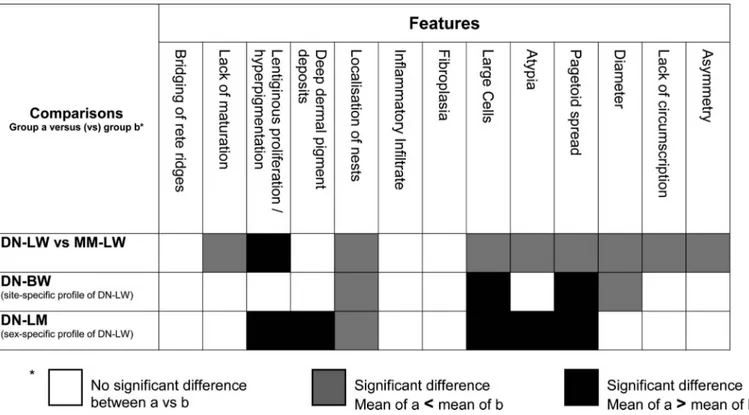
TABLE 3.Significant Difference of the Mean Scores (Mann-hitney Test:P,0.05) Between 3 Groups
q2010 Lippincott Williams & Wilkins www.amjdermatopathology.com | 601
Am J DermatopatholVolume 32, Number 6, August 2010 Dysplastic Melanocytic Nevi of the Lower Leg
In a recent study of melanocytic nevi of the ankle, 4 lesion types were identified—benign melanocytic nevi, dysplastic nevi, MM, and melanocytic nevi of the ankle with atypical features. This latter group was most commonly seen in women, were small (average diameter 3.0 mm) and showing severe architectural disorder, prominent single-cell prolifera- tion, and lack of circumscription. The majority showed mild cytological atypia. On follow-up, these lesions had a benign course.16 This group shows some similarity to the DN-LW, although dysplastic nevi had been specifically separated off.
In this study of melanocytic lesions of the lower leg, a set of histopathology features has been presented specifically distinguishing DN in women from other melanocytic lesions on this location. DN-LW showed significant differences from DN-BW and from the DN-LM suggesting that this is another special situation. DN-LW were predominantly junctional with prominent pagetoid spread and a dermal pigment band, compared with those in men which were compound nevi with little or no pagetoid spread or pigment band. DN-LW were smaller and more likely to show pagetoid spread than those on the back. Such common features in DN of the lower leg make
distinction from early MM in this site in women difficult as there is considerable overlap. Elastosis, a marker of chronic sun damage, was seen in less than 30% of CN-LW, DN-LW, and MM-LW specimens and was not a distinguishing feature.
This is consistent with the current theory that MM in usually protected sites such as the back, result from acute rather than chronic sun damage.17In addition, the age range was similar in all groups, with MM occurring as young as 25 years. So patient age was also not helpful to distinguish between MM and DN. However, one useful clue that has emerged from this study is lesion size. In addition to assessing the usual histological features, an assessment of diameter may provide additional information aiding diagnosis. A large histologically atypical melanocytic lesion on the lower leg of a woman is more likely to be a MM than a DN.
REFERENCES
1. Zalaudek I, Hofmann-Wellenhof R, Kittler H, et al. A dual concept of nevogenesis: theoretical considerations based on dermoscopic features of melanocytic nevi.J Dtsch Dermatol Ges. 2007;5:985–992.
2. Roesch A, Burgdorf W, Stolz W, et al. Dermatoscopy of ‘‘dysplastic nevi’’:
a beacon in diagnostic darkness.Eur J Dermatol. 2006;16:479–493.
3. Hosler GA, Moresi JM, Barrett TL. Nevi with site-related atypia: a review of melanocytic nevi with atypical histologic features based on anatomic site.J Cutan Pathol. 2008;35:889–898.
4. Clark LN, Shin DB, Troxel A, et al. Association between the anatomic distribution of melanoma and sex. J Am Acad Dermatol. 2007;56:
768–773.
5. Okun MR. Histological demarcation of lateral borders: an unsupportable criterion for distinguishing malignant melanoma from Spitz naevus and compound naevus.Histopathology. 1998;33:158–162.
6. Kerl H, Trau H, Ackerman AB. Differentiation of melanocytic nevi from malignant melanomas in palms, soles, and nail beds solely by signs in the cornified layer of the epidermis.Am J Dermatopathol. 1984;6(Suppl):
159–160.
7. Ackerman AB, Cerroni L, Kerl H.Pitfalls in Histopathologic Diagnosis of Malignant Melanoma. Philadelphia, PA: Lea and Febiger; 1994.
8. Christensen WN, Friedman KJ, Woodruff JD, et al. Histologic character- istics of vulvar nevocellular nevi.J Cutan Pathol. 1987;14:87–91.
9. Clark LN, Shin DB, Troxel A, et al. Association between the anatomic distribution of melanoma and sex.J Am Acad Dermatol. 2007;56:768–773.
10. Friedman RJ, Ackerman AB. Difficulties in the histological diagnosis of melanocytic nevi on the vulvae of premenopausal women. In: Ackerman AB, eds.Pathology of Malignant Melanoma. New York, NY: Masson Publications; 1981:119–127.
11. Rongioletti F, Ball RA, Marcus R, et al. Histopathological features of flexural melanocytic nevi: a study of 40 cases.J Cutan Pathol. 2000;27:
215–217.
12. Saad AG, Patel S, Mutasim DF. Melanocytic nevi of the auricular region:
histologic characteristics and diagnostic difficulties.Am J Dermatopathol.
2005;27:111–115.
13. Lazova R, Lester B, Glusac EJ, et al. The characteristic histopathologic features of nevi on and around the ear.J Cutan Pathol. 2005;32:40–44.
14. Blessing K. Benign atypical naevi: diagnostic difficulties and continued controversy.Histopathology. 1999;34:189–198.
15. Foucar E, Bentley TJ, Laube DW, et al. A histopathologic evaluation of nevocellular nevi in pregnancy.Arch Dermatol. 1985;121:350–354.
16. Khalifeh I, Taraif S, Reed JA, et al. A subgroup of melanocytic nevi on the distal lower extremity (ankle) shares features of acral nevi, dysplastic nevi, and melanoma in situ.Am J Surg Pathol. 2007;31:1130–1136.
17. Curtin JA, Fridlyand J, Kageshita T, et al. Distinct sets of genetic alterations in melanoma.N Engl J Med. 2005;353:2135–2147.
FIGURE 1.A, Typical profile of a DN-LW. Such lesions are small, usually ,3 mm, and considerable pagetoid spread and enlarged atypical single cells can be found. B, Another frequent finding in DN-LW is deep dermal deposits of pigment accompanied by a moderate inflammatory response.
602 | www.amjdermatopathology.com q2010 Lippincott Williams & Wilkins
Coras et al Am J DermatopatholVolume 32, Number 6, August 2010
- 5 -
Dysplastische melanozytäre Nävi des Unterschenkels:
Geschlechts- und lokalisationsspezifische Histopathologie
Einleitung
In vielen Dermatopathologie- bzw. Pathologielehrbüchern überschneiden sich die Kriterien des initialen malignen Melanoms mit denen des schwer dysplastischen melanozytären Nävus, so dass sich besondere Schwierigkeiten in der Abgrenzung ergeben. Auch einige lokalisationsspezifische Kriterien für melanozytäre Nävi ähneln den Melanommerkmalen.
Zur lokalisationsspezifischen Histopathologie des melanozytären Nävus wurden bereits einige Studien publiziert, welche auf die Besonderheiten an Akren, Ohrregion, Beugeseiten der Extremitäten und im Genitalbereich eingehen.
In den letzten Jahren zeigte sich darüber hinaus, dass auch die Unterschenkel zu diesen besonderen Lokalisationen zu rechnen sind.
Bei Frauen finden sich aber an diesem Ort auch gehäuft superfiziell spreitende maligne Melanome, was zusätzlich die Differentialdiagnose kompliziert macht.
Für den Dermatopathologen/Pathologen besteht daher eine große Herausforderung darin, den dysplastischen Nävus des Unterschenkels vom superfiziell spreitenden malignen Melanom (Level 1 bis 2) zu unterscheiden. Kann ein initiales malignes Melanom bei einer atypischen Läsion nicht ausgeschlossen werden, wird diese manchmal unnötigerweise mit größerem Sicherheitsabstand exzidiert.
In der vorliegenden Studie steht der dysplastische Nävus des Unterschenkels (zwischen Knie und Fußgelenk) im Mittelpunkt (einzige Publikation).
- 6 -
Fragestellung des Projektes
Ausgehend von den Literaturdaten ergaben sich für diese Arbeit folgende Fragestellungen 1. Welche Unterschiede bestehen zwischen den melanozytären Nävi am Unterschenkel und Rücken bei Frauen?
2. Welche Unterschiede bestehen zwischen den dysplastischen Nävi am Unterschenkel zwischen Frauen und Männern?
3. Welche Kriterien sind geeignet melanozytäre Nävi, dyplastische Nävi und maligne Melanome am Unterschenkel von Frauen zu differenzieren?
In dieser retrospektiven histopathologischen Studie verglichen wir dysplastische Nävi am Unterschenkel von 42 Frauen (DN-LW, L = lower leg, W = women) mit Nävi bzw.
Melanomen von Patienten aus verschiedenen Kontrollgruppen. Dazu zählten 20 Männer mit dysplastischen Nävi am Unterschenkel (DN-LM, M = men), 20 Frauen mit dysplastischen Nävi am Rücken (DN-BW, B = back), 40 Frauen mit unspezifischen Nävi am Unterschenkel (CN-LW, CN = common nevus) und 20 Frauen mit initialen superfiziell spreitenden Melanomen (Level 1-2) am Unterschenkel (MM-LW, MM = malignant melanoma).
Ziel war es, zum einen zu beurteilen, ob DN-LW lokalisations- und geschlechtsspezifische Eigenschaften aufweisen und zum anderen die histologischen Kriterien zu definieren, die den DN vom initialen MM am Unterschenkel bei Frauen unterscheidet.
- 7 -
Material und Methoden
Aus der Datenbank der Dermatologischen Klinik der Universität Regensburg wurden die
"melanozytären Nävi am Unterschenkel" bezogen auf den Zeitraum von 2004 bis 2006 gesichtet. Hierbei war mit "Unterschenkel" die Region zwischen Knie und Sprunggelenk definiert. Spitznävi, blaue Nävi, intradermale Nävi und unklassifizierte Läsionen wurden ausgeschlossen. Die Läsionen mussten im Gesunden exzidiert sein.
Der "dysplastische Nävus" wurde als ein Nävus mit breitflächigen architektonischen und/oder zytologischen Unregelmäßigkeiten definiert. Die Definition wurde zunächst von drei Untersuchern/Dermatohistologen, den Autoren Brigitte Coras, Thomas Vogt und Michael Landthaler, unabhängig voneinander formuliert und abschließend in einer Konsensuskonferenz abgestimmt. Der "dysplastische Nävus des Unterschenkels bei der Frau" (DN-LW) wurde als spezifische Untergruppe identifiziert. Zum Vergleich wurden folgende Gruppen herangezogen: der dysplastische Nävus am Unterschenkel des Mannes (DN-LM), der dysplastische Nävus am Rücken der Frau (DN-BW), der unspezifische Nävus ("common nevus") am Unterschenkel der Frau (CN-LW) und das superfiziell spreitende maligne Melanom am Unterschenkel der Frau (MM-LW). Das Mindestalter lag bei 21 Jahren.
Jeder Untersucher beurteilte semiquantitativ die einzelnen Veränderungen anhand vordefinierter Kriterien: Symmetrie, Begrenzung, Durchmesser, pagetoide Durchsetzung, zytologische Atypien, Größe der Melanozyten, Fibroplasie, entzündliche Infiltrate, Lokalisation der Nester, Verteilung der dermalen Pigmentablagerung, lentiginöse Hyperplasie und Hyperpigmentierung, "Reifung" der dermalen Melanozyten, Brücken- bildung zwischen benachbarten Reteleisten und Elastose. Nach Diskussion wurde dann ein
- 8 -
Konsensus-Score etabliert, der jeweils mit den Punktwerten 0, 1 oder 2 versehen war (z.B.
Asymmetrie: 0 = nicht vorhanden, 1 = vorhanden, oder Atypie: 0 = nicht vorhanden, 1 = moderate Ausprägung, 2 = schwere Ausprägung). (Tabelle1). Das Auswertungs-System wurde kalibriert, indem ein Set von Referenzläsionen mit verschiedenen Punktwerten (Scores) erstellt wurde. Dieses Set wurde dann vergleichend während der gesamten Evaluation verwendet.
- 9 -
Tabelle 1
Histologische Kriterien
*Okun-Definition: Laterale unscharfe Begrenzung bedeutet, dass atypische Melanozyten in Einzelformationen an der Junktionszone (oder suprabasal) über das letzte erkennbare Melanozytennest in der Epidermis hinaus angeordnet sind
** moderate Ausprägung: durchschnittliche Anzahl ≤ 5, schwere Ausprägung:
durchschnittliche Anzahl > 5 in drei aufeinander folgenden beurteilten Serienschnitten Vordefinierte Kriterien Definition des Scores
Symmetrie 0 (nicht
vorhanden)
1 (vorhanden)
Begrenzung* 0 (nicht
vorhanden)
1 (vorhanden)
Durchmesser mm
Pagetoide Durchsetzung 0 (nicht vorhanden)
1 (moderate Ausprägung)
2 (schwere Ausprägung)**
Zytologische Atypien 0 (nicht vorhanden)
1 (moderate Ausprägung)
2 (schwere Ausprägung)**
Große Melanozyten 0 (nicht
vorhanden)
1 (moderate Ausprägung)
2 (schwere Ausprägung)**
Fibroplasie 0 (nicht
vorhanden)
1 (fokal) 2 (diffus) Entzündliche Infiltrate 0 (nicht
vorhanden)
1 (fokal) 2 (bandartig) Lokalisation der Nester 1 (junktional) 2 (compound)
Verteilung der dermalen Pigmentablagerung
0 (normal) 1 (bandartige Pigmentierung in der Dermis) Lentiginöse Hyperplasie/
Hyperpigmentierung
0 (nicht vorhanden)
1 (vorhanden) Fehlende “Reifung” der Melanozyten 0 (nicht
vorhanden)
1 (vorhanden)
Brückenbildung 0 (nicht
vorhanden)
1 (vorhanden)
Elastose 0 (nicht
vorhanden)
1 (vorhanden)
- 10 -
Statistische Analyse
Das SPSS 13.0 Software-Paket wurde verwendet, um die grundlegenden statistischen Eigenschaften zu analysieren, den Durchschnittswert und ein 95%-Konfidenzintervall des Durchschnitswertes festzulegen. Für Unterschiede des Durchschnittswertes wurde der nicht parametrische Mann-Whitney-U-Test eingesetzt. Die Ergebnisse wurden als signifikant angesehen, wenn ein P ≤ 0,05 erreicht war.
Ergebnisse
In der Datenbank wurden 996 melanozytäre Hautveränderungen des Unterschenkels anhand der oben definierten Ein- und Ausschlusskriterien identifiziert.
Diese waren überwiegend benigne junktionale melanozytäre Nävi. Die lokalisations- spezifische Charakteristika der melanozytären Läsionen bei Frauen wurden beurteilt durch den Vergleich von 42 Läsionen mit unspezifischen Nävi („common nevi“ der gleichen Lokalisation, N = 40), dysplastische Nävi am Rücken (DN-BW, N = 20) und maligne Melanome des Unterschenkels der Frau (MM-LW, N =20). Durch den Vergleich der DN- LM (N = 20) und der DN-LW (N = 42) konnten geschlechtsspezifische Charakteristika festgestellt werden. Der Altersdurchschnitt (21-60 Jahre) war in allen Gruppen gleich hoch. Die histologischen Unterschiede sind in den Tabellen 2 und 3 zusammengefasst. CN- LW und DN-LW waren ähnlich in Bezug auf Durchmesser der Läsion, Elastose, „Reifung“
und Brückenbildung zwischen den Reteleisten. Es wurden erwartungsgemäß Unterschiede in Symmetrie und Begrenzung festgestellt. Darüberhinaus im Einklang mit der Definition des „dysplastischen Nävus“ zytologische Atypien und der Nachweis von großen Melanozyten. Die pagetoide Durchsetzung wurde in etwa die Hälfte der DN-LW beobachtet und war schwer ausgeprägt in einem Drittel der Fälle. Fibroplasie war in DN- LW häufig und in der Regel eher fokal als diffus. Eine charakteristische bandartige
- 11 -
dermale Pigmentierung wurde in weniger als die Hälfte der Fälle der dysplastischen Nävi, aber nicht in den unspezifischen Nävi beobachtet. Im Vergleich mit den DN-BW waren DN-LW deutlich kleiner (4,99 vs 2,8 mm) und zeigten häufiger eine pagetoide Durch- setzung (P ≤ 0,05). Die dysplastischen Nävi der Männer (DN-LM) waren überwiegend Nävi vom „Compound Typ“ mit nur geringfügiger oder fehlender pagetoider Durch- setzung. Zum Vergleich waren die dysplastischen Nävi der Frauen (DN-LW) vorranging junktional mit prominenter pagetoider Durchsetzung und einer bandartigen dermalen Pigmentierung.
Da die Abgrenzung zwischen dem superfiziell spreitenden malignem Melanom (MM) Level 1-2 und dem dysplastischen Nävus auch eine Frage des Begutachters ist, wurde ein Set von malignen Melanomen als Kontrolle definiert. Die wichtigsten Unterschiede waren, dass die MM-LW größer waren (mittlerer Durchmesser 4,28 mm) als DN-LW (mittlerer Durchmesser 2,8 mm). MM-LW zeigten auch schwerere zytologische Atypien (65%) als DN-LW (14,28%) und häufiger große Melanozyten (80%) verglichen mit DN-LW (47,6%). Sowohl MM-LW als auch DN-LW wiesen eine Pigmentablagerung in der darunter liegenden Dermis auf. Elastose wurde in weniger als 30% der MM-LW und der DN-LW gesehen und war kein besonderes Merkmal. Zusammenfassend zeigte sich, dass eine Läsion die größer als 4 mm ist und große atypische Melanozyten zeigt eher als ein MM diagnostiziert wurde. Die pagetoide Durchsetzung war kein entscheidendes Merkmal in kleinen, überwiegend junktionalen Läsionen.
- 12 -
Tabelle 2
Vergleich der lokalisations- und geschlechtsspezifischen Charakteristika (%)
Lokalisationsspezifisch
Geschlechtsspezifisch DN-BW
(n=20)
DN-LW (n=42)
DN-LM (n=20) Symmetrie
vorhanden nicht vorhanden
65.00 35.00
61.95 38.10
70.00 30.00 Begrenzung
vorhanden nicht vorhanden
65.00 35.00
38.09 61.90
65.00 35.00
Durchmesser (mm) Durchschnitt 4.99 2.8 3.06
Pagetoide Durchsetzung 0
1 2
50.00 50.00
0
23.80 42.85 33.33
60.00 40.00
0 Zytologische Atypien
0 1 2
65.00 35.00
0
42.85 42.85 14.28
95.00 5.00
0 Große Melanozyten
0 1 2
10.00 90.00
0
21.42 30.95 47.60
35.00 65.00
0 Fibroplasie
0 1 2
55.00 45.00
0
4.76 64.28 30.95
10.00 50.00 40.00 Entzündliche Infiltrate
Fokal Bandartig
75.00 25.00
85.71 14.28
90.00 10.00 Lokalisation der Nester
Compound Junktional
65.00 35.00
16.66 83.33
50.00 50.00 Verteilung des dermalen Pigmentes
0 1
70.00 30.00
45.23 54.76
85.00 15.00 Lentiginöse Hyperpigmentierung
vorhanden nicht vorhanden
55.00 45.00
73.80 26.19
45.00 55.00
« Reifung » vorhanden nicht vorhanden
100 0
95.23 4.76
90.00 10.00 Brückenbildung
vorhanden nicht vorhanden
80.00 20.00
54.76 45.23
30.00 70.00
- 13 -
Tabelle 3
Signifikante Unterschiede des Mittelwertes (Mann-Whitney-Test: P≤ 0,05) zwischen drei Gruppen
Unterschiede Gruppe a versus
(vs) Gruppe b
Charakteristika
Brückenbildung Fehlende “Reifung” derMelanozyten Lentiginöse Hyperplasie / Hyperpigmentierung Verteilung der dermalenPigmentablagerung Lokalisation der Nester Entzündliche Infiltrate Fibroplasie Große Melanozyten Zytologische Atypien Pagetoide Durchsetzung Durchmesser Unscharfe Begrenzung Asymmetrie
DN-LW vs MM-LW DN-LW vs DN-BW (Lokalisationspezifische Profil des DN-LW) DN-LW vs DN-LM (Geschlechtsspezifisches Profil des DN-LW)
Diskussion
Wie bei melanozytären Nävi der Akren, der Ohrregion, der Beugeseiten der Extremitäten und des Genitalbereichs zeigt diese Studie, für dysplastische Nävi am Unterschenkel von Frauen auch lokalisationsspezifische Merkmale und zusätzlich geschlechtsspezifische Unterschiede.
Melanozytäre Nävi an den Handflächen und Fußsohlen können lang ausgezogene Reteleisten und eine kontinuierliche Proliferation der Melanozyten an der Junktionszone zeigen. Einzelstehende oder zu Nestern aggregierte Melanozyten können sich auch in den oberen Epidermisetagen finden. Die Läsionen sind scharf oder unscharf begrenzt.
Kein signifikanter
Unterschied a vs b Signifikanter Unterschied Mean of a
<
mean of bSignifikanter Unterschied Mean of a
>
mean of b- 14 -
Die melanozytären Nävi im Genitalbereich (Vulva und Perineum) sind breite Läsionen. Es können erhebliche Unterschiede in der Größe und Form der melanozytären Nester bestehen, mit Konfluenz der Nester. Melanozyten werden auch in den epithelialen Strukturen der Adnexe gesehen und die intraepidermalen Melanozyten können zytologischen Atypien zeigen. Diese genitalen Nävi wurden als "atypische Nävuszellnävi im Genitalbereich" bezeichnet. Darüber hinaus können die melanozytären Nävi der Beugenseiten der Extremitäten (Axilla, Nabel, inguinal, Mons pubis, skrotal und Perianalbereich) ähnliche Merkmale wie die melanozytäre Nävi im Genitalbereich aufweisen. Bei einer Reihe von jungen Patienten mit Nävi an den Beugen, die aus kosmetischen Gründen entfernt worden sind, wurden große, unregelmäßige, aber konfluierende Nester von Melanozyten an der dermo-epidermalen Junktionszone gesehen, oft mit Beteiligung der adnexialen epithelialen Struktur. Zum Teil wurde ebenfalls Fibroplasie beschrieben.
Auch in den aurikulären und periaurikulären melanozytäre Nävi, die aus verschiedenen Gründen exzidiert worden sind, wurden Kriterien des malignen Melanoms beschrieben.
Ein gemeinsames Merkmal ist die Irregularität der Nester an der dermo-epidermalen Junktionszone mit Variabilität in Form und Größe, mit Nestern sogar zwischen den Reteleisten. Mittelschwere bis schwere zytologischen Atypien der Melanozyten mit blassem fein granulärem Zytoplasma sowie pagetoide Durchsetzung der Epidermis wurden ebenfalls nachgewiesen. Allerdings wurden Mitosen und apoptotische Melanozyten nicht gesehen.
In einer von Khalifeh et. al publizierten Studie über melanozytäre Nävi des Sprunggelenks wurden vier Typen von Läsionen identifiziert: benigne Nävuszellnävi, dysplastische Nävi, maligne Melanome und Nävuszellnävi des Sprunggelenks mit atypischen Merkmalen.
Diese letztere Gruppe wurde am häufigsten bei Frauen beobachtet. Die Läsionen waren
- 15 -
klein (mittlerer Durchmesser 3,0 mm) und wiesen schwere architektonische Unregelmäßigkeiten, prominente Einzelzellproliferationen und keine scharfe Begrenzung, aber meistens nur geringe zytologische Atypien auf.
Im Follow-up zeigten sich bei den als dysplastische melanozytäre Nävi diagnostizierten Fälle keine Lokalrezidive, was von den Autoren als Bestätigung für ihre Kriterien angesehen wurde. Diese Gruppe zeigt eine gewisse Ähnlichkeit mit dem dysplastischen Nävus des Unterschenkels der Frau (DN-LW), obwohl diese dysplastischen Nävi speziell abgetrennt wurden.
Einige Autoren vermuten, dass die zytologischen und architektonischen Veränderungen der melanozytären Läsionen in besonderen Lokalisationen durch die Einwirkung von äußeren Faktoren, wie z. B. UV-Exposition, Druck als auch durch intrinsische Faktoren wie z. B. Alter sowie durch hormonelle Einflüsse zu erklären sind.
Im Vergleich zu den dysplastischen Nävi des Rückens, waren die dysplastischen Nävi am Unterschenkel der Frau kleiner im Durchmesser und zeigten darüber hinaus einen signifikant höheren Score bezüglich der pagetoiden Durchsetzung (P< 0,05). Beim dysplastischen Nävus des Unterschenkels bei Frauen (DN-LW) fielen geschlechts- spezifische Unterschiede zu den Männern (DN-LM) auf. Diese Unterschiede bezogen sich auf 1. die pagetoide Durchsetzung (P < 0,05), 2. die zytologische Atypien (P < 0,05), 3. das Auftreten von großen Melanozyten (P < 0,05) und 4. eine bandartige Pigmentierung in der Dermis (54% der DN-LW vs. 15% der DN-LM).
Zusammenfassend konnten in dieser Studie sowohl lokalisations- und geschlechts- spezifische Eigenschaften des dysplastischen Nävus am Unterschenkel der Frau als auch einige histopathologische Besonderheiten melanozytärer Läsionen des Unterschenkels herausgearbeitet werden:
- 16 -
DN-LW waren vorrangig junktional mit prominenter pagetoider Durchsetzung und einer bandartigen dermalen Pigmentierung. Im Vergleich dazu handelte es sich bei den Männern (DN-LM) um Nävi vom "Compound Typ" mit nur geringfügiger oder fehlender pagetoider Durchsetzung. Auch die bandartige dermale Pigmentierung war nur gering ausgeprägt oder fehlte ganz.
DN-LW waren kleiner und zeigten häufiger eine pagetoide Durchsetzung als dysplastische Nävi am Rücken (DN-BW).
Gemeinsamkeiten zwischen dem dysplastischen Nävus des Unterschenkels der Frau und dem initialen malignen Melanom in dieser Lokalisation erschweren die Differenzial- diagnose. Die Elastose, ein Marker für chronische Schäden durch Sonneneinstrahlung, war in der Differentialdiagnose kein wegweisendes Merkmal, denn sie konnte nur in weniger als 30% der DN-LW, CN-LW und MM-LW festgestellt werden. Diese Beobachtung stimmt mit der aktuellen Theorie überein, dass das maligne Melanom zum Beispiel in einer von der Sonne geschützten Region wie am Rücken das Ergebnis eines akuten und nicht eines chronischen Sonnenschadens ist.
Da in der Melanomgruppe auch Patienten unter 30 Jahren zu beobachten waren und auch der Altersdurchschnitt in allen Gruppen ähnlich lag, liefert dieses Kriterium keine zusätzliche Information. Viel wichtiger ist die Größe der Hautveränderung. Eine melanozytäre Läsion des Unterschenkels bei Frauen mit mehr als 4 mm ist mit höherer Wahrscheinlichkeit ein malignes Melanom als ein dysplastischer Nävus.
Dermatohistologen/Pathologen sollten berücksichtigen, dass der dysplastische Nävus des Unterschenkels der Frau lokalisations- und geschlechtsspezifische Charakteristika aufweist. Dadurch kann das Risiko von Überdiagnosen bei diesen dysplastischen Nävi vermieden werden.
- 17 -
Publikationen
1. Coras B, Burgdorff T, Landthaler M, Stolz W: Kombinierte Immun- /Chemotherapie-eine Therapiebeobachtung. Der Deutsche Dermatologe 2001;8:555-7.
2. Coras B, Glaessl A, Kinateder J, et al. Teledermatoscopy in daily routine-results of the first 100 cases. Curr Probl Dermatol 2003;32:207-12.
3. Glaessl A, Coras B, Popal H, et al. A survey among dermatologist in practice about teledermatology. Curr Probl Dermatol 2003; 32:172-5.
4. Coras B, Hohenleutner U, Landthaler M, et al. Spindle cell hemangioma. Dermatol Surg 2003;29:875-8.
5. Coras B, Vogt T, Hohenleutner U. et al. Porokeratotic eccrine ostial and dermal duct naevus (PEODDN). JDDG 2003;10:797-800.
6. Coras B, Hohenleutner S, Raff K Lanthaler M, Hohenleutner U. Das „rote
Melanom“- eine seltene Variante des amelanotischen malignen Melanoms. JDDG 2004;7:597-600.
7. Coras B, Hafner C, Reichle A, Hohenleutner U, Szeimies RM, Landthaler M, Vogt T. Antiangiogenic therapy with pioglitazone, rofecoxib, and trofosfamide in a patient with endemic Kaposi sarcoma. Arch Dermatol 2004;140:1504-7.
8. Bongartz T, Coras B, Vogt T, Schölmerich, Müller-Ladner U. Treatment of active psoriatic arthritis with the PPAR ligand pioglitazone: an open-label pilot study.
Rheumatology 2005;44:126-9.
9. Coras B, Vogt T, Ulrich H, Landthaler M, Hohenleutner U. Fumaric acid esters therapy-a new treatment modality in pityriasis rubra pilaris? Br J Dermatol 2005;152:388-9.
10. Coras B, Eßbauer S, Pfeffer M, Meyer H, Schröder J, Stolz W, Landthaler M, Vogt T. Cowpox and a cat. Lancet 2005;365:446.
11. Coras B, Hohenleutner U, Landthaler M, Hohenleutner S. Comparison of two absorbable monofilament polydioxanone threads in intradermal buried sutures.
Dermatol Surg 2005;31:331-3.
- 18 -
12. Coras B, Landthaler M, Hofstaedter F, MeiselC , Hohenleutner U. Fibroadenoma of the axilla. Dermatol Surg 2005;31:1152-4.
13. Coras B, Hafner C, Roesch A, Vogt T, Landthaler M, Hohenleutner U. Congenital cartilaginous rests of the neck (wattles). Dermatol Surg 2005;31:1349-50.
14. Coras B, Hohenleutner U, Landthaler M, Hohenleutner S. Early recurrence of eruptive vellus hair cysts after Er:YAG laser therapy: case report and review of the literature. Dermatol Surg 2005;31:1741-4.
15. Coras B, Roesch A, Koschorreck L, Landthaler M, Vogt T. Post-traumatic iatrogenic aggravation of eruptive collagenomas in a child. Eur J Dermatol 2006;16:196-7.
16. Coras B, Michel S, Landthaler M, Hohenleutner U. Rosai-Dorfman disease with cutaneous manifestation (sinus histiocytosis with massive lymphadenopathy. Eur J Dermatol 2006;16:293-6.
17. Coras B, Vogt T, Hafner C, Landthaler M, Reichle A. Antiangiogenic therapy in metastatic prostate carcinoma complicated by cutaneous lupus erythematodes.
Lancet Oncol 2006;7:695-7.
18. Coras B, Vogt T, Roesch A, Landthaler M, Hohenleutner U. Bowen`s disease on porokeratotic eccrine ostial and dermal duct nevus. Dermatol Surg 2007;33:496-9.
19. Hafner C, Schmiemann V, Ruetten A, Coras B, Landthaler M, Reifenberger J, Vogt T. PCTH mutations are not mainly involved in the pathogenesis of sporadic trichoblastomas. Human Pathol 2007;38:1496-500.
20. Reichle A, Vogt T, Coras B, Terheyden P, Neuber K, Trefzer U, Schultz E, Berand A, Bröcker EB, Landthaler M, Andreesen R. Targeted combined anti-inflammatory and angiostatic therapy in advanced melanoma: a randomized phase II trial.
Melanoma Res 2007;17:360-4.
21. Schroeder JA, Weingart C, Coras B, Hausser I, Reinhold S, Mack M, Seybold V, Vogt T, Banas B, Hofstaedter F, Krämer BK. Ultrastructural evidence of dermal gadolinium deposits in a patient with nephrogenic systemic fibrosis and end-stage renal disease. Clin J Am Soc Nephrol 2008;3:968-75.
22. Coras B, Weigert U, Ramrath K, Guther S, Stolz W.Das maligne Melanom.
Aktuelle Aspekte zu Diagnostik und Therapie. TZM News 2008;2:4-10.
- 19 -
23. Eigentler TK, Radny P, Hauschild A, Gutzmer R, Linse R, Pföhler C, Wagner SN, Schadendorf D, Ellwanger U, Garbe C; German Dermatologic Cooperative
Oncology Group. Adjuvant treatment with vindesine in comparison to observation alone in patients with metastasized melanoma after complete metastasectomy: a randomized multicenter trial of the German Dermatologic Cooperative Oncology Group. Melanoma Res 2008;18:353-8.
24. Coras B, Landthaler M, Stolz W, Vogt T. Dysplastic melanocytic nevi of the lower leg: sex-and site-specific histopathology. Am J Dermatopathol 2010;32:599-602.
25. Miltenburg C, Peters B, Coras B, Stolz W. Glistening white erosions on the buccal mucosa with tonsillar edema more marked on the right than the left. J Dtsch
Dermatol Ges 2010;8:1021-3.
26. Geissler S, Dyall-Smith D, Coras B, Guther S, Peters B, Stolz W. Unique brown star shape on dermatoscopy of generalized Dowling-Degos disease. Australas J Dermatol 2011;52:151-3.
27. Klein A, Coras B, Landthaler M, Babilas P. Off-label use of fumarate therapy for granulomatous and inflammatory skin diseases other than psoriasis vulgaris: a retrospective study. J Eur Acad Dermatol Venereol. 2012;26:1400-6.
28. Coras-Stepanek B, von Portatius A, Dyall-Smith D, Stolz W. Dermatoscopy of pigmented extramammary Paget disease simulating melanoma. J Am Acad Dermatol. 2012;67:144-6.
29. Isbary G, Coras-Stepanek B, Stolz W. Pigmented lesion of the left nipple in a 49- year-old woman. J Dtsch Dermatol Ges 2013;Jul:23.
30. Isbary G, Coras-Stepanek B, Guther S, Tillmann A, Stolz W. Five patients with melanosis of the nipple and areola JEADV (2013 accepted)
31. Fischer I, Dyall-Smith D, Peters B, Stolz W, Coras-Stepanek B. Waldenström macroglobulinaemia presenting as tingling ulcers on the feet. Dermatol Pract Conc.
(2013 accepted) Buchbeiträge
1 Coras B, Landthaler M, (2008) Leon Goldman. In:Löser Ch, Plewig G (Hrsg) Pantheon der Dermatologie, Springer, Heidelberg S 357-61.
- 20 -
2 Coras B, Landthaler M, (2013) Leon Goldman. In: Löser Ch, Plewig G,Burgdorf W (eds). Pantheon of Dermatology, Springer, Heidelberg pp 384-7.
3 Coras B, Landthaler M, (2010) Hämangiome. In Szeimies RM, Hauschild A, Garbe C, Kaufmann R, Landthaler M (Hrsg) Tumoren der Haut. Grundlagen, Diagnostik und Therapie in der dermatologischen Onkologie, Thieme, Stuttgart, New York S 622-31.
- 21 -
Gewidmet meiner Familie