Multiple ocular manifestations in a case of cat scratch disease without systemic signs
Abstract
A 43-year-old woman presented with impaired vision and redness in her left eye of 2 weeks duration. She had a pet cat that scratched her
Kanako Annoura
1Ichiya Sano
1forehead 3 weeks before she presented to us. She had no systemic
Shinji Makino
1signs such as lymphadenopathy, fever, or fatigue. The respective right
Hidetoshi Kawashima
1and left corrected visual acuities were 20/16 and 20/2000. The anterior chamber of the left eye exhibited inflammation; a fundus examination of that eye revealed optic disc swelling and a serous macular detach-
1 Department of
Ophthalmology, Jichi Medical ment with hard stellate exudates. Based on the recent cat scratch and
the ocular findings, cat scratch disease (CSD) was suspected. The results
University, Shimotsuke-shi, Japan
of serologic testing showed elevated titres of IgM and IgG antibodies toBartonella henselae. Administration of doxycycline and a steroid was initiated. This report describes the occurrence of multiple ocular mani- festations of CSD in both the posterior and the anterior segment.
Keywords:cat scratch disease, anterior uveitis, serous macular detachment
Introduction
Cat scratch disease (CSD), a rare infection caused by Bartonella henselae, typically presents as subacute lymphadenopathy, fever, malaise, chills, and small skin lesions following a cat scratch or bite. Ocular involvement occurs in 5% to 10% of infected patients [1]. The clinical manifestations of ocular involvement include Parinaud oculoglandular syndrome (POGS), neuroretinitis, serous retinal detachment, retinal infiltrates, choroiditis, branch retinal vessel occlusion, endophthalmitis, and anterior uveitis. The presence of ocular symptoms without system- ic CSD is uncommon, as are fundus manifestations in patients with POGS or anterior uveitis [2]. We report a rare case of CSD in which there were multiple ocular manifestations but no systemic signs.
Case description
A 43-year-old woman presented with impaired vision in her left eye for the previous 2 weeks. She had a non- traumatic unilateral red eye, chemosis, and untreated hypertension. At the initial visit, the respective right and left corrected visual acuities were 20/16 and 20/2000, and the bilateral intraocular pressure was 20 mm Hg.
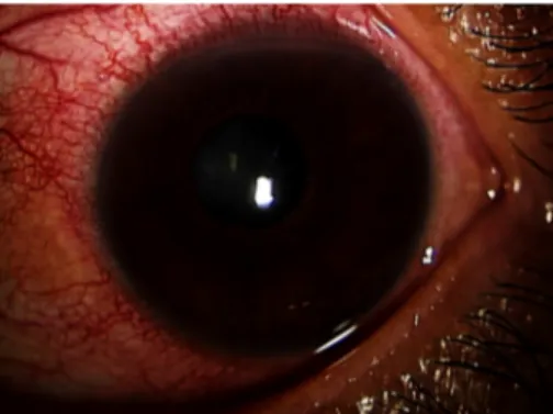
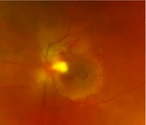
The chemosis was accompanied by serous discharge, keratic precipitates, partial synechia of the iris and cells (2+) in the anterior chamber (Figure 1, Figure 2), and vitreous of the left eye. A fundus examination of the left eye showed optic disc swelling, a focal retinitis lesion at the temporal margin of the optic disc, macular exudates in a star pattern, a flame-like retinal haemorrhage, and a serous macular detachment (Figure 3, Figure 4).
Fluorescein angiography showed leakage from the optic disc of the left eye (Figure 5). No signs of inflammation were apparent in the right eye. The patient reported that her pet cat had scratched her forehead 3 weeks before she presented for examination. She did not have a fever, any detectable skin lesions, or lymphadenopathy. An in- fectious disease specialist performed a systemic examin- ation and found no evidence of systemic CSD or any other infectious disease.
Figure 1: The chemosis with serous discharge
Figure 2: Keratic precipitates and cells in the anterior chamber
1/4 GMS Ophthalmology Cases 2020, Vol. 10, ISSN 2193-1496
Case Report
OPEN ACCESS
Figure 3: Optic disc swelling, a focal retinitis lesion at the temporal margin of the optic disc, macular exudates in a star
pattern, and a flame-like retinal haemorrhage
Figure 4: A serous macular detachment
Figure 5: Leakage from the optic disc of the left eye (fluorescein angiography)
Since conjunctivitis and iridocyclitis were present in addition to the aforementioned fundus findings, extensive investigations for uveitis were conducted that included measurement of the total blood count and erythrocyte sedimentation rate (ESR) as well as tests for auto- antibodies, syphilis, viruses, and toxoplasma antibody.
Except for a slightly elevated ESR rate (26 mm/hour), the results were unremarkable. Angiotensin-converting enzyme, soluble interleukin-2 receptor, and chest x-ray parameters were all within the normal limits. A blood sample was sent to a pathology laboratory for serology testing forB. henselaeantibody.
Since optic disc edema is associated with a macular star, it is important to exclude other causes such as malignant hypertension, sarcoidosis, toxoplasmosis, tuberculosis, and spirochetal disease [2]. The systemic blood pressure was 160/91 mm Hg, and all blood test results were
within the normal limits, with the exception of the slightly elevated ESR. QuantiFERON TB (Qiagen, Germantown, MD, USA) was negative. Serology testing forB. henselae antibodies was the only positive test result (IgM titre 1:16, IgG titre 1:256). Serology tests for cytomegalovirus, toxoplasma, and human immunodeficiency virus were negative.
Given the clinical ocular findings and the reported cat scratch, CSD was suspected even without systemic ill- ness. Oral doxycycline 200 mg/day and prednisolone 20 mg/day with tapering were prescribed. Topical corti- costeroid and mydriatic treatments were administered for the anterior chamber inflammation.
After the initiation of treatment, the optic disc edema, focal retinal infiltrates, retinal haemorrhage, serous retinal detachment, and macular exudates gradually decreased (Figure 6). The cells in the anterior chamber and vitreous resolved over the treatment course. However, it is note- worthy that the focal retinitis persisted and doxycycline was prescribed for 3 months. Six months after the start of treatment, macular atrophy developed and the visual acuity remained 20/40.
Figure 6: After the treatment, optic disc edema, focal retinal infiltrates, retinal haemorrhage, serous retinal detachment,
and macular exudates decreased.
Discussion
Three aspects of the current case are particularly note- worthy. One pertains to various complications involved.
POGS is reportedly the most common of the potential ocular manifestations of CSD, occurring in approximately 5% of patients with CSD [1]. Others include neuroretinitis (1–2%), and, less frequently, choroiditis, uveitis, retinal branch artery occlusion, retinal vein occlusion, and serous retinal detachment [3]. Even though each of these indi- vidual manifestations are rare in CSD, the current patient exhibited all of these ocular manifestations in the current case. POGS, anterior uveitis, neuroretinitis, a serous ret- inal detachment, and a flame-like retinal haemorrhage indicating retinal vein occlusion were all detected. POGS usually presents as unilateral conjunctivitis with or without conjunctival granulomata [4]. Although granulomatous
2/4 GMS Ophthalmology Cases 2020, Vol. 10, ISSN 2193-1496
Annoura et al.: Multiple ocular manifestations in a case of cat scratch ...
lesions in the conjunctiva were not detected in the current patient, chemosis and conjunctival injection with serous discharge were seen. Simultaneous neuroretinitis is un- common in patients with POGS [5], and anterior uveitis occurring in association with CSD is also rare [6]. In a previously reported case in a child, repetitive anterior uveitis was followed by neuroretinitis [7]. Thus, the simul- taneous occurrence of anterior and posterior complica- tions are noteworthy. Another notable feature of the cur- rent case was that the ocular involvement manifested without systemic disorders.
The current case is interesting in that neither lymphatic disease nor systemic symptoms developed. Classical CSD presents as a febrile illness associated with lymph- adenopathy, skin lesions, and fever. Lymphadenopathy has been considered a diagnostic criterion of CSD [8], [9]. The presence of ocular symptoms without systemic CSD is unusual, although some cases with ocular symp- toms but no systemic illness have been reported [10].
The current patient had no evidence of systemic disease.
We suspected CSD based on her report of a relatively recent cat scratch and various ocular findings in conjunc- tion with negative laboratory test results related to other possible diseases.
Unfortunately, the prognosis was not good despite the fact that the condition was most likely benign. Since CSD is regarded as benign, it is not uncommon for patients to be observed without treatment. Treatment remains con- troversial due to the self-limiting nature of the disease.
Neuroretinitis usually follows a benign course and visual function recovers in just a few months [11]. However, notably, ocular CSD can be accompanied by complications resulting in permanent visual loss. A strong clinical suspi- cion of CSD as the cause of vision-threatening intraocular inflammation is necessary before initiation of prompt antibiotic treatment. Reports have suggested that early treatment can improve visual outcomes [11], [12]. Since the current patient exhibited severe ocular complications, we prescribed both antibiotic and corticosteroid treat- ments. The efficacy of systemic corticosteroids remains controversial, but in patients with severe inflammation, systemic corticosteroids should be beneficial. Despite the early intervention and the long treatment duration in the current patient, the visual acuity recovery was not as good as we anticipated.
Conclusion
Ocular CSD is associated with various findings in the an- terior and posterior segments, but simultaneous anterior and posterior complications are rare. We report a rare case of CSD in which there were simultaneous multiple ocular manifestations but no systemic signs. CSD should be considered in patients with ocular signs, even in the absence of systemic signs.
Notes
Competing interests
The authors declare that they have no competing in- terests.
References
1. Carithers HA. Cat-scratch disease. An overview based on a study of 1,200 patients. Am J Dis Child. 1985 Nov;139(11):1124-33.
DOI: 10.1001/archpedi.1985.02140130062031 2. Jones DB. Cat-scratch disease. In: Pepose JS, Holland GN,
Wilhelmus KR, editors. Ocular Infection and Immunity. St. Louis:
Mosby Year Book; 1996. p. 1389-97.
3. Cunningham ET, Koehler JE. Ocular bartonellosis. Am J Ophthalmol. 2000 Sep;130(3):340-9. DOI: 10.1016/s0002- 9394(00)00573-0
4. Domínguez I, Cartes C, Sabat P, Ortiz O, Matus G, Traipe L.
Isolated conjunctival granuloma as a first manifestation of Parinaud’s oculoglandular syndrome: A case report. Am J Ophthalmol Case Rep. 2019 Jun;14:58-60. DOI:
10.1016/j.ajoc.2019.02.007
5. Wong MT, Dolan MJ, Lattuada CP Jr, Regnery RL, Garcia ML, Mokulis EC, LaBarre RA, Ascher DP, Delmar JA, Kelly JW.
Neuroretinitis, aseptic meningitis, and lymphadenitis associated with Bartonella (Rochalimaea) henselae infection in
immunocompetent patients and patients infected with human immunodeficiency virus type 1. Clin Infect Dis. 1995 Aug;21(2):352-60. DOI: 10.1093/clinids/21.2.352 6. Ur Rehman S, Metcalfe TW, Barnham M. Anterior uveitis
associated with cat scratch disease. Br J Ophthalmol. 1998 May;82(5):587-8. DOI: 10.1136/bjo.82.5.584d
7. Martínez-Osorio H, Calonge M, Torres J, González F. Cat-scratch disease (ocular bartonellosis) presenting as bilateral recurrent iridocyclitis. Clin Infect Dis. 2005 Mar;40(5):e43-5. DOI:
10.1086/427881
8. Anderson BE, Neuman MA. Bartonella spp. as emerging human pathogens. Clin Microbiol Rev. 1997 Apr;10(2):203-19. DOI:
10.1128/CMR.10.2.203-219.1997
9. Spach DH, Koehler JE. Bartonella-associated infections. Infect Dis Clin North Am. 1998 Mar;12(1):137-55. DOI:
10.1016/s0891-5520(05)70414-1
10. Celiker H, Kazokoglu H, Eraslan M, Cerman E, Karabas L.
Bartonella henselae Neuroretinitis in Patients without Cat Scratch. Jpn J Infect Dis. 2018 Nov;71(6):397-401. DOI:
10.7883/yoken.JJID.2017.518
11. Reed JB, Scales DK, Wong MT, Lattuada CP Jr, Dolan MJ, Schwab IR. Bartonella henselae neuroretinitis in cat scratch disease.
Diagnosis, management, and sequelae. Ophthalmology. 1998 Mar;105(3):459-66. DOI: 10.1016/S0161-6420(98)93028-7 12. Rolain JM, Brouqui P, Koehler JE, Maguina C, Dolan MJ, Raoult
D. Recommendations for treatment of human infections caused by Bartonella species. Antimicrob Agents Chemother. 2004 Jun;48(6):1921-33. DOI: 10.1128/AAC.48.6.1921-1933.2004
3/4 GMS Ophthalmology Cases 2020, Vol. 10, ISSN 2193-1496
Annoura et al.: Multiple ocular manifestations in a case of cat scratch ...
Corresponding author:
Ichiya Sano, MD, PhD
Department of Ophthalmology, Jichi Medical University, 3311-1 Yakushiji, Shimotsuke-shi, Tochigi 329-0498, Japan
lepapaigalagala@gmail.com
Please cite as
Annoura K, Sano I, Makino S, Kawashima H. Multiple ocular manifestations in a case of cat scratch disease without systemic signs. GMS Ophthalmol Cases. 2020;10:Doc45.
DOI: 10.3205/oc000172, URN: urn:nbn:de:0183-oc0001721
This article is freely available from
https://www.egms.de/en/journals/oc/2020-10/oc000172.shtml Published:2020-12-15
Copyright
©2020 Annoura et al. This is an Open Access article distributed under the terms of the Creative Commons Attribution 4.0 License. See license information at http://creativecommons.org/licenses/by/4.0/.
4/4 GMS Ophthalmology Cases 2020, Vol. 10, ISSN 2193-1496
Annoura et al.: Multiple ocular manifestations in a case of cat scratch ...