• Keine Ergebnisse gefunden

RESEARCH ARTICLE

N/A
N/A
Protected

Academic year: 2021

Aktie "RESEARCH ARTICLE"

Copied!
17
0
0

Wird geladen.... (Jetzt Volltext ansehen)

Volltext

(1)

RESEARCH ARTICLE

Effects of histamine on human periodontal ligament fibroblasts under simulated

orthodontic pressure

Marcella Groeger

1

, Gerrit Spanier

2

, Michael Wolf

3

, James Deschner

4

, Peter Proff

1

, Agnes Schro ¨ der

1☯

, Christian Kirschneck ID

1☯

*

1 Department of Orthodontics, University Hospital Regensburg, Regensburg, Germany, 2 Department of Cranio-Maxillo-Facial Surgery, University Hospital Regensburg, Regensburg, Germany, 3 Department of Orthodontics, University Hospital RWTH Aachen, Aachen, Germany, 4 Department of Periodontology and Operative Dentistry, University of Mainz, Mainz, Germany

☯ These authors contributed equally to this work.

* christian.kirschneck@ukr.de

Abstract

As type-I-allergies show an increasing prevalence in the general populace, orthodontic patients may also be affected by histamine release during treatment. Human periodontal lig- ament fibroblasts (PDLF) are regulators of orthodontic tooth movement. However, the impact of histamine on PDLF in this regard is unknown. Therefore PDLF were incubated without or with an orthodontic compressive force of 2g/cm

2

with and without additional hista- mine. To assess the role of histamine-1-receptor (H1R) H1R-antagonist cetirizine was used.

Expression of histamine receptors and important mediators of orthodontic tooth movement were investigated. PDLF expressed histamine receptors H1R, H2R and H4R, but not H3R.

Histamine increased the expression of H1R, H2R and H4R as well as of interleukin-6, cyclo- oxygenase-2, and prostaglandin-E2 secretion even without pressure application and induced receptor activator of NF-kB ligand (RANKL) protein expression with unchanged osteoprotegerin secretion. These effects were not observed in presence of H1R antagonist cetirizine. By expressing histamine receptors, PDLF seem to be able to respond to fluctuat- ing histamine levels in the periodontal tissue. Increased histamine concentration was asso- ciated with enhanced expression of proinflammatory mediators and RANKL, suggesting an inductive effect of histamine on PDLF-mediated osteoclastogenesis and orthodontic tooth movement. Since cetirizine inhibited these effects, they seem to be mainly mediated via his- tamine receptor H1R.

Introduction

In the dental specialty of orthodontics malpositioned teeth, which give rise to functional prob- lems as well as affect facial aesthetics, are moved into their correct physiological position within the alveolar bone by fixed or removable orthodontic appliances via the application of mechanical forces in the direction of required movement [1]. These forces promote the a1111111111

a1111111111 a1111111111 a1111111111 a1111111111

OPEN ACCESS

Citation: Groeger M, Spanier G, Wolf M, Deschner J, Proff P, Schro¨der A, et al. (2020) Effects of histamine on human periodontal ligament fibroblasts under simulated orthodontic pressure.

PLoS ONE 15(8): e0237040. https://doi.org/

10.1371/journal.pone.0237040

Editor: David M. Ojcius, University of the Pacific, UNITED STATES

Received: May 4, 2020 Accepted: July 17, 2020 Published: August 7, 2020

Peer Review History: PLOS recognizes the benefits of transparency in the peer review process; therefore, we enable the publication of all of the content of peer review and author responses alongside final, published articles. The editorial history of this article is available here:

https://doi.org/10.1371/journal.pone.0237040 Copyright: © 2020 Groeger et al. This is an open access article distributed under the terms of the Creative Commons Attribution License, which permits unrestricted use, distribution, and reproduction in any medium, provided the original author and source are credited.

Data Availability Statement: All relevant data are

within the paper and its Supporting Information

files.

(2)

formation of tensile and pressure zones in the periodontal ligament, which connects teeth to their surrounding alveolar bone. As a result, a sterile inflammatory reaction occurs in the peri- odontal ligament, which is mainly mediated by periodontal ligament fibroblasts (PDLF) [2,3], but also involves cells of the immune system such as macrophages, lymphocytes and T cells [4,5]. Stimulation by mechanical forces induces secretion of proinflammatory enzymes, cyto- kines and chemokines by PDLF [3,6,7]. Furthermore PDLF enhance receptor activator of NF- kB ligand (RANKL) expression and reduce osteoprotegerin secretion upon pressure applica- tion [3,6,8], thus promoting differentiation of osteoclast progenitor cells to bone-resorptive osteoclasts [7,9]. According to the biphasic theory of orthodontic tooth movement (OTM), movement is achieved via mechanically stressed PDLF- and lymphocyte-regulated bone resorption processes with subsequent bone formation via osteoclast-stimulated osteoblasts with numerous cell-cell interactions [2,3,10]. Despite the importance of orthodontic treatment for patient health, many aspects of orthodontic therapy have so far been poorly understood.

Nutrition is reported to influence the oral microflora in that an oral health-optimized diet can reduce inflammatory processes associated with gingivitis and periodontitis [11]. An influ- ence of diet-induced obesity on periodontal bone loss has also been demonstrated with adipos- ity and the applied fatty acid profile modulating bone metabolism [12,13]. Histamine, on the one hand, can be absorbed through food, but is also released in the body as part of innate immunity. In food, histamine is produced by the bacterial degradation of the amino acid histi- dine [14]. Biochemically histamine is a biogenic amine, just as tyramine, serotonin, dopamine, epinephrine, norepinephrine or octopamine. It is formed by elimination of carbon dioxide from the amino acid histidine and stored in particular in mast cells, basophilic granulocytes and nerve cells [15]. Histamine is a natural product that acts as a tissue hormone and neuro- transmitter in the human or animal organism and is also widely found in plants and bacteria [16]. In humans, histamine plays a central role in allergic reactions and is involved in the immune system and in the defense against foreign substances [15]. In the human body, hista- mine is produced by mast cells and released after an immune reaction [15,17]. It can cause a drop in blood pressure and allergic reactions such as itching or redness. Dietary histamine may, under certain conditions, also lead to such reactions, including poisoning [18].

Four different histamine receptors are currently known: H1R, H2R, H3R and H4R [19,20].

They are distinguished by their function, structure, distribution, and their affinity to histamine [21–23]. Histamine can have pro-inflammatory and anti-inflammatory effects, which are mediated by different histamine receptor subtypes and cell types [23]. The receptors H1, H2 and H4 are particularly responsible for the effects of histamine in defense reactions such as mast cell activation, release of interleukins, recruitment of leukocytes, erythema, gastric acid secretion, vomiting and enlargement and increase of permeability of small blood vessels [20].

H1R is involved in allergy and inflammation and responsible for cell migration, vasodilatation and nociception [24,25]. H2R is known to modify vascular permeability [26]. H3R plays an important role in neuro-inflammatory diseases [22]. Like H1R, H4R is involved in allergy and inflammation and mediates mast cell activation [27] In addition to H1R, H3R mediates the neurotransmitter functions of histamine [22,23].

Histamine was reported to promote osteoclastogenesis directly through autocrine and para- crine action on osteoclast progenitor cells and indirectly by increasing the RANKL/OPG ratio in osteoblasts indicating specific roles of H1R and H2R [28]. Furthermore H1R seems to be expressed in PDLF playing a role in Ca

2+

signalling [29]. As PDLF play an important role in the regulation of orthodontic tooth movement due to their mechanically induced expression of mediators resulting in alveolar bone remodeling, a possible impact of histamine on these cells in the context of OTM is of clinical interest, as repercussions on OTM velocity and possi- ble side effects such as dental root resorption and periodontal bone loss could be relevant in

Funding: This study was supported by the Verein zur Fo¨rderung der wissenschaftlichen

Zahnheilkunde in Bayern e.V. (C.K., grant nr.

Kirschneck 2018, http://www.vfwz.de) and the German Orthodontic Society DGKFO (A.S., grant nr. Schro¨der 2019, https://www.dgkfo-vorstand.de/

).

Competing interests: The authors have declared

that no competing interests exist.

(3)

patients with allergies. It is, however, currently still unknown, whether increased histamine concentrations have an impact on PDLF-mediated orthodontic tooth movement in pressure zones of of the periodontal ligament.

Material and methods

Isolation of periodontal ligament fibroblasts (PDLF)

We isolated primary periodontal ligament fibroblasts (PDLF) from periodontal tissue of the middle third of human wisdom teeth, which were free of decay and extracted at our dental facility for medical reasons such as retention or displacement. We performed all experiments in accordance with relevant guidelines and regulations. We obtained approval to collect and use PDLF from the ethics committee of the University of Regensburg, Germany (approval number 12-170-0150). Informed consent was obtained from all participants and/or their legal guardian/s. A pool of PDLF from six gender-mixed patients (aged 17–27 years) was used to maximize data generalisability. PDLF cells of each individual subjects included into the pool were tested for increased COX-2 and RANKL gene expression upon compressive force treat- ment (S1 Fig). Tissue samples were incubated in 6-well-plates at 37˚C, 5% CO

2

, 100% H

2

O in full media (dulbecco’s modified eagle medium DMEM–high glucose, D5671, Sigma Aldrich, Munich, Germany), 10% FBS (fetal bovine serum, P30-3306, PAN-Biotech, Aidenbach, Ger- many), 10% L-Glutamine (G7513, Sigma Aldrich, Munich, Germany), 1% ascorbic acid (A8960, Sigma Aldrich, Munich, Germany), 1% antibiotics/antimycotics (A5955, Sigma Aldrich, Munich, Germany) until proliferation of fibroblasts [30]. We characterized the cells by fibroblast-specific marker gene expression and morphology, as described before [30]. Until use, they were frozen in liquid nitrogen and 90% FBS, 10% DMSO (dimethyl sulfoxide, A994.1, Carl Roth, Karlsruhe, Germany).

Experimental design of cell culture experiments

PDLF of 3rd to 5th passage were used for the experiments. A total of 70,000 PDLF in 2 ml DMEM per well were randomly seeded onto 6-well-plates, and preincubated for 24 h with or without addition of 100 μM of histamine (H7125, Sigma Aldrich, Munich, Germany) (50, 100 and 200 μM in receptor expression experiments). After that time PDLF were left untreated or a glass plate (2g/cm

2

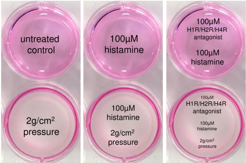
) was applied for another 48 h to simulate orthodontic pressure in the periodontal ligament according to an established and published in-vitro model [30,31] (Fig 1).

To test for histamine receptor (HR) interaction, we additionally incubated PDLF with 100 μM of H1R antagonist cetirizine (C3618, Sigma Aldrich, Munich, Germany), H2R antagonist ranitidine (R101, Sigma Aldrich, Munich, Germany) or H4R antagonist JNJ7777120 (J3770, Sigma Aldrich, Munich, Germany), respectively, two hours prior to histamine application.

The used antagonist concentrations were adopted from the concentration of a H1R antagonist previously used and published in experiments on nasal fibroblasts [32]. Then PDLF were pre- incubated for 24 h followed by pressure application for another 48 h as described above. We then analyzed gene expression (RT-qPCR) and protein expression (Western Blot, ELISA).

Determination of cell number

We harvested PDLF with a cell scraper in 1 ml PBS and quantified cell number using a Beck-

man Coulter Counter Z2™ (Beckman Coulter, Krefeld, Germany) according to the manufac-

turer’s instructions.

(4)

Cytotoxicity assay (LDH release)

To determine cytotoxicity we used lactate dehydrogenase (LDH) assays (04744926001, Sigma Aldrich, Munich, Germany) following the manufacturer’s instructions. Briefly, we added 100 μl of freshly prepared LDH solution containing of 22 μl catalyst mixed with 1 ml dye to 100 μl cell culture supernatant and incubated the mixture for 30 min in the dark at room tem- perature. We stopped the reaction by adding 50 μl stop solution. An ELISA reader (Multiscan GO Microplate Spectrophotometer, Thermo Fisher Scientific, Waltham, MA, USA) was used to measure LDH activity (absorbance at 490 nm), subtracting background absorbance at 690 nm.

Isolation of total RNA

Total RNA from PDLF was isolated using 500 μl TriFast (peqGOLD, PEQLAB Biotechnology Erlangen, Germany) for each sample according to the manufacturer’s instructions. The RNA pellet was eluted in 25 μl nuclease-free water (T143, Carl Roth, Karlsruhe, Germany) and RNA concentration was determined by measuring OD at 260 nm (NanoPhotometer, Implen, Munich, Germany).

Fig 1. In vitro simulation of compressive force application to periodontal ligament fibroblasts (PDLF) occurring during orthodontic tooth movement. After a preincubation time of 24 h with or without histamine (100 μM, 50/100/200 μM in receptor expression experiments) and an H1R/H2R/H4R antagonist (100 μM), a pressure of 2g/cm

2

was applied to PDLF by means of a sterile glass disc (ø33cm, 17.1g) for 48 h according to an established and published in vitro model.

https://doi.org/10.1371/journal.pone.0237040.g001

(5)

cDNA synthesis

For cDNA synthesis we mixed 1 μg of RNA with nuclease-free water to get a volume of 11 μl.

This compound was applied to a mixture of 4 μl 5xM-MLV-buffer (M1705, Promega, Madison, WI, USA), 1 μl Oligo

dt

primer (SO131, Thermo Fisher Scientific, Waltham, MA, USA), 1 μl random hexamer primer (SO142, Thermo Fisher Scientific, Waltham, MA, USA), 1 μl 10 mM dNTP (L785.2, Carl Roth, Karlsruhe, Germany), 1 μl (40 U) RNase Inhibitor (EO0381, Thermo Fisher Scientific, Waltham, MA, USA) and 1μl (200 U) M-MLV Reverse Transcriptase (M1705, Promega, Madison, WI, USA) [6]. All samples were incubated at 37˚C for 1 h and at 95˚C for 2 min to inactivate the transcriptase. They were stored at -20˚C until use. To minimize experimental variations, all components were prepared as a master mix and cDNA synthesis was performed at the same for all samples.

Semiquantitative PCR

We performed semiquantitative PCR and agarose gel electrophoresis to get information regarding histamine receptor expression in PDLF. For this purpose we mixed 2 μl of cDNA with 2 μl 10xFastStart PCR buffer with 20 mM MgCl

2

(12161567001, Sigma Aldrich, Munich, Germany), 0.5 μl of the appropriate forward and reverse primer respectively (Table 1), 0.4 μl dNTPs (L785.2, Carl Roth, Karlsruhe, Germany) and 0.2 μl FastStart Taq polymerase

(12032929001, Sigma Aldrich, Munich, Germany) and added H

2

O

dd

to a total volume of 20 μl.

We used histamine receptor primer combinations according to the study of Park et al. [32]

(Table 1). RPL22 was used as reference gene, as it has been shown to be stably expressed before [30,33]. The samples were heated in a thermocycler (VWR, Radnor, PA, USA) at 95˚C for 5 minutes and went through 40 cycles at 60˚C for 30 seconds each. For agarose gel electrophore- sis, we used a 1.5% agarose gel, which was prepared with agarose powder (T145.3, Carl Roth, Karlsruhe, Germany), 1xTris acetate EDTA buffer and gel red buffer (41003, Biotrend, Cologne, Germany). 7 μl of each sample were mixed with a 2 μl sucrose buffer and carefully pipetted into the pockets of the agarose gel. A voltage of 120 V was applied for 40 min in TAE buffer. The evaluation was then carried out using the gel documentation system Genoplex 2 and its software GenoSoft (VWR, Radnor, PA, USA). Densitometric analysis of specific bands was performed with ImageJ (ver. 1.47, Wayne Rasband, National Institutes of Health, USA).

Quantitative real-time polymerase chain reaction (RT-qPCR)

We pipetted 7.5 μl SYBR

1

Green Jumpstart Taq ready mix (S4438, Sigma Aldrich, Munich, Germany), 5.25 μl nuclease-free water (T143, Carl Roth, Karlsruhe, Germany), 0.75 μl of a

Table 1. Primer data for target genes and reference genes (PPIB, RPL22) for semiquantitative and RT-qPCR.

Gene symbol Gene name Accession Number 5´-forward primer-3´ 5´-reverse primer-3´

H1R histamine 1 receptor NM_001098213.1 GTCTAACACAGGCCTGGATT GGATGAAGGCTGCCATGATA

H2R histamine 2 receptor NM_001131055.1 ATTAGCTCCTGGAAGGCAGC CTGGAGCTTCAGGGGTTTCT

H3R histamine 3 receptor NM_007232.3 TCGTGCTCATCAGCTACGAC AAGCCGTGATGAGGAAGTAC

H4R histamine 4 receptor NM_021624.4 GGCTCACTACTGACTATCTG CCTTCATCCTTCCAAGACTC

COX2 cyclooxygenase 2 NM_000963.3 GAGCAGGCAGATGAAATACCAGTC TGTCACCATAGAGTGCTTCCAAC

IL6 interleukin 6 NM_000600.3 TGGCAGAAAACAACCTGAACC CCTCAAACTCCAAAAGACCAGTG

TNFRSF11B (OPG)

osteoprotegerin NM_002546.4 TGTCTTTGGTCTCCTGCTAACTC CCTGAAGAATGCCTCCTCACAC

PPIB peptidylprolyl isomerase A NM_000942.4 TTCCATCGTGTAATCAAGGACTTC GCTCACCGTAGATGCTCTTTC

TNFSF11 (RANKL) receptor activator of NFκB ligand NM_003701.3 ATACCCTGATGAAAGGAGGA GGGGCTCAATCTATATCTCG

RPL22 ribosomal protein L22 NM_000983.3 TGATTGCACCCACCCTGTAG GGTTCCCAGCTTTTCCGTTC

https://doi.org/10.1371/journal.pone.0237040.t001

(6)

corresponding primer pair (0.375 μ l / primer) and 1.5 μ l cDNA, previously diluted to 1:10, per well onto a 96-well plate (712282, Biozym, Hessisch Oldendorf, Germany) in duplicates. To ensure equal concentrations, all components except the cDNA solution were prepared as a master mix. Amplification was performed with a Mastercycler ep realplex-S thermocycler (Eppendorf, Hamburg, Germany). At the beginning, the plate was heated to 95˚C for 5 min and went through 45 cycles with 95˚C each for 10 sec, 60˚C for 8 sec and 72˚C for 8 sec. At the end of each step fluorescence was quantified at 520 nm. C

q

values were determined using the software realplex (CalqPlex algorithm, automatic baseline). Normalization of target genes was based on two reference genes (RPL22 and PPIB), which were validated before for PDLF and the used in vitro model [30,33]. We calculated relative gene expression as 2

-ΔCq

[34] with ΔC

q

= C

q

(target gene)–C

q

(mean RPL22/PPIB). All gene specific primers (Table 1) and gene nucle- otide sequences were constructed according MIQE guidelines [35] using NCBI (National Cen- tre for Biotechnology Information) PrimerBLAST and additional software considering final concentration of qPCR components [36]. Primers were synthesized by Eurofins MWG Operon LLC (Huntsville, AL, USA).

Western Blot

Protein from PDLF was isolated using 100 μl CelLytic M (C2978; Sigma Aldrich, Munich, Ger- many) containing proteinase inhibitors (87786, Carl Roth, Karlsruhe, Germany) per well. We determined protein concentration with RotiQuant (K015.3; Carl Roth, Karlsruhe, Germany) according to the manufacturer’s instructions. We separated equal amounts of total protein on a 10% SDS-polyacrylamide gel under reducing conditions and transferred the proteins onto a polyvinylidene diflouride (PVDF) membrane (T830, Carl Roth, Karlsruhe, Germany). Mem- branes were blocked with 5% nonfat milk in tris-buffered saline and 0.1% Tween 20, pH 7.5 (TBS-T) at 4˚C over night and incubated with anti-RANKL (TA306362, OriGene, Rockville, MD, USA) diluted 1:2,000 in 0.5% milk in TBS-T, or 1:500 anti-HSP90 (s-13119, Santa Cruz Biotechnology, Dallas, TX, USA) for 1 h. After washing three times in TBS-T, we incubated the blots for 1 h with anti-mouse IgG-κBP-HRP (sc-516102, Santa Cruz Biotechnology, Dallas, TX, USA) diluted 1:5,000 or anti-rabbit IgG HRP (611–1302, Rockland immunochemicals, Gilbertsville, PA, USA) diluted 1:5,000 in 5% milk in TBS-T horseradish peroxidase-conju- gated anti-rabbit IgG (Thermo Fisher Scientific, Waltham, MA, USA), diluted 1:5000 in 0.5%

milk in TBS-T at room temperature. After washing, antibody binding was visualized by the gel documentation system Genoplex 2 and its software (VWR, Radnor, PA, USA). Densitometric quantification of specific bands was performed with ImageJ (ver. 1.47, Wayne Rasband, National Institutes of Health, USA).

Enzyme-linked immunosorbent assay (ELISA)

We used commercially available enzyme-linked immunosorbent assay (ELISA) kits (IL6:

CSB-E04638h, Cusabio, Houston, TX, USA; osteoprotegerin (OPG): EHTNFRSF11B, Thermo Fisher Scientific, Waltham, MA, USA; RANKL: RD193004200R, BioVendor, Brno, Czech Republic) according to the manufacturers’ instructions and measured absorbance using an ELISA plate reader (Multiskan Go, Thermo Fisher Scientific, Waltham, MA).

Statistical methods

IBM SPSS Statistics 24 (IBM

1

, Armonk, NY, USA) was used for statistical analysis. Each sym- bol in figures represents a data point. Horizontal lines represent the mean ± standard error of mean. Data were validated by Welch-corrected ANOVAs with Games-Howell posthoc tests.

All differences were considered statistically significant at p � 0.05.

(7)

Results

Expression of different histamine receptors (HR) and effects of HR- antagonists in PDLF

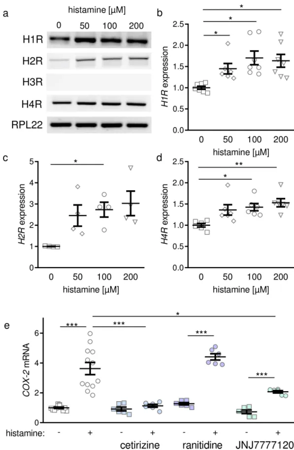
First, we focused on the expression levels of histamine receptors with variable concentrations of histamine. PDLF expressed histamine 1 receptor (H1R), histamine 2 receptor (H2R) and histamine 4 receptor (H4R, Fig 2A–2D). Histamine 3 receptor (H3R) was not expressed in PDLF (Fig 2A). Increasing histamine concentrations led to a significant increase of all expressed histamine receptors in PDLF using a concentration of 100 μM histamine (Fig 2A–

2D). Application of 100 μM histamine increased COX-2 gene expression significantly (Fig 2E).

To determine which histamine receptor was responsible for this upregulation, we tested cetiri- zine which is a H1R antagonist, ranitidine as H2R antagonist and JNJ777210, which acts as H4R antagonist. We observed a significant reduction of COX-2 gene expression after applica- tion of 100 μM histamine, when inhibiting H1R with cetirizine (Fig 2E). Neither ranitidine nor JNJ777210 seemed to inhibit histamine-induced COX-2 upregulation at the mRNA level (Fig 2E).

Effects of histamine and H1R antagonist cetirizine on cell number and cell viability

Next we investigated the effect of histamine on PDLF without and with compressive force treatment occurring during orthodontic tooth movement. Furthermore we investigated via which receptor histamine-induced effects are mediated. Histamine application increased PDLF number significantly without and with mechanical loading (Fig 3A). Inhibition of H1R with cetirizine truncated this effect (Fig 3A). Compressive force treatment reduced cell num- ber significantly under all tested conditions (Fig 3A). In line with that, cytotoxicity was increased with pressure application under all tested conditions (Fig 3B), whereas LDH release was reduced after addition of histamine (Fig 3B). Again, treatment with cetirizine limited the histamine-induced effect (Fig 3B).

Effects of histamine and H1R antagonist cetirizine on expression of proinflammatory genes in PDLF

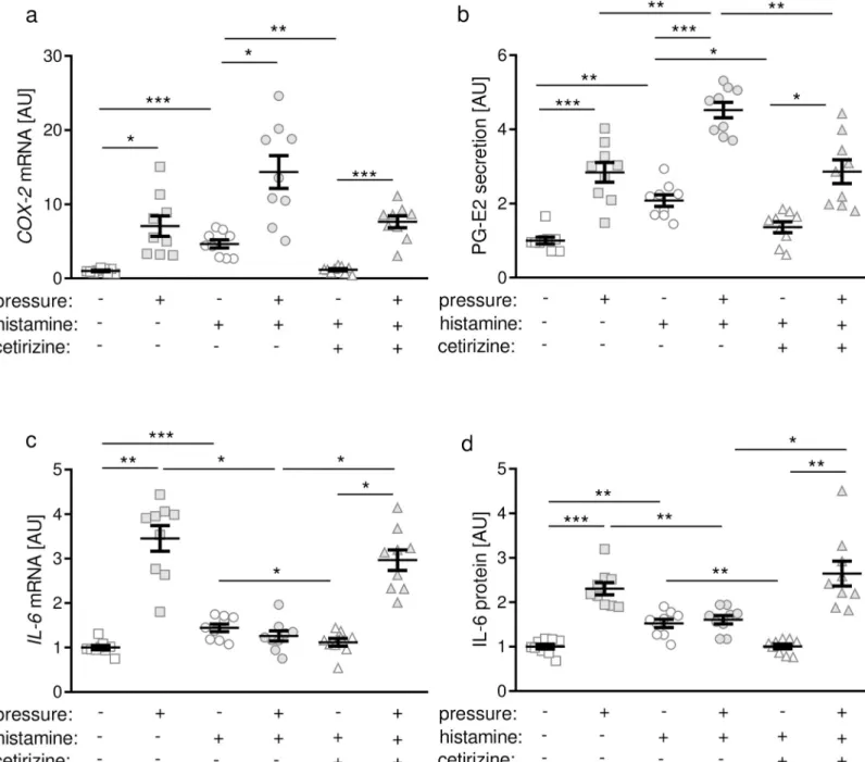
Next, we investigated gene and protein expression of proinflammatory genes. Compressive force treatment increased COX-2 gene expression significantly (Fig 4A). Stimulation of PDLF with 100 μM histamine led to an enhanced gene expression of COX-2 under control conditions without pressure application (Fig 4A). Inhibition of H1R with cetirizine or fexofenadine (S2 Fig) reduced the histamine-induced COX-2 expression to the control level without histamine (Fig 4A). We observed a pressure-induced upregulation of COX-2 gene expression indepen- dent of histamine or cetirizine application (Fig 4A). In line with that PG-E2 secretion was enhanced after compression independent of treatment with histamine or cetirizine (Fig 4B).

We observed increased PG-E2 secretion after treatment with histamine under control condi- tions and compressive force treatment, which was inhibited after adding cetirizine (Fig 4B).

Under control conditions without pressure application histamine treatment increased IL-6 gene expression and protein secretion significantly (Fig 4C and 4D). This effect could be inhib- ited by application of cetirizine (Fig 4C and 4D) or fexofenadine (S2 Fig). As expected, pres- sure application increased IL-6 gene and protein expression in PDLF (Fig 4C and 4D).

Histamine application, however, truncated this pressure-induced IL-6 gene and protein

expression (Fig 4C and 4D). Addition of cetirizine inhibited this histamine-induced effect at

the mRNA and protein level (Fig 4C and 4D).

(8)

Fig 2. Histamine-dependent expression of histamine receptors and COX-2 by periodontal ligament fibroblasts. (a)

Representative pictures of gene expression of histamine receptors in PDLF. Expression of histamine-3-receptor (H3R) could

(9)

Effects of histamine and H1R antagonist cetirizine on the RANKL/OPG system in PDLF

Next we were interested in the impact of histamine on the PDLF-mediated remodeling of alve- olar bone during simulated orthodontic tooth movement. Therefore we investigated the RANKL/OPG system. Compressive force treatment did not affect OPG (osteoprotegerin) mRNA expression in PDLF (Fig 5A). Histamine, however, increased OPG gene expression independent of pressure treatment. This effect was inhibited by cetirizine application (Fig 5A).

In contrast to OPG gene expression we observed a decrease of secreted OPG protein in the media after pressure application (Fig 5B). Addition of histamine to cell culture media reduced OPG protein secretion with and without compression (Fig 5B). This reduction was counter- acted by treatment with cetirizine. Pressure application resulted in significant RANKL gene expression under control conditions without histamine or cetirizine inhibition (Fig 5C). Hista- mine increased RANKL gene expression in PDLF without compression and reduced it to the control level after compressive force treatment (Fig 5C). Next we investigated RANKL secre- tion and protein expression of membrane-bound RANKL in PDLF. We observed enhanced RANKL secretion after mechanical loading in PDLF under control conditions (Fig 5D). Hista- mine treatment, however, reduced RANKL secretion without and with pressure application (Fig 5D). H1R inhibition via cetirizine administration restored this histamine-induced trunca- tion of RANKL secretion without compression (Fig 5D). In line with soluble RANKL secre- tion, expression of membrane-bound RANKL on PDLF increased with pressure application (Fig 5E). Moreover, histamine enhanced membrane-bound RANKL expression with and with- out compressive force treatment (Fig 5E). Cetirizine application reversed this histamine effect on RANKL protein expression (Fig 5E). Next, we calculated RANKL/OPG mRNA ratio to directly assess the changes due to histamine or cetirizine treatment (Fig 5F). Under control conditions compression resulted in an increased RANKL/OPG mRNA ratio. Without pressure histamine elevated RANKL/OPG mRNA ratio significantly mediated by the H1R, as this effect was truncated by cetirizine (Fig 5F).

Discussion

In this study we investigated the effect of histamine and histamine 1 receptor antagonist cetiri- zine on PDLF. Cetirizine has been shown to have an exquisite anti-H1R specificity exerting its effects only on H1R [37] and to facilitate bone formation by suppressing osteoclastic activity [38]. We determined that histamine receptors 1 (H1R), 2 (H2R) and 4 (H4R) are expressed by PDLF, whereas type 3 (H3R) is not. Gene expression of H1R and H2R significantly increased with histamine treatment. Furthermore histamine enhanced expression of interleukin-6 (IL- 6), cyclooxygenase-2 (COX-2) and the secretion of prostaglandin E2 (PG-E2) by PDLF even without compression. RANKL protein expression was also induced, whereas OPG secretion remaining unaffected. Histamine significantly increased cell number and reduced LDH release. All mentioned effects were not observed during simultaneous incubation with the H1R antagonist cetirizine indicating that histamine effects were transmitted through H1R.

Despite some interindividual variation of PDLF characteristics and expression behaviour, our results derived from a pool of PDLF from six gender-matched patients should be generalisable,

not be determined. Densitometric analysis of semiquantitative PCR for histamine-1-receptor (H1R, b), histamine-2-receptor (H2R, c) or histamine-4-receptor (H4R, d) after treatment with different histamine concentrations. (e) COX-2 gene expression after stimulation with histamine and inhibition of histamine receptor interaction using different inhibitors. AU:

arbitrary units;

p � 0.05;

��

p � 0.01;

���

p � 0.001. Statistics: Welch-corrected ANOVA with Games-Howell posthoc tests.

Each symbol in figures represents a data point. Horizontal lines represent the mean ± standard error of mean.

https://doi.org/10.1371/journal.pone.0237040.g002

(10)
(11)

as comparable upregulating effects by compressive force treatment were observed in all indi- vidual cell lines.

Our experiments indicate that PDLF only express H1R, H2R and H4R and not H3R. The reason could be that H3R is normally expressed by neurons and thus rather involved in neuro

Fig 3. Assessment of cell number (a), cytotoxicity (b) and viability (c) after compression with or without histamine or inhibition with cetirizine. AU: arbitrary units;

p � 0.05;

��

p � 0.01;

���

p � 0.001. Statistics: Welch-corrected ANOVA with Games-Howell posthoc tests. Each symbol in figures represents a data point. Horizontal lines represent the mean ± standard error of mean.

https://doi.org/10.1371/journal.pone.0237040.g003

Fig 4. COX-2 gene expression (a), PG-E2 secretion (b), IL-6 gene expression (c) and IL-6 secretion (d) after compression with or without histamine or inhibition with cetirizine. AU: arbitrary units;

p � 0.05;

��

p � 0.01;

�� �

p � 0.001. Statistics: Welch-corrected ANOVA with Games-Howell posthoc tests. Each symbol in figures represents a data point. Horizontal lines represent the mean ± standard error of mean.

https://doi.org/10.1371/journal.pone.0237040.g004

(12)
(13)

diseases [39] than in orthodontic tooth movement. The highest receptor expression was observed regarding H2R, when adding 200 μg of histamine. At the same time we could not see any inhibiting effect when applying H2R-antagonist ranitidine. In addition, cetirizine showed the highest reversion of the histamine-induced effect. This was in line with results by Park et al. [32]. These authors investigated gene expression of nasal fibroblasts and determined H1R to be the most distinctly expressed receptor and a H1R antagonist having the highest inhibiting effect. Furthermore, our study indicates that histamine stimulates proliferation of PDLF.

These results were in line with Hong et al., who reported an increasing cell number after apply- ing histamine on nasal fibroblasts [40].

One of the first responses to orthodontic pressure is the synthesis of prostaglandins [41].

This is mediated by COX-2, an enzyme, which enhances inflammatory reactions [42]. As expected, gene expression of COX-2 and secretion of PG-E2 increased in PDLF upon hista- mine and pressure treatment indicating an enhanced proinflammatory response at the begin- ning of orthodontic tooth movement. These data were in line with the study of Grimm et al., who reported a significant upregulation of COX-2 and IL-6 gene expression in PDLF within three hours [43] and with Niisato et al, who reported increased PG-E2 secretion with hista- mine treatment [29]. Studies with a similar setup showed an increased COX-2-induced PG-E2 expression during compressive force treatment [6,44,45]. Other studies explained the impor- tant role of PG-E2 for bone resorption [46] and its impact on RANKL expression [47].

Interleukin 6 (IL-6) plays an important role in host defense [9] and has an effect on bone resorption [48]. Histamine seems to have a decreasing effect on IL-6 expression during force treatment, which is less pronounced without pressure. Schroeder et al. reported an increase of IL-6 expression during the first 48 h under orthodontic compressive forces and a decrease after 72 hours [6]. It is known that IL-6 is also regulated by IL1α/β and TNFα [44]. Okada et al. also reported that IL1α/β- or TNFα- induced IL-6 production can be inhibited by PG-E2.

This could be a reason for the decrease of IL-6 expression after histamine treatment. Histamine enhanced expression of proinflammatory cytokines. Meh et al. (2011) found a correlation between tooth movement, histamine and cetirizine in rats. Tooth movement was increased by histamine and inhibited by cetirizine in the last period of orthodontic tooth movement [49].

In contrast, Kriznar et al. (2008) observed that cetirizine inhibited tooth movement in the first period of orthodontic tooth movement [50].

RANKL and its decoy receptor osteoprotegerin play an important role in bone formation and resorption [51]. RANKL binds to its receptor RANK on osteoclast precursor cells to stim- ulate osteoclast formation and activation [9]. Schroeder et al. found an increasing effect on RANKL expression during the first 72 h of orthodontic force treatment [6]. Nam et al.

reported an upregulation of RANKL in the serum and nasal mucosal tissue of allergic rhinitis patients [52]. This is in line with our data, as RANKL gene expression and protein expression are significantly increased when adding histamine.

Based on these in vitro results, it is likely that increased histamine levels as occuring in patients with allergies may boost orthodontic tooth movement velocity, which is a sterile pseudo-inflammatory reaction dependent on an increase in inflammatory cytokines and RANKL expression leading to elevated osteoclastogenesis in direction of movement [3]. On the other hand, it is also possible that the elevated release of proinflammatory cytokines and RANKL by PDLF under the influence of histamine may trigger uncontrolled osteoclastogenesis

Fig 5. Effects of histamine and H1R antagonist cetirizine on OPG (a,b) and RANKL (c-e) gene and protein expression as well as on RANKL/OPG mRNA ratio (f). AU: arbitrary units;

p � 0.05;

��

p � 0.01;

���

p � 0.001. Statistics: Welch-corrected ANOVA with Games-Howell posthoc tests. Each symbol in figures represents a data point. Horizontal lines represent the mean ± standard error of mean.

https://doi.org/10.1371/journal.pone.0237040.g005

(14)

leading to severe side effects such as dental root resorptions and periodontal bone loss, which merits investigation in further in vivo studies.

Conclusions

By expressing H1R, H2R and H4R, PDLF are likely to be able to detect fluctuating histamine levels in the periodontal ligament. Increased histamine levels seem to be associated with increased expression of proinflammatory mediators and RANKL, suggesting an inductive effect of histamine on PDLF-mediated osteoclastogenesis and thus orthodontic tooth move- ment, which requires resorption of the alveolar bone in direction of movement, but may also be associated with side effects such as dental root resorptions or periodontal bone loss during orthodontic therapy, which are caused by increased and uncontolled osteoclast activity. Since cetirizine as specific H1R inhibitor cancels these effects, the histamine effect seems to be pre- dominantly mediated via the H1R.

Supporting information

S1 Fig. Fold changes in COX-2 and RANKL gene expression by PDLF due to pressure application for 48 h for each individual subject included into the used pool of PDLF.

(DOCX)

S2 Fig. Effects of histamine and 50 μM H1R antagonist fexofenadine (F9427, Sigma- Aldrich) on COX-2 (a) and IL-6 (b) gene expression. AU: arbitrary units; p � 0.05; ��

p � 0.01. Statistics: Welch-corrected ANOVA with Games-Howell posthoc tests. Each symbol in figures represents a data point. Horizontal lines represent the mean ± standard error of mean.

(DOCX) S1 Dataset.

(XLSX)

S1 Raw Images.

(DOCX)

Acknowledgments

The authors thank Kathrin Bauer and Eva Zaglauer for their technical support.

Author Contributions

Conceptualization: Marcella Groeger, Gerrit Spanier, Michael Wolf, James Deschner, Peter Proff, Agnes Schro ¨der, Christian Kirschneck.

Data curation: Marcella Groeger.

Formal analysis: Agnes Schro ¨der, Christian Kirschneck.

Funding acquisition: Agnes Schro ¨der, Christian Kirschneck.

Investigation: Marcella Groeger, Agnes Schro ¨der.

Methodology: Marcella Groeger, James Deschner, Agnes Schro ¨der, Christian Kirschneck.

Project administration: Peter Proff, Christian Kirschneck.

Resources: Gerrit Spanier.

(15)

Software: Agnes Schro ¨der, Christian Kirschneck.

Supervision: Michael Wolf, James Deschner, Peter Proff, Agnes Schro ¨der, Christian Kirschneck.

Validation: Gerrit Spanier, Michael Wolf, James Deschner, Peter Proff, Agnes Schro ¨der, Christian Kirschneck.

Visualization: Agnes Schro ¨der, Christian Kirschneck.

Writing – original draft: Marcella Groeger, Agnes Schro ¨der, Christian Kirschneck.

Writing – review & editing: Marcella Groeger, Gerrit Spanier, Michael Wolf, James Deschner, Peter Proff, Agnes Schro ¨der, Christian Kirschneck.

References

1. Graber LW, Vanarsdall RL, Vig KWL, Huang GJ. Orthodontics. Current principles and techniques.

St. Louis, Missouri: Elsevier; 2017.

2. Krishnan V, Davidovitch Z. Cellular, molecular, and tissue-level reactions to orthodontic force. Am J Orthod Dentofacial Orthop. 2006; 129: 469.e1–32. https://doi.org/10.1016/j.ajodo.2005.10.007 PMID:

16627171

3. Meikle MC. The tissue, cellular, and molecular regulation of orthodontic tooth movement: 100 years after Carl Sandstedt. Eur J Orthod. 2006; 28: 221–240. https://doi.org/10.1093/ejo/cjl001 PMID:

16687469

4. Wolf M, Lossdo ¨ rfer S, Marciniak J, Ro¨mer P, Kirschneck C, Craveiro R, et al. CD8+ T cells mediate the regenerative PTH effect in hPDL cells via Wnt10b signaling. Innate Immun. 2016; 22: 674–681. https://

doi.org/10.1177/1753425916669417 PMID: 28071181

5. Jiang C, Li Z, Quan H, Xiao L, Zhao J, Wang Y, et al. Osteoimmunology in orthodontic tooth movement.

Oral Dis. 2015; 21: 694–704. https://doi.org/10.1111/odi.12273 PMID: 25040955

6. Schro¨der A, Bauer K, Spanier G, Proff P, Wolf M, Kirschneck C. Expression kinetics of human periodon- tal ligament fibroblasts in the early phases of orthodontic tooth movement. J Orofac Orthop. 2018; 79:

337–351. https://doi.org/10.1007/s00056-018-0145-1 PMID: 30019109

7. Wolf M, Lossdo ¨ rfer S, Ro¨mer P, Kirschneck C, Ku¨pper K, Deschner J, et al. Short-term heat pre-treat- ment modulates the release of HMGB1 and pro-inflammatory cytokines in hPDL cells following mechan- ical loading and affects monocyte behavior. Clin Oral Investig. 2016; 20: 923–931. https://doi.org/10.

1007/s00784-015-1580-7 PMID: 26358476

8. Kanzaki H, Chiba M, Shimizu Y, Mitani H. Dual regulation of osteoclast differentiation by periodontal lig- ament cells through RANKL stimulation and OPG inhibition. J Dent Res. 2001; 80: 887–891. https://

doi.org/10.1177/00220345010800030801 PMID: 11379890

9. Proff P, Ro¨mer P. The molecular mechanism behind bone remodelling: a review. Clin Oral Investig.

2009; 13: 355–362. https://doi.org/10.1007/s00784-009-0268-2 PMID: 19319579

10. Alikhani M, Sangsuwon C, Alansari S, Nervina JM, Teixeira CC. Biphasic theory: breakthrough under- standing of tooth movement. J World Fed Orthod. 2018; 7: 82–88. https://doi.org/10.1016/j.ejwf.2018.

08.001

11. Woelber JP, Bremer K, Vach K, Ko¨nig D, Hellwig E, Ratka-Kru ¨ ger P, et al. An oral health optimized diet can reduce gingival and periodontal inflammation in humans—a randomized controlled pilot study.

BMC Oral Health. 2016; 17: 28. https://doi.org/10.1186/s12903-016-0257-1 PMID: 27460471 12. Muluke M, Gold T, Kiefhaber K, Al-Sahli A, Celenti R, Jiang H, et al. Diet-Induced Obesity and Its Differ-

ential Impact on Periodontal Bone Loss. J Dent Res. 2016; 95: 223–229. https://doi.org/10.1177/

0022034515609882 PMID: 26450512

13. Alsahli A, Kiefhaber K, Gold T, Muluke M, Jiang H, Cremers S, et al. Palmitic Acid Reduces Circulating Bone Formation Markers in Obese Animals and Impairs Osteoblast Activity via C16-Ceramide Accumu- lation. Calcif Tissue Int. 2016; 98: 511–519. https://doi.org/10.1007/s00223-015-0097-z PMID:

26758875

14. Biegański T. Biochemical, physiological and pathophysiological aspects of intestinal diamine oxidase.

Acta Physiol Pol. 1983; 34: 139–154. PMID: 6416024

15. Branco ACCC, Yoshikawa FSY, Pietrobon AJ, Sato MN. Role of Histamine in Modulating the Immune Response and Inflammation. Mediators Inflamm. 2018; 2018: 9524075. https://doi.org/10.1155/2018/

9524075 PMID: 30224900

(16)

16. Heeschen W. Pathogene Mikroorganismen und deren Toxine in Lebensmitteln tierischer Herkunft.

Hamburg: Behr; 1989.

17. O’Mahony L, Akdis M, Akdis CA. Regulation of the immune response and inflammation by histamine and histamine receptors. J Allergy Clin Immunol. 2011; 128: 1153–1162. https://doi.org/10.1016/j.jaci.

2011.06.051 PMID: 21824648

18. Moneret-Vautrin DA. False food allergies: non-specific reation to foodstuffs. In: Lessof MH, editor. Clini- cal reactions to food. Chichester: Wiley; 1983.

19. Cataldi M, Borriello F, Granata F, Annunziato L, Marone G. Histamine receptors and antihistamines:

from discovery to clinical applications. Chem Immunol Allergy. 2014; 100: 214–226. https://doi.org/10.

1159/000358740 PMID: 24925401

20. Hattori Y, Seifert R, editors. Histamine and Histamine Receptors in Health and Disease. 1st ed. Cham:

Springer International Publishing; Springer; 2018.

21. Leurs R, Chazot PL, Shenton FC, Lim HD, Esch IJP de. Molecular and biochemical pharmacology of the histamine H4 receptor. Br J Pharmacol. 2009; 157: 14–23. https://doi.org/10.1111/j.1476-5381.

2009.00250.x PMID: 19413568

22. Singh M, Jadhav HR. Histamine H3 receptor function and ligands: recent developments. Mini Rev Med Chem. 2013; 13: 47–57. PMID: 22931528

23. Thangam EB, Jemima EA, Singh H, Baig MS, Khan M, Mathias CB, et al. The Role of Histamine and Histamine Receptors in Mast Cell-Mediated Allergy and Inflammation: The Hunt for New Therapeutic Targets. Front Immunol. 2018; 9: 1873. https://doi.org/10.3389/fimmu.2018.01873 PMID: 30150993 24. Bakker RA, Jongejan A, Sansuk K, Hacksell U, Timmerman H, Brann MR, et al. Constitutively active

mutants of the histamine H1 receptor suggest a conserved hydrophobic asparagine-cage that con- strains the activation of class A G protein-coupled receptors. Mol Pharmacol. 2008; 73: 94–103. https://

doi.org/10.1124/mol.107.038547 PMID: 17959710

25. Bakker RA, Schoonus SB, Smit MJ, Timmerman H, Leurs R. Histamine H(1)-receptor activation of nuclear factor-kappa B: roles for G beta gamma- and G alpha(q/11)-subunits in constitutive and ago- nist-mediated signaling. Mol Pharmacol. 2001; 60: 1133–1142. https://doi.org/10.1124/mol.60.5.1133 PMID: 11641442

26. Seifert R, Strasser A, Schneider EH, Neumann D, Dove S, Buschauer A. Molecular and cellular analysis of human histamine receptor subtypes. Trends Pharmacol Sci. 2013; 34: 33–58. https://doi.org/10.

1016/j.tips.2012.11.001 PMID: 23254267

27. Thurmond RL. The histamine H4 receptor: from orphan to the clinic. Front Pharmacol. 2015; 6: 65.

https://doi.org/10.3389/fphar.2015.00065 PMID: 25873897

28. Biosse-Duplan M, Baroukh B, Dy M, Vernejoul M-C de, Saffar J-L. Histamine promotes osteoclastogen- esis through the differential expression of histamine receptors on osteoclasts and osteoblasts. Am J Pathol. 2009; 174: 1426–1434. https://doi.org/10.2353/ajpath.2009.080871 PMID: 19264900 29. Niisato N, Ogata Y, Furuyama S, Sugiya H. Histamine H1 receptor-stimulated Ca2+ signaling pathway

in human periodontal ligament cells. J Periodont Res. 1996; 31: 113–119. https://doi.org/10.1111/j.

1600-0765.1996.tb00472.x PMID: 8708938

30. Kirschneck C, Batschkus S, Proff P, Ko¨stler J, Spanier G, Schro ¨ der A. Valid gene expression normali- zation by RT-qPCR in studies on hPDL fibroblasts with focus on orthodontic tooth movement and peri- odontitis. Sci Rep. 2017; 7: 14751. https://doi.org/10.1038/s41598-017-15281-0 PMID: 29116140 31. Schro¨der A, Ku¨chler EC, Omori M, Spanier G, Proff P, Kirschneck C. Effects of ethanol on human peri-

odontal ligament fibroblasts subjected to static compressive force. Alcohol. 2019; 77: 59–70. https://

doi.org/10.1016/j.alcohol.2018.10.004 PMID: 30336201

32. Park I-H, Um J-Y, Cho J-S, Lee SH, Lee SH, Lee H-M. Histamine Promotes the Release of Interleukin- 6 via the H1R/p38 and NF-κB Pathways in Nasal Fibroblasts. Allergy Asthma Immunol Res. 2014; 6:

567–572. https://doi.org/10.4168/aair.2014.6.6.567 PMID: 25374757

33. Kirschneck C, Proff P, Fangha ¨ nel J, Wolf M, Rolda´n JC, Ro¨mer P. Reference genes for valid gene expression studies on rat dental, periodontal and alveolar bone tissue by means of RT-qPCR with a focus on orthodontic tooth movement and periodontitis. Ann Anat. 2016; 204: 93–105. https://doi.org/

10.1016/j.aanat.2015.11.005 PMID: 26689124

34. Koretsi V, Kirschneck C, Proff P, Ro¨mer P. Expression of glutathione peroxidase 1 in the spheno-occipi- tal synchondrosis and its role in ROS-induced apoptosis. Eur J Orthod. 2015; 37: 308–313. https://doi.

org/10.1093/ejo/cju045 PMID: 25312980

35. Taylor S, Wakem M, Dijkman G, Alsarraj M, Nguyen M. A practical approach to RT-qPCR-Publishing data that conform to the MIQE guidelines. Methods. 2010; 50: S1–5. https://doi.org/10.1016/j.ymeth.

2010.01.005 PMID: 20215014

(17)

36. Ye J, Coulouris G, Zaretskaya I, Cutcutache I, Rozen S, Madden TL. Primer-BLAST: a tool to design target-specific primers for polymerase chain reaction. BMC Bioinformatics. 2012; 13: 134. https://doi.

org/10.1186/1471-2105-13-134 PMID: 22708584

37. Bernheim J, Arendt C, Vos C de. Cetirizine: more than an antihistamine. Agents Actions Suppl. 1991;

34: 269–293. PMID: 1686525

38. Hwang S, Chung CJ, Choi YJ, Kim T, Kim K-H. The effect of cetirizine, a histamine 1 receptor antago- nist, on bone remodeling after calvarial suture expansion. Korean J Orthod. 2020; 50: 42–51. https://

doi.org/10.4041/kjod.2020.50.1.42 PMID: 32042719

39. Arrang JM, Garbarg M, Lancelot JC, Lecomte JM, Pollard H, Robba M, et al. Highly potent and selective ligands for histamine H3-receptors. Nature. 1987; 327: 117–123. https://doi.org/10.1038/327117a0 PMID: 3033516

40. Hong S-M, Park I-H, Um J-Y, Shin J-M, Lee H-M. Stimulatory effects of histamine on migration of nasal fibroblasts. Int Forum Allergy Rhinol. 2015; 5: 923–928. https://doi.org/10.1002/alr.21555 PMID:

26097205

41. Kanzaki H, Chiba M, Shimizu Y, Mitani H. Periodontal ligament cells under mechanical stress induce osteoclastogenesis by receptor activator of nuclear factor kappaB ligand up-regulation via prostaglan- din E2 synthesis. J Bone Miner Res. 2002; 17: 210–220. https://doi.org/10.1359/jbmr.2002.17.2.210 PMID: 11811551

42. Meeran NA. Cellular response within the periodontal ligament on application of orthodontic forces. J Indian Soc Periodontol. 2013; 17: 16–20. https://doi.org/10.4103/0972-124X.107468 PMID: 23633766 43. Grimm S, Wolff E, Walter C, Pabst AM, Mundethu A, Jacobs C, et al. Influence of clodronate and com-

pressive force on IL-1ß-stimulated human periodontal ligament fibroblasts. Clin Oral Investig. 2020; 24:

343–350. https://doi.org/10.1007/s00784-019-02930-z PMID: 31102041

44. Okada N, Kobayashi M, Mugikura K, Okamatsu Y, Hanazawa S, Kitano S, et al. Interleukin-6 production in human fibroblasts derived from periodontal tissues is differentially regulated by cytokines and a gluco- corticoid. J Periodont Res. 1997; 32: 559–569. https://doi.org/10.1111/j.1600-0765.1997.tb00932.x PMID: 9401927

45. Proff P, Reicheneder C, Faltermeier A, Kubein-Meesenburg D, Ro¨mer P. Effects of mechanical and bacterial stressors on cytokine and growth-factor expression in periodontal ligament cells. J Orofac Orthop. 2014; 75: 191–202. https://doi.org/10.1007/s00056-014-0212-1 PMID: 24825831 46. Klein DC, Raisz LG. Prostaglandins: stimulation of bone resorption in tissue culture. Endocrinology.

1970; 86: 1436–1440. https://doi.org/10.1210/endo-86-6-1436 PMID: 4315103

47. Yamaguchi M. RANK/RANKL/OPG during orthodontic tooth movement. Orthod Craniofac Res. 2009;

12: 113–119. https://doi.org/10.1111/j.1601-6343.2009.01444.x PMID: 19419454

48. Kurihara N, Bertolini D, Suda T, Akiyama Y, Roodman GD. IL-6 stimulates osteoclast-like multinucle- ated cell formation in long term human marrow cultures by inducing IL-1 release. J Immunol. 1990; 144:

4226–4230. PMID: 2341718

49. Meh A, Sprogar S, Vaupotic T, Co¨r A, Drevensˇek G, Marc J, et al. Effect of cetirizine, a histamine (H(1)) receptor antagonist, on bone modeling during orthodontic tooth movement in rats. Am J Orthod Dento- facial Orthop. 2011; 139: e323–9. https://doi.org/10.1016/j.ajodo.2009.11.013 PMID: 21457838 50. Kriznar I, Sprogar S, Drevensek M, Vaupotic T, Drevensek G. Cetirizine, a histamine H1 receptor antag-

onist, decreases the first stage of orthodontic tooth movement in rats. Inflamm Res. 2008; 57 Suppl 1:

S29–30. https://doi.org/10.1007/s00011-007-0615-1. PMID: 18345503

51. Huang H, Williams RC, Kyrkanides S. Accelerated orthodontic tooth movement: molecular mecha- nisms. Am J Orthod Dentofacial Orthop. 2014; 146: 620–632. https://doi.org/10.1016/j.ajodo.2014.07.

007 PMID: 25439213

52. Nam S-Y, Kim H-Y, Min J-Y, Kim H-M, Jeong H-J. An osteoclastogenesis system, the RANKL/RANK signalling pathway, contributes to aggravated allergic inflammation. Br J Pharmacol. 2019; 176: 1664–

1679. https://doi.org/10.1111/bph.14615 PMID: 30737962

Abbildung

Fig 1. In vitro simulation of compressive force application to periodontal ligament fibroblasts (PDLF) occurring during orthodontic tooth movement
Table 1. Primer data for target genes and reference genes (PPIB, RPL22) for semiquantitative and RT-qPCR.
Fig 2. Histamine-dependent expression of histamine receptors and COX-2 by periodontal ligament fibroblasts
Fig 3. Assessment of cell number (a), cytotoxicity (b) and viability (c) after compression with or without histamine or inhibition with cetirizine

Referenzen

ÄHNLICHE DOKUMENTE

Despite much higher coccolithophore abundance in the greenhouse treatment, particulate inorganic carbon production (calcification) was significantly decreased by the combination

Eine Auswertung aller Metaanalysen zur therapeutischen Hypothermie durch die Brain Trauma Foundation hat zeigen können, dass eine therapeutische Hypo - thermie zwar nicht

(dee) MCF-7 and 2 different shLAMP2A cell lines were cultured in ovo, followed by staining and scoring of the tumor cells for LAMP2A (upper panel) and Ki-67 (lower panel)

Increased valvular expression of NF-κB in diabetic individuals is associated not only with serum HbA 1c and fructosamine levels but also with AVA and transvalvular gradient,

To evaluate patient clinical conditions and the effect of landiolol hydrochloride treatment on contrast media-induced anaphylactic shock treatment response, we reviewed the

However, compounds reported to release histamine from cells and thereby leading to arrhythmias in humans, namely morphine, ketamine, and fentanyl, failed to induce a more

The speeifity of the method was proved by thinlayer chromatography, degradation b y diamine oxidase (pig kidney) and histamine methyltransferase (guinea-pig brain),

Summary. High histamine concentrations and histamine methyl transferase activity were demonstrated in the gastric mueosa of man, dog, pig and cow. Modified methods for