The Molecular Mechanisms Underlying the Cytochrome C Oxidase Dysfunction-Induced
Accelerated Apoptosis
Inaugural-Dissertation Zur
Erlangung des Doktorgrades
der Mathematisch-Naturwissenschaftlichen Fakultät der Universität zu Köln
vorgelegt von Stephan Schüll
aus Hilden
Berichterstatter: Prof. Dr. Elena Rugarli Prof. Dr. Rudolf Wiesner
Tag der mündlichen Prüfung: 21.05.2014
Abbreviations III
Abstract V
Zusammenfassung VI
Introduction 1
APOPTOSIS, THE CONTROLLED DEMOLITION OF A CELL 1
POINT OF NO RETURN:MITOCHONDRIAL OUTER MEMBRANE PERMEABILIZATION 3
DYSREGULATION OF APOPTOSIS 5
INHIBITION OF APOPTOSIS AND CANCER 6
INCREASED APOPTOSIS AND DEGENERATIVE DISEASES 7
THE MITOCHONDRIAL RESPIRATORY CHAIN 8
THE MITOCHONDRIAL RESPIRATORY CHAIN AND DEGENERATIVE DISEASES 10
THE MITOCHONDRIAL RESPIRATORY CHAIN AND CELL DEATH 12
AIM OF THE STUDY 13
Material and Methods 15
CELL CULTURE 15
CHEMICALS AND REAGENTS 15
MEASUREMENT OF OXYGEN CONSUMPTION 15
DNA CONSTRUCTS AND SIRNA-MEDIATED KNOCKDOWN 16
CELL DEATH 17
MEASUREMENT OF SUPEROXIDE FORMATION 17
SAMPLE PREPARATION AND IMMUNOBLOTTING (IB) 18
IMMUNOPRECIPITATION (IP) OF ACTIVE BAX 19
LIPID ANALYSIS 20
FLUORESCENCE MICROSCOPY 20
STATISTICAL ANALYSIS 21
CHEMICAL INHIBITION OF CYTOCHROME C OXIDASE SELECTIVELY ENHANCES ROS-INDUCED CELL DEATH 22 CYTOCHROME C OXIDASE DYSFUNCTION ENHANCES OXIDATIVE STRESS-INDUCED APOPTOSIS 25 MTDNA DEPLETION-DERIVED COX DEFICIENCY ENHANCES MITOCHONDRIAL APOPTOSIS IN RESPONSE TO
OXIDATIVE STRESS. 30
GENOMIC ABLATION OF COX10 ENHANCES MITOCHONDRIAL APOPTOSIS IN RESPONSE TO OXIDATIVE
STRESS 31
SPHINGOLIPID METABOLISM IS ALTERED UPON COX DYSFUNCTION 33 INHIBITION OF CERAMIDE C16:0 DE NOVO SYNTHESIS COMPLETELY RESCUES COX
DYSFUNCTIONAL CELLS FROM ROS INDUCED APOPTOSIS 38
GENERATION OF LIVER-SPECIFIC COX10 KNOCKOUT MICE ON CERS6 KNOCKOUT AND WILD TYPE
BACKGROUND 43
Discussion 45
INHIBITION OF COX REVEALS SELECTIVE SUSCEPTIBILITY TO OXIDATIVE STRESS 45 ACCUMULATION OF CERAMIDES SENSITIZES COX-DEFICIENT CELLS TOWARDS OXIDATIVE STRESS-INDUCED
APOPTOSIS 47
CERAMIDE SYNTHASE 6 IS THE KEY MEDIATOR OF OXIDATIVE STRESS-INDUCED APOPTOSIS IN COX-
DEFICIENCY 49
PHYSIOLOGICAL RELEVANCE 51
References 54
Appendix 64
VECTOR MAP 64
Erklärung 65
III
Abbreviations
(Frequently used terms only)
AD Alzheimer’s disease
aSMase acid Sphingomyelinase 3-NP 3-Nitropropionic acid Bak Bcl-2 antagonist killer 1 Bax Bcl-2 associated x protein Bcl-2 B-cell lymphoma 2
Bcl-xL B-cell lymphoma extra-large
BH Bcl-2 homology
Bid BH3-interacting domain death antagonist BSA Bovine serum albumin
CerS Ceramide synthase
CL Cardiolipin
COX Cytochrome c oxidase Cybrid Cellular hybrid
e- Electron
ER Endoplasmic reticulum
H hours
H2O2 Hydrogen peroxide
IMM Inner mitochondrial membrane
kDa Kilodalton
DMSO Dimethyl sulfoxide DKO Double knockout DTT Dithiothreitol
IV EDTA Ethylenediaminetetraacetic acid
FB1 Fumonisin B1
IMS Intermembrane space
IF Immunofluorescence
IP Immunoprecipitation
M (p/n/µ/m) Molar; pico-, nano-, mikro-, milli-
MOMP Mitochondrial outer membrane permeabilization mtDNA mitochondrial DNA
nDNA nuclear DNA
nSMase neutral Sphingomyelinase OMM Outer mitochondrial membrane
ON Over night
OXPHOS Oxidative phosphorylation O2- Superoxide anion
PAGE Polyacrylamide gel electrophoresis PBS Phosphate buffered saline
PCR Polymerase chain reaction PD Parkinson’s disease ROS Reactive oxygen species RC Respiratory chain
RT Room temperature
SDS Sodium dodecyl sulfate SEM Standard error of the mean STS Staurosporine
WB Western blot
V
Abstract
Apoptosis, the physiological cell death, is an essential component of cellular homeostasis and tissue regeneration, and the dysregulation of apoptosis culminates in multiple human diseases.
Whereas the failure to execute timely programmed cell death in renovating tissues contributes to cancer, excessive apoptosis in post-mitotic tissues precipitates degenerative states, aging and aging-associated diseases.
Research work within the last two decades has shown that tissue degeneration caused by mitochondrial oxidative phosphorylation (OXPHOS) defects is associated with excessive apoptosis. However, it is unclear how OXPHOS-dysfunctions interfere with the apoptotic machinery and how this impacts on tissue degeneration.
In this context we demonstrate that solely the chemical induction of cytochrome c oxidase (COX) deficiency, which is among the most common defects found in mitochondrial diseases, exclusively and dramatically increases the Bcl-2 dependent apoptotic response towards oxidative stress. Furthermore, we could widen and reconfirm our findings in COX-deficient cybrids harboring an mtDNA-encoded deletion of COX subunit I as well as in murine fibroblast devoid of COX-assembly factor COX10. These findings indicate a general mechanism of COX-deficiency induced apoptosis by oxidative stress. Moreover, our data highlight that COX-deficiency is accompanied by an increased de novo synthesis and accumulation of the sphingolipid species ceramide. Due to their chemical properties especially ceramides with an acyl side-chain length of C16:0 have conclusively and repeatedly been demonstrated to induce apoptosis by pore- formation and by contributing to pro-apoptotic Bax and Bak induced permeabilization of the mitochondrial outer membrane (MOMP). Correspondingly, we show that the inhibition of endoplasmic reticulum-resident specific ceramide synthases abrogates enhanced apoptosis induced by mitochondrial respiratory chain-dysfunction. In particular, we identify ceramide synthase 6 (CerS6) as the key mediator of C16:0 ceramide induced apoptosis to oxidative stress:
While down-regulation of CerS6 protects COX-deficient cells from oxidative stress, overexpression of CerS6 introduces susceptibility towards oxidative stress in COX-functional cells.
In summary, our findings identify ceramide accumulation and in particular CerS6 as important components of the apoptotic response of COX-deficient cells towards oxidative stress and provide new insights into how mitochondrial dysfunction interferes with the apoptotic machinery and may impact on tissue homeostasis.
VI
Zusammenfassung
Apoptose, besser bekannt als physiologischer Zelltod, ist ein wichtiger Bestandteil der Zell- Homöostase sowie der Geweberegeneration. Ein Ungleichgewicht im physiologischen Zelltod kann die Entstehung multipler Krankheiten beim Menschen begünstigen. So ist beispielsweise eine verminderte oder gänzlich reduzierte Apoptose ursächlich für die Entstehung von Krebs, wohingegen exzessive und deregulierte Apoptose in postmitotischen Gewebe zu degenerativen Krankheiten bzw. altersbedingten Krankheitserscheinungen führen kann.
Die Forschungsarbeit der letzten zwei Jahrzehnte hat aufgezeigt, dass die Gewebedegeneration aufgrund defekter mitochondrialer oxidativer Phosphorylierung (OXPHOS) mit erhöhter Apoptose assoziiert ist. Hierbei ist jedoch unklar, wie genau sich Störungen der OXPHOS auf den Wirkungsmechanismus der Apoptose auswirken und wie dies dann zur Gewebedegeneration beiträgt.
In diesem Zusammenhang können wir zeigen, dass ausschließlich der durch chemische Inhibition hervorgerufene Defekt der Cytochrome C Oxidase (COX), eine der am häufigsten auftretenden Defekte bei mitochondrialen Erkrankungen, ursächlich für eine erhöhte Bcl-2 abhängige Apoptose aufgrund von oxidativem Stress ist. Diese Befunde konnten durch weitere Untersuchungen in einer COX-defizienten Cybridlinie, welche einen mitochondrial-kodierten Defekt der strukturellen COX-Untereinheit I enthält, sowie in murinen Knockout-Fibroblasten des Assembly-Faktors COX10 bestätigt und untermauert werden. Unsere Untersuchungen deuten daher auf einen generellen Wirkungsmechanismus der durch COX-Defizienz induzierten Apoptose nach oxidativem Stress hin.
Des Weiteren belegen unsere Untersuchungen, dass Defekte der COX mit einer erhöhten de novo Synthese und der Anhäufung von Ceramiden einhergehen. Für Ceramide, insbesondere solcher mit einer C16:0 Seitenkette, konnte in der Vergangenheit wiederholt gezeigt werden, dass sie bei der Permeabilisierung der äußeren mitochondrialen Membran (engl. MOMP) mittels der pro-apoptotischen Proteine Bax und Bak eine wichtige Funktion als Porenbildner einnehmen. In diesem Zusammenhang zeigt die hier vorliegende Arbeit, dass die durch oxidativen Stress hervorgerufene Apoptose in COX-defizienten Zellen durch die Blockade der sich am endoplasmatischen Retikulum befindenden Ceramide-Synthasen vollständig aufgehoben werden kann. Insbesondere können wir hierbei zeigen, dass die Ceramide-Synthase 6 (CerS6) eine Schlüsselrolle in der Apoptose von COX-defizienten Zellen auf oxidativen Stress einnimmt:
VII Während eine Verminderung der CerS6-Expression in COX-defizienten Zellen vor oxidativem Stress schützt, bewirkt die Überexpression von der CerS6, dass normal respirierende Zellen ohne COX-Defekt gegenüber oxidativem Stress anfällig werden.
Zusammenfassend zeigt die vorliegende Arbeit, dass die Akkumulation von Ceramiden sowie die CerS6 essentielle Bestandteile der durch oxidativen Stress-induzierten Apoptose in Zellen mit defekter COX sind. Unsere Arbeit ermöglicht neue Einblicke in die Auswirkungen von mitochondrialen Defekten auf den Prozess des physiologischen Zelltods und wie sich dies auf die Gewebe-Homöostase auswirken könnte.
1
Introduction
Apoptosis, the controlled demolition of a cell
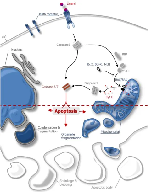
The indispensable physiological process of programmed cell death was first described by Kerr, Wyllie and Currie around forty years ago (Kerr et al., 1972). Based on its characteristic morphological changes including chromatin condensation, fragmentation of the nucleus, organelle degradation as well as cell shrinkage and blebbing it was termed apoptosis, referring to the Greek term for “dropping off” petals from flowers (Williams et al., 1974; Wyllie et al., 1980). The result of these morphological changes is the formation of cellular fragments termed apoptotic bodies which are finally cleared by phagocytes, preventing damage to the surrounding tissue by uncontrolled spilling of cellular debris and allowing the recovery of precious cellular constituents (Reddien et al., 2001; Ravichandran & Lorenz, 2007). These processes are mainly, but not exclusively, orchestrated by members of a family of cysteine proteases known as caspases. Caspases cleave their substrates after specific aspartate residues (the “asp” in the word caspase), while the hydrolysis of the peptide bond is catalyzed by a cysteine in the active site (the “c” in the word caspase) (Yuan et al., 1993; Nicholson & Thornberry, 1997). The activation of caspases occurs via two major pathways, referred to as extrinsic and intrinsic pathway of apoptosis (Figure 1.1).
The extrinsic pathway of apoptosis is activated by the binding of extracellular death ligands of the tumor necrosis factor (TNF) family such as FAS-ligand, TNF- or TRAIL to death receptors on the surface of the plasma membrane. Ligand-binding provokes receptor oligomerization and the recruitment of adapter proteins like FAS associated death domain (FADD), which in turn recruits the initiator caspase 8 to form the death inducing signaling complex (DISC) (Kischkel et al., 1995; Boatright et al., 2003). Caspase 8 becomes activated and then directly cleaves and activates the downstream executioner caspases 3 and 7 which conduct protein degradation and apoptosis.
The intrinsic pathway of apoptosis can be initiated cell-autonomously by overwhelming intracellular stress such as DNA damage, viral infection or growth factor withdrawal. Because intrinsic apoptosis arises from the mitochondrial outer membrane permeabilization (MOMP) followed by the release of pro-apoptotic intermembrane space (IMS) proteins like cytochrome c into the cytosol, it is also referred to as the mitochondrial pathway of apoptosis. The release of
2 cytochrome c into the cytosol stimulates the ATP-dependent oligomerization with apoptotic protease activating factor 1 (APAF1) to form the apoptosome, which in turn catalyzes the auto- activation of initiator caspase 9 (Liu et al., 1996; Li et al., 1997). Activated caspase 9, like caspase 8, then directly cleaves and activates executioner caspases. Both pathways are interconnected by the capability of caspase 8 to proteolytically activate Bid, a BH3-only member of the Bcl-2 protein family, which upon truncation (then called tBid) translocates to mitochondria and initiates MOMP (Figure 1.1).
Figure 1.1 | Extrinsic and intrinsic pathway of apoptosis
Upon binding of a death ligand to its specific receptor on the surface of the plasma membrane, the DISC complex constitutes and initiator caspase 8 becomes activated. Caspase 8 either directly activates
3 executioner caspases 3/7 or indirectly by cleaving BH3-only protein Bid. tBid activates pro-apoptotic Bax/Bak on the outer mitochondrial membrane while activation of Bax/Bak is antagonized by anti- apoptotic Bcl-2 proteins. Bax/Bak induce MOMP, resulting in the release of IMS proteins like cytochrome c and activation of initiator caspase 9. In turn caspase 9 cleaves and activates caspase 3/7, thereby executing the apoptotic demise of the cell. PM, plasma membrane; Cyt c, cytochrome c.
Point of no return: Mitochondrial outer membrane permeabilization
MOMP is considered as the point of no return, making it profoundly important to provide well- balanced regulatory mechanisms which are carried out by proteins of the B- cell lymphoma 2 (Bcl-2) family. Based on their role in conducting apoptosis, Bcl-2 proteins are subdivided into three classes: the class of pro-apoptotic Bcl-2 proteins like Bax and Bak which are responsible for MOMP, the class of anti-apoptotic Bcl-2 proteins such as Bcl-2, Bcl-xL and Mcl-1 which counteract MOMP by directly binding and inhibiting Bax or Bak, and the divergent class of BH3- only proteins including Bad, Bik, Bid, Bim, Bmf, Noxa and Puma that can bind and regulate the activity of pro- and anti-apoptotic Bcl-2 proteins likewise. Recent evidence suggests that BH3- only proteins de-repress and liberate Bax and Bak by direct binding and inhibition of anti- apoptotic family members (Westphal et al., 2014). By contrast, an opposing model postulates direct activation of Bax and Bak by BH3-only proteins including Bim, tBid and Puma (Tait &
Green 2010).
The mitochondrial apoptotic pathway depends on the activation of Bax and Bak, either of which is sufficient to drive MOMP in the majority of cells. Upon cytotoxic stress, the hydrophobic BH3- domain of Bax and Bak becomes exposed and facilitates the generation of stable homo- oligomers that insert into the outer mitochondrial membrane, forms membrane pores and thereby provokes the release of pro-apoptotic IMS-proteins (Zha et al., 1996; Dewson et al., 2008; Kim et al., 2009). The insertion of Bax and Bak into the outer mitochondrial membrane represents a central prerequisite for their pro-apoptotic action which is directed by a C-terminal tail anchor (TA). The tail anchor potentiates the association of cellular proteins mainly with mitochondria or the endoplasmic reticulum (ER), where they are inserted into the lipid membrane via a single membrane span. Pro-apoptotic Bak resides as an inactive monomer in the outer mitochondrial membrane where it is constantly bound and inactivated by anti- apoptotic Mcl-1 and Bcl-xL (Willis et al. 2005). Unlike Bak and the majority of TA-proteins, including the anti-apoptotic Bcl-2 family members Bcl-2 and Bcl-xL that are constitutively bound
4 to their target membranes, Bax in its monomeric inactive state predominantly appears in the cytosol and translocates to the mitochondria upon activation (Hsu & Youle 1998). Whereas only a minor fraction of Bax has been shown to be loosely attached to the outer mitochondrial membrane, the solubility of Bax is effectively regulated by the reduced exposure of its hydrophobic C terminus (Suzuki et al., 2000). Recent data conclusively demonstrated that Bax exists in a dynamic equilibrium between the cytosolic and mitochondrial compartment and that the subcellular localization of Bax is constantly maintained by Bcl-xL (Schellenberg et al. 2013;
Edlich et al. 2011).
Previous work revealed that the pore formation in the outer mitochondrial membrane is not only controlled by the expression status of Bcl-2 protein family members, but that in addition the lipid composition of the mitochondrial outer membrane is also critical for the action of Bcl-2 proteins.
The vast majority of membrane phospholipids and lipid precursors are synthesized in the ER and are delivered via vesicle transport or lipid-transfer proteins (LTP) for further processing into the Golgi apparatus, peroxisomes and mitochondria (Lev 2012). Initial studies performed on unilamellar vesicles mimicking outer mitochondrial membranes provided conceptional advances concerning the interplay of Bcl-2 proteins and mitochondrial lipids. Further work has demonstrated that the embedment of pro-apoptotic Bax together with tBid (Lovell et al. 2008;
Shamas-Din et al. 2013) as well as anti-apoptotic Bcl-xL (Billen et al., 2008) into membrane compartments mimicking the outer mitochondrial membrane is necessary for the regulation of Bax.
The inner and outer mitochondrial membranes are predominantly composed by the two phospholipids phosphatidylcholine (PC) and phosphatidylethanolamine (PE), representing approximately 70-80% of the total mitochondrial membrane lipid content. Phosphatidylinositol (PI) and the diglycerophospholipid cardiolipin (CL) account for 10-15%, while other phospholipids including sphingomyelins are only detectable in trace amounts (Horvath & Daum, 2013). CL is unique among the mitochondrial phospholipids in that it is mitochondria-exclusive and mainly resides in the inner mitochondrial membrane. Beside its function in membrane fluidity, supercomplex assembly and stability (Pfeiffer et al. 2003), CL has been demonstrated to be essential for tBid mediated Bax oligomerization and pore formation in liposomes (Lutter et al., 2000; Kuwana et al., 2002) and early oxidation of CL in apoptosis is critical for the dissociation of cytochrome c from the inner mitochondrial membrane and release into the cytosol (Kagan et al., 2005; Choi et al., 2007). Accordingly, overexpression of phospholipid
5 scramblase 3 (PLS3), a mitochondrial flippase responsible for the transfer of CL between inner and outer mitochondrial membrane compartments, increased CL exposure on the mitochondrial surface and enhanced mitochondrial apoptosis upon TNF- treatment (Liu et al., 2008).
Furthermore, perturbation of CL biosynthesis is sufficient to sensitize mitochondria to both extrinsic and intrinsic apoptotic stress (Potting et al. 2013).
In addition to CL, distinct sphingolipids species have been shown not only to be involved in membrane architecture, but also in numerous and diverse cellular processes such as proliferation, differentiation, senescence, inflammation as well as in the induction and inhibition of apoptosis (Hannun & Obeid 2008). In this regard, ceramides have become the center of attention since the early discoveries of the molecular principles of apoptosis (Obeid et al., 1993) and the pivotal function of ceramides in mediating apoptosis has been intensely described (Tirodkar & Voelkel-Johnson 2012; Chipuk et al. 2012). The vast majority of mammalian sphingolipids have a C18:0 sphingoid base linked to a varying side chain, whereas the head group is either unmodified (ceramides), contains a phosphocholine group derived from phosphatidylcholine (sphingomyelins) or is linked to mono- or oligosaccharides (cerebrosides and gangliosides, respectively). Ceramides exert their physiological role in a fatty acyl side-chain specific manner. Here, especially ceramides containing a C16:0 side chain have been shown to participate in Bax/Bak dependent MOMP by versatile stress stimuli including death ligand binding (Matsko et al., 2001), UV light (Kashkar et al., 2005) and irradiation (Lee et al. 2011), though the exact biochemical nature of the interplay between ceramides and Bcl-2 proteins in pore formation remains controversial (discussed in Renault & Chipuk, 2014).
Dysregulation of apoptosis
In order to maintain the cellular orchestra of constant renovation and demise, multicellular organisms are not only highly dependent on accurate regeneration and replacement of cells, but also on their controlled degradation. Therefore, the prerequisite of tissue homeostasis is that if cells are constantly renewed, an equal amount must necessarily die. Initial experiments in the model organism Caenorhabditis elegans illustrated the critical role of apoptosis in the embryogenesis and development of organisms (Ellis & Horvitz 1986). Similar observations in flies and vertebrates like zebrafish and rodents underscored the fundamental role of apoptosis in embryogenesis and development. For example, pro-apoptotic Bax and Bak were shown to be crucial for embryonic development in mice, given that the majority of double knockout animals
6 die prenatally or display multiple developmental defects (Lindsten et al. 2000). Additionally, the generation of caspase knockout mice further underlined the importance of effective apoptosis in embryogenesis and development, revealing prenatal mortality or neurological defects in adulthood in caspase 3, 8 and 9 knockout mice (reviewed in Kumar, 2007). In addition to its involvement in embryonic development, imbalanced apoptosis in adult tissues may cause defects in tissue homeostasis and prelude the progression of numerous different diseases.
Accordingly, failure to precisely execute timely controlled cell death in renovating tissues contributes to the formation of cancer. On the other hand, excessive programmed cell death particularly in post-mitotic tissue is assumed to precipitate degenerative states and is implicated in the progression of aging and aging associated diseases like Alzheimer’s disease and Parkinson.
Inhibition of Apoptosis and cancer
There is a general consensus that the transformation of healthy cells into tumorigenic and malignant cells is characterized by the acquisition of eight biological capabilities in a multistep process caused by genomic instabilities: 1.) Reprogramming of energy metabolism, 2.) escaping immune response, 3.) sustaining proliferative signaling and 4.) evading growth suppressors, 5.) inducing angiogenesis, 6.) activating invasion and metastasis, 7.) enabling replicative immortality and 8.) resisting cell death (Hanahan & Weinberg 2011). The importance of apoptosis and the implications of its alteration in the progression of different malignancies has accompanied the advances made in understanding the molecular mechanism, regulation and physiological role of apoptosis ever since (Evan & Littlewood 1998). Indeed, already in the initial work done by Kerr and coworkers, apoptosis was correlated to therapeutically induced tumor regression (Kerr et al. 1972). Accordingly, apoptosis reduction in cancer progression and maintenance can be associated with defective death receptor signaling, dysregulated caspase activation and misbalances in the interaction of pro- and anti-apoptotic Bcl-2 proteins. Here, overexpression of anti-apoptotic Bcl-2 proteins like Bcl-2, Mcl1 and Bcl-xL and transcriptional and/or post-transcriptional repression of pro-apoptotic Bcl-2 members like Bax and Bak and BH3-only proteins have been demonstrated as iterant traits of cancer cells (Beroukhim et al.
2010; Kelly & Strasser 2011). On the basis of these findings, enormous efforts have been made in utilizing pro-apoptotic compounds targeting anti-apoptotic Bcl-2 proteins in the hope to reintroduce the apoptotic potential in tumors and tumor environments (Lessene et al., 2008). In
7 this context, therapeutic strategies have already entered the clinical application with great promise in treating patients suffering hematologic malignancies (Brinkmann & Kashkar 2014).
Increased apoptosis and degenerative diseases
Unopposed or increased apoptosis is considered as a cause of tissue degeneration in aging and aging-associated diseases. In particular, increased apoptosis and the alterations of neuronal tissues such as Alzheimer’s and Parkinson’s disease are well described. Alzheimer’s disease (AD) is the most common neurodegenerative disease, characterized by the progressive decline of neurons and synapses in the cerebral cortex and subcortical regions. AD is described by the accumulation of amyloid-ß peptide deposits in extracellular plaques and hyperphosphorylation of the microtubule-associated protein tau, eventually leading to the degeneration of affected regions. Apoptosis has been shown to be essential for the onset of the disease, especially in the caspase-mediated proteolysis of the amyloid precursor protein (APP) resulting in the aggregation of amyloid-ß peptides (Gervais et al., 1999; Garwood et al., 2011). Furthermore, Rohn and colleagues could demonstrate that by inhibiting apoptosis through overexpression of anti-apoptotic Bcl-2 in a mouse model of AD, the pathology could be drastically reduced (Rohn et al. 2008).
Parkinson’s disease (PD) is the second most prevalent neurodegenerative disease and is characterized by a loss of specific dopaminergic neurons of the substantia nigra. PD has been linked to mutations in several genes necessary for efficient autophagic recovery of defective mitochondria and apoptosis regulation such as parkin and (PTEN)-induced kinase 1 (PINK1) (Kitada et al., 1998; Valente et al., 2004). The E3-ligase parkin is involved in the ubiquitination and degradation of Bax and other pro-apoptotic proteins and hence acts as an anti-apoptotic sensitizer at the mitochondria, while PINK1 was shown to be necessary for the recruitment of parkin to mitochondria. PINK1 and parkin deletion were reported to enhance the levels of Bax translocation to the mitochondria and release of cytochrome c in the cytoplasm, resulting in constitutively elevated levels of active caspases (Wood-Kaczmar et al. 2008).
In addition to AD and PD, degenerative diseases caused by mitochondrial oxidative phosphorylation (OXPHOS) defects are also assumed to be associated with elevated apoptosis primarily in affected tissues with high energy demands (Breuer et al. 2013). In this context, mutations in nuclear DNA (nDNA) as well as mitochondrial DNA (mtDNA) encoding factors
8 involved in the respiratory chain are known to be causative in versatile severe degenerative states, premature aging phenotypes and premature mortality (Ylikallio & Suomalainen 2012).
These findings highlight the role of mitochondrial integrity in the formation of degenerative states and raised attention to the impact of mitochondrial respiratory chain defects in the onset of a vast range of degenerative diseases.
The mitochondrial respiratory chain
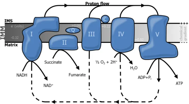
According to the chemiosmotic theory proposed by Peter Mitchell in 1961, the demand for energy in form of ATP by cells and whole organisms is predominantly achieved by the translation of energy from energetic molecules by a concerted transfer of electrons (e-) across an impermeable membrane, generating a chemical gradient which propels the synthesis of ATP (Mitchell 1961). This highly conserved process, termed oxidative phosphorylation, is located to the inner mitochondrial membrane (IMM) and composed of five large intermembrane complexes as well as the electron carriers ubiquinone and the inner membrane associated cytochrome c (Figure 1.2).
Figure 1.2 | Mitochondrial electron transport chain and oxidative phosphorylation
ATP production at the inner mitochondrial membrane (IMM) is coupled to the oxidation of nicotinamide adenine dinucleotide (NADH) at NADH-dehydrogenase (complex I) and flavin adenine dinucleotide
9 (FADH2) at Succinate dehydrogenase (complex II). Electrons (e-) from complex I and II are transferred via reduction of ubiquinone (Q), which is then oxidized by Cytochrome c reductase (complex III). e- are transferred to Cytochrome c oxidase (complex IV) by consecutive reduction and oxidation of cytochrome c (C) and finally reduce molecular oxygen and protons to water at the matrix-side of complex IV. The energy released during the electron transfer drives the translocation of protons from the matrix to the intermembrane space (IMS) across complex I, III and IV, generating a membrane potential across the IMM which drives the generation of ATP at the ATP synthase (complex V) by reflux of protons into the mitochondrial matrix. Electric potential differences are depicted at several steps of the respiratory chain.
Pi, inorganic phosphate
The initial step of the oxidative phosphorylation cascade is the oxidation of the tricarboxylic acid cycle (TCA) metabolite nicotinamide adenine dinucleotide (NADH) by NADH-dehydrogenase (also referred to as complex I) (Hatefi & Stempel, 1969; Efremov et al., 2010; Hunte et al., 2010). Oxidation of NADH is the result of a higher redox-potential of the matrix-faced prosthetic group flavin mononucleotide (FMN), and transfer of two e- to ubiquinone (or coenzyme Q10) is accompanied by a still controversial mechanism of proton translocation across the IMM (reviewed in Brandt et al., 2003). A second entry point into the electron transport chain is provided by succinate-dehydrogenase (complex II) at which succinate is oxidized to fumarate by the prosthetic group flavin adenine dinucleotide (FAD) and e- are subsequently transferred to ubiquinone by iron-sulfur clusters (Coles et al. 1979; Sun et al. 2005). Reduced ubiquinone is then oxidized by iron clusters within the heme group of cytochromes of cytochrome c reductase (complex III) (Hatefi et al., 1962; Xia et al., 1997), resulting in the dislocation of protons from the matrix side to the IMS and the transfer of e- to cytochrome c (Leung & Hinkle 1975). In the last step, cytochrome c is reoxidized by cytochrome c oxidase (complex IV), repeatedly leading to the translocation of protons and finalizing the e- cascade by the reduction of molecular oxygen and protons to water (Wikström, 1977; Tsukihara et al., 1996). As a result, the transformation of the electric potential of high-energetic metabolites originating from glycolysis, fatty acid oxidation and protein degradation generates a chemical gradient across the IMM known as proton motive force (PMF). This chemical gradient provokes a passive reflux of protons through the transmembrane channel of the FO subunit of the ATP-synthase (complex V) (Soper et al., 1979; Ogawa & Lee, 1984), thus powering alternating conformational changes in the F1 subunit of ATP-synthase and ultimately generating ATP from ADP and inorganic phosphate (Watt et al., 2010).
10 The mitochondrial respiratory chain and degenerative diseases
Mitochondria resemble their bacterial ancestry in terms of size, membrane architecture and dynamics. Most importantly, during their endosymbiotic evolution, intense expansive and reductive rearrangement of the mitochondrial proteome and massive transfer of the mitochondrial genome to the nucleus has left the mitochondrial network with traces of mtDNA (Nass & Nass, 1963; Clayton & Vinograd, 1967), which encode indispensable structural subunits of all OXPHOS complexes, except for complex II, as well as tRNAs and ribosomal RNAs (Anderson et al. 1981). In contrast to OXPHOS-defects resulting from mutations of nuclear encoded structural subunits and assembly factors or by autosomal mutations in nuclear encoded genes responsible for mtDNA maintenance, mitochondrial disorders resulting from sporadic or maternally inherited mutations of the mtDNA are always the result of threshold effects, given that the copy number of mtDNA varies between hundreds or thousands within a single cell (Elliott et al., 2008). While both, autosomal or x-linked nDNA and maternally inherited mtDNA mutations can manifest degenerative diseases at any age, the onset of age-associated diseases is predominantly the consequence of accumulated mutations or deletions of mtDNA within the lifetime of organisms (Lin et al., 2002; Kraytsberg et al., 2006).
Since the first descriptions of the molecular basis of disease-causing mutations of nuclear (Bourgeron et al., 1995) and mtDNA (Holt et al., 1988; Wallace et al., 1988) encoded OXPHOS proteins, the knowledge of degenerative diseases caused by mitochondrial dysfunction has widely expanded and to date more than 150 distinct genetic mitochondrial syndromes caused by approx. 300 mutations are known (Schon et al., 2012; Vafai & Mootha, 2012;
www.mitomap.org/MITOMAP). The complex and diverse clinical appearance of distinct genetic mutations of OXPHOS complexes together with the bipartite origin of OXPHOS subunits has long time puzzled the understanding of mitochondrial disorders. Enormous advantages in molecular and cellular techniques like the standardized analysis of mtDNA mutations using cellular hybrids (cybrids) (King & Attardi 1989), the development of mouse models of nuclear encoded OXPHOS subunits and assembly factors (Vempati et al., 2009) and huge progress in genetic screenings have contributed to our current understanding of degenerative diseases (Ylikallio & Suomalainen 2012). The currently known mtDNA and nDNA encoded disease-causing structural subunits and assembly factor are summarized in Table 1.1.
11 Table 1.1: Common disease-encoding OXPHOS genes either encoding structural
subunits (mtDNA and nDNA) or assembly factors (nDNA only) of the respective OXPHOS Complexes (not included: mutations in mitochondrial tRNAs and rRNAs).
Complex Genetic defect Genes
I
mtDNA ND1-ND6
nDNA structural gene NDUFS1-NDUFS8; NDUFV1, 2;
NDUFA1,2,11 nDNA assembly factor NDUFAF1,2; C6orf66;C8ORF38
II
nDNA structural gene SDHA,SDHB,SDHC,SDHD
nDNA assembly factor SDHAF1,2
III
mtDNA CYTB
nDNA structural gene UQCRB,UQCRQ nDNA assembly factor BCS1L, TTG19
IV
mtDNA COX1-3
nDNA structural gene COX6B1, COX4I2 nDNA assembly factor SURF1, SCO1 & 2, COX10,
COX15, C2orf64
V
mtDNA ATP6 & 8
nDNA structural gene ATP5E
nDNA assembly factor ATP12, TMEM70
Most OXPHOS defects are the result of nDNA mutations, given that the vast majority of OXPHOS complexes and assembly factors are encoded by the nucleus. Accordingly, defects in complex I, by far the largest of all OXPHOS complexes, represent most of the known OXPHOS defects (reviewed in Zhu et al., 2009). Mutations in nDNA and in mtDNA encoded OXPHOS subunits as well as in mitochondrial tRNAs and rRNAs precipitate many different and often fatal degenerative states like, amongst others, myopathy, cardiomyopathy, encephalopathy, hepatopathy and stroke (reviewed in Breuer et al., 2013). In spite of the diversity of OXPHOS defects-derived syndromes, most frequent and common to all OXPHOS complexes, especially complex I and IV, is Leigh’s syndrome (LS) (Leigh 1951), a disease which is associated with lactic acidosis, muscle atrophy as well as bilateral, focal lesions of the basal ganglia and the brain-stem and early mortality (Baertling et al. 2014). French Canadian Leigh syndrome (LSFC), a type of LS frequently found in the region of Quebec and characterized by complex IV deficiency, is caused by mutations in the mitochondrial mRNA stability protein leucine-rich pentatricopeptide repeat motif-containing (LRPPRC) (Mootha et al., 2003; Debray et al., 2011).
12 Leber’s hereditary optic neuropathy (LHON), a disease characterized by the degeneration of retinal ganglion cells, optic atrophy and loss of sight is the result of specific mtDNA mutations affecting complex I and, to lesser extent, complex III (Wallace et al., 1988; Bénit et al., 2009).
Another disease shared by diverse mtDNA mutations is mitochondrial encephalomyopathy, lactic acidosis and stroke-like episodes (MELAS), a multisystem disorder affecting versatile tissues such as brain, muscle, heart and the endocrine system (Pavlakis et al., 1984) and mainly arises from a distinct mutation (“A3243G”) in the mitochondrial tRNALeu (Majamaa et al. 1998) as well as from mutations in complex I (Dimauro & Davidzon 2005) and mtDNA encoded subunits of complex IV (Manfredi et al., 1995; Rossmanith et al., 2008).
Degenerative diseases can also be the result of large scale deletions of mtDNA, first documented in patients suffering from Kearns-Sayre syndrome (KSS) by Moraes and colleagues in the early 1990s (Moraes et al., 1991; Moraes et al., 1992). More recent studies identified defects in proteins responsible for maintaining the mitochondrial nucleotide pool (Nishino et al., 1999), nucleotide transport (Kaukonen et al., 2000), and DNA replication (Spelbrink et al., 2001;
Van Goethem et al., 2001) respectively, as source for large mtDNA deletions.
The mitochondrial respiratory chain and cell death
Within the last two decades, advantages in molecular techniques tremendously supported our understanding of the clinical features of degenerative diseases caused by defective OXPHOS and it became evident that tissue degeneration is associated with enhanced cell death. In LS, tissue degeneration mainly arises from necrotic cell death, though several investigations have linked the loss of cerebral tissue to enhanced apoptosis both histochemically (Formichi et al., 2004) or in cultured cybrids (Carrozzo et al. 2004), while a shift from apoptosis to necroptosis has also been reported (Quintana et al., 2010). Apoptotic cell death was also detected in peripheral blood lymphocytes of patients with LHON (Battisti et al. 2004), in cybrids containing different mtDNA mutations encoding complex I subunits ND1, 4 and 6 (Danielson et al. 2002; Ghelli et al. 2003), in patients suffering from KSS, MELAS and chronic progressive external ophthalmoplegia (CPEO) (Mirabella et al., 2000; Auré et al., 2006) and in patients with complex IV deficiency (Di Giovanni et al. 2001). Chemical inhibition of complex I in mice and utilizing complex I deficient cybrids revealed oxidative damage induced cell death, presumably by sensitizing affected cells to Bax-induced apoptosis (Perier et al. 2005). In addition, skin fibroblasts from patients suffering from complex V deficiency derived neurogenic ataxia retinitis pigmentosa (NARP) also displayed
13 superoxide-induced apoptosis (Geromel et al. 2001), while complex II deficiency seems to be generally linked to apoptotic demise by oxidative damage (reviewed in Grimm, 2013).
A growing contribution to our understanding of the correlation between defective OPXHOS compartments and increased apoptosis comes from increasing numbers of mouse models describing mitochondrial mutations. Roughly a decade ago the work of Trifunovic and colleagues and in parallel by Kujoth et al underlined the importance of mtDNA integrity in the generation of aging-associated degenerations. By generating mice with defective mtDNA-specific polymerase proofreading subunits, the authors could demonstrate that accumulating mtDNA mutations cause a loss of respiratory chain capacity, severe onset of premature aging and early lethality by tissue malfunction due to enhanced apoptosis (Trifunovic et al., 2004; Kujoth et al., 2005). In addition, knockout of the mitochondrial transcription factor A (TFAM) revealed massive apoptosis in knockout embryos and in the heart of TFAM tissue-specific knockout animals (Wang et al., 2001).
Furthermore, mouse models of defective respiratory chain subunits and assembly factors have highlighted the importance of OXPHOS integrity: Enhanced apoptosis preceding fatal hepatopathy was demonstrated in mice with a liver-specific knockout of COX10, an indispensable assembly factor of the prosthetic heme a of complex IV (Diaz et al., 2008). A muscle-specific knockout of COX10 revealed premature aging characteristics like weight loss, progressive myopathy and early mortality (Diaz et al., 2005). Embryonic lethality and early mortality was also linked to complex IV deficiency by the knockout of assembly factor SURF1 (Agostino et al., 2003) as well as for a knockout of SDHD, a membrane-anchoring subunit of complex II (Piruat et al. 2004). Knockout-mice of the complex I subunit NDUFS4 appeared healthy but developed fatal encephalomyopathy at the age of 5 weeks (Kruse et al. 2008).
Aim of the study
Apoptosis is a vital process in cell homeostasis and tissue-regeneration and has constantly been shown to be manipulated by diverse mitochondrial dysfunctions. Even though numerous findings in the recent years have shed light on the correlation between the decline of OXPHOS and the increase of cell death by apoptosis in various degenerative and aging-associated diseases (Breuer et al. 2013), the exact molecular mechanisms that trigger this mitochondrial dysfunction-associated tissue demise remain elusive.
14 Overwhelming generation of reactive oxygen species (ROS) has been long-standing associated with defective mitochondrial respiration, though the causality of excessive ROS generation and the decline of mitochondrial respiratory activity is still heavily debated. However, oxidative stress has often been implicated in the onset of OXPHOS-degenerative states as well as aging and aging-associated diseases (Cui et al., 2012). In this context, the present work aims to dissect the role of the distinct complexes of the mitochondrial respiratory chain towards oxidative stress induced apoptosis and tries to substantiate our understanding of the molecular mechanisms underlying OXPHOS-dysfunction induced enhanced apoptosis.
15
Material and Methods
Cell culture
Human cell lines Hela (CCL-2, obtained from American Type Culture Collection (ATCC), Rockville, MD, USA), Hela Bcl-2 (Kashkar et al.,2005), 143B control and 143BCOX (gift from R. Wiesner, Cologne, Germany) as well as adult murine fibroblasts of COX10 knockout mice (gift from C.
Moraes, Miami, FL, USA) and murine embryonic fibroblasts (MEFs) of Bax and Bak single and Bax/Bak double-knockout mice (gift from A. Villunger, Innsbruck, Austria) were cultured in Dulbecco’s modified Eagle’s medium (DMEM, Gibco (Life Technologies), Darmstadt, Germany), supplemented with 10% fetal calf serum (FCS, Gibco (Life Technologies), Darmstadt, Germany), 100 µ/ml streptomycin and 100 U/ml penicillin (Biochrom (Merck), Darmstadt, Germany).
DMEM of 143BCOX, COX10 knockout fibroblasts or mammalian cell lines under conditions of chemical suppression of the mitochondrial respiratory chain (RC) was supplemented with 1mM sodium pyruvate (Biochrom (Merck), Darmstadt, Germany) and 50µg/ml uridine (Sigma-Aldrich, Seelze, Germany).
Chemicals and reagents
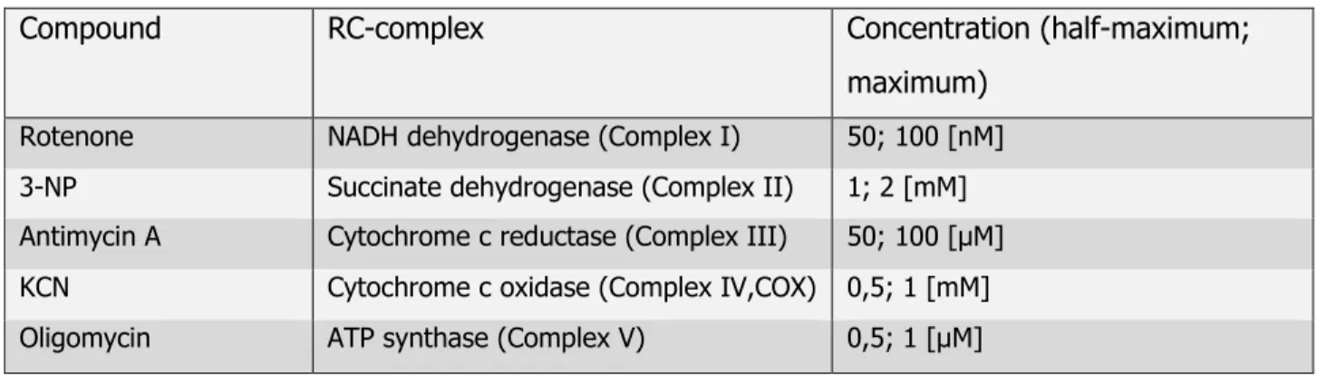
All chemicals were purchased from Merck (Darmstadt, Germany), Roth (Karlsruhe, Germany) and Sigma-Aldrich (Seelze, Germany), unless indicated otherwise. Staurosporine, zLEHD-fmk, zIETD-fmk and zVAD-fmk were obtained from Enzo Life Sciences (Lörrach, Germany). Rotenone, 3-nitropropionic acid (3-NP), antimycin A, KCN, oligomycin, and H2O2 were purchased from Sigma-Aldrich (Seelze, Germany).
Measurement of oxygen consumption
Measurement of oxygen consumption was performed with a Clark-like oxygen electrode system (Hansatech Instruments, Norfolk, UK). 4x10⁶ cells were diluted in Buffer E and placed in a water-filled, air-saturated chamber at 37oC. Water-dissolved oxygen was reduced by the addition of trace amounts of dithionite (Na2S2O4). Oxygen consumption under conditions of chemical inhibition of the respective respiratory chain complexes was recorded, related to total cell number and expressed as femtomoles O2 per cell per minute.
16 Table 2.1: Buffers for oxygen consumption
Buffer E 300 mM Mannitol; 5 mM MgCl2; 10 mM KCl; 10 mM KH2PO4, pH 7.4; 1mg/ml bovine serum albumin, fatty acid free
Table 2.2: Respiratory chain-inhibitors and concentrations
Compound RC-complex Concentration (half-maximum;
maximum) Rotenone NADH dehydrogenase (Complex I) 50; 100 [nM]
3-NP Succinate dehydrogenase (Complex II) 1; 2 [mM]
Antimycin A Cytochrome c reductase (Complex III) 50; 100 [µM]
KCN Cytochrome c oxidase (Complex IV,COX) 0,5; 1 [mM]
Oligomycin ATP synthase (Complex V) 0,5; 1 [µM]
DNA constructs and siRNA-mediated knockdown
For the construction of human ceramide synthase (CerS) GFP fusion proteins, open reading frames encoding human ceramide synthase 3 and 6 were amplified by PCR with appropriate restriction sites and cloned into pEGFP-N3 vector (Clontech, Saint-Germain-en-Laye, France).
For the generation of CerS6R131A, site-directed mutagenesis PCR was performed on pEGFP- CerS6. Cells were transfected with TurboFect transfection reagent (Thermo Scientific, Rockford, IL, USA) according to manufacturer’s manual and incubated for at least 24h.
Table 2.3: Primers for DNA constructs
Construct Primer (‘5-3’)
CerS3 forward (5’-EcoRI) GATCGAATTCATGTTTTGGACGTTTAAAGAATGGTTCTG CerS3 reverse (3’-BamHI) GATCGGATCCATGGCCATGCTGGCCATT
CerS6 forward (5’-EcoRI) GATCGAATTCATGGCAGGGATCTTAGCC CerS6 reverse (3’-BamHI) GATCGGATCCATCATCCATGGAGCAGGAGCC CerS6R131A sense TTCTGTGAGAGCATGTGGAGATTT
CerS6R131A anti-sense GGCCGTCAGCGTGCTTGGCTTCTCCTG
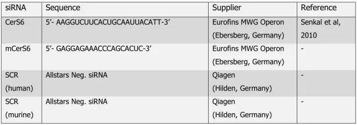
17 For siRNA mediated knockdown of human CerS6 as well as murine CerS6 (mCerS6), suitable amounts of cells were transfected with 100 pmol/ml siRNA using LipofectamineTM RNAimax transfection reagent (Life Technologies, Darmstadt, Germany) and incubated for 48h before cytotoxic treatment. All siRNAs were purchased from Eurofins MWG Operon (Ebersberg, Germany).
Table 2.4: siRNAs used in this work
siRNA Sequence Supplier Reference
CerS6 5’- AAGGUCUUCACUGCAAUUACATT-3’ Eurofins MWG Operon (Ebersberg, Germany)
Senkal et al, 2010 mCerS6 5’- GAGGAGAAACCCAGCACUC-3’ Eurofins MWG Operon
(Ebersberg, Germany) -
SCR (human)
Allstars Neg. siRNA Qiagen
(Hilden, Germany)
-
SCR (murine)
Allstars Neg. siRNA Qiagen
(Hilden, Germany)
-
Cell death
Cell death was measured by trypan blue exclusion using an automated cell counter (CountessTM, Life Technologies, Darmstadt, Germany) according to manufacturer’s instructions. In brief, approx. 0.75-1.5x10⁵ cells were seeded in 24-well chambers and incubated under desired conditions. Before counting, cells were briefly detached with trypsin and diluted to appropriate working concentrations. Cell suspension was then diluted with trypan blue in a ratio of 1:1 (v/v) and dead cells were measured.
Measurement of superoxide formation
For immunofluorescence analysis of superoxide (O2-) formation, MitoSoxTM Red mitochondrial superoxide kit (Life Technologies, Darmstadt, Germany) was used according to manufacturer’s instructions. In brief, 3x10⁵ cells were seeded on sterile glass bottom live cell dishes (Greiner Bio-One, Frickenhausen, Germany), incubated with 5 µM MitoSoxTM reagent working solution and washed three times with warm HBSS/Ca/Mg buffer (Life Technologies, Darmstadt,
18 Germany). Samples were analyzed with an Olympus Flowview 1000 confocal microscope (Olympus, Hamburg, Germany). Additionally, 3x10⁵ Hela cells were incubated with 5 µM MitoSoxTM reagent working solution and washed three times with warm HBSS/Ca/Mg buffer and analyzed by flow cytometry (BD FACSCaliburTM, BD Biosciences, Heidelberg, Germany).
Sample preparation and immunoblotting (IB)
Whole cell lysates were prepared by incubating cell pellets in CHAPS lysis buffer on ice for 20 min and subsequent centrifugation at 14.000 x g for 20 min at 4oC to recover the supernatant.
To obtain cytosolic extracts, cells were incubated for 20 min in ice-cold HEP buffer following mechanical disruption by repeated passaging (x12-15) through a 26 x G 1/2`` needle, the supernatant was recovered after centrifugation at 20.000 x g for 20 min at 4oC. For Western blotting, equal amounts of protein, determined by BCA protein assay (Thermo Scientific, Rockford, IL, USA) were prepared in 1x Laemmli buffer containing 4% ß-mercaptoethanol, separated by SDS-PAGE and transferred onto nitrocellulose membranes (Protran BA85, GE Healthcare, Freiburg, Germany). After blocking and appropriate incubation in primary and secondary antibodies, protein signals were visualized by enhanced chemiluminescence (ECL;
Thermo Scientific, Rockford, IL, USA).
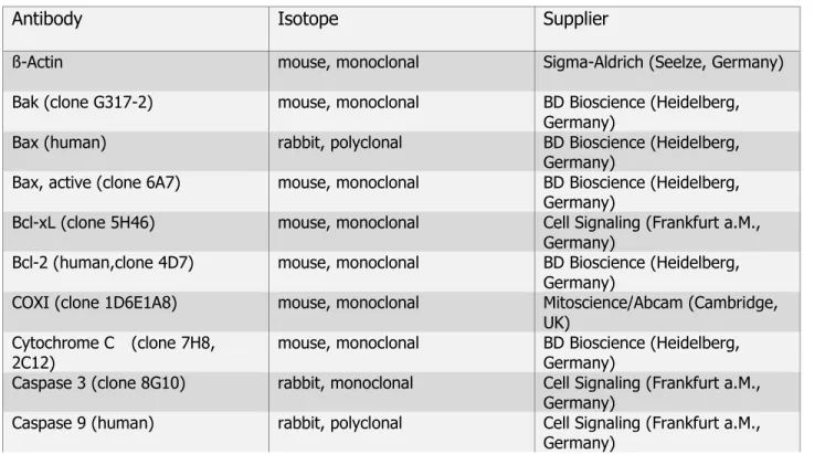
Table 2.5: Primary and secondary antibodies (IB)
Antibody Isotope Supplier
ß-Actin mouse, monoclonal Sigma-Aldrich (Seelze, Germany)
Bak (clone G317-2) mouse, monoclonal BD Bioscience (Heidelberg, Germany)
Bax (human) rabbit, polyclonal BD Bioscience (Heidelberg,
Germany)
Bax, active (clone 6A7) mouse, monoclonal BD Bioscience (Heidelberg, Germany)
Bcl-xL (clone 5H46) mouse, monoclonal Cell Signaling (Frankfurt a.M., Germany)
Bcl-2 (human,clone 4D7) mouse, monoclonal BD Bioscience (Heidelberg, Germany)
COXI (clone 1D6E1A8) mouse, monoclonal Mitoscience/Abcam (Cambridge, UK)
Cytochrome C (clone 7H8, 2C12)
mouse, monoclonal BD Bioscience (Heidelberg, Germany)
Caspase 3 (clone 8G10) rabbit, monoclonal Cell Signaling (Frankfurt a.M., Germany)
Caspase 9 (human) rabbit, polyclonal Cell Signaling (Frankfurt a.M., Germany)
19 Caspase 9 (mouse) rabbit, polyclonal Cell Signaling (Frankfurt a.M.,
Germany)
Ceramide Synthase 6 mouse, polyclonal Abnova (Heidelberg, Germany) GAPDH (clone D16H11) rabbit, monoclonal Cell Signaling (Frankfurt a.M.,
Germany)
Mcl-1 rabbit, polyclonal Cell Signaling (Frankfurt a.M.,
Germany)
Anti-mouse IgG HRP-linked goat Sigma-Aldrich (Seelze, Germany)
Anti-rabbit IgG HRP-linked goat Sigma-Aldrich (Seelze, Germany)
Table 2.6: Buffers for sample preparation and immunoblotting
Antibody dilution buffer 50 mM Tris, pH 7.6; 150 mM NaCl; 0,1% Tween-20; 5% BSA
Blocking buffer 10 mM Tris-Hcl, pH 7.4-7.6; 150 mM NaCl; 5% milk powder; 2%
BSA; 0.1% Tween-20
Blot transfer buffer 25 mM Tris-HCl; 190 mM glycine; 20% methanol
CHAPS lysis buffer 10 mM HEPES, pH 7.4; 150 mM NaCl; 1% CHAPS; protease inhibitor HEP buffer 20 mM HEPES, pH 7.5; 10 mM KCl; 1.5 mM MgCl2; 1 mM EDTA; 10
µM cytochalasin B; 1 mM DTT; protease inhibitor
Laemmli sample buffer (5x) 0.6 M Tris-HCl, pH 6.8; 144 mM SDS; 25% glycerol; 0.1%
bromophenol blue; 5% ß-mercaptoethanol SDS running buffer 190 mM glycine; 20 mM Tris; 0.1% SDS
S-PBS 120 mM NaCl; 10 mM NaH2PO4; 30 mM K2HPO4, pH 7.6
Immunoprecipitation (IP) of active Bax
100 µg of whole cell lysates were brought to a final volume of 500 µl with CHAPS lysis buffer containing 150mM KCl and incubated with 2 µg of monoclonal, active Bax- specific antibody 6A7 over night at 4oC. The antigen-antibody complex was immobilized by the addition of GammaBind-G-Sepharose (GE Healthcare, Freiburg, Germany) and subsequent gentle rotation for 2h at 4oC. Samples were washed twice with CHAPS/ 150mM KCl and the complexes were pelleted by centrifugation at 500 x g for 1 min. The resolved pellet was washed twice with CHAPS/ 150 mM KCl and further subjected to SDS-Page and western blotting.
20 Lipid analysis
Total amount of cellular sphingolipid levels were determined from whole cell homogenates of at least 1x10⁶ cells. For ceramide synthase activity measurement, 2,5x10⁶ cells were incubated with 2 µM C17-sphinganine (Avanti Polar Lipids, Alabaster, AL, USA) for 2h prior to cell homogenization. Lipids were extracted in methanol/chloroform 2:1 (v/v) overnight at 48oC, purified and analyzed by Liquid Chromatography coupled to Electrospray Ionization Tandem Mass Spectrometry (LC-ESI-MS/MS) by Dr. Susanne Brodesser at the Cecad Lipidomics Facility.
Activity of neutral and acid sphingomyelinase (n/aSMase) was measured by Katja Krönke- Wiegmann from crude cytosolic extracts (nSMase) or total cell lysates (aSMase) of at least 3x10⁶ cells in appropriate buffer using 0.2 µCi/ ml [14C]sphingomyelin (Amersham/ GE Healthcare, Freiburg, Germany). The amount of sphingolipid hydrolysis derived phosphorylcholin was measured by thin layer chromatography (TLC) and scintillation counting (Zinsser Analytic, Frankfurt a.M., Germany)
Table 2.7: Buffers for lipid analysis
nSMase sample buffer 20 mM HEPES, pH 7.4; 10 mM MgCL2; 2 mM EDTA; 5 mM DTT, 0.1 mM Na3VO4; 0.1 mM Na2MoO4; 30 mM p-nitrophenylphosphate; 10 mM ß-glycerophosphate;
750 µM ATP; 1 mM PMSF; 10 µM leupeptin; 10 µM pepstatin; 0.2% Triton X-100
aSMase sample buffer 0.2% Triton X-100
nSMase activity buffer 20 mM HEPES; 1 mM MgCl2, pH 7.4
aSMase activity buffer 250 mM sodium acetate; 1 mM EDTA, pH 5.0
Fluorescence microscopy
For immunofluorescence (IF) analyses of Bax activation and cytochrome c release, 3x10⁵ Hela cells were transfected with 1 µg of the particular pEGFP-N3 constructs and incubated for 24h before subjection to H2O2 for 4h. Cells were washed twice with PBS, fixed in 3%
paraformaldehyde and simultaneously permeabilized and blocked with blocking buffer for 30 min. Cells were incubated with monoclonal active Bax 6A7 antibody over night at 4oC. Cells were
21 washed three times for 10 min with washing buffer and incubated with Alexa FluorTM 647 secondary antibody for 1h at room temperature. Cells were washed three times with washing buffer for 10 min. For staining of cytochrome c, directly Alexa FluorTM 555 labeled cytochrome c antibody was utilized according to manufacturer’s instructions. Cells were washed three times with washing buffer for 10 min and Dapi was added to the second washing step for nuclei staining. Samples were mounted onto cover slides using Mowiol mounting medium and examined with a spinning disk confocal microscope (Perkin Elmer, Rodgau, Germany).
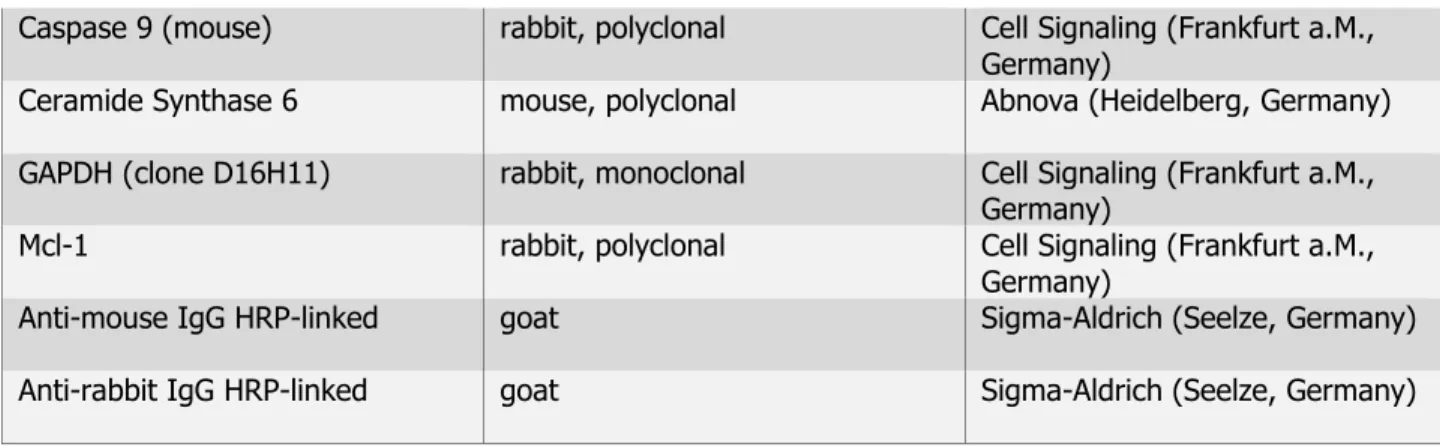
Table 2.8: Primary and secondary antibodies (IF)
Antibody/Compound Isotope Supplier
Cytochrome C Alexa FluorTM 555 mouse, monoclonal BD Bioscience (Heidelberg, Germany) Anti-mouse Alexa FluorTM 647 goat Life Technologies (Darmstadt, Germany)
Dapi - Life Technologies (Darmstadt, Germany)
Statistical Analysis
Data are presented as mean of at least three independent experiments. Standard deviation (SD) and standard error of the mean (SEM) were calculated as indicated in the figures. Statistical significance was calculated by two tailed unpaired student’s t-test.
Software
Vector NTI (Plasmids)
FlowJo (Flow cytometry)
Photoshop CS2 (IF, IB)
ImageJ, Volocity (IF)
Illustrator CS2 (data processing)
22
Results
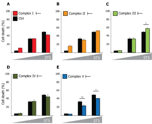
Chemical inhibition of cytochrome c oxidase selectively enhances ROS-induced cell death
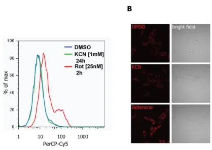
The decline of mitochondrial respiratory capacity and the incidence of cellular death resulting in tissue degeneration have been extensively described (Breuer et al. 2013). However, it is still unclear how excessive apoptosis within a degenerative tissue impacts on tissue damage caused by mitochondrial dysfunction. To investigate how specific alterations of the mitochondrial respiratory chain (RC) as well as ATP-synthase impact on apoptosis, the activity of each of the five oxidative phosphorylation (OXPHOS) complexes was chemically inhibited in Hela cells (Figure 3.1). Oxygen consumption was monitored as a read-out for the efficacy of the chemical inhibition of OXPHOS complexes, revealing a reduction in respiration between 15 and 24% of untreated conditions (Figure 3.1 B). Inhibitor concentrations were chosen based on maximal efficacy without cytotoxic side effects in a period of 48h as measured by trypan blue exclusion assay (Figure 3.1 A).
23 Figure 3.1 | Chemical inhibition of mitochondrial respiratory chain complexes
(A) Hela cells were incubated with increasing concentrations of the respective OXPHOS inhibitors and cell death was assessed after 48h by trypan blue exclusion. Error bars represent mean ± SEM (n=3).
(B) Hela cells were incubated with two concentrations of specific inhibitors as listed in the table and oxygen consumption rate was instantly measured with a Clark-like oxygen electrode system. EtOH (vehicle) concentration was 1 and 2% (v/v), respectively. Error bars represent mean ± SEM of at least four experiments.
Oxidative damage within an aging tissue (e.g. by mitochondrial dysfunction) has been considered as one of the central mechanisms causing tissue degeneration (Turrens, 2003;
Balaban et al., 2005; Sena & Chandel, 2012). Therefore, we first investigated whether the inhibition of OXPHOS complexes interferes with the susceptibility of cells towards oxidative stress by exposing Hela cells to RC inhibitors together with increasing amounts of H2O2 (Figure 3.2). In striking contrast to the inhibition of all other OXPHOS complexes, inhibition of cytochrome c oxidase (COX/complex IV) revealed a dramatic increase in susceptibility to H2O2 in a dose dependent manner (Figure 3.2).
24 Figure 3.2 | Inhibition of cytochrome c oxidase enhances H2O2-induced cell death
Hela cells were left untreated or treated with OXPHOS complex inhibitors; (A) rotenone [100 nM], (B) 3- nitropropionic acid [2 mM], (C) antimycin A [100 µM], (D) KCN [1 mM], (E) oligomycin [1 µM]. After 24h, cells were subjected to increasing concentrations of H2O2 [0; 50; 150 µM] and cell death was assessed after 20h by trypan blue exclusion. Error bars represent mean ± SEM (n=6).Statistical significance was measured by two tailed unpaired t-test (including all following figures). *p<0.05, ***p<0.001
Importantly, the inhibition of the OXPHOS complexes did not interfere with susceptibility to the well-known apoptosis-inducer staurosporine (STS) (Figure 3.3).
Figure 3.3 | Inhibition of RC complexes does not influence STS-induced cell death
Hela cells were left untreated or treated with OXPHOS complex inhibitors; (A) rotenone, (B) 3- nitropropionic acid, (C) antimycin A, (D) KCN, (E) oligomycin as described in Figure 3.2. After 24h, cells were subjected to increasing concentrations of STS [0; 25; 50 nM] and cell death was assessed after 20h by trypan blue exclusion. Error bars represent mean ± SEM (n=3). *p<0.05, **p<0.01