A molecular basis of ELF3 action in the Arabidopsis circadian clock
Inaugural-Dissertation zur
Erlangung des Doktorgrades
der Mathematisch-Naturwissenschaftlichen Fakultät der Universität zu Köln
vorgelegt von
Eva Herrero Serrano aus Vitoria, Spanien
Köln 2011
- II -
Die vorliegende Arbeit am Max-Planck Institut für Pflanzenzüchtungsforschung Köln, in der Arbeitsgruppe von Dr. Seth J. Davis, Abteilung für Entwicklungsbiologie der Planzen (Direktor Prof. Dr. George Coupland) angefertigt.
Berichterstatter: Prof. Dr. George Coupland
Prof. Dr. Ute Höcker
Prüfungsvorsitzender: Prof. Dr. Martin Hülskamp Tag der mündlichen Prüfung 20 Mai 2011
- II -
Abstract
The circadian clock anticipates daily environmental changes and optimizes timing of physiological events. Circadian systems are present in most living organisms.
In Arabidopsis, circadian components are arranged in positive/negative regulatory feed-back loops. The core loop is arranged by the morning transcription factors LHY and CCA1, and the evening pseudo-response regulator TOC1. The morning loop reciprocally connects LHY and CCA1 to the TOC1-sequence-related components PRR9 and PRR7. Other genes, when mutated, display a clock phenotype, but not all of such genes have been placed into the clock model. For instance, three evening genes, ELF3, ELF4, and LUX, have been found to be essential for circadian function. Clock- and light-signaling networks are tightly interconnected. Light input to the clock is mediated by photoreceptors, such as the phytochromes. ELF3 and ELF4 play a pivotal role in the generation of circadian rhythms and in the integration of light signal to the clock mechanism. Both encoded proteins are reported to be located in the nucleus, and both the elf3 and the elf4 mutants display a similarly arrested clock.
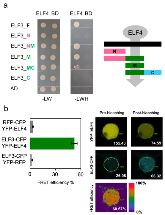
In this thesis, ELF3 was found to be genetically downstream of ELF4 within the same clock signaling pathway required to sustain circadian rhythms. Moreover, I found that ELF3 and ELF4 proteins physically interact. This interaction correlated with an increase of ELF3 nuclear localization. These observations are consistent with a role of ELF4 as an effector that promotes ELF3 activity to lengthen circadian periodicity. A functional complementation approach identified three functional modules in the ELF3 encoded protein. The N-terminus and middle domains mediate interaction with phyB and ELF4, respectively. The C-terminus domain was found to be required for ELF3 nuclear localization. Thus, ELF3 is a multifunctional protein that interacts with both light-signaling and clock components.
The molecular function of ELF3 had previously remained elusive. PRR9 expression was found to be down-regulated in ELF3 and ELF4 over-expressors.
Interestingly, I found that ELF3 physically associated with the same conserved region in the PRR9 promoter as the transcription factor LUX. I found that LUX was genetically downstream of ELF4, and that LUX required ELF3. Taken together, I proposed that ELF3, ELF4, and LUX are part of an evening-clock complex required to repress PRR9 expression, and to sustain circadian oscillations.
Abstract
- ii -
ELF3 has been reported to be crucial to buffer light input to the oscillator.
Photoreceptors and ELF3 play an opposite role in light-mediated acceleration of circadian periodicity, where photoreceptors shortens, and ELF3 lengthens, circadian period under constant light. Interestingly, I found that the N-terminus of ELF3 was not essential for ELF3 circadian function, but that mediated the physical interaction of ELF3 to phyB. An elf3 complementation line deleted for its N-terminus displayed hyposensitivity to the period-shortening effect induced by constant-red light. Therefore, I hypothesized that phyB interaction to the N-terminus of ELF3 mediates light- repression of ELF3 action in circadian-periodicity.
In chapter 4, further characterization of the weak allele elf3-12 supported the role of ELF3 as a decelerator of circadian periodicity. The elf3-12 mutation encodes an amino-acid replacement in a conserved box within the ELF4-binding domain. The elf3-12 coding region led to robust expression of ELF3-12 protein, and ELF3-12 retained the capacity to bind both ELF4 and phyB. elf3-12 displayed light-dependent short-period phenotype that was enhanced by phytochrome over-expression.
Moreover, elf3-12 displayed hypersensitive to red-light-resetting pulses. Thus, I found that elf3-12 is attenuated in its function to repress light input to the clock and/or and increased phy-mediated repression of ELF3 function. elf3-12 was the first described elf3 weak allele. My characterization of a collection of elf3 TILLING alleles led to the identification of novel short- and long-period alleles that I predict will expand current understanding of the role of ELF3 as an integrator of light signals and as a core-clock component.
Taken together, my thesis has placed ELF3 within the circadian mechanism.
ELF3, ELF4, and LUX are part of an evening-repressor complex required to sustain circadian function. The genetic interaction of these three genes is consistent with a hierarchy of complex assembly. In this, I propose that ELF4 works as an effector protein that activates ELF3, possibly by increasing the ELF3 nuclear pool. Then, the association of both ELF3 and LUX to the PRR9 promoter is required for transcriptional repression of PRR9. Additionally, I propose that ELF3 function in circadian periodicity is modulated by its interaction partners by a competition between a positive effect of ELF4 and a light-mediated-negative effect of phyB. This is consistent with ELF3 being a multifunctional protein that integrates light signals as a core-oscillator component.
Zusammenfassung
Die innere zirkadiane Uhr antizipiert die täglichen Umweltveränderungen und optimiert die zeitliche Koordinierung physiologischer Abläufe. Zirkadiane Systeme existieren in den meisten lebenden Organismen. In der Modellpflanze Arabidopsis sind die Komponenten der zirkadianen Uhr in positiv/negativen Rückkopplungsschleifen arrangiert. Der sogenannte Morgenschaltkreis besteht aus den Transkriptionsfaktoren LHY und CCA1, sowie dem abends expremierten Pseudo-response Regulator TOC1.
Der morgendliche Schaltkreis verbindet reziprok LHY und CCA1 mit den Komponenten PRR9 und PRR7, deren Sequenzen mit der von TOC1 verwandt sind. Andere Gene zeigen bei Mutation ebenfalls einen Phänotyp bezüglich der zirkadianen Uhr, aber nicht alle von ihnen sind bisher in das Modell der inneren Uhr integriert worden. So sind beispielsweise die drei abends expremierten Gene ELF3, ELF4 und LUX essentiell für die Funktion der inneren Uhr. Die Netzwerke der inneren Uhr und des Lichtsignalweges sind eng miteinander verbunden. Das Lichtsignal wird über Photorezeptoren, wie beispielsweise die Phytochrome, an die innere Uhr weitergegeben. ELF3 und ELF4 spielen dabei eine ausschlaggebende Rolle bei der Erzeugung von zirkadianen Rhythmen, aber auch bei der Integration von Lichtsignalen in das System der zirkadianen Uhr. Von beiden Proteinen wird berichtet, dass sie im Nukleus lokalisiert sind. Mutanten beider Gene zeigen eine ähnlichen Phänotyp, der eine Arretierung der inneren Uhr zur Folge hat.
In dieser Dissertation konnte gezeigt werden, dass ELF3 genetisch downstream von ELF4 im selben Signalweg der inneren Uhr arbeitet und dazu benötigt wird die zirkadiane Rhythmik aufrecht zu erhalten. Darüber hinaus wurde gezeigt, dass die Proteine ELF3 und ELF4 physisch miteinander interagieren. Diese Interaktion korrelierte mit einer vermehrten Lokalisation von ELF3 im Zellkern. Diese Beobachtungen decken sich mit dem Effekt, dass ELF4 die Aktivität von ELF3 fördert und dadurch die zirkadiane Periode verlängert. Mit Hilfe eines funktionellen Komplementierungsansatzes konnten drei funktionelle Abschnitte im ELF3 Protein identifiziert werden. Der N-Terminus vermittelt die Interaktion mit PhyB, während der mittlere Proteinabschnitt für die Interaktion mit ELF4 verantwortlich ist. Für den C- Terminus konnte festgestellt werden, dass er für die Lokalisierung von ELF3 im Nukleus verantwortlich ist. Daraus folgt, dass ELF3 ein mutlifuntionelles Protein ist, das sowohl mit dem Lichtsignalweg als auch mit Komponenten der inneren Uhr interagiert.
Zusammenfassung
- iv -
Die molekulare Funktion von ELF3 war bisher unbekannt. Man wusste lediglich, dass die Expression von PRR9 in Überexpressoren von ELF3 und ELF4 verringert war.
Interessanter Weise konnte in dieser Arbeit gezeigt werden, dass ELF3 physisch mit der selben konservierten Region im PRR9 Promotor interagiert wie der Transkriptionsfaktor LUX. Außerdem konnte gezeigt werden, dass LUX genetisch downstream von ELF4 arbeitet und dass LUX ELF3 benötigt um zu funktionieren.
Zusammenfassend lässt sich daher annehmen, dass ELF3, ELF4 und LUX Teil eines abends gebildeten Komplexes der inneren Uhr sind, der benötigt wird um die Expression von PRR9 zu verhindern und daher für die Aufrechterhaltung von zirkadianen Oszillationen benötigt wird.
Von ELF3 wird berichtet, dass es ausschlaggebend ist für die Abpufferung der Lichtsignale an den Hauptoscillator der inneren Uhr. Die Photorezeptoren und ELF3 spielen entgegengesetzte Rollen bei der Licht vermittelten Beschleunigung der zirkadianen Periodizität. Währen die Photorezeptoren die Periode verkürzen, verlängert ELF3 die Periode unter konstanten Lichtbedingungen. In dieser Arbeit konnte gezeigt werden, dass der N-Terminus zwar für die zirkadiane Funktion von ELF3 entbehrlich ist, dennoch aber die physische Interaktionsebene für ELF3 und PhyB darstellt. Eine elf3 Komplementationslinie, die keinen N-Terminus mehr besaß, zeigte einen hyposensitiven Phänotyp bezüglich der Verkürzung der zirkadianen Periode unter konstantem Rotlicht. Daraus wurde gefolgert, dass die Interaktion von PhyB mit dem N-Terminus von ELF3 die Licht vermittelte Unterdrückung der zirkadianen Periodizität ermöglicht.
In Kapitel 4 wurde ein schwaches Allel von ELF3, elf3-12, charakterisiert. Die durchgeführten Untersuchungen lieferten weitere Anhaltspunkte für eine entschleunigende Wirkung von ELF3 auf die zirkadiane Periode. Die elf3-12 Mutation sorgt für einen Aminosäureaustausch in einem konservierten Bereich innerhalb der ELF4-Bindedomäne. Die kodierende Region von elf3-12 erzeugte eine robuste Expression des ELF3-12 Proteins, welches die Fähigkeit ELF4 und PhyB zu binden behielt. elf3-12 hatte einen lichtabhängigen Phänotyp mit einer verkürzten Periode, welcher durch die Überexpression von Phytochrom B noch verstärkt wurde. Darüber hinaus war elf3-12 hypersensitiv für Lichtpulse, die den Oszillator zurücksetzen.
Daraus lässt sich schließen, dass elf3-12 nur eine abgeschwächte Funktion bei der Unterdrückung von Lichtsignalen an die innere Uhr hat. Möglich ist auch eine Verstärkung der durch PhyB vermittelte Unterdrückung der ELF3 Aktivität. elf3-12 war das erste schwache Allel von ELF3, das beschrieben wurde. Durch die Charakterisierung weiter sogenannter TILLING Allele von ELF3 konnten weitere Allele
Zusammenfassung
mit verlängerter oder verkürzter Periode identifiziert werden, die zum künftigen weiteren Verständnisses der Rolle von ELF3 als Integrator von Lichtsignalen und als Hauptkomponente der inneren Uhr beitragen werden.
Zusammenfassend lässt sich sagen, dass durch die Untersuchungen in dieser Arbeit ELF3 eine Rolle im Mechanismus der inneren Uhr zugeschrieben werden konnte. ELF3, ELF4 und LUX sind Teil eines Abendkomplexes mit reprimierender Funktion, der für die Aufrechterhaltung der zirkadianen Rhythmik von Nöten ist. Die genetische Interaktion dieser drei Gene ist vereinbar mit der Hierarchie der Bildung des Komplexes. Das könnte bedeuten, dass ELF4 als ein Effektorprotein funktioniert, das ELF3 aktiviert, was möglicherweise durch eine Erhöhung der im Nukleus befindlichen Menge von ELF3 bewerkstelligt wird. Darüber hinaus wird der Effekt von ELF3 auf die zirkadiane Periode vermutlich durch seine Interaktionspartner moduliert. So könnte es zu einem Konkurrieren von ELF4 mit positivem Effekt auf die innere Uhr und dem negativen, durch Licht vermittelten Effekt von PhyB kommen. Dies wiederum wäre vereinbar mit der Rolle von ELF3 als multifunktionelles Protein, das als eine Kernkomponente der inneren Uhr Lichtsignale integriert.
Table of contents
Abstract i
Zusammenfassung iii
Table of contents vi
List of figure elements viii
Abbreviations x
Chapter 1 Introduction 1
1.1 Introduction to circadian rhythms 2
1.2 The Arabidopsis circadian clock 4
1.2.1 Tools to investigate clock function 4
1.2.2 The circadian clock model 6
1.3 Three clock genes are essential for sustained circadian oscillations 9
1.3.1 ELF3 9
1.3.2 ELF4 10
1.3.3 LUX 10
1.4 Outputs of the clock 11
1.5 Photoperiodism and plant development 12
1.6 Light signaling is tightly interconnected with the circadian clock 14 1.6.1 Photoreceptors are not core-clock components 15
1.6.2 Light regulation of gene expression 15
1.6.3 Entrainment 16
1.6.4 Aschoff’s rule 18
1.6.5 Gating of light signals by the clock 19
1.7 Conserved mechanisms in circadian systems: chromatin remodeling and regulation of
sub-cellular distribution 19
1.7.1 Chromatin remodeling 19
1.7.2 Regulation of sub-cellular distribution of clock proteins 20 1.7.3 Light and clock signaling proteins localize in nuclear bodies 23
1.8 The TILLING approach 24
1.9 Thesis objectives 25
Chapter 2 Material and methods 26
2.1 Materials 27
2.1.1 Mutant lines and genetic markers 27
2.1.2 Chemicals 29
2.1.3 Reagents for each method 31
2.2 Methods 38
2.2.1 Seed sterilization 38
2.2.2 Bioluminescence assays 38
2.2.3 Molecular biology 39
Table of contents
- vii -
2.2.6 Tobacco agro-infiltration 45
2.2.7 Chromatin immunoprecipitation (ChIP) 45
2.2.8 Confocal imaging 47
2.2.9 Analysis of ELF3 encoded sequence 48
Chapter 3 Characterization of ELF3-ELF4 complex 49
3.1 Introduction 50
3.2 Results 51
3.2.1 ELF3 and ELF4 physically and genetically interact 51 3.2.2 ELF4 constrains ELF3 to nuclear distribution 55 3.2.3 The ELF4 binding site of ELF3 is required for sustaining circadian period 58 3.2.4 ELF4 is localized preferentially in the nucleus in Arabidopsis 64 3.2.5 ELF3 represses PRR9 by physical association to its promoter 65 3.2.6 LUX is a component of ELF3/ELF4 signaling 68
3.3 Discussion 70
Chapter 4 An insight of ELF3 function in the light input to the clock 73
4.1 Introduction 74
4.2 Results 75
4.2.1 Characterization of elf3-12 allele 75
4.2.2 Identification of novel elf3 alleles 81
4.3 Discussion 96
Chapter 5 Final discussion and perspectives 99
5.1 Summary 100
5.1.1 ELF4 acts as an effector for ELF3 100
5.1.2 The N-terminus of ELF3 is involved in Aschoff’s rule 102
5.1.3 ELF3 localizes in nuclear bodies 102
5.1.4 ELF3, ELF4, and LUX form a transcriptional repressor complex 103 5.1.5 Activation and repression in the PRR9 promoter 105 5.1.6 elf3-12 in the middle of phyB-ELF4 competition 106
5.2 Perspectives 107
5.2.1 ELF3-ELF4 interaction 107
5.2.2 Functional analysis of ELF3 nuclear bodies 108
5.2.3 ELF3-ELF4 as transcriptional repressors 109
5.2.4 Mechanism of cooperative LUX and ELF3 action 110
5.2.5 ELF3 biochemical activity 112
5.2.6 Towards a mechanism for Aschoff’s rule 113
5.2.7 ELF4-phyB competition for ELF3 113
Chapter 6 References 115
Appendix 1 125
Acknowledgments 132
Erklärung 133
Curriculum vitae 134
List of figure elements
Figures
Figure 1.1. The plant circadian clock system. 3
Figure 1.2. Analysis of circadian rhythms. 5
Figure 1.3. One Arabidopsis circadian clock model. 7
Figure 1.4. Clock components are interconnected by transcriptional loops of activation and repression. 8
Figure 1.5. Phase response curve (PRC). 17
Figure 1.6. Sub-cellular localization dynamics are at the core of circadian systems. 22 Figure 3.1. The middle domain of ELF3 mediates the physical interaction with ELF4. 52
Figure 3.2. ELF3 is genetically downstream of ELF4. 54
Figure 3.3. ELF3-OX ELF4-OX double over-expressor retains circadian rhythms. 55 Figure 3.4. ELF3 fragments have different sub-cellular distribution and ELF4 is nuclear
localized. 56 Figure 3.5. ELF4 constrains ELF3 to nuclear distribution. 57 Figure 3.6. Complementation of elf3-4 with YFP-ELF3 fragments. 58
Figure 3.7. Bioluminescence of LHY:LUC under LL. 60
Figure 3.8. The N-terminus of ELF3 mediates interaction with phyB. 61 Figure 3.9. ELF3N accelerates circadian periodicity under Rc and in DD. 62 Figure 3.10. Constitutive expression of YFP-ELF4 is localized preferentially in the nucleus.
64 Figure 3.11. PRR9 is down-regulated in ELF3-OX, and ELF4-OX. 65 Figure 3.12. ELF3 associates with conserved region of PRR9 promoter. 67 Figure 3.13. YFP-LUX nuclear localization is not affected in the elf3-4 and elf4-1. 68
Figure 3.14. LUX is downstream of ELF4 action. 69
Figure 4.1. ELF3-12 protein is nuclear localized, and can bind to ELF4 and phyB 76 Figure 4.2. Mutant clock-period properties are light-dependent in elf3-12 77 Figure 4.3. PHY-OX enhances the elf3-12 short period phenotype under LL. 79 Figure 4.4. ELF3-12 is hypersensitive to RL resetting pulses. 80 Figure 4.5. Distribution of the residue changes on the elf3 TILLING lines within the
encoded ELF3 protein. 82
Figure 4.6. elf3-225 is a loss of function allele. 84
Figure 4.7. Circadian rhythms of GI:LUC in elf3-TILLING-lines under LL after LD
entrainment. 87 Figure 4.8. Circadian rhythms of GI:LUC in elf3-TILLING-lines under LL after WC
entrainment. 88 Figure 4.9. Circadian rhythms of GI:LUC in elf3-TILLING-lines in DD after LD entrainment.
89 Figure 4.10. Circadian rhythms of GI:LUC in elf3-TILLING-lines in DD after WC entrainment.
90 Figure 4.11. Circadian rhythms of GI:LUC in elf3-TILLING-lines Rc. 91 Figure 4.12. Circadian rhythms of GI:LUC in elf3-TILLING-lines under Bc. 92
List of figure elements
- ix -
Figure 4.14. Allelic test of elf3 lines 210, 211, 212 and 218. 95
Figure 5.1. ELF3 is a multifunctional protein. 100
Figure 5.2. Model of the assembly of ELF3, ELF4, and LUX repressor complex. 104 Figure 5.3. Activation and repression in the PRR9 promoter. 105
Figure 5.4. ELF4-phyB competition hypothesis. 106
Figure 5.5. ELF3-ELF4 interaction essential for circadian function. 108 Figure 5.6. Understanding LUX-ELF3 cooperative action. 111
Tables
Table 2.1. Luciferase lines. 27
Table 2.2. Mutant and transgenic lines in the Ws background 28 Table 2.3. Genotyping of mutant lines in the C24 background. 28
Table 2.4. Primers for genotyping transgenic lines 29
Table 2.5. Antibiotics for plant selection 31
Table 2.6. Plasmid used for molecular cloning 32
Table 2.7. Primers for cloning cDNAs into pDONR 201/207 (Invitrogen) 33 Table 2.8. Primers for cloning ELF3 into pDONR 201/207 (Invitrogen) 33
Table 2.9. Primers for Multi-gateway cloning 34
Table 2.10. Primer for elf3-12 mutagenesis 34
Table 2.11. Antibiotics for bacteria selection 34
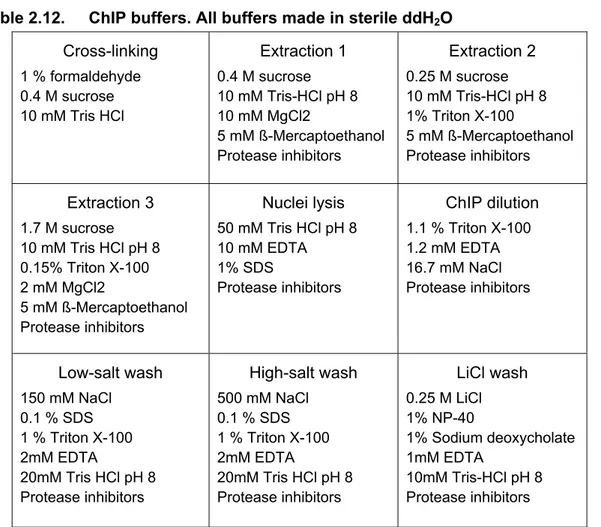
Table 2.12. ChIP buffers. All buffers made in sterile ddH2O 36 Table 2.13. Primers for amplification of PRR9 and PRR7 promoters for ChIP 37 Table 2.14. Light and entrainment conditions in the growth cabinets. 38
Table 2.15. Gateway® reactions set up. 41
Table 2.16. Site-directed mutagenesis set up. 42
Table 3.1 Period and RAE values of LHY:LUC rhythms in DD, under R+Bc, Rc, and Bc. 63
Table 4.1 elf3-TILLING lines 83
Table 4.2 Summary of period and phase phenotypes of elf3-TILLING lines under different
entrainment and free-running conditions. 93
App1 Table 1 Period and phase values of GI:LUC for elf3-TILLING lines under LD to LL. 126 App1 Table 2 Period and phase values of GI:LUC for elf3-TILLING lines under WC to LL. 127 App1 Table 3 Period and phase values of GI:LUC for elf3-TILLING lines under LD to DD. 128 App1 Table 4 Period and phase values of GI:LUC for elf3-TILLING lines under WC to DD. 129 App1 Table 5 Period and phase values of GI:LUC for elf3-TILLING lines under LD to Rc. 130 App1 Table 6 Period and phase values of GI:LUC for elf3-TILLING lines under LD to Bc. 131
Abbreviations
ACE ACGT- containing element ATP Arabidopsis TILLING Project Bc Continuous Blue Light
bHLH Basic-Helix-Loop-Helix
BL Blue Light
BMAL BRAIN AND MUSCLE ARNT-LIKE
bZIP Basic-Region Leucine-Zipper
CAB CHLOROPHYLL A/B BINDING PROTEIN CAPS Cleaved Amplified Polymorphic Sequence
CAT3 CATALASE 3
CCA1 CIRCADIAN CLOCK ASSOCIATED 1
CCG Clock-control gene
CCR2 COLD AND CLOCK REGULATED 2 CLK CLOCK
CO CONSTANS
COP1 CONSITITUTIVELY PHOTOMORPHOGENIC 1 CRY CRYPTOCHROME
CT Circadian time
CYC CYCLE
dCAPS Derived Cleaved Amplified Polymorphic Sequence
DD Continuous dark
DET1 DAMAGED DNA-BINDING PROTEIN 1
EE Evening Element
ELF3 EARLY FLOWERING 3
ELF4 EARLY FLOWERING 4
EMS ETHYL METHANESULFONATE
FAR1 FAR-RED IMPAIRED RESPONSE 1 FBS FHY3 Binding Site
FHY3 FAR-RED ELONGATED HYPOCOTYL 3
FRL Far-Red Light
FRc Continuous Far-Red Light FRQ FREQUENCY
FT FLOWERING LOCUS T
GCN5 SAGA Complex Histone Acetyltransferase Catalytic Subunit Gcn5 GI GIGANTEA
Abbreviations
- xi -
H3K27me3 Tri-methylation on Lysine of N-terminus tail of HISTONE 3 H3Lys14 Acetylation on Lysine 14 of N-terminus tail of HISTONE 3 H3Lys9 Acetylation on Lysine 9 of N-terminus tail of HISTONE 3
HAD6 HISTONE DEACETYASE 6
HAT HISTONE-ACETYLTRANSFERASE
HD1 HISTONE DEACETYLASE 1
HMR HEMERA
HY5 LONG HYPOCOTYL 5
JMD5 JUMONJI DOMAIN CONTAINING 5 LBS LUX Binding Site
LD Light/dark cycles
LHY LATE AND LONG HYPOCOTYL
LL Continuous light
LOV LIGHT OXYGEN VOLTAGE
LUC LUCIFERASE
LUX LUX ARRYTHMO
NES Nuclear Export Signal NLS Nuclear Localization Signal OX Over-expressor PER PERIOD
PHY PHYTOCHROME
PIF PHYTOCHROME INTERACTING FACTOR PLC1 PHYTOCLOCK
PNB Phytochrome Nuclear Body PRC Phase Response Curve
PRR PSEUDO RESPONSE REGULATOR
Rc Continuous Red Light
RL Red Light
sPNB Stable Phytochrome Nuclear Body
TILLING Target Induced Local Lesions in Genomes TIM TIMELESS
TOC1 TIME OF CAB EXPRESSION
tPNB Transient Phytochrome Nuclear Body
WC Warm/cool cycles
WC WHITE COLLAR
WCC WHITE COLLAR COMPLEX
ZT Zeitgeber time
ZTL ZEITLUPE
Chapter 1 Introduction
Chapter 1 Introduction
1.1 Introduction to circadian rhythms
Diurnal life rhythms are shaped by circadian clocks. The Earth’s rotation has been a constant generator of rhythmic oscillations in environmental conditions. These are notably light - dark, warm - cool, and dampness rhythms. Therefore, physiological processes in most organisms have adapted to be rhythmic. Organism evolved circadian clocks to anticipate these predictable diurnal oscillations in environmental conditions. Circadian rhythms are endogenously generated and enable overall optimization of the timing of physiological processes. Circadian oscillations have a periodicity of ≈ 24 hours to match the duration of a day (Dunlap et al., 2004).
Although biological rhythms had been observed for many centuries, the first circadian experiment was performed in the 18th century. The French astronomer DeMarian showed that rhythms of leaf movement persist in constant dark conditions, providing the first evidence that circadian rhythms are endogenously generated. Since that experiment, the existance of circadian rhythms has extended to many diverse organisms, such as cyanobacteria, yeast, insects, birds, mammals, and plants. Hence, circadian clocks are ubiquitous mechanisms in living organism (Dunlap et al., 2004).
Chronobiology studies the occurrence and generation of life rhythms. Circadian oscillations are part of our everyday. For example, they are found in the patterns of sleeping and awaking, mental concentration, hormone levels, and body-temperature homeostasis. In most organisms, core biological processes, such as cell division and changes in gene expression, occur in a circadian fashion (Dunlap et al., 2004). These processes are outputs of the circadian clock. In plants, examples of clock outputs are leaf movement, growth, metabolic pathway, and stress responses [Figure 1.1, (Harmer, 2009)].
The fitness advantage of an endogenous clock has been demonstrated in several model systems. Plants with an internal-clock length matching the day length of the environment had increased fitness through increasing levels of photosynthesis, growth, and survival (Dodd et al., 2005). Similar conclusions have been observed in cyanobacteria (Ouyang et al., 1998). Moreover, plant mutants that loss circadian regulation had increased levels of mortality (Green et al., 2002) and susceptibility to pathogen infections (Wang et al., 2011). Importantly, in man, serious diseases, such as diabetes, cancer, and depression, have been associated to circadian defects (Dunlap
Chapter 1 Introduction
- 3 -
et al., 2004). These observations highlight the biological relevance of internal time- keeping.
Importantly, external cues reset the circadian clock daily, keeping internal and external times synchronized. This process is called entrainment and it is mediated by the so-called zeitgebers (‘time-givers’). In most organisms, the main zeitgebers are light and temperature signals (Jones, 2009). Entrainment is mediated by the clock-input pathways. Notably, these input pathways are simultaneously controlled as clock outputs (Figure 1.1). This enables circadian clocks to modulate the susceptibility for entrainment cues throughout the day. Therefore, entrainment is a complex process that requires interaction of the circadian clock with both input and output pathways.
Figure 1.1. The plant circadian clock system.
Circadian rhythms are generated by a central oscillator. The timing of many physiological processes is regulated by the circadian clock through the output pathways (green arrows). Moreover, the oscillator is reset daily by the input pathways (pink arrows). Simultaneously, the input pathways are circadian regulated through the output pathways, increasing the robustness of the oscillator to external and internal perturbations.
Chapter 1 Introduction
Entrainment allows circadian clocks to keep track of seasonal changes in day length, in a process termed photoperiodism. Organisms living in extreme latitudes are particularly challenged by seasonal environmental oscillations (Dunlap et al., 2004).
Hence, photoperiod is widely used as an external cue to predict the most favorable time for developmental choices. For example, in plants, photoperiod pathways controls flowering time (de Montaigu et al., 2010). Also, photoperiodism controls the timing of bird and monarch butterflies migrations (Gwinner, 2003; Kyriacou, 2009). Photoperiod perception is achieved cooperatively by integrating the timing of the information derived from the circadian clock and light-input pathways (de Montaigu et al., 2010).
The molecular components of the clock are not conserved between kingdoms, suggesting that circadian systems have multiple evolutionary origins. In red-blood cells and the microscopic algae Ostreococcus tauri, circadian oscillations persist in the absence of transcription (O'Neill and Reddy, 2011; O'Neill et al., 2011). Nevertheless, the molecular mechanism in most multi-cellular circadian systems involves interconnected transcriptional-translational feedback loops between clock components.
Circadian clocks are a fascinating example of convergence evolution (Dunlap et al., 2004). The next sections will focus on the state of the circadian clock in the plant Arabidopsis thaliana (Arabidopsis).
1.2 The Arabidopsis circadian clock
1.2.1 Tools to investigate clock function
Circadian waves can be mathematically described. Several properties of circadian oscillations are of particular interest. The period is the duration of a single oscillation and defines the speed of the circadian rhythm. The phase describes the timing of specific events within the circadian day. The amplitude is defined as half the difference between the maximum and the minimum value of an oscillation (Figure 1.2).
These three properties can be genetically separable (Harmer, 2009). Finally, the accuracy of the circadian oscillation is described by its robustness. Here, the term arrhythmicity is used when circadian rhythms are not sustained. Period, phase, amplitude, and robustness are used to characterize circadian mutants, and to describe input signals that affect the endogenous oscillator.
The main tool to investigate circadian function in plants is the monitoring of
Chapter 1 Introduction
- 5 -
control-genes (CCG). In the early 90s, the use of firefly-luciferase based circadian reporters (CCG:LUC) boosted circadian research in Arabidopsis (Millar et al., 1992). In these reporters, the promoter of a CCG was fused to the luciferase coding sequence.
In the presence of the luciferase substrate (luciferin) plants then emit bioluminescence in a circadian fashion. In such experiments, plants harboring CCG:LUC are grown for few days under entraining cycles of light or temperature (Light/Dark, LD or Warm/Cool, WC, respectively), and then they are released to so-called free-running conditions of continuous light (LL) or darkness (DD) (Figure 2A). The bioluminescence emission is acquired by highly sensitive cameras that can detect low intensity light. Then, the obtained data can be analyzed by using mathematical and statistical tools.
Constant conditions Period Amplitude Phase
0 24 48 72 96 120 144
Entraining cycles (LD)
Time (h)
Bioluminescence CCG:LUC
Constant conditions Period Amplitude Phase
0 24 48 72 96 120 144
Entraining cycles (LD)
Time (h)
Bioluminescence CCG:LUC
Figure 1.2. Analysis of circadian rhythms.
The figure shows the main features of bioluminescence-based circadian experiments.
Under diurnal or entraining cycles of time-givers, such as temperature or light/dark cycles, the period of the clock is about 24 hours. Under constant conditions, circadian period-length deviates from 24 hours. Phase refers to the timing of a physiological event within the circadian cycle. Normally, it is refers to the highest luminescence value within the oscillation (peak). Amplitude is half the difference between the highest and the lowest value of the oscillation.
Several CCG promoters have been fused to luciferase to study circadian oscillations. One example is CHLOROPHYLL A/B BINDING PROTEIN (CAB). CAB has a complex regulation by both light and the circadian clock. Hence, CAB:LUC was the first luciferase reporter to be used for studying the interaction between light and clock (Millar et al., 1995a). Moreover, CAB:LUC expression was used to identify
Chapter 1 Introduction
circadian mutants with aberrant reporter expression (Millar et al., 1995b; Millar et al., 1995a). The dampening of CAB expression in the dark limits the use of CAB:LUC for DD experiments. However, robust rhythmic expression persist in DD for other CCG genes, such as the COLD AND CIRCADIAN REGULATED (CCR2, also known as AtGRP7) (Heintzen et al., 1997; Covington et al., 2001; Hanano et al., 2006;
McWatters et al., 2007; Schoning et al., 2007). Finally, most of the core-clock components (see below) are also CCG, and their promoters have also been used within the luciferase platform to build up the complex interconnections of the circadian- clock model.
1.2.2 The circadian clock model
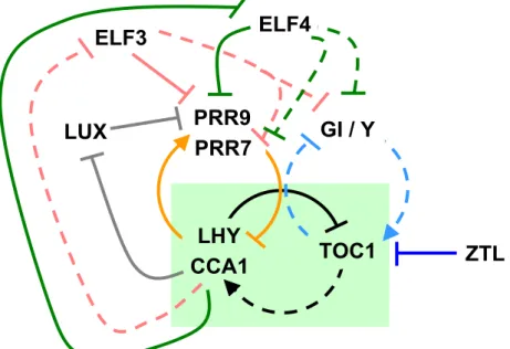
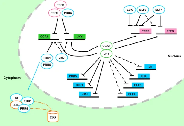
In Arabidopsis, the first loop to be defined was the so-called core loop. The core loop connects LATE AND ELONGATED HYPOCOTYL (LHY) (Schaffer et al., 1998) and CIRCADIAN CLOCK ASSOCIATED 1 (CCA1) (Wang and Tobin, 1998) to TIME OF CAB EXPRESSION (TOC1) (Strayer et al., 2000) (Figure 1.3). LHY and CCA1 encode two myb-like transcription factors that are expressed in the morning. LHY and CCA1 act additively as transcriptional repressors by associating to the promoter of TOC1, confining TOC1 expression to dusk (Alabadi et al., 2001; Perales and Mas, 2007) (Figure 1.4). Consistently, the over-expression of LHY or CCA1 led to clock arrest with basal levels of TOC1 expression (Wang and Tobin, 1998; Matsushika et al., 2002b). Conversely, the pseudo response regulator (PRR) TOC1 promotes the expression of CCA1 and LHY by an unknown mechanism [Figure 1.3, (Alabadi et al., 2001; Pruneda-Paz et al., 2009)]. This thus closes the core of the clock. Notably, in the absence of the core loop the circadian clock arrests (Ding et al., 2007).
Subsequent experimental and mathematical approaches expanded the plant clock to the so-called three-loop-model (Locke et al., 2006; Zeilinger et al., 2006). In this model, a morning and evening loop were added interlocking with the central loop (Figure 1.3). This was extended and confirmed with molecular genetics and biochemical experiments. In here, three TOC1-sequence-related clock components, PRR9, PRR7, and PRR5, were placed within the core mechanism. During the day, PRR9 and PRR7 associate sequentially with the promoter of LHY and CCA1 to repress their transcription [Figure 1.4, (Nakamichi et al., 2005; Nakamichi et al., 2010)].
Reciprocally, LHY and CCA1 promote the expression of PRR9 and PRR7 by direct association to promoter cis-elements [Figure 1.4, (Farre et al., 2005; Portoles and Mas, 2010)]. Notably, the function of TOC1 as an activator of LHY and CCA1 is opposite to
Chapter 1 Introduction
- 7 -
oscillator, one as direct repressor of LHY and CCA1 (Nakamichi et al., 2010), and a second one as stabilizer of TOC1 protein-nuclear pool [Figure 1.4, (Wang et al., 2010)].
This second role of PRR5 seems to be more prominent since both toc1 and prr5 null alleles were shown to have short-period phenotypes, and this was opposite to prr9, prr7, and the double mutant prr7 prr9, all of which displayed a long-period phenotypes (Alabadi et al., 2001; Yamamoto et al., 2003; Farre et al., 2005). Recent mathematical modeling efforts validated the PRR9, PRR7, and PRR5 repressor wave to LHY and CCA1 (Pokhilko et al., 2010). Consequently, the triple mutant prr975 was found to be arrhythmic for all circadian outputs (Nakamichi et al., 2005).
PRR7 PRR9 PRR7 PRR9
CCA1 LHY CCA1
LHY CCA1
LHY
GI / Y
TOC1 ELF4
ZTL ELF3
LUX
Figure 1.3. One Arabidopsis circadian clock model.
Circadian-clock components are arranged in interconnected feedback loops. In the core loop (black arrows, green box), LHY and CCA1 negatively regulate TOC1 expression, and feedback from TOC1 promotes LHY and CCA1. In the morning loop (orange arrows), PRR9 and PRR7 negatively regulate LHY and CCA1, and LHY and CCA1 feedback to promote PRR9 and PRR7. In the evening loop (light blue arrows), TOC1 negatively regulates GI/Y expression, and Y activates TOC1 expression. ZTL promotes TOC1 protein degradation. CCA1 and LHY represses ELF3, ELF4, and LUX expression (pink, green and blue, respectively). LUX, ELF3, and ELF4 repress PRR9 expression. Additionally, ELF3 and ELF4 repress GI and PRR7. Continuous line indicates regulatory link is direct, dashed line indicates direct link has not been determined.
An evening loop was proposed between an unknown component Y and TOC1 (Figure 1.3). The component Y is light induced and acts as a promoter of TOC1 expression (Locke et al., 2006; Zeilinger et al., 2006). Partial Y function was assigned to the evening gene GIGANTEA (GI). However, high levels of TOC1 found in the gi mutants suggested additional components may assist in Y function (Martin-Tryon et al., 2007). GI encodes for a protein of unknown function and is unique to plants. Different
Chapter 1 Introduction
gi mutants showed pleiotropic defects in flowering time, hypocotyl growth, and stress response (Huq et al., 2000; Cao et al., 2005; Mizoguchi et al., 2005). This suggests that GI has a wide role in plant physiology.
PRR9 PRR7
PRR9 PRR7
PRR5
LHY CCA1
PRR5 TOC1
GI
JMJ
ZTL
TOC1 JMJ
ELF3 ELF4 CCA1
LHY
PRR5
GI
LUX
LUX ELF3 ELF4
TOC1
26S PRR5
Nucleus
Cytoplasm
Figure 1.4. Clock components are interconnected by transcriptional loops of activation and repression.
Squares and circles represent clock promoters and clock proteins, respectively.
Continuous lines indicate links are direct and have been experimentally validated.
Dashed lines refer to indirect or not experimentally confirmed links. Links ending on arrows or horizontal lines represent activation or repression, respectively. Squared line refers to target 26S-proteasome-mediated degradation. The model is explained in the text (Sections 1.2.2 and 1.3).
GI function in the circadian clock is mediated by its physical interaction with ZEITLUPE (ZTL) [Figure 1.4, (Kim et al., 2007)]. ZTL is a F-box protein that facilitates ubiquitination by E3-ligase of TOC1 and PRR5, targeting them for degradation by the 26S-proteasome [Figure 1.4; (Mas et al., 2003; Kiba et al., 2007; Baudry et al., 2010;
Wang et al., 2010)]. Consequently, loss of ZTL function lengthened circadian period.
ZTL protein interactions are mediated by the LOV (Light, Oxygen, or Voltage) domain that also functions as a blue-light photoreceptor. Hence, blue light modulates the affinity of ZTL to its protein interactors (Mas et al., 2003; Kiba et al., 2007; Baudry et al., 2010; Wang et al., 2010). For instance, blue light induces ZTL and GI protein
Chapter 1 Introduction
- 9 -
the cytoplasm, the interactions of ZTL, GI, and TOC1 in the cytoplasm is thought to regulate active levels of nuclear TOC1 protein [Figure 1.4, (Kim et al., 2007)].
Therefore, GI negatively regulates TOC1 levels by stabilizing ZTL.
1.3 Three clock genes are essential for sustained circadian oscillations
The function of any individual clock components of the three-loop model is not essential for circadian function (Locke et al., 2006; Zeilinger et al., 2006). To date, only a small group of clock components have been found to be essential for sustained circadian function in Arabidopsis. These three genes are EARLY FLOWERING-3 (ELF3), EARLY FLOWERING-4 (ELF4), and LUX ARRYTHMO/PHYTOCLOCK1 (LUX/PCL1) (Hicks et al., 1996; Doyle et al., 2002; Hazen et al., 2005; Onai and Ishiura, 2005).
1.3.1 ELF3
ELF3 is required for core-clock function since elf3 loss-of-function mutants are arrhythmic for circadian outputs under LL (Covington et al., 2001) and in DD (Covington et al., 2001; Thines and Harmon, 2010). ELF3 encodes for an unknown protein unique to plants. It was reported to localize in the nucleus (Liu et al., 2001).
ELF3 protein accumulation is rhythmic and peaks at subjective night (dusk) under LD (Liu et al., 2001). ELF3 protein possess features of a transcriptional regulator, such as a proline-rich region (residues 440-540), an acidic region (residues 206-320), a threonine-rich region (residues 636-652), a putative nuclear-targeting signal starting at residue 591, and a glutamine-stretch (residues 544-585). The glutamine stretch is polymorphic in length between different Arabidopsis accessions (Hicks et al., 2001;
Tajima et al., 2007). The timing of ELF3 protein accumulation, ELF3 nuclear localization, and ELF3 protein features suggest a role in clock-mediated transcriptional regulation at dusk. elf3 null alleles affect the expression of the core-loop components, with low levels of LHY and CCA1, and high levels of TOC1 (Kikis et al., 2005).
Moreover, expression levels of PRR9, PRR7, and GI were found to be constantly high in elf3 loss-of-function mutants (Dixon et al., 2011). Interestingly, ELF3 protein was recently found to associate to the promoter of PRR9, but not to PRR7 or GI, indicating a direct transcriptional repression of ELF3 on PRR9 expression [Figure 1.3 and 1.4, (Dixon et al., 2011)]. Plants over-expressing ELF3 (ELF3-OX) have rhythmic circadian
Chapter 1 Introduction
oscillations with long period (Covington et al., 2001). Therefore, ELF3 functions within the core oscillator as a transcriptional repressor of PRR9, and a negative regulator of clock periodicity.
1.3.2 ELF4
ELF4 is a small homodimeric nuclear protein unique to plants (Khanna et al., 2003; Kolmos et al., 2009). Structural modeling predicts ELF4 to consist of a single protein-protein interaction domain, and hence, ELF4 was predicted to function as an effector for a target protein (Kolmos et al., 2009). Similar to ELF3, loss of ELF4 function leads to clock arrest both under LL and in DD, as well as, arrhythmic low levels of LHY and CCA1 expression with high levels of TOC1 (Doyle et al., 2002; Kikis et al., 2005;
McWatters et al., 2007). Hypomorphic alleles of ELF4 had accelerated circadian rhythms, and a long periodicity resulted by ELF4-OX. Hence, ELF4 decelerated circadian periodicity (McWatters et al., 2007; Kolmos et al., 2009). Additionally, the transcript accumulation of the secondary loop components PRR9, PRR7, and GI was found to be constitutively high in elf4 loss of function, and this was also found to a lesser extent in weak elf4 alleles (Kolmos et al., 2009). The expression phenotypes of LHY, CCA1, and TOC1 in the elf4 mutant can be mimicked computationally by the simultaneous increase of the expression levels of PRR9 and GI in the three-loop mathematical model (Kolmos et al., 2009). Taken together, ELF4 is core-clock component that has two entry points to the oscillator, by repressing both morning and evening secondary loop components [Figure 1.3 and 1.4, (Kolmos et al., 2009)].
1.3.3 LUX
The GARP-type transcription factor LUX is pivotal to sustain circadian rhythms.
LUX loss of function is arrhythmic for all tested circadian outputs, and this phenotype is more severe under LL (Hazen et al., 2005; Onai and Ishiura, 2005). LUX has been recently found to work as a transcriptional repressor by associating to a DNA motif found in the promoter of PRR9, called the LUX BINDING SITE (LBS, nucleotide sequence GATTCG) [(Helfer et al., 2011), Figure 1.3 and 1.4]. Lack of repression of PRR9 expression is likely to cause abnormal expression levels of core-loop clock components: low expression of LHY and CCA1 and high levels of TOC1 (Hazen et al., 2005; Onai and Ishiura, 2005), which are also found in elf3 and elf4 loss of function mutants (Kikis et al., 2005; McWatters et al., 2007). Since PRR9 over-expression does
Chapter 1 Introduction
- 11 -
2002a), additional lack of repression of other targets of LUX are likely to participate in the lux severe clock phenotypes.
Sustained circadian rhythms in Arabidopsis require ELF3, ELF4, and LUX function. The three genes act as transcriptional repressors at dusk. Their loss function phenotypes are similar. Therefore, these three genes could act closely in a genetic pathway essential for the circadian-clock mechanism.
1.4 Outputs of the clock
In plants, circadian oscillations can be found in many physiological processes.
These include gene expression, hypocotyl growth, photosynthesis, and pathogen responses (Hanano and Davis, 2007; Nozue et al., 2007; Covington et al., 2008;
Michael et al., 2008; Roden and Ingle, 2009; Graf et al., 2010). The transcript accumulation of about 40 % of Arabidopsis genes is circadian regulated, as shown by microarray analysis (Covington et al., 2008; Hubbard et al., 2009). Such an extensive circadian regulation is achieved by the rhythmic expression of transcription factor families, such as MYB, bHLH, and bZIP (Hanano et al., 2008). Clock regulation extends to hormone production, magnitude of hormone and cold responses, and primary metabolic pathways (Hanano et al., 2006; Covington and Harmer, 2007;
Covington et al., 2008; Fukushima et al., 2009). Clock transcriptional regulation of gene expression is extensive in Arabidopsis.
Multiple cis-elements in the promoter of CCG genes mediate association of positive and negative regulators that shape their circadian oscillation (Michael and McClung, 2002; Harmer and Kay, 2005; Michael et al., 2008; Helfer et al., 2011).
Computational analyses have shown the importance of the interplay of the circadian clock with temperature- and light-signaling pathways (Michael et al., 2008).
Additionally, several cis-elements have found to be over-represented in the promoter of CCG (Covington et al., 2008; Michael et al., 2008). The most studied cis-element is the so-called Evening Element (EE, nucleotide sequence AAAATATCT), over-represented in the promoter of genes that peak at dusk, such as TOC1, GI, ELF4, and LUX (Harmer, 2009; Li et al., 2011). The binding of LHY and CCA1 to the EE mediates transcriptional repression during the subjective day and confers evening-phase expression. Notably, the EE also mediates activation of the expression of morning phased genes, such as PRR9, PRR7, and CAB (Farre et al., 2005; Lu et al., 2009;
Portoles and Mas, 2010). This indicates that LHY and CCA1 can work both as
Chapter 1 Introduction
activators and repressors, possibly by participating in different protein complexes depending on promoter context (Lu et al., 2009). In summary, different clock transcription factors mediate transcriptional activation or repression by associating to the promoters of CCG.
Phylogenetic shadowing of promoter sequences can reveal evolutionary conserved cis-elements required for transcriptional regulation (Boffelli et al., 2003). For this, the extent of sequence conservation is examined in an alignment of homologous sequences of related species. Sequence conservation indicates selective pressure to keep DNA motifs of functional importance (Picot et al., 2010), and can be then validated by experimental approaches. The use of this process as a technique is exemplified by the identification of regulatory elements in the LHY promoter (Spensley et al., 2009).
1.5 Photoperiodism and plant development
Light- and clock-signaling pathways control diurnal plant growth and the transition from vegetative to reproductive phase (de Montaigu et al., 2010). The regulation of hypocotyl elongation has been studied as a model for diurnal-growth control. Hypocotyl growth rate is controlled by the circadian clock. Hypocotyl growth arrests at the beginning of the day. In arrhythmic mutants, such as elf3, hypocotyls are constantly growing. Therefore, these mutants displayed long hypocotyls when grown under LD conditions (Dowson-Day and Millar, 1999; Nozue et al., 2007). Light also inhibits hypocotyl elongation independently of the clock. long hypocotyl 1 (hy) mutant displayed rhythmic-hypocotyl elongation, but still had long hypocotyl due to lack of light perception through the phytochrome photoreceptors (Dowson-Day and Millar, 1999).
The PHYTOCHROME INTERACTING FACTORS (PIF) PIF4 and PIF5 promote hypocotyl growth. These encode bHLH transcription factors that bind to the active form of phytochromes. The clock controls transcript accumulation of PIF4 and PIF5 that is highest at the end of the night. PIF4 and PIF5 expression peaks coincide with maximal hypocotyl growth rate. In the absence of clock function, PIF4 and PIF5 levels are constitutively high leading to continuous growth (Nozue et al., 2007). At dawn, light signaling arrests hypocotyl growth by degrading PIF4 and PIF5. The combined regulation by clock and by light restricts maximal diurnal hypocotyl elongation to the end of the night, when PIFs highest transcript accumulation coincides with protein
Chapter 1 Introduction
- 13 -
Interestingly, loss of ELF3 or PHYTOCRHOME B (PHYB) functions were found to lead to similar developmental defects, such as such as long hypocotyls, elongated petioles, and pale leaves (Zagotta et al., 1996; Liu et al., 2001). The loss of elf3 had long hypocotyls under constant red light (Rc), and to less extent under constant blue light (Bc), whereas no phenotype was observed under constant far-red light (FRc). This indicates that ELF3 plays a more important role in phyB mediated red-light signals to inhibit hypocotyl elongation (Reed et al., 2000; Liu et al., 2001). Light inhibition of hypocotyl growth is fluence-rate dependent, i.e. increasing light intensities lead to an increase in the inhibition rate. Notably, elf3 PHYB-OX mutant responded to increasing red light intensities, but showed longer hypocotyls than PHYB-OX (Reed et al., 2000).
Overall, ELF3 affects hypocotyl elongation as a component of the clock mechanism and phyB signaling, whereas phyB role is likely limited to RL signaling.
The circadian clock participates in regulation of the transition from vegetative to reproductive development through the photoperiodic pathway (de Montaigu et al., 2010). In the external coincidence model, the clock controls the phase of CONSTANS (CO) expression (Suarez-Lopez et al., 2001). Under short day, CO expression occurs in the dark phase, and CO protein is targeted to degradation by the E3-ligase CONSTITUTIVE MORPHOGENENIC 1 (COP1) (Jang et al., 2008). However, under long days, maximal CO expression coincides with the light phase. CO protein is stable in the light and activates the transcription of FLOWERING LOCUS T (FT) (Valverde et al., 2004). Finally, FT moves from the leaf to the apex and triggers the transition to flowering (Corbesier et al., 2007).
The elf3, elf4, and lux loss of function mutants are early-flowering mutants regardless of photoperiod (Zagotta et al., 1996; Hicks et al., 2001; Doyle et al., 2002;
Hazen et al., 2005). A weaker early-flowering phenotype has been also observed in phyB mutants. Notably, elf3 and phyB mutations were additive in reducing flowering time (Reed et al., 2000). This suggests that ELF3 and phyB delay flowering time by at least partially independent mechanisms.
The elf3 early-flowering phenotype was shown to be independent of CO, and did not require high levels of FT expression. This suggest that ELF3 may repress flowering independently of the CO-dependent pathway (Kim et al., 2005). GI also posses separable functions in the clock and as a promoter of flowering (Martin-Tryon et al., 2007). GI is an activator of CO transcription and consequently gi mutants are late flowering. Moreover, elf3 gi double mutant is late flowering, but displayed a long hypocotyl like elf3 (Chou and Yang, 1999). This suggests the existence of separate