Anoctamins in Volume Regulation, Proliferation and Apoptotic Cell Death
DISSERTATION ZUR ERLANGUNG DES DOKTORGRADES DER NATURWISSENSCHAFTEN (DR.RER.NAT.)
DER FAKULTÄT FÜR BIOLOGIE UND VORKLINISCHE MEDIZIN DER UNIVERSITÄT REGENSBURG
Vorgelet von
Wanitchakool Podchanart Aus Nonthaburi, Thailand
im Oktober 2016
Das Promotionsgesuch wurde eingereicht am:
14-10-2016
Die Arbeit wurde angeleitet von:
Prof. Dr. Karl Kunzelmann Unterschrift:
Promotionsgesuch eingereicht am: Oktober 2016
Die Arbeit wurde angeleitet von: Prof. Dr. med. Karl Kunzelmann
Prüfungsausschuss:
Vorsitzender: Prof. Dr. Gernot Längst
1.Gutachter: Prof. Dr. med. Karl Kunzelmann 2.Gutachter: Prof. Dr. Bernhard weber
3.Prüfer: Prof. Dr. med. Ernst R. Tamm Ersatzperson: Prof. Dr. Rainer Schreiber
SUMMARY
The family of anoctamins consists of ten different proteins (called ANO1-10 or TMEM16A- K). A variety of functions have been attributed to anoctamins such as that of Ca2+ activated Cl- channels controlling Cl- secretion, cell volume, cell migration and proliferation, and that of phospholipid scramblases contributing to apoptotic cell death. Some anoctamin members are overexpressed in tumors and have been linked to developmental defects. Mutations in anoctamins can cause genetic diseases and cancer. However, it is unknown how anoctamins fulfill these various functions.
Before ANO1 was identified as a protein forming a chloride channel, it was known as the cancer marker DOG1. DOG1/ANO1 is expressed in gastrointestinal stromal tumors (GIST) and particularly in head and neck squamous cell carcinoma (HNSCC), and is not detected at very high levels in other tissues. We found that ANO1 strongly augments cell proliferation, cell migration and metastasis. We also found that ANO1 expression is controlled by histone deacetylases (HDAC), corresponding well to the known role of HDAC in HNSCC. As ANO1 did not enhance proliferation in every cell type, its function is perhaps modulated by cell- specific factors. In contrast to ANO1, we found that ANO6, by operating as a Ca2+ activated membrane phospholipid scramblase, supports cellular apoptosis and necroptosis rather than promoting proliferation.
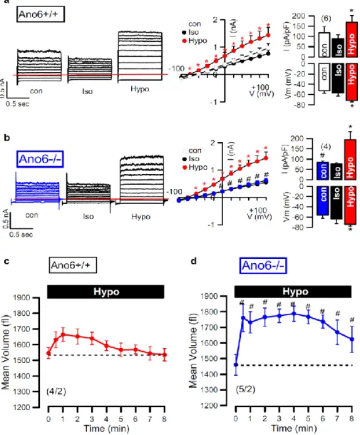
Earlier findings from our laboratory and other groups indicated that ANO6 is a Cl- channel, a phospholipid scramblase and a nonselective cation channel activated by intracellular Ca2+ and pro-apoptotic stimuli. Moreover, previous studies characterized ANO6 as an outwardly rectifying Cl- channel (ORCC) and volume-regulated anion channel (VRAC) that is activated during apoptotic cell death. In the experiments presented here I found a major role of ANO6 for both regulatory volume decrease (RVD) as well as apoptotic volume decrease (AVD).
RVD and AVD were examined in four different cell types under physiological conditions, i.e.
in the presence of physiological intracellular and extracellular bath solutions and at 37 °C.
Moreover swelling activated whole cell currents and volume regulation was assessed in
freshly isolated intestinal epithelial cells from wild type and anoctamin 6 knockout mice. The data indicate that ANO6 generates a VRAC and is activated during RVD by membrane stretch, Ca2+ influx through TRP channels and activation of phospholipase A2 (PLA2), which generates membrane lysophospholipids (LPL). Accumulation of LPL in the plasma membrane enhances membrane tension, which possibly induces a conformational change of ANO6, thereby opening the Cl- conductive pore and causing RVD. Remarkably, RVD was largely reduced in intestinal epithelial cells from ANO6 knockout mice. The results also demonstrate that ANO6 is activated by low intracellular Cl-, which suggests that ANO6 operates as an osmosensor.
ANO6 was further demonstrated to participate in the innate immune response in macrophages. We could demonstrate that in macrophages Ca2+ influx through P2X7 channels leads to initial cell shrinkage, phospholipid scrambling, cell membrane blebbing and ultimately cell death. Cell migration and phagocytic activity was found to be largely reduced in Ano6-/- macrophages. We proposed that ANO6 plays a central role for the immune defense realized by macrophages. The data also indicate that P2X7-induced cell death in macrophages is due to activation of apoptosis as well as necrosis.
Another member of the anoctamin family, ANO10, was also demonstrated to be expressed in macrophages and to participate in innate immune response. In collaboration with the Max Planck institute of experimental Medicine in Göttingen we showed that the genomic ANO10 mutation R263H increases serum titers of antibodies directed against Lyme disease causing Borrelia. We suggest that ANO10-R263Henhances susceptibility towards Borrelia infection and attenuates clearance of spirochetes after tick bites. ANO10-R263H caused a reduced VRAC activity and RVD. Loss of ANO10 expression in macrophages compromised phagocytosis and clearance of Borrelia, attenuated volume regulation and reduced apoptotic cell death. Taken together the results demonstrate that anoctamins participate in the regulation of proliferation and cell death and play important roles in cancer and innate immune defense.
Zusammenfassung
Die Anoctamin-Familie besteht aus zehn verschiedenen Proteinen (ANO1-10 oder TMEM16A-K). Inzwischen wurde den Anoctaminen eine Vielzahl von Funktionen zugeordnet. So bilden Anoctamine Ca2+-aktivierte Chloridkanäle, nehmen Einfluß auf das Zellvolumen, die Zellmigration und Zellproliferation und bilden Membranphospholipid- Scramblasen, womit sie einen Beitrag zum apoptotischen Zelltod leisten. Einige Anoctamine sind in Tumoren überexprimiert und werden mit Entwicklungsstörungen in Verbindung gebracht. Anoctamin-Mutationen können genetisch bedingte Krankheiten und Krebs verursachen. Es ist jedoch weitgehend unbekannt, wie Anoctamine diese verschiedenen Funktionen erfüllen.
Bevor Anoctamin 1 (ANO1) als Chloridionenkanal identifiziert wurde, war es als der Krebsmarker DOG1 bekannt. DOG1/ ANO1 ist in gastrointestinalen Stromatumoren (GIST) und vor allem in Weichteiltumoren des Kopfbereichs (HNSCC) derart hoch exprimiert, wie man dies in gesunden Geweben nie vorfindet. Wir fanden heraus, dass ANO1 die Zellproliferation, Zellmigration und Tumormetastasierung fördert. Wir fanden ebenfalls, dass die ANO1-Expression durch Histon-Deacetylasen (HDAC) kontrolliert wird. Dies korreliert sehr gut mit der bekannten Rolle von HDAC in Kopftumoren. Da ANO1 nicht in jedem Zelltyp die Proliferation stimuliert, ist anzunehmen, dass seine Funktion durch zellspezifische Faktoren moduliert wird. Im Gegensatz zu ANO1 fanden wir, dass ANO6 als Ca2+-aktivierte Plasmamembran-Phospholipidscramblase die zelluläre Apoptose und Nekroptose unterstützt, anstatt Proliferation zu fördern.
Ergebnisse aus unserem Labor und von anderen Arbeitsgruppen zeigen, dass ANO6 offensichtlich gleichzeitig ein Chloridionenkanal, eine Phospholipidscramblase und einen nichtselektiven Kationenkanal bildet und durch intrazelluläre Calciumionen und proapoptotische Stimuli aktiviert wird. Weiterhin kennzeichnen frühere Studien ANO6 als auswärts-gleichrichtenden Chloridionenkanal (ORCC) und als volumenregulierten
Anionenkanal (VRAC). Diese Ionenkanalströme werden während des apoptotischen Zelltods aktiviert. In den hier vorgestellten Experimenten fand ich eine dominante Rolle von ANO6 sowohl für regulatorische Volumenabnahme (RVD), als auch für den apoptotischen Volumenverlust (AVD). RVD und AVD wurden in vier verschiedenen Zellarten unter physiologischen Bedingungen, d.h. in Gegenwart von physiologischen intra- und extrazellulären Ionenkonzentrationen und bei 37 ° C, untersucht. Darüber hinaus wurden Chloridionenströme und Volumenregulation in frisch isolierten Darmepithelzellen von Wildtyp-Mäusen und ANO6-Knockoutmäusen untersucht. Die Ergebnisse zeigen, dass ANO6 einen VRAC generiert und während RVD durch Dehnung der Plasmamembran, Ca2+
Einstrom durch Transient Receptor Potential (TRP)- Kanälen und Aktivierung von Phospholipase A2 (PLA2) aktiviert wird. PLA2 führt zur Bildung und Akkumulation von Lysophospholipiden (LPL) in der Plasmamembran, was zu einer Spannungsänderung innerhalb der Plasmamembran bewirkt und eine Konformationsänderung von ANO6 zur Folge hat. Diese Konformationsänderung induziert die Öffnung des ANO6-Kanals und die Aktivierung von RVD. Bemerkenswerterweise war die Kanalaktivierung und RVD in intestinalen Epithelzellen von ANO6-Knockoutmäusen deutlich reduziert. Weitere Ergebnisse zeigen, dass ANO6 durch eine niedrige intrazelluläre Chloridionenkonzentration aktiviert wird, was darauf hindeutet, dass ANO6 als Osmosensor funktioniert.
Weiterhin konnte eine Beteiligung von ANO6 bei der Immunreaktion in Makrophagen demonstriert werden. Wir konnten zeigen, dass in Makrophagen der Ca2+ Einstrom durch P2X7 Kanäle zur initialen Zellschrumpfung führt und danach Phospholipidscrambling, Zellmembran-Blebbing und schließlich Zelltod induziert wird. Die Zellmigration und Phagozytose-Aktivität war deutlich reduziert in Ano6-Knockout-Makrophagen. Hieraus schlossen wir, dass ANO6 eine zentrale Rolle bei der Immunabwehr durch Makrophagen spielt. Die Daten zeigen ebenfalls, dass der P2X7-induzierte Zelltod in Makrophagen durch gleichzeitige Aktivierung von Apoptose und Nekrose zustande kommt.
Ein weiteres Mitglied der Anoctaminfamilie, ANO10, wird ebenfalls in Makrophagen exprimiert. In Zusammenarbeit mit dem Max-Planck-Institut für experimentelle Medizin in Göttingen konnten wir zeigen, dass die genomische ANO10 Variante R263H zu erhöhten Serum-Titern von Antikörpern gegen Borrelia führt, welche die Lyme-Krankheit verursachen.
Wir vermuten, dass ANO10-R263H die Anfälligkeit für Borrelia-Infektionen erhöht und deren Elimination nach Übertragung durch Zeckenbisse reduziert. ANO10-R263H inhibiert die VRAC-Aktivität und vermindert RVD. Ein Verlust der ANO10-Expression in Makrophagen hemmt die Phagozytose und Elimination von Spirochäten, beeinträchtigt RVD und hemmt Apoptose. Zusammengefasst zeigen die Ergebnisse, dass Anoctamine Proliferation und Zelltod kontrollieren und eine wichtige Rolle bei Krebs und angeborener Immunabwehr spielen.
CONTENTS
SUMMARY……….i
ZUSAMMENFASSUNG………...iii
CONTENTS………iv
CHAPTER 1 Introduction………...1
CHAPTER 2 Role of anoctamins in cancer and apoptosis………..……….12
CHAPTER 3 Cellular volume regulation by anoctamin 6: Ca2+, phospholipase A2 and osmosensing ………..………...….26
CHAPTER 4 Anoctamin 6 mediates effects essential for innate immunity downstream of P2X7-Receptors in macrophages ………...48
CHAPTER 5 A coding variant of ANO10, affecting volume regulation of macrophages, is associated with Borrelia seropositivity……….……...78
CHAPTER 6 Discussion……….106
CONCLUSION………...………116
REFERENCES……….……….………..117
ACKNOWLEDGEMENTS………146
ERKLÄRUNGEN………...……….147
CURRICULUM VITAE……….….…….148
CHAPTER 1 Introduction
Ion channels form transmembrane pores, which have mechanisms for gating to control the movement of particular ions species. Ions are driven by their concentration gradient to move from one side of plasma membrane to the other (1). Membrane ion channels are in charge of numerous physiological functions. In fact mutations in ion channels cause either a loss or a gain of channel function, which may lead to very different cellular problems, the so called ion channel diseases (1).
Chloride channels
Chloride (Cl-) channels consist of a large family of functionally and structurally different anion-selective channels in plasma membranes or intracellular organelles (2). Some Cl- channels are not only permeable for chloride, but also for other anions such as bicarbonate, bromide, and iodide, as well as small organic osmolytes like taurine (3). Their functions involve membrane excitability in neurons, skeletal muscle, cardiac muscle, and smooth muscle cells. In addition Cl- channels are necessary to counterbalance the electrogenic transport by the H+-ATPase, which is required for acidification of internal and extracellular compartments (2). Cl- currents are also essential for transepithelial salt transport, as well as for the regulation of the cell volume. Moreover, Cl- channels are in charge of cellular homeostasis during cell proliferation and differentiation (4). Cl- channels can be classified into five functional groups (4): i) Ligand-gated Cl- channels are made as pentameric structures and are excitatory in embryonic neurons and inhibitory in the differentiated neuronal tissues of the central nervous system (1). For example GlyR and GABAAR respond to the inhibitory neurotransmitters, glycine (Gly) and γ-aminobutyric acid (GABA) (3). ii) The cAMP/PKA activated channel which is the well-known cystic fibrosis transmembrane conductance regulator (CFTR), a member of the ATP-binding cassette (ABC) transporter superfamily. CFTR is most prominently expressed in the apical surface of various epithelial cells and is regulated by cyclic AMP-dependent protein kinase A (PKA) and ATP-binding.
CFTR is demonstrated to regulate volume-regulated anion channels (VRAC), as well as other Cl- channels, such as outwardly rectifying chloride channel (ORCC), and calcium-activated Cl- channels (CaCC). CFTR also functions as a regulator of other ion channels and transporters, such as amiloride-sensitive epithelial Na+ channels (ENaC), inwardly rectifying potassium channels like the renal outer medullary potassium channel (ROMK) and various ion exchangers (5;6). The mutation ΔF508 (deletion of phenylalanine at position 508) is the most common mutation causing cystic fibrosis due to misfolding of the protein and consequently targeting for degradation within endoplasmic reticulum (ER) (2;6). iii) Voltage- gated Cl- channels are found either in the plasma membrane or in intracellular compartments of several cell types. The so called CLC channels form an important group of voltage-gated Cl- channels (3;5). This family has been demonstrated to play a role in a variety of physiological processes, including maintainance of resting membrane potentials, pH regulation, cell volume regulation, ion transport, cell migration, cell proliferation and differentiation (1;7;8). iv) Calcium-activated Cl- channels (CaCCs) exhibit slow activation, subsequent membrane depolarization, outward rectification of the steady-state current voltage relationship and an anion permeability sequence of I- > Cl-. Various stimuli (such as cholinergic agonists) directly activate CaCCs by rising intracellular Ca2+ ([Ca2+]i) via inositol 1,4,5-triphosphate (IP3) signaling and Ca2+ influx through plasma membrane voltage-gated Ca2+ channels (6). CaCCs are abundantly expressed in most eukaryotic cells and are associated with a large number of physiological functions, such as epithelial fluid secretion, sensory transduction, neuronal and cardiac excitability, regulation of smooth muscle contraction and nociception, cell proliferation, phospholipid scrambling and regulation of cell death (5). v) Volume-regulated Cl- channels are activated by cell swelling in response to hypotonic shock. Regulation of the cell volume always involves activation of volume- regulated K+ and Cl- channels, resulting in a release intracellular ions and water efflux to re- shrink the cells. This is called the regulatory volume decrease (RVD) (2). These channels, called ICl,swell, are generally produce outwardly-rectifying Cl- currents and show time- dependent inactivation at positive membrane potentials (9). Various mediators and signaling
pathways have been reported to activate these channels, such as extracellular-regulated kinase ERK1/2, the cytoskeleton, phospholipase A2-dependent pathways and intracellular ATP or Ca2+ (10-13). In mammalian cells, volume-regulated Cl- channels are important for cell volume regulation, immunological responses, cell proliferation, cell differentiation and cell death (9;14-16). Cells have mechanisms to control their volume during cell cycle progression.
It has been reported that volume-regulated Cl- channels are involved in survival signaling to control downstream target proteins in G0/G1 and S phases of cell cycle (14;17). Additionally, changes of cell volume are related to cell death, particularly apoptosis (14;18). Cell shrinkage has been shown to be the hallmark of the initial step in apoptosis termed apoptotic volume decrease (AVD) which is induced by several apoptotic compounds and pathological situations (16;19;20).
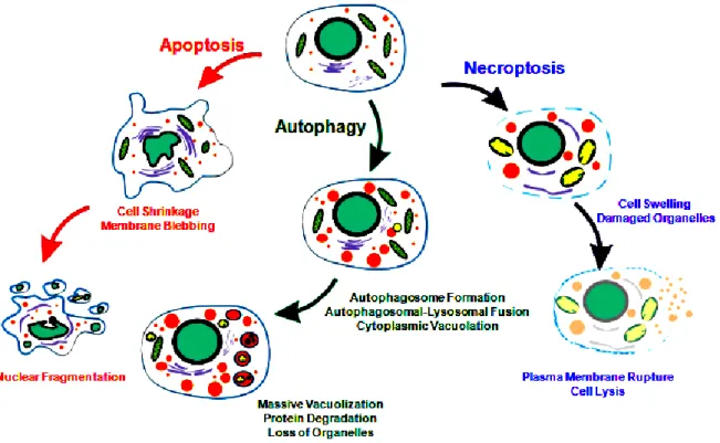
Fig. 1: Apoptosis, Autophagy and Necroptosis. Morphological features of distinct programmed cell death (PCD) (21).
Cell death
Programmed cell death (PCD) is an essential biological process during development,
homeostasis and immunity of multicellular organisms. The type of PCD is depended on stimuli and background of individual cells. PCD is required for elimination of individual cells during homeostasis of adult organisms, tissue remodeling and organ development. However, PCD is also involved in various diseases, such as cancers, autoimmune and immunodeficiency disorders. To date, PCD is described by morphological criteria into three types. PCD type I is called apoptosis (Fig. 1). Apoptosis is described by unique morphological and biochemical changes consisting of cell shrinkage (pyknosis), DNA fragmentation (karyorrhexis), nuclear condensation, activation of proteolytic enzymes caspases and blebbing of the plasma membrane. The internal cell components are tightly packed in apoptotic bodies and then rapidly engulfed by phagocytes (22;23). PCD type II is named autophagy (Fig. 1).
Autophagy is typically triggered by nutrient starvation. Autophagy is initiated by formation of unique organelles termed „autophagosome‟. After fusion with lysosomes cell organelles and proteins are degraded in vacuoles by lysosomal or vascular hydrolases after (25). PCD type III is called necrosis or necroptosis (Fig. 1). After stimulation of the TNF receptor (TNFR) by TNF the recruitment of the TNF receptor-associated death domain (TRADD) and receptor- interacting protein kinase 1 (RIPK1) is initiated. In the absence of active caspase 8, RIPK1 and RIPK3 forms a complex called necrosome, which activates the pro-necroptotic protein mixed lineage kinase domain-like (MLKL) (26). Active MLKL is translocated to the plasma membrane and into the membranes of intracellular organelles (such as ER, mitochondria and lysosome), forms nonspecific pores and Na+ and Ca2+ influxes which is required for necroptosis induced cell death (27).
Apoptosis
Apoptosis is a well-organized sequence of morphological events and energy dependent processes. Apoptotic cells are recognized by phagocytes and rapidly engulfed without inflammatory responses. Apoptosis has been considered as an irreversible process. Because activation of caspases (caspase-3, -6 and -7) commit cells to a „point-of-no-return‟ (28;29).
Caspases-dependent apoptosis pathways are mediated by extrinsic (receptor-mediated) or intrinsic (mitochondria-mediated) signaling pathways (30).
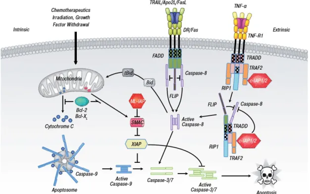
The extrinsic or receptor-mediated pathway
Two mechanisms of the direct initiation of apoptosis have been suggested: the TNF- induced signal pathway and the Fas-Fas ligand-mediated signal pathway. Both pathways involve receptors of the TNF receptor (TNFR) family (Fig. 2, 30;31). Activation of these receptors leads to the formation of two different death-inducible signaling complexes (DISC).
Complex I is associated with TNFR1, TNFR-associated death domain (TRADD), TRAF2, RIP1, cIAP1 and cIAP2 in the plasma membrane. Complex II requires Fas-associated death domain (FADD), TRADD and caspase-8 (30). Autoproteolytic of caspase-8 causes activation of executioner caspases cascade (32).
Fig. 2 Major distinct pathways of apoptosis. The extrinsic (receptor-mediated) pathway acts via death receptors while the intrinsic pathway acts via release of mitochondrial proteins.
Activation of both pathways leads to cell death by activation of caspases. Crosstalk between the pathways occurs via the Bcl-2 family member, Bid (33).
The intrinsic or mitochondria-mediated pathway
The intrinsic or mitochondria-mediated pathway (Fig. 2) is activated by various stimuli, such as DNA damage, irradiation, cytotoxic agents, growth deprivation, endoplasmic reticulum (ER) stress, intracellular Ca2+ overload and oxidative stress (29). The central role of this
pathway is dysfunction of mitochondrial, including the opening of a permeability transition pore (PTP), mitochondrial matrix swelling and outer mitochondrial membrane (OMM) rupture (34). Generally, mitochondria integrity is maintained by Bcl-2 family proteins, including anti-apoptotic (Bcl-2, Bcl-xL and Bcl-w) and pro-apoptotic (Bax, Bak, Bad, Bid and Bim) members (31). In the presence of apoptotic stimuli, oligomerization of Bax and Bak translocate and form pore into OMM, leading to release of mitochondrial components, such as cytochrome C, Smac/Diablo and apoptosis-inducing factor (AIF). Consequently, cytochrome C forms the complex with protease-activating factor-1 (Apaf-1) and caspase-9, called
„apoptosome‟ complex (29). Activated caspase-9 induces downstream executioner caspases-3, -6 and -7. However, there is crosstalk between extrinsic and intrinsic pathway in certain cell types. Subsequent activation of caspase-8 results in cleavage of pro-apoptotic protein „Bid‟.
Truncated Bid then translocate to mitochondria, causing the release of cytochrome C (32).
Clearance of death cells
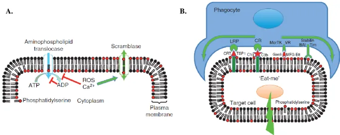
After cell death processes, phagocytosis is a crucial termination process for death cells to prevent surrounding tissue from the release of intracellular immunogenic contents (36). Bever et al. reported the present of phosphatidylserine (PS) on the outer cell surface is one major signal “eat me” to attract phagocytes (Fig, 3, 37-39). Consistent with apoptosis and tumorigenic cells, large amount of PS exposure triggers the recognition of macrophages and phagocytosis. However, asymmetrical distribution of phospholipids is composed of phosphatidylcholine (PC) and sphingomyelin (SM) in the outer leaflet, while PS, phosphatidylinositol (PI) and phosphatidylethanolamine (PE) in the inner leaflet of plasma membrane. This lipid asymmetry is maintained by the action of three distinct activities, including two of ATP-dependent and one of Ca2+-dependent manner. ATP-dependent enzymes have been known as flippases and floppases to translocate phospholipids in and out of cell surface, respectively (39). The transiently and rapidly translocation of PS and PE into the outer leaflet of plasma membrane is regulated by Ca2+-dependent manner, called scramblase (39;40).
Fig. 3 The regulation and physiology of membrane phospholipid asymmetry. A.) The aminophospholipid translocase uses ATP to pump phosphatidylserine (PS) from the outer to inner leaflet of the plasma membrane, whereas the calcium-activated phospholipid scramblase can cause PS exposure by randomizing of phospholipid distribution. B.) „Eat-me‟ signaling.
PS activates phagocytosis via lipoprotein receptor-related protein (LRP) and other receptors on the phagocyte (35).
For example, the impairment of scramblase activity is found in Scott syndrome, an inherited bleeding disorder (41;42). According to PS-recognized by macrophages, PS exposure on apoptotic cells can bind to lipoprotein receptor-related protein (LRP) and other receptors on surface of macrophages (39). After clearance process, phagocytes produce anti- inflammatory cytokines or immune suppressor proteins (36;43;44). Interestingly, it has been shown in earlier finding that Cl- channels strongly regulate several cellular functions of macrophages (45;46).
Role of anoctamins in cell death
Anoctamins are homologous plasma membrane protein family consists of ten different members (TMEM16A to K, excluding I or ANO1-ANO10) with several splice variants. They are composed of ten putative transmembrane-spanning domains and the NH2 and COOH termini protruding into cytosol (46-50). Anoctamins reveal tissue-specific expression. ANO1, 6, 7, 8, 9 and 10 are expressed in various tissues, whereas ANO2, 3, 4 and 5 are specifically expressed in neuronal and musculoskeletal tissues (51-53). Anoctamins are involved in ion
A. B.
transport, cell proliferation, phospholipid scrambling and cell death (50;51;53-55).
ANO1 is frequently amplified on chromosomal region 11q13 which is associated with growth, proliferation, metastasis and poor prognosis in cancer cells, including head, and neck squamous cell carcinomas (HNSCC), pancreatic cancer, gastric cancer, prostate cancer and breast cancer (56-60). This region has been found to harbor several genes that have potential to drive oncogenes (55;58). ANO1 has been defined in multiple names depended on different cancer cells, such as gastrointestinal stromal tumors protein1 (DOG1) and tumor-amplified and over expressed sequence2 (TAOS2) (60). ANO1 plays a role in several tissues, including smooth muscles, secretory epithelia (in airway, exocrine glands and intestine) and sensory neurons (57;61-68). Knockout of ANO1 is embryonically lethal because it is an important in disease states, such as pulmonary diseases, hypertension and diarrhea (53;69). There are evidences showed that overexpression of ANO1 contributed tumorigenesis through the activation of EGF receptor (FGFR), CAMK, AKT (70;71) and MAPK signaling pathways (58;72;73). Consistent with previous studies, ANO1 is pro-proliferative protein which controls G1/S transition of cell cycle and cancer progression (74). It has been demonstrated that ANO1 interacts with signaling/scaffold actin-binding regulatory proteins, such as ezrin, radixin, moesin and RhoA, accompanied with localized cell shrinkage at the rear part in migrating cells (75-79). Moreover, it has been reported that ANO1 is an important for both RVD and AVD (79).
ANO6 has been shown to be expressed in various tissues, such as the respiratory system, the gastrointestinal tract, kidney, ovary, skeleton, and skin (51;54;80). ANO6 plays a role in skeletal development in embryonic tissues, cell migration, metastasis, cell volume regulation and phosphatidylserine (PS) exposure during early phase of apoptosis (70;78;81-83). It has been reported to act as Ca2+-activated nonselective cation channel, Ca2+-activated Cl- channel, Ca2+-dependent phospholipid scrambling (70;83-85). Additionally, ANO6 is identified to be a volume-regulated Cl- channel, which generates an outwardly rectifying Cl- current (ORCC) to contribute RVD and AVD (86;87). Our previous studies showed knockdown of ANO6 significantly inhibited Fas- and staurosporine-induced ORCC but not Ca2+ dependent chloride
channels in lung epithelial cells (87). Nevertheless, Shumilina et al. showed Ca2+ -dependent ORCC, which is an important for migration in dendritic cells. The authors suggested ANO6 may form variation of oligomeric complexes caused different ORCC functions in specific cell types. Interestingly, Suzuki et al. demonstrated PS scrambling in Ba/F3 lymphocytes that is induced by overexpression of ANO6. Patients with Scott syndrome have a loss-of-function mutation in ANO6 characterized by a deficiency in platelet pro-coagulant activity, causing the congenital bleeding disorder (56;60). There is intriguing evidence that ANO6 mutation in Scott patients impairs not only PS exposure but also decrease cell blebbing and shedding of microparticals from membrane in platelets, red blood cells and lymphocytes (44;83;88-91).
Moreover, recently finding has reported that a deficiency of PS scrambling in ANO6 knock- out mice reduce the production of calcified bone matrix during bone mineralization (82;92).
ANO10 comprises like other anoctamins a DUF590 domain of unknown function. In mouse tissues, ANO10 is frequently expressed in the same tissues with ANO1 and Ano6, especially in neuronal and muscle tissues (53;80;93;94). In adult human, ANO10 is predominantly expressed in frontal and cerebellum in contrast to fetal brain. It has been reported that ANO10 certainly encodes a subunit of Ca2+-regulated Cl- channel and plays a role in Purkinje cells of cerebellum (93). The results from the combination of homozygosity mapping together with targeted array-based sequencing showed mutations in ANO10 are associated with autosomal-recessive cerebellar ataxia (93). Cerebellar degeneration has been suggested to involve mitochondrial- and DNA-repair dysfunction. Previously, it has reported ANO10 is probable role in cellebellar ataxia and schizophrenia (95). Furthermore, Drosophila ortholog of ANO10 has been also found to play a role in spindle formation and cell cycle progression, termed „Axs‟ gene. This gene is proposed to be a mediator of meiotic spindle assembly and chromosome segregation (53).
Apart from ANO1, ANO6 and ANO10, there is less available evidence for other anoctamins. Interestingly, several independent groups have reported that other anoctamins (ANO2, ANO3, ANO8 and ANO9) exhibited their function as Ca2+-activated Cl- channels which are associated with murine embryogenesis on several organs (52;53;80). ANO8 and
ANO9 were also implicated in the cell volume regulation (94). ANO3, 4, 7 and 9 also play a role in phospholipid scramblases (28). High levels of other anoctamins expression are also related to cancers and diseases. For example, ANO5 (GDD1) is associated with the proliferation and differentiation of skeletal muscle. Mutation of ANO5 effects the repairment of muscle, causing muscular dystrophy (97). ANO7 (NGEP gene) is highly expressed at cell:cell contact regions in prostate cancer cells (98). ANO9 is located in chromosome 11 p15 which plays a role in Ca2+-mediated phospholipid scrambling. In recent study, it found ANO9 acts as a metastasis-related gene which exhibit the inhibiting colorectal cancer progression (99). Because of deeply mechanism of anoctamins with diseases is remaining to be puzzled, it is therefore an intriguing issue to be investigated.
Intention and outline of the present thesis
According to physiological evidences, RVD plays a role in cell proliferation, cell death, migration, immune response and cancer. Although, several Cl- channels have been proposed as candidates for RVD, ANO1, 6 and 10 are shown to correlate to cellular volume regulation but the underlying mechanisms is still unclear. Therefore, this thesis will investigate the role of ANO1, 6 and 10 for RVD under physiological and under pathological conditions.
In chapter 2, experiments were aimed to understand the function of ANO1 for RVD in cancer. Therefore, the role of ANO1 in development of human head and neck stromal cell carcinoma (HNSCC) and colonic cancer will be investigated.
In chapter 3 and 5, we investigated several signal pathways which induce RVD and activation of ANO6 and ANO10 using the Xenopus oocytes expression system and primary cells from ANO6 and ANO10 knock-out mice.
In chapter 4, we ask the question, whether ANO6 and RVD could play a role in innate immune system in macrophages in response to bacterial infection.
In chapter 5, we investigated an ANO10-R263H mutation found in patients with neuroborreliosis and ask for the role of ANO10 during Borrelia infection.
CHAPTER 2
Role of anoctamins in cancer and apoptosis Abstract
Anoctamin 1 (TMEM16A, ANO1) is a recently identified Ca2+-activated chloride channel and a member of a large protein family comprising 10 paralogues. Before ANO1 was identified as a chloride channel protein, it was known as the cancer marker DOG1. DOG1/ANO1 is expressed in gastrointestinal stromal tumours (GIST) and particularly in head and neck squamous cell carcinoma, at very high levels never detected in other tissues. It is now emerging that ANO1 is part of the 11q13 locus, amplified in several types of tumour, where it is thought to augment cell proliferation, cell migration and metastasis. Notably, ANO1 is upregulated through histone deacetylase (HDAC), corresponding to the known role of HDAC in HNSCC. As ANO1 does not enhance proliferation in every cell type, its function is perhaps modulated by cell-specific factors, or by the abundance of other anoctamins. Thus ANO6, by regulating Ca2+-induced membrane phospholipid scrambling and annexin V binding, supports cellular apoptosis rather than proliferation. Current findings implicate other cellular functions of anoctamins, apart from their role as Ca2+-activated Cl- channels.
Key words: anoctamin 1, anoctamin 6, TMEM16A, TMEM16F, cancer, head and neck stromal cell carcinoma
Published in: Podchanart Wanitchakool1, Luisa Wolf1, Gudrun E. Koehl2, Lalida Sirianant1, Rainer Schreiber1, Sucheta Kulkarni3, Umamaheswar Duvvuri3 and Karl Kunzelmann1Role of anoctamins in cancer and apoptosis. Philosophical transactions of the royal society:
3;369(1638):20130096 (2014).
Own experimental contribution: Most western blot, cell proliferation and migration experiment performed in cell lines.
Own written contribution: Methods, Results, Parts of Introduction and Discussion.
Other contributions: Designed experiments and analyzed data.
Introduction
Anoctamin 1 (ANO1, TMEM16A) is a novel Ca2+-activated chloride channel (CaCC) with important physiological functions in epithelial cells and other cell types (1-4). It was also shown to be activated during cell swelling, probably secondary to an increase in intracellular Ca2+ (5). While some detected a role of Ca2+-dependent anoctamins, such as ANO1 or ANO6, to volume regulation, others did not (5;6). Upregulation of endogenous Ca2+-activated Cl- channels in proliferating cells has been observed recently, but the role of these channels for proliferation has remained unclear (7,8). Before ANO1 was identified as CaCC, it was already known as a protein that is coexpressed with the morphogen sonic hedgehog during embryonic development (9). ANO1 was also known as DOG1, a protein strongly expressed in gastrointestinal stromal tumours and head and neck squamous cell carcinoma (HNSCC).
Expression of DOG1 correlates with poor outcome in oesophageal squamous cell cancer (10- 12). Interstitial cells of Cajal (ICC) are another predominant site for ANO1 expression (13- 15). It has been reported that mice lacking ANO1 had fewer proliferating ICC (16).
Moreover, additional data suggested that ANO1 regulates proliferation at the G1/S transition of the cell cycle. Assuming such a pro-proliferative role, targeting of ANO1 has been proposed as a novel method for the treatment of malignant tumours (7;17;18).
Materials and Methods
Animals: Heterozygous APCMin+/− pups (Fodde R et al, (1994) Proc Natl Acad Sci U S A 91:8969–8973) (Jackson Laboratory, USA) were weaned at 4–5 weeks of age and genotyped by PCR. Animals were fed with complete chow with 15% corn oil (Sniff, Soest, Germany), which has an adverse effect on polyp numbers in these animals (Wasan et al (1997) Proc Natl Acad Sci U S A 94:3308–3313). APCMin+/− were sacrificed after a weight loss of 10%.
Wildtype (wt) animals of the same age served as controls. Crypt cell isolation and expression analysis of ion channels in colonic epithelial cells. Colonic crypts were isolated from an inverted colon in Ca2+ free buffer solution. Animal studies were conducted according to the guidelines of the National Institute of Health and the German laws on protection of animals.
Cell Culture, siRNA and Transfection: BHY, CAL-33, T84 and HT29 cell lines were grown in Opti-MEM or DMEM (Gibco) supplemented with 10% (v/v) heat inactivated fetal bovine serum (FBS, Gibco, Karlsruhe, Germany) at 5% CO2 and 37°C. LipofectamineTM2000 Transfection Reagent (Invitrogen, Karlsruhe, Germany) was used for transfection of hANO siRNA (Stealth siRNA, Invitrogen, 59-GGUUCCCAGCCUACCUC ACUAACUU-39, 59- AGUUAGUGAGGUAGGCUGGGAACC-39) or Negative Control High GC (Invitrogen) according to the manufacture‟s guidelines. Experiments were performed 48 h-72 h after transfection.
Reverse Transcriptase PCR: Total RNA was isolated from T84 cells using RNeasy Mini- Kit Qiagen; Hilden, Germany). 2 mg of total RNA was reverse-transcribed in 50 ml for 1 h at 40°C using random primer and RT. 30 cycles of RT-PCR was performed using standard procedures (GoTaq DNA Polymerase, Promega), 1 ml RT and primers for anoctamis (Ruiz et al (2012) 7: e43265, PLoS one). For semi-quantitative comparison, ß-actin was co-amplified (primer 0.05 mM). Products were analyzed on ethidium bromide-stained 2% agarose gels using densitometry. Densitometric ratios for ANO1 and ß-actin were calculated as a measure for relative expression of ANO1.
Patch Clamp: Cells were grown on glass cover slips and mounted on a perfused bath on the stage of an inverted microscope (IM35, Zeiss) and kept at 37 ºC. The bath was perfused continuously with Ringer solution (mM: NaCl 145, KH2PO4, K2HPO4 1.6, D-glucose 6, MgCl2 1, Ca2+-gluconate 1.3, pH 7.4) at about 5 ml/min. For fast whole-cell patch clamping pipettes were filled with intracellular like solution containing (mM: KCl 30, potassium gluconate 95, NaH2PO4 1.2, Na2HPO4 4.8, EGTA 1, calcium gluconate 0.758, MgCl2 1.034, D-glucose 5, ATP 3, pH was 7.2). For all solutions Ca2+ concentrations were adjusted to 0.1 μM (pipette) and 1.3 mM (bath). During experiments cells were current clamped and membrane voltages were measured. At intervals cells were voltage clamped to ± 50 mV in steps of 10 mV for 1 s. The applied clamp voltages were corrected for liquid junction potentials. Because current voltage relationships were rather linear within the clamp voltage range, membrane conductances were calculated according to Ohm‟s law.
Impedance based xCELLigence Attachement / Proliferation / Migration assay: The xCELLigence invasion assay (Roche, Germany) is based on changes in electrical impedance
at the interphase between cell and electrode as migrating cells move through a barrier (Rahim S, Uren A (2011). A real-time electrical impedance based technique to measure invasion of endothelial cell monolayer by cancer cells. J Vis Exp. 2011 Apr 1;(50). pii: 2792). These changes were directly correlated with the migrative capacity of BHY and CAL33 cells. The technique provides an advantage over existing methods such as boyden chamber and matrigel assays, and standard proliferation assays, respectively, since the data is obtained continuously in real-time, when compared to endpoint analysis in other methods. For migration assays cells were seeded at a density of 10,000 cells/well on CIM-plates-16 migration plates. The xCELLigence attachment/proliferation assay is based on assessment of impedance during attachment (early phase) and proliferation (late phase) as detected by real-time electrical impedance measurement. For attachment and proliferation assays cells were seeded at a density of 5,000 cells/well on E16 plates (Roche, Germany).
Western Blot: Freshly isolated mouse intestinal cells or cultured HT29, T84, BHY, and UMSCC cells were lysed with an appropriate buffer (150 mM NaCl, 50 mM Tris–HCl, 1 mM EDTA, 1% NP-40, protease inhibitor, 100 mM DTT; pH 7.4) and DNA was sheared by sonication. Samples were quantified using a Bio-rad protein assay (Biorad) and the same amount of protein (50 mg). Bands were analyzed using densitometry. Densitometric ratios for ANO1 and ß-actin were calculated as a measure for relative expression of ANO1.
Apoptosis assay: Apoptosis was assessed by NucViewTM 488 Caspase-3 assay kit (Biotum, Germany). Briefly, cells were incubated with DEVD-NucView488 substrate and apoptotic cells were analyzed using the plate reader NOVOstar.
Proliferation assay: For proliferation assays, cells were plated at a density of 2,000 cells/0.35 cm2 and incubated 2 days later with various concentrations of rapamycin or TSA as indicated.
Cell proliferation was assessed by 5-bromo-2-deoxyuridine (BrdUrd) incorporation using an enzyme-linked immunosorbent assay kit (Roche Diagnostics, Penzberg, Germany) and by cell counting. The cell number was assessed after fixation in 3.7% formaldehyde and 0.5% Triton X-100 for 30 min at room temperature and after staining with Mayer‟s hemalaun (Merck, Darmstadt, Germany) for 5 min. Digitized microscopic images were taken (Fluovert FS, Leitz), and nuclei were counted using imaging software (TINA).
Immunohistochemistry of ANO1-expression: Primary tissue samples were collected after obtaining informed consent and approval from the Institutional Review Board. Normal adjacent mucosa is defined as histologically benign appearing mucosa (as judged by an experienced pathologist) acquired from the margins of the tumor resection. Staining was conducted with anti-TMEM16A antisera and scored using a semiquantitative system (IHC index). The index was defined as the relative intensity, scored on a 0 to 3 scale, multiplied by the percentage of positively stained cells. High and low expressors were categorized as having H-scores above or below the median.
Results and Discussion
ANO1 is located on the 11q13 amplicon
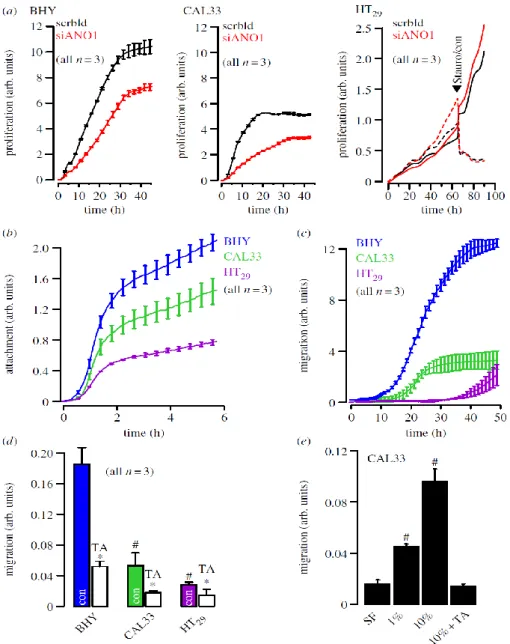
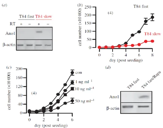
The coding sequence of ANO1 is located within the 11q13 region, a chromosomal locus that is frequently amplified in a number of different human cancers, such as urinary bladder cancer, breast cancer and HNSCC (100). The 11q13 amplicon contains a stretch of proteins related to cell cycle, proliferation and apoptosis (cyclin D1, ORAOV1, FGF19, FGF4, TMEM16A, Fas-associated via death domain, PPFIA1, cortactin (101). The complex structure of this amplicon has mostly been studied in breast cancer, where multiple genes have been suggested as driver genes (102;103). These findings implied a link between ANO1 expression, cell-cycle regulation and proliferation, which has recently been demonstrated in HNSCC and other cancer cells (55;72-74;104). Surprisingly, downregulation of ANO1 contributes to cerebrovascular remodeling by promoting basilar smooth muscle cell proliferation, which is through inhibition of expression of cyclin D1 and cyclin E (68). We performed additional experiments and compared proliferation of two HNSCC (CAL33, BHY) and one colonic epithelial cell line (HT29) using an online impedance-based xCELLigence proliferation assay system (79) (figure 1a). Proliferation was clearly higher in the HNSCC cell lines, which express much higher levels of ANO1, when compared with HT29 cells (figure 3a) (79). In a previous study, we did not detect inhibition of proliferation of BHY cells by siRNA (79). Using improved Stealth siRNA, these experiments were repeated. Proliferation was suppressed by siRNA-knockdown of ANO1 expression in HNSCC but not in HT29 cells.
These results suggest that very effective suppression of ANO1 expression is necessary to eliminate the pro-proliferative effects of ANO1, suggesting that low expression of ANO1 is sufficient to induce this effect. While siRNA had no effect in HT29 cells, 1 mM staurosporine (figure 1a black arrow, dashed lines) inhibited proliferation and induced cell apoptosis in colonic carcinoma cells, as demonstrated in an earlier publication (105). Thus, enhanced expression of ANO1 does not seem to enhance proliferation in every cell type (58;79).
Fig. 1 ANO1 controls proliferation: (a) Cell proliferation measured online by impedance- based xCELLigence proliferation assay system in HNSCC (BHY, CAL33) and colonic epithelial (HT29) cells (see electronic supplementary material). siRNA-knockdown of ANO1 expression reduced proliferation in both BHY and CAL33 cells, but slightly augmented proliferation in HT29 cells. Staurosporine (1 µg ml-1) strongly inhibited proliferation. (b) Attachment of BHY, CAL33 and H29 cells after seeding in xCELLigence chambers (see electronic supplementary material). (c) Migration of BHY, CAL33 and H29 cells after seeding
Fig. 2 Anoctamins facilitate cell volume decrease and support diapedesis and cell migration: model for diapedesis and migration of a tumour cell expressing anoctamin Cl- channels, which allow Cl- release and osmotic cell shrinkage. Ano1 is regulated by actin and possibly by a number of proteins related to signalling/scaffolding, such as ezrin, radixin, moesin and proteins related to cell attachment and migration, such as zyxin, fibulin 1, S100A11, twinfilin and catenin.
in the xCELLigence migration chamber (see electronic supplementary material). (d ) Summary of the rate of cell migration between 10 and 30 h after seeding and inhibition of migration by TA(10 mM). (e) Migration of CAL33 cells and effect of fetal calf serum and TA. Mean±s.e.m. (number of cells). Symbol # denotes significant difference when compared with BHY and serum free, respectively ( p < 0.05; ANOVA). Asterisks (*) denote significant inhibition by TA ( p , 0.05; paired t-test).
However, there is increasing consent that high levels of ANO1lead to enhanced cell motility, distal metastasis and poor prognosis (55;58;72;79). Also in this study, cell attachment and migration was enhanced with increasing expression of ANO1 (BHY, CAL33, HT29). It is further demonstrated that migration could be significantly inhibited by tannic acid, a blocker of ANO1 and other anoctamins (figure 1c,d). Moreover, migration was strongly dependent on the presence of serum in the migration chamber and was inhibited by the ANO1 inhibitor tannic acid (TA, figure 1e). Although proliferation, attachment and migration were positively correlated with expression of ANO1, this does not prove that ANO1 is directly responsible for the change of these properties.
Role of ANO1 for metastasis
How does ANO1 control the ability of tumour cells to migrate and form distal metastasis? The relationship between ion channel currents, cell volume regulation, migration and metastasis is well established (106-109). Previous findings indicate that ANO1 is activated during hypotonic cell swelling and contributes to regulatory volume decrease (RVD), which certainly requires a rise in intracellular Ca2+ (51;79;96).
A rise in intracellular Ca2+ may activate ANO1 together with K+ channels to release intracellular K+ and Cl- ions. Resulting osmotic loss of intracellular water will cause rapid cell shrinkage and allow passage through narrow gaps like those formed by endothelial cells (109;110) (figure 2). Ca2+-dependent activation of ANO1 would support cell shrinkage at the rear end of migrating cells, thereby further reducing cell volume and facilitating diapedesis.
Inhibition of Cl- channels impedes cell volume changes and thereby compromises tumour cell invasion. This has been demonstrated for ANO1 (79) as well as other Cl- channels (110).
Importantly, cell migration requires constant depolymerization and repolymerization of the actin cytoskeleton, which permanently changes cell-matrix adhesions (76;111). Our earlier findings suggested that ANO1 Cl- currents are controlled by the actin cytoskeleton (75). This was supported by a subsequent report indicating that ANO1 associates with the signaling/scaffolding proteins ezrin, radixin, moesin and RhoA, which are known to connect plasma membrane proteins to the cytoskeleton (112). Moreover, results from a two hybrid split ubiquitin screening suggested interaction of ANO1 with a number of proteins related to cell attachment and migration, such as zyxin, fibulin 1, S100A11, twinfilin and catenin (unpublished results from the Kunzelmann laboratory, K. Kunzelman 2012, figure 2).
It was shown that members of the Rho GTPase family exert effects on cell shape and motility by regulating actin cytoskeleton; the activation of Rac1 organizes cortical actin cytoskeleton and promotes formation of lamellipodia at the leading edge, a hallmark of a motile cell, while the activation of RhoA at the rear influences acto-myosin complexes to allow retraction of the trailing end. Spatial and temporal regulation of the activity at each end create an unequal distribution of membranous, cytoskeletal and cytoplasmic contents to induce a highly polarized, motile shape that is suitable for movement and metastasis (108).
Thus, the presence of ANO1 at the plasma membrane, its ability to regulate cell shape and volume, and its connections to cytoplasmic/cytoskeletal elements is likely to contribute to cell movement and metastasis.
Regulation of expression of ANO1 by histone deacetylase and clinical implications
Recruitment of histone acetyltransferases and histone deacetylases (HDACs) is a key element in the dynamic regulation of genes controlling cellular proliferation and differentiation during normal development as well as carcinogenesis (113;114). A number of
anti-cancer treatments are based on the inhibition of HDAC. HDAC inhibitors promote expression of p21 in breast cancer cells, which inhibits the action of cyclin D1. HDAC inhibitors may therefore also be useful for the treatment of those HNSCC that show overexpression of ANO1 and concomitant activation of cyclin D1 (55).
Fig. 3 HDAC regulates expression of ANO1: (a,b) western blots and densitometric analysis indicating expression of ANO1 in HT29 colonic epithelial and BHY HNSCC cells, and inhibition by HDAC inhibitors valproic acid (3 mM) and butyrate (4 mM). ß-actin was used as a loading control. (c) Real-time PCR analysis of ANO1-mRNA expression in HT29 and BHY cells. (d,e) Induction of apoptosis and inhibition of proliferation of BHY cells by valproic acid and butyric acid, as measured by apoptosis assays (see electronic supplementary material) and cell counting. (f) Whole cell ANO1 Cl- currents (Vc = +100 mV) activated by an increase in intracellular Ca2+ owing to stimulation with the purinergic agonist ATP (100 mM).
(g) Expression of ANO1 in human HNSCC samples and normal tissue as measured by
immunohistochemistry (cf. electronic supplementary material). (h) Inhibition of proliferation of UM-SCC cancer cells by various concentrations of TSA. Mean±s.e.m. (number of cells).
Symbol # denotes significant difference when compared with control, normal tissue, HT29
cells or absence of HDAC inhibitors, respectively (p < 0.05; unpaired t-test).
In fact, HDAC inhibitors have already entered preclinical evaluation (115;116). In recent experiments, we found that pronounced expression of ANO1 in the HNSCC cell line BHY was largely inhibited by treatment with the HDCA inhibitors valproic acid or butyric acid, along with inhibition of cell survival and ANO1-dependent whole cell currents (figure 3a-f).
Although this does not prove that HDAC inhibitors act through downregulation of ANO, these novel results again demonstrate a correlation between ANO1 expression and proliferation. Corresponding to the data discussed in figure 1, which did not identify a role ANO1 expression for proliferation of HT29 colonic epithelial cells, expression levels for ANO1 were much lower and were not affected by HDCA inhibitors. As pointed out, expression levels for ANO1 are much higher in HNSCC compared with normal tissues (figure 3g). We also found a dose-dependent inhibition of proliferation of another HNSCC cell line, UMSCC cancer cells, by a third type of HDAC-inhibitor, trichostatin (TSA) (figure 3h). UM- SCC cells contain amplification of the ANO1 gene locus similar to BHY cells. The results confirm that the inhibitory effect of HDAC inhibitors on ANO1 expression is independent of the HNSCC cell line used. While valproic acid and butyric acid are rather broad, non- selective inhibitors of HDAC, TSA selectively suppresses class I/II, suggesting that ANO1 expression is regulated by these HDACs. Although the mechanisms by which TSA promotes loss of cell survival/growth in these cells is incompletely understood, the present results support the use of HDAC inhibitors for the treatment of HNSCC, which may act in part through inhibition of ANO1 expression.
ANO1 and sonic hedgehog
Interesting links exist between ANO1 and the sonic hedgehog (Hh) signaling pathway.
Hh is coexpressed with ANO1 in the zone of polarizing activity in mouse limb buds during E10.5 and E11.5 (117). Hh signaling controls many aspects of development and also regulates cell growth and differentiation in adult tissues. It is activated in a number of human malignancies. Hh and Wnt signaling frequently act together in controlling cell growth and