(a-Br-TMC) Inhibits the JAK/STAT Signaling Pathway
Sophia Pinz1., Samy Unser1., Susanne Brueggemann1, Elisabeth Besl1, Nafisah Al-Rifai2, Hermina Petkes2¤, Sabine Amslinger2*, Anne Rascle1*
1Stat5 Signaling Research Group, Institute of Immunology, University of Regensburg, Regensburg, Germany,2Institute of Organic Chemistry, University of Regensburg, Regensburg, Germany
Abstract
Signal transducer and activator of transcription STAT5 and its upstream activating kinase JAK2 are essential mediators of cytokine signaling. Their activity is normally tightly regulated and transient. However, constitutive activation of STAT5 is found in numerous cancers and a driving force for malignant transformation. We describe here the identification of the synthetic chalcone a-Br-29,3,4,49-tetramethoxychalcone (a-Br-TMC) as a novel JAK/STAT inhibitor. Using the non- transformed IL-3-dependent B cell line Ba/F3 and its oncogenic derivative Ba/F3-1*6 expressing constitutively activated STAT5, we show that a-Br-TMC targets the JAK/STAT pathway at multiple levels, inhibiting both JAK2 and STAT5 phosphorylation. Moreover,a-Br-TMC alters the mobility of STAT5A/B proteins in SDS-PAGE, indicating a change in their post-translational modification state. These alterations correlate with a decreased association of STAT5 and RNA polymerase II with STAT5 target genes in chromatin immunoprecipitation assays. Interestingly, expression of STAT5 target genes such as Cisandc-Mycwas differentially regulated bya-Br-TMC in normal and cancer cells. While both genes were inhibited in IL-3- stimulated Ba/F3 cells, expression of the oncogenec-Mycwas down-regulated and that of the tumor suppressor geneCis was up-regulated in transformed Ba/F3-1*6 cells. The synthetic chalconea-Br-TMC might therefore represent a promising novel anticancer agent for therapeutic intervention in STAT5-associated malignancies.
Citation:Pinz S, Unser S, Brueggemann S, Besl E, Al-Rifai N, et al. (2014) The Synthetica-Bromo-29,3,4,49-Tetramethoxychalcone (a-Br-TMC) Inhibits the JAK/STAT Signaling Pathway. PLoS ONE 9(3): e90275. doi:10.1371/journal.pone.0090275
Editor:Michael Nevels, University of Regensburg, Germany
ReceivedDecember 22, 2013;AcceptedJanuary 27, 2014;PublishedMarch 3, 2014
Copyright:ß2014 Pinz et al. This is an open-access article distributed under the terms of the Creative Commons Attribution License, which permits unrestricted use, distribution, and reproduction in any medium, provided the original author and source are credited.
Funding:This work was supported by the Deutsche Forschungsgemeinschaft (Grant No. RA 2010/2-1 to AR), the Deutsche Krebshilfe (Grant No. 109750 to AR), institutional research funds (Foerderlinie C to AR), the Fonds der Chemischen Industrie (Liebig scholarship to SA) and the DAAD (Doctoral scholarship to NA). The funders had no role in study design, data collection and analysis, decision to publish, or preparation of the manuscript.
Competing Interests:The authors have declared that no competing interests exist.
* E-mail: anne.rascle@klinik.uni-regensburg.de (AR); sabine.amslinger@chemie.uni-regensburg.de (SA) .These authors contributed equally to this work.
¤ Current address: Department of Chemistry, Babes-Bolyai University, Cluj-Napoca, Romania
Introduction
The signal transducer and activator of transcription STAT5 is a key regulator of immune responses, cell proliferation, differenti- ation, survival and oncogenesis [1,2]. STAT5 proteins normally reside as latent transcription factors in the cytoplasm of unstimulated cells. Following cytokine, growth factor or hormone stimulation, the receptor-associated JAK2 tyrosine kinase gets auto-phosphorylated (trans-phosphorylation) before it phosphory- lates the receptor intracellular domain, creating docking sites for STAT5, which is in turn phosphorylated by activated JAK2. STAT5 phosphorylation allows its dimerization and translocation into the nucleus where it binds to specific recognition sites, and ultimately activates transcription of specific target genes (e.g. c- Myc, Pim-1, Bcl-x, Cis) [1,3–5]. STAT5 activity is regulated through multiple post-translational modifications [1,6–8] and through interactions with cofactors, other transcription factors or STAT5 itself (tetramerization) [1,9–15]. STAT5 activation under physiological conditions is rapid and transient. Attenuation of the STAT5 response is controlled mainly through dephosphorylation by specific phosphatases (SHP-1) and a negative feedback loop mediated by proteins of the SOCS family (CIS, SOCS-1/-3)
[16,17]. However, constitutively activated STAT5 (caSTAT5) has been observed in a wide variety of human cancers, from hematologic malignancies (leukemia, lymphoma, myeloma) to solid tumors such as breast cancer, squamous cell carcinoma of the head and neck (SCCHN) or melanoma [16–18]. Constitutive activation of STAT5 directly contributes to oncogenesis, mainly through the up-regulation of oncogenes driving cell proliferation and survival (Bcl-x, c-Myc, Pim-1) [4,19–23], but also through the down-regulation of tumor suppressor genes and inhibitors of the pathway, such as SHP-1 and CIS/SOCS family members, via epigenetic silencing [17,18,24–28].
Therefore, the JAK/STAT pathway in general and STAT5 proteins in particular represent attractive targets for therapeutic intervention in human cancers [18,29–31]. Numerous inhibitors of the Janus Kinase family members (JAK1, JAK2, JAK3, Tyk2, and the JAK2V617F active mutant responsible for myeloproliferative neoplasia) and of STAT5 have been described [30,32–45]. Most of the JAK inhibitors are small molecules acting as tyrosine kinase inhibitors by targeting their ATP-binding catalytic domain. The JAK1/JAK2 inhibitor Ruxolitinib has been approved by the U.S.
Food and Drug Administration for the treatment of myelofibrosis while several others are being currently evaluated in clinical trials
for the treatment of various diseases [32,33]. Reported STAT5 inhibitors target STAT5 activity at multiple levels, from its activation by phosphorylation to its DNA binding activity, independently of upstream activating kinases [30,34,35,38, 39,45]. We were the first to show that deacetylase inhibitors such as sodium butyrate, trichostatin A (TSA) or suberoylanilide hydroxamic acid (SAHA) inhibit IL-3-mediated STAT5 transcrip- tional activity in the mouse pro-B cell line Ba/F3 [46]. We demonstrated that deacetylase inhibitors interfere neither with STAT5 initial activation (phosphorylation or nuclear transloca- tion) nor with STAT5 binding to DNAin vivo. Instead, these drugs prevent the proper assembly of the basal transcription machinery ultimately resulting in inhibition of transcription [46,47]. The deacetylase inhibitor SAHA (Vorinostat) has been approved for the treatment of cutaneous T cell lymphoma while many other deacetylase inhibitors are currently evaluated in clinical trials for the treatment of various types of cancers [48]. Therefore our findings support the attractive possibility that this class of drugs might be efficient in targeting STAT5-associated cancers.
Accordingly, tyrosine kinase inhibitor and deacetylase inhibitor combination therapies have recently proven to be more effective in the treatment of cancers with constitutive active STAT5 [49–53].
More recently, we turned to the identification of new types of STAT5 inhibitors, with a particular focus on natural and synthetic chalcones.
Chalcones, which are naturally abundant in fruits, vegetables and spices, area,b-unsaturated carbonyl compounds well known for their multiple biological activities as antioxidant, antibacterial, antifungal, anti-inflammatory and anticancer agents [54]. Natural and synthetic chalcones have been shown to inhibit the function of a variety of tyrosine kinases (JAKs, ERKs, Src) and transcription factors (NF-kB, STAT3) implicated in the response to inflamma- tion and the control of cell proliferation and survival [54–58]. We therefore hypothesized that the JAK2/STAT5 signaling pathway might be affected by chalcones. We previously described the synthesis of a-X-29,3,4,49-tetramethoxychalcones (a-X-TMCs) with different X substituents (X = H, F, Cl, Br, I, CN, Me, p- NO2-C6H4, Ph, p-OMe-C6H4, NO2, CF3, COOEt, COOH) at the a-position of the a,b-unsaturated carbonyl unit [59]. We formerly showed that thesea-X-TMCs have distinct electrophilic reactivities [59–61] which correlate well with their anti-inflamma- tory activity [59]. In this study, the ability of foura-X-TMCs (with X = CN, Br, Cl, H) to regulate STAT5 function was investigated.
Five natural chalcones, namely 29-hydroxy-3,4,49-trimethoxychal- cone (HTMC), 29,49-dihydroxy-3,4-dimethoxychalcone (DHDM C), 29,3,49-trihydroxy-4-methoxychalcone (THMC), butein, and isoliquiritigenin (ISL), as well as curcumin, another natural product carrying twoa,b-unsaturated carbonyl groups, were also investigated (Figure 1A).
We describe here a-Br-29,3,4,49-tetramethoxychalcone (a-Br- TMC) as a novel inhibitor of the STAT5 signaling pathway. We show thata-Br-TMC inhibits both JAK2 and STAT5 phosphor- ylation, leading to the inhibition of expression of downstream STAT5-dependent and -independent genes in IL-3-stimulated Ba/F3 cells. Moreover,a-Br-TMC alters the mobility of STAT5A and STAT5B proteins in SDS-PAGE, both in cells expressing regulated (Ba/F3) and constitutively active (Ba/F3-1*6 and K562) STAT5, presumably reflecting a change in STAT5 post-transla- tional modifications. Nevertheless, a-Br-TMC treatment affects differentially STAT5 transcriptional activity in normal and cancer cells. Altogether, this study identified a novel inhibitor of STAT5 signaling targeting both STAT5 and its upstream activating kinase JAK2, and affecting the regulation of downstream oncogenes and tumor suppressor genes.
Materials and Methods Chemicals
Chalcones were prepared by published procedures [59,60].
Dimethyl sulfoxide (DMSO), trichostatin A (TSA) and curcumin were purchased from SIGMA (D2650, T8552 and B6938 respectively). Imatinib was from Cayman Chemical (No. 13139).
Compounds were dissolved in DMSO at a final concentration of 1 mM (TSA) or 100 mM (other compounds).
Thiol assay
Kinetic thiol assay for TSA (Biotrend BN0742) was performed as described [60] using 500-fold excess cysteamine as a thiol substrate and an incubation time of 48 hours. The presence of the addition product was detected by liquid chromatography-mass spectrometry (not shown). Second-order rate constants k2values for chalcones and curcumin are taken from the literature [59,60].
Cell lines and drug treatments
All cell lines were grown in RPMI 1640 (PAN-Biotech P04- 16500) supplemented with 10% heat-inactivated fetal calf serum (FCS; PAN-Biotech), penicillin/streptomycin (PAN-Biotech) (thereafter designated as RPMI-based medium) and cultivated at 37uC under 5% CO2in a humidified incubator. K562 cells (a kind gift from Daniela Ma¨nnel, University of Regensburg, Germany;
[62]) were maintained in RPMI-based medium. The non- tumorigenic immortalized interleukin-3 (IL-3)-dependent mouse pro-B cell line Ba/F3 (a kind gift from Jacqueline Marvel, IFR 128 BioSciences Gerland-Lyon Sud, France; [63]) was grown in RPMI-based medium supplemented with 2 ng/mL rmIL-3 (ImmunoTools). The Ba/F3-1*6 cell line stably expressing the constitutively active mouse STAT5A-1*6 mutant [64] was generated according to German GenTSV (genetic engineering safety regulations; authorization AZ.55.1-8791.7.52) by electropo- rating Ba/F3 cells with a pcDNA3-based expression vector allowing expression of a mSTAT5A-1*6-FLAG fusion protein.
Stably transfected cells were selected in IL-3-free medium in the presence of 500mg/mL G418 (PAA). Individual IL-3-independent clones were isolated and characterized to verify mSTAT5A-1*6 transgene expression and constitutive activity, as originally described [64]. Clone Ba/F3-1*6 F7 was used for this study. Cells were maintained in RPMI-based medium supplemented with 500mg/mL G418.
For cytokine stimulation of Ba/F3 cells, cells were washed twice in RPMI 1640 and rested in RPMI-based medium for 12 hours before addition of 5 ng/mL IL-3. Duration of IL-3 stimulation was contingent upon the downstream assay, as indicated in the figure legends. For chalcone and other inhibitor treatments, Ba/F3 cells were incubated with compounds or DMSO (vehicle) for 30 minutes prior to cytokine stimulation. Ba/F3-1*6 and K562 cells were treated for 60–90 minutes with the various compounds or DMSO (vehicle), as indicated. With the exception of cell viability assays (see below), all experiments were performed in the presence of equal amounts of DMSO.
Gene expression analysis by quantitative RT-PCR Following inhibitor and cytokine treatments, cells were harvest- ed, lysed in the iScript RT-qPCR sample preparation reagent (170–8899, Bio-Rad Laboratories) and subjected to cDNA synthesis using the iScript cDNA Synthesis kit (170–8891, Bio- Rad Laboratories), following the manufacturer’s instructions.
Quantitative PCR was performed on a RotorGene Q (Qiagen) with SYBR Green I and HotStarTaq (Qiagen). Data were normalized to either S9 ribosomal mRNA (Ba/F3 and Ba/F3-
1*6 cell lines) or human Lamin A/C (LMNA) mRNA (K562 cell line), and expressed as relative mRNA levels, as previously described [3,46,47]. Real-time PCR primers specific for the following mouse (m) or human (h) cDNAs have been described:S9 (m),Cis(m),c-Myc(m/h),Pim-1(m),Osm(m),c-Fos(m),Jun-B(m),
36b4(m) and LMNA (h) [46,65]. Forward (fwd) and reverse (rev) real-time PCR primers specific for human Cis cDNA were 59- CTGCTGTGCATAGCCAAGACC-39 (fwd) and 59-GTGCCT- TCTGGCATCTTCTGC-39 (rev), and for mouse Ho-1 cDNA were 59-GCTGGTGATGGCTTCCTTGT-39 (fwd) and 59- Figure 1.a-Br-TMC inhibits IL-3-mediated induction of STAT5-dependent and -independent genes in Ba/F3 cells.(A) Structure of the natural and synthetic chalcones used in this study. Second-order rate constants k2values of compounds obtained using cysteamine in TRIS-HCl buffer pH 7.4: ethylene glycol 20:80 are taken from refs. [59,60]. (B) Ba/F3 cells were pre-treated 30 minutes with 0.2mM (TSA) or 20mM (all other compounds) candidate inhibitors and stimulated 60 minutes with 5 ng/mL IL-3, as described in Materials and Methods. Following cell harvest, expression of the STAT5 target geneCisand of the housekeeping gene36b4were measured by quantitative RT-PCR, as described in Materials and Methods. Together with TSA,a-Br-TMC was the only compound able to inhibit expression of the STAT5 target geneCis. (C) Ba/F3 cells were pre- treated 30 minutes with the indicated concentrations of TSA anda-Br-TMC and further stimulated with 5 ng/mL IL-3 for 30 minutes. Expression of STAT5-dependent (Cis,Osm,c-Myc) and -independent (JunB,Ho-1,36b4) genes was analyzed by quantitative RT-PCR. DMSO (vehicle) was adjusted to 0.02% final concentration in all conditions. Curcu., curcumin.
doi:10.1371/journal.pone.0090275.g001
GGTTCTGCTTGTTGCGCTCT-39 (rev). Data shown are representative of at least three independent experiments.
Cytotoxicity assays
WST-1 assays (11 644 807 001, Roche) were performed as described in the manufacturer’s protocol. The tetrazolium salt WST-1 is cleaved into the formazan dye by mitochondrial dehydrogenase enzymes. Changes in metabolically active mito- chondrial dehydrogenases as a result of TSA- or a-Br-TMC- induced cytotoxicity was evaluated by measuring formazan dye production upon incubation with the WST-1 reagent. Rested Ba/
F3 and growing Ba/F3-1*6 and K562 cells were pre-treated for 30 minutes with the indicated TSA anda-Br-TMC concentrations or with DMSO (vehicle), as in our gene expression analysis experiments. WST-1 reagent was added to the cells either alone (Ba/F3-1*6, K562) or together with IL-3 (Ba/F3). Absorbance was measured in a microplate reader (Mithras LB 940, Berthold Technologies) at 450/620 nm after incubation at 37uC under 5%
CO2in a humidified incubator for 90 minutes. This duration of treatment was chosen as it is both optimal for the WST-1 assay (not shown) and close to the maximal duration of inhibitor treatment in our gene expression studies. A positive control for no mitochondrial enzyme activity (1% Triton X-100) was included in every experiment. Data are expressed as a percentage of cytotoxicity relative to DMSO (vehicle). Small molecule inhibitor treatment was carried out 2 to 3 times for each cell line, and the WST-1 assay was performed in duplicate. Results of one representative experiment are shown.
Cell viability assays
Equal cell numbers of growing Ba/F3, Ba/F3-1*6 and K562 cells were incubated for 24 and 48 hours in the presence of the indicated TSA and a-Br-TMC concentrations. Since very low DMSO concentrations were used (0.0005% to 0.01% final), no DMSO vehicle control was included. The total number of living and dead cells was evaluated by Trypan Blue exclusion. Viable cell number, reflecting cell proliferation and survival, was plotted as a function of time for each treatment. Data shown are representative of at least three independent experiments.
Quantitative chromatin immunoprecipitation (ChIP) assays
Chromatin immunoprecipitation was performed as previously described [3,46,47] with the following modifications: sonication was performed on a BRANSON SONIFIER 250 and cell samples were subjected to 6 pulses of 60 seconds each (output control: 5;
duty cycle: 60%); Immunoprecipitated genomic DNA was purified on a Nucleospin clean-up column (740609, Macherey-Nagel) following their recommended protocol for SDS-containing sam- ples. STAT5 and RNA polymerase II antibodies, as well as real- time PCR primers specific for the STAT5 binding sites of the mouseCisgene and for the transcription start site (tss) of the mouse Cis and Osm genes, have been described [46]. Real-time PCR primers specific for the mouseOsmSTAT5 binding sites (2184/
2122 relative to tss) were 59-CATCATCCTTGGGCGTGG- GGC-39(fwd) and 59-CGCTCCTCCTCCCGTTTTCTTCG-39 (rev). Data shown are representative of at least three independent experiments.
Protein analysis
Whole-cell Brij protein lysis, nuclear and cytosolic protein extractions, and Western-blot analyses were performed as described [46,65]. Antibodies used for detection of the respective
proteins and their relevant dilutions were: pSTAT5 (#9351, Cell Signaling Technology; 1:1000), STAT5A (L-20, sc-1081, Santa- Cruz Biotechnology; 1:1000), STAT5B (G-2, sc-1656, Santa-Cruz Biotechnology; 1:200), STAT5A+B (C-17, sc-835, Santa-Cruz Biotechnology; 1:1000), pJAK2 (Cell Signaling Technology,
#3771; 1:200), JAK2 (#3230, Cell Signaling Technology;
1:500), a-tubulin (DM1A, sc-32293, Santa-Cruz Biotechnology;
1:200), Anti-Rabbit IgG-Peroxidase (SIGMA A0545; 1:10,000), Anti-Mouse IgG-Peroxidase (SIGMA A8924; 1:10,000). Apparent molecular weight of detected proteins was as predicted by the antibody manufacturers, i.e. 125 kDa for JAK2, 90 kDa for STAT5 (with STAT5A running slightly slower than STAT5B in SDS-PAGE, as confirmed by the STAT5A+B immunoblots) and 55 kDa for a-tubulin. Chemoluminescence detection was performed using Amersham ECL Prime (RPN2232, GE Health- care Life Sciences) or SuperSignal West Femto (34095, Thermo Fisher Scientific) for weaker signals, and images were captured on an ImageQuant LAS 4000 mini imaging system (GE Healthcare Life Sciences). Immunoblots shown are representative of at least three independent experiments.
Results
Thea-Br-chalconea-Br-TMC inhibits expression of IL-3- induced STAT5-dependent and -independent genes in mouse Ba/F3 cells
In our initial screening for potential STAT5 inhibitors we chose a variety of natural and synthetic chalcones characterized towards their chemical reactivity in Michael additions with thiols. The respective second-order rate constants (k2) of a-X-TMCs (with X = CN, Br, Cl, H) using cysteamine as substrate [59–61] revealed differences of more than four orders of magnitude (Figure 1A), pointing to large variations in electrophilic reactivity. We also included the natural product curcumin, which is a weak electrophile (k2= 0.0660 M21s21) [60], and the synthetic lysine deacetylase inhibitor trichostatin A (TSA), which serves as a reference compound for STAT5 inhibition [46,47]. A kinetic thiol assay on TSA, which contains ana,b,c,d-unsaturated carbonyl unit, revealed very low amounts of the expected thiol adduct by mass spectrometry after 48 hours incubation with 500-fold excess of cysteamine (data not shown). This indicates that TSA main activity is not based on an electrophilic behavior, as anticipated from its electron-rich carbonyl functionality.
To identify novel inhibitors of the STAT5 signaling pathway, these ten natural and synthetic chalcones as well as curcumin were tested for their ability to suppress IL-3-mediated induction of the STAT5 target geneCis. The geneCiswas selected for the initial screen as it is a well characterized STAT5-dependent gene [3,46,47,66] and a potential therapeutic target in STAT5- associated cancers [67].
Ba/F3 cells withdrawn from IL-3 for 12 hours (STAT5 pathway turned off) were pre-treated for 30 minutes with 0.2mM TSA or with 20mM natural and synthetic chalcones or curcumin, before being stimulated with IL-3 for 1 hour (Figure 1B). Out of the 10 compounds tested, one (a-Br- 29,3,4,49-tetramethoxychalcone thereafter called a-Br-TMC) ex- erted an 80% inhibition of IL-3-mediated induction of Cis expression, comparable to that exerted by TSA, while the housekeeping gene36b4remained unaffected (Figure 1B). In the following experiments, we therefore focused on characterizinga- Br-TMC activity, using TSA as a reference inhibitory compound.
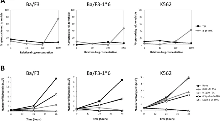
First, we ensured the absence ofa-Br-TMC-mediated cytotox- icity in the investigated time frame (up to 90 minutes treatment) in IL-3-stimulated Ba/F3 cells (Figure 2A). No cytotoxicity was
detected up to 10mM a-Br-TMC. However, strong cytotoxicity was evidenced at an a-Br-TMC concentration of 100mM (Figure 2A). Thusa-Br-TMC was further used at concentrations not above 10mM. We also investigated the long term effect ofa- Br-TMC treatment on cell proliferation and survival of IL-3- growing Ba/F3 cells (Figure 2B). Similarly to TSA, a-Br-TMC impaired cell growth and viability in a dose-dependent manner.
Ba/F3 cells cultured in 5mM a-Br-TMC for 24 and 48 hours stopped dividing and died, while cells grown in 0.5mMa-Br-TMC showed limited cell death and reduced cell proliferation (Figure 2B and data not shown).
We then analyzed expression of a series of STAT5 target genes (Cis,Osm,c-Myc) as well as STAT5-independent genes (JunB,Ho-1, 36b4) in dose-response experiments using 0.4 to 10mM a-Br- TMC. Rested Ba/F3 cells were incubated 30 minutes with the respective compounds and stimulated with IL-3 for 30 minutes (Figure 1C). Induction of all three STAT5 target genes was inhibited bya-Br-TMC in a dose-dependent manner. In addition, the IL-3-inducible STAT5-independent gene JunB, while unaf- fected by TSA as expected [46], was also inhibited bya-Br-TMC in a dose-dependent manner (Figure 1C). It is thought thatJunBis induced through the JAK2/MAPK pathway [68,69], therefore suggesting thata-Br-TMC is targeting the upstream JAK/STAT pathway rather than STAT5 itself.a-Br-TMC was shown to up- regulate the Nrf2-dependent anti-inflammatory HO-1 protein levels in the mouse macrophage cell line RAW264.7 [59]. In Ba/
F3 cells however, althoughHo-1gene expression was slightly up- regulated by IL-3 and TSA, no dose-dependent effect of a-Br- TMC was observed (Figure 1C), suggesting that the Nrf2-
dependent anti-inflammatory response pathway is - if at all - only modestly activated and not influenced bya-Br-TMC. In contrast to IL-3-regulated genes, expression of the control gene 36b4 remained unaffected bya-Br-TMC treatment (Figure 1C), further supporting specificity of action.
Together, these gene expression data suggest that a-Br-TMC might target the JAK/STAT pathway for inhibition. We therefore monitored the effect ofa-Br-TMC on STAT5 and JAK2 protein phosphorylation.
a-Br-TMC inhibits both JAK2 and STAT5 phosphorylation STAT5 proteins are expressed in two forms, STAT5A and STAT5B, with both redundant and unique functions and encoded by two related genes. STAT5A and STAT5B exhibit 91% identity in their amino acid sequence, the C-terminal transactivation domain being most divergent [3]. STAT5 phosphorylation in IL- 3-stimulated Ba/F3 cells pre-treated with either 0.2mM TSA or 0.4 to 10mMa-Br-TMC was investigated by Western-blot using a phospho-STAT5-specific antibody (pSTAT5) (Figure 3A). IL-3- induced STAT5 phosphorylation was not affected by TSA, in agreement with our previous report [46]. By contrast, IL-3- induced STAT5 phosphorylation was decreased bya-Br-TMC in a dose-dependent manner. Interestingly, both STAT5A and STAT5B proteins showed a dose-dependent downward mobility shift in SDS-PAGE, with STAT5B being more affected than STAT5A (Figure 3A). This mobility shift suggests thata-Br-TMC induces a protein modification targeting both STAT5 proteins.
Analysis of cytosolic and nuclear fractions of unstimulated and stimulateda-Br-TMC-treated cells, revealed that thisa-Br-TMC-
Figure 2. Effect ofa-Br-TMC on cytotoxicity and viability of normal (Ba/F3) and transformed (Ba/F3-1*6, K562) cells.(A) Cells were pre-treated 30 minutes with 0.001, 0.01, 0.1 and 1mM TSA or with 0.1, 1, 10 and 100mMa-Br-TMC before starting the WST-1 assay. IL-3 (5 ng/mL) was added to rested Ba/F3 cells at the same time as the WST-1 reagent to mimic the IL-3 stimulation conditions used in other assays. DMSO (vehicle) concentration was adjusted to 0.1% final in all conditions. OD measurement was performed after 90 minutes incubation with the WST-1 reagent, and the percentage of cytotoxicity was normalized to the vehicle control. (B) Growing Ba/F3, Ba/F3-1*6 and K562 cells were incubated for 24 and 48 hours in the presence of the indicated concentrations of TSA anda-Br-TMC. Cell viability was measured by Trypan Blue exclusion assay.
doi:10.1371/journal.pone.0090275.g002
mediated protein modification is taking place in the cytoplasm of unstimulated cells (Figure 3B) and hence is IL-3-independent.
Analysis of cytosolic and nuclear fractions ofa-Br-TMC-treated cells also confirmed that STAT5 phosphorylation and nuclear translocation are reduced (Figure 3B), thus supporting the idea that STAT5 activation is impaired upona-Br-TMC treatment. Of note, the detection of shifted bands by the pSTAT5 antibody in the cytosolic and nuclear fractions of a-Br-TMC-treated IL-3- stimulated cells, suggests that the modified STAT5 protein can be to some extent phosphorylated at the expected tyrosine residue (Y694/699 in STAT5A/B respectively) and translocated into the nucleus.
STAT5 activation by IL-3 relies on the initial activation of the cellular tyrosine kinase JAK2 [70]. Our gene expression data revealing an inhibitory effect ofa-Br-TMC on expression of the JAK2/MAPK-dependentJunBgene (Figure 1C) suggest thata-Br- TMC might also target the upstream JAK2 tyrosine kinase. We therefore investigated whether IL-3-induced phosphorylation of JAK2 was inhibited by a-Br-TMC. Ba/F3 cells were treated as before with 10mMa-Br-TMC and briefly stimulated with IL-3 to analyze JAK2 phosphorylation by Western-blot using a pJAK2- specific antibody (Figure 3C). Following a-Br-TMC treatment, JAK2 phosphorylation was strongly diminished. By contrast to STAT5A and STAT5B proteins, JAK2 protein mobility was not affected bya-Br-TMC (Figure 3C).
Altogether, these experiments demonstrated that the a-Br- chalcone a-Br-TMC has the ability to inhibit the JAK/STAT pathway at multiple levels, by targeting both JAK2 and STAT5 proteins in IL-3-stimulated Ba/F3 cells.
a-Br-TMC inhibits STAT5-mediated transcriptional activity The effect ofa-Br-TMC on STAT5 transcriptional activity was further investigated by chromatin immunoprecipitation (ChIP).
We previously showed that the deacetylase inhibitor TSA inhibits STAT5-mediated transcription at a step subsequent to STAT5 binding to its target genes by preventing recruitment of the
transcriptional machinery [46]. STAT5 and RNA polymerase II ChIP assays were performed on TSA- anda-Br-TMC-treated Ba/
F3 cells and protein recruitment to the mouse Cis and Osm promoters was examined (Figure 4). In agreement with our former report [46], RNA polymerase II recruitment to theCisand Osm promoters was abrogated upon TSA treatment, without notably affecting STAT5 DNA binding activity. Although the level of nuclear pSTAT5 was strongly reduced ina-Br-TMC-treated cells (Figure 3B), the proportion of STAT5 bound to theCisandOsm promoters was only moderately affected (20% and 58% reduced respectively; Figure 4A and B). This indicates that the nuclear pool of STAT5 proteins ina-Br-TMC-treated Ba/F3 cells might still support efficient binding to target sites. On the other hand, in correlation with the reduced Cis and Osm mRNA levels (Figure 1), recruitment of the RNA polymerase II to the Cis and Osm transcription start sites was markedly impaired (55% and 94%
respectively; Figure 4A and B), pointing to an a-Br-TMC- mediated transcriptional inhibition of the Cis and OsmSTAT5 target genes in Ba/F3 cells.
a-Br-TMC differentially affects JAK/STAT signaling in cells transformed by constitutively active STAT5
We showed that a-Br-TMC can inhibit IL-3-mediated JAK2 and STAT5 activation in normal mouse Ba/F3 cells. We next addressed whether a-Br-TMC can also hinder JAK/STAT activity in cancer cells exhibiting constitutive activation of STAT5.
For this purpose, we compared gene expression patterns of normal Ba/F3 cells to those of transformed Ba/F3-1*6 and K562 cells.
Ba/F3-1*6 cells stably express a mutant form of mouse STAT5A (so-called 1*6) with two amino acids substitutions within its N-terminal and C-terminal transactivation domains respective- ly. These point mutations result in constitutive STAT5 phosphor- ylation, nuclear localization and transactivation properties [64].
Expression of constitutively active STAT5A-1*6 is sufficient to confer IL-3-independent growth to Ba/F3 cells in vitro and tumorigenicity to bone marrow cellsin vivo[64,71]. Importantly,
Figure 3. a-Br-TMC inhibits both JAK2 and STAT5 phosphorylation. Ba/F3 cells were pre-treated 30 minutes with the indicated concentrations of TSA anda-Br-TMC (A) or with 10mMa-Br-TMC and DMSO vehicle (B, C) and further stimulated with 5 ng/mL IL-3 for 30 minutes (A), 15 minutes (B) or 5 minutes (C). Final DMSO concentration was 0.02% in (A) and 0.01% in (B, C). Protein whole cell extracts (panels A, C) or cytosolic (C) and nuclear (N) extracts (panel B) were prepared as described in Materials and Methods and analyzed by Western-blot using antibodies specific for phospho-STAT5 (pSTAT5), phospho-JAK2 (pJAK2), STAT5A, STAT5B, STAT5A and B, JAK2 anda-tubulin (loading control and cytosolic- specific marker). SDS-PAGE in (C) was shorter, explaining why the STAT5 mobility patterns (a-Br-TMC-induced shift and STAT5A and B doublet) are not as apparent as on immunoblots in (A, B).
doi:10.1371/journal.pone.0090275.g003
JAK2 is not activated in Ba/F3-1*6 cells as a consequence of IL-3- independent growth [4,64]. The mechanism of constitutive activation of STAT5A-1*6 is unclear. Whether basal JAK2 activity or an unidentified tyrosine kinase is responsible for STAT5A-1*6 phosphorylation is unknown [4,64]. Increased stability of the phosphorylated STAT5A-1*6 mutant via an uncharacterized mechanism also contributes to its constitutive activity [64]. The human K562 leukemia cell line expresses a constitutively active BCR-ABL tyrosine kinase. BCR-ABL onco- genic fusion protein constitutively phosphorylates STAT5A and STAT5B proteins, directly contributing to oncogenesis [72–74].
Ba/F3, Ba/F3-1*6 and K562 cells were treated with 0.2mM TSA or 10mM a-Br-TMC for 90 minutes, with Ba/F3 being stimulated 60 minutes with IL-3 following 30 minute inhibitor pre- treatment, as before. In addition, K562 cells were treated with 1mM of the BCR-ABL specific inhibitor Imatinib, as a positive control for inhibition of STAT5 phosphorylation [75]. Expression of STAT5 target genes (Cis,Osm,c-Myc, Pim-1), of JAK2/MAPK-
regulated STAT5-independent genes (JunB, c-Fos), and of a housekeeping control gene (36b4) was evaluated by quantitative RT-PCR (Figure 5). As predicted upon expression of constitutively active STAT5-1*6 [46], expression of the STAT5 target genes was up-regulated in growing Ba/F3-1*6 cells in comparison to unstimulated Ba/F3 cells (Figure 5A–B). As anticipated, expres- sion of the STAT5 target genes in all three cell lines was inhibited by TSA (Figure 5A–C). Similarly, STAT5 target genes were down-regulated in Imatinib-treated K562 cells (Figure 5C).
Expression of all STAT5-target genes investigated in IL-3- stimulated Ba/F3 cells, including Cis, Osm, c-Myc, Pim-1 (Figure 5A) as well asSpi2.1andSOCS-1(not shown) was inhibited by a-Br-TMC. While expression of STAT5 target genes was down-regulated in a-Br-TMC-treated Ba/F3 cells (Figure 5A), distinct effects ofa-Br-TMC were noted in Ba/F3-1*6 and K562 cells (Figure 5B–C). For instance, expression ofCisand Osmwas up-regulated in a-Br-TMC-treated Ba/F3-1*6 (Figure 5B) and K562 (Figure 5C) cells. c-Myc and Pim-1 mRNA levels were Figure 4. RNA polymerase II recruitment to theCisandOsmpromoters is impeded in Ba/F3 cells treated witha-Br-TMC.Ba/F3 cells were pre-treated 30 minutes with 0.2mM TSA or 10mMa-Br-TMC before being stimulated with 5 ng/mL IL-3 for 30 minutes. DMSO (vehicle) was adjusted to 0.02% in all conditions. Chromatin immunoprecipitation (ChIP) was performed as described in Materials and Methods using antibodies directed against STAT5 or RNA polymerase II (RNA Pol II) proteins. Co-precipitated genomic DNA was analyzed by quantitative PCR using primers specific for the STAT5 binding sites (STAT5 ChIP) or the transcription start site (RNA Pol II ChIP) of the mouseCis(A) andOsm(B) genes.
doi:10.1371/journal.pone.0090275.g004
partially reduced in a-Br-TMC-treated Ba/F3-1*6 cells (Figure 5B), while c-Myc mRNA level was slightly increased in a-Br-TMC-treated K562 cells (Figure 5C). Therefore, thea-Br- 29,3,4,49-tetramethoxychalcone exerts distinct effects on STAT5 target gene expression in normal Ba/F3 vs. its transformed counterpart Ba/F3-1*6, as well as in the oncogenic K562 cell line.
Both JAK2-mediated MAPK-dependent genesJunBand c-Fos were induced by IL-3 in Ba/F3 cells (Figure 5A). Their expression was either unaffected or upregulated by TSA respectively, as previously reported [46]. Expression of JunB and c-Fos was inhibited by a-Br-TMC, probably as a consequence of JAK2 inhibition (Figure 5A). In contrast to Ba/F3 cells,JunBand c-Fos genes were expressed at background levels in Ba/F3-1*6 cells (Figure 5B), most likely as a result of the absence of JAK2/MAPK activation [4]. Treatment of Ba/F3-1*6 cells witha-Br-TMC did not affect JunB expression while c-Fos was slightly up-regulated (Figure 5B), although mRNA levels remained low in comparison to that detected in IL-3-stimulated Ba/F3 cells (Figure 5A). The observation thata-Br-TMC was able to up-regulate the expression of c-Fos - and of other genes - in Ba/F3 cells transformed with STAT5A-1*6 but not in Ba/F3 parental cells, suggests thata-Br- TMC might target other factor(s) involved in gene regulation in Ba/F3-1*6 cells.
To further evaluate the effect of a-Br-TMC on normal and transformed cells, cytotoxicity and cell viability assays were performed as before (Figure 2A–B). The effect ofa-Br-TMC on proliferation and survival of Ba/F3-1*6 and K562 cells was comparable to that observed with normal Ba/F3 cells. All cells died in the presence of 5mM a-Br-TMC, while reduced proliferation and minimal cell death were observed in the presence of 0.5mM a-Br-TMC over 48 hours (Figure 2B and data not shown). On the other hand, a-Br-TMC-induced cytotoxicity (at 100mM) was reduced by approximately 50% in the transformed Ba/F3-1*6 and K562 cell lines in comparison to normal Ba/F3 cells (Figure 2A; 75% cytotoxicity in Ba/F3 cells vs. 47% and 42%
in Ba/F3-1*6 and K562 cells respectively). This indicates a distinct chemosensitivity of the cancer cell lines toa-Br-TMC. Together with the observation that a-Br-TMC differentially affects gene expression in the transformed cells, our results suggest that its activity is regulated in a distinct manner in the investigated normal vs. transformed cell lines.
In conclusion, our gene expression analysis demonstrated that a-Br-TMC differentially regulates expression of STAT5 target genes in normal and cancer cells. Since we showed that a-Br- TMC hinders STAT5 transcriptional activity in Ba/F3 cells by inhibiting both STAT5 and JAK2 phosphorylation and possibly by modifying STAT5 proteins, we examined the effect of a-Br- TMC on JAK2 and STAT5 proteins in the transformed Ba/F3- 1*6 and K562 cells.
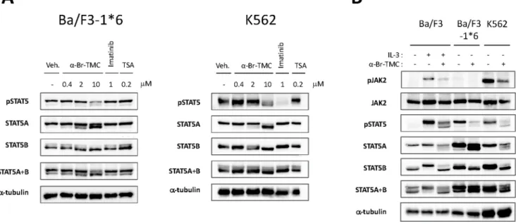
a-Br-TMC-induced STAT5 protein modification differentially modulates STAT5 activity in normal and cancer cells
STAT5 phosphorylation in Ba/F3-1*6 and K562 cells was assessed by Western-blot analysis as before, on cells treated with 0.4 to 10mMa-Br-TMC or 0.2mM TSA. Both cell lines were also treated with 1mM Imatinib, as a positive control for pSTAT5 inhibition in K562 cells and a negative control for Ba/F3-1*6 cells (Figure 6A). As expected, TSA did not affect STAT5 phosphor- ylation in Ba/F3-1*6 and K562 cells. Similarly, the BCR-ABL inhibitor Imatinib did not affect STAT5 phosphorylation in Ba/
F3-1*6 cells, while drastically inhibiting STAT5 phosphorylation in K562 cells, as previously reported [75]. As observed in Ba/F3 cells, treatment of Ba/F3-1*6 and K562 cells witha-Br-TMC led
to a decrease in STAT5 phosphorylation and a downward mobility shift of both STAT5A and STAT5B proteins, in a dose- dependent manner (Figure 6A and 6B). Again, STAT5B appeared to be more affected than STAT5A.
We next assessed the effect ofa-Br-TMC on JAK2 phosphor- ylation in Ba/F3-1*6 and K562 cells (Figure 6B). In agreement with previous reports [4,64], pJAK2 was barely detectable in Ba/
F3-1*6 cells. The absence of JAK2 phosphorylation in Ba/F3-1*6 cells is also in agreement with our gene expression data, revealing only basal expression levels of the JAK2/MAPK-dependent genes c-FosandJunB(Figure 5B). The observation that STAT5 proteins undergo a-Br-TMC-mediated dephosphorylation and mobility shift in the absence of JAK2 supports the idea that these modifications occur in a JAK2-independent manner. Finally, like in Ba/F3 cells, JAK2 phosphorylation in K562 cells was inhibited upona-Br-TMC treatment (Figure 6B), further indicating thata- Br-TMC targets both JAK2 and STAT5 proteins.
Discussion
In the search for novel STAT5 inhibitors, we examined the effect of a series of natural and synthetic chalcones as well as of curcumin, another naturala,b-unsaturated carbonyl compound, on JAK2/STAT5 activation and expression of downstream STAT5 target genes in normal (Ba/F3) and caSTAT5-trans- formed (Ba/F3-1*6, K562) cell lines.
We show here that the synthetic a-Br-29,3,4,49-tetramethox- ychalcone (a-Br-TMC) is a potent inhibitor of both JAK2 tyrosine kinase and STAT5 transcription factor activities. None of the natural products investigated (curcumin and five natural chal- cones) affected STAT5-mediated transactivation of theCisgene in IL-3-stimulated Ba/F3 cells at a concentration of 20mM. Amongst the four synthetic chalconesa-X-TMCs (with X = CN, Br, Cl, H) tested, a-Br-TMC was the only active STAT5 inhibitor. We showed before thata-Br-TMC,a-Cl-TMC and to a lesser extent a-H-TMC exhibit anti-inflammatory activities in RAW364.7 cells, in particular inhibiting both Nrf2- and NF-kB-dependent path- ways [59]. We could not detect an effect ofa-Br-TMC on the expression of the Nrf2-dependent Ho-1 gene in Ba/F3 cells, possibly due to the short duration of treatment. Nevertheless, taken together, our data indicate thata-Br-TMC is able to target multiple pathways controlling inflammation, proliferation and survival. The natural compound sophoraflavanone G was recently reported to exhibit a similar pleiotropic activity against both the JAK/STAT signaling pathway and pathways controlling inflam- mation and infection in murine and human cell lines [36]. Since interconversion between flavanones and 29-OH-chalcones was recently evidenced in cell culture [76], it might explain why these two classes of molecules exhibit similar biological activities.
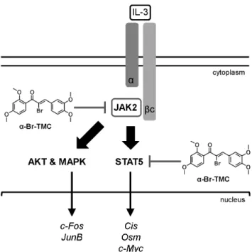
In accordance with the pleiotropic activity of a-Br-TMC, we found that it targets the JAK2/STAT5 pathway at multiple levels (Figure 7). First, a-Br-TMC inhibits JAK2 tyrosine kinase activation, thus indirectly inhibiting activation of pathways downstream of JAK2, in particular STAT5 and mitogen-activated protein kinase (MAPK). Second,a-Br-TMC inhibits constitutive STAT5 phosphorylation in cells showing basal JAK2 activation (Ba/F3-1*6). Although we cannot rule out that basal JAK2 activation is responsible for the constitutive STAT5 phosphory- lation in Ba/F3-1*6 cells [4], our data suggest thata-Br-TMC also inhibits STAT5 phosphorylation - directly or indirectly - in a JAK2-independent manner. Third,a-Br-TMC might alter a post- translational modification on STAT5 proteins, as revealed by a downward mobility shift in SDS-PAGE. a-Br-TMC treatment changed the mobility of both STAT5A and STAT5B proteins in
SDS-PAGE, although STAT5B appears more strongly affected than STAT5A. Moreover, this effect takes place within the cytoplasm and independently of IL-3 stimulation and STAT5 phosphorylation. In fact, the faster-migrating STAT5 forms could still be phosphorylated and translocated into the nucleus to some extent. Since this protein modification correlates with a reduced phosphorylation of STAT5A-1*6, possibly independent of JAK2, it is tempting to speculate that a-Br-TMC-induced STAT5 modification interferes - directly or indirectly - with STAT5 activation and hence STAT5 transcriptional activity.
As a result of reduced JAK2 and STAT5 activation, transcrip- tion of STAT5 target genes such asCisandOsmwas impaired, in agreement with a diminished recruitment of RNA polymerase II to their respective transcription start site. Somewhat surprisingly, STAT5 binding to DNA was marginally diminished upon a-Br- TMC treatment. It suggests that the modified nuclear STAT5 proteins still have the ability to bind to DNA and hence might compete with transcriptionally competent unmodified STAT5 proteins.
We found that although STAT5 proteins were similarly modified by a-Br-TMC in normal and transformed cells based on their mobility profile in SDS-PAGE, the consequence on expression of downstream target genes was different. Indeed, while expression ofCisand Osmwas down-regulated in Ba/F3 cells, it was up-regulated in Ba/F3-1*6 and K562 cells. Meanwhile, expression of thec-Myconcogene was reduced in both Ba/F3 and
Ba/F3-1*6 cells but slightly increased in K562 cells. According to the differential effect ofa-Br-TMC on gene expression, the three cell lines exhibited a distinct chemosensitivity toa-Br-TMC. These observations suggest that a-Br-TMC might target additional signaling and/or transcription factors in cells transformed by constitutively active STAT5 (mouse Ba/F3-1*6 and human K562), resulting in distinct gene expression patterns. The observation that basal expression of c-Fos in Ba/F3-1*6 cells in the absence of Jak2 activation was slightly increased upona-Br- TMC treatment would support this hypothesis. On the other hand, we cannot exclude thata-Br-TMC-mediated modification of STAT5 protein differentially affects STAT5 transcriptional activity in normal and cancer cells. Given that STAT5 DNA binding activity was only weakly impaired ina-Br-TMC-treated cells, one could indeed envisage that DNA-bound STAT5 protein complexes exhibit distinct transcriptional activities depending on the cellular context. For instance, STAT5 is known to regulate transcription through interactions with positive and negative cofactors and with other transcription factors bound on adjacent sites, as well as through tetramerization [1,9–11], which could be differentially regulated in transformed cells. Clearly, additional experiments are needed to determine whether STAT5 is also bound to the Cis promoter in Ba/F3-1*6 and K562 cells and further unravel the underlying molecular mechanism.
The question remains as to how a-Br-TMC inhibits JAK2 activation. We cannot rule out at this point thata-Br-TMC acts as Figure 5.a-Br-TMC exerts distinct effects in normal and cancer cells.Ba/F3 (A), its caSTAT5-transformed counterpart Ba/F3-1*6 (B) and human leukemic K562 (C) cells were treated 90 minutes with 0.2mM TSA, 10mMa-Br-TMC or 1mM Imatinib. Ba/F3 cells (A) were stimulated with 5 ng/mL IL-3 after an initial 30 minute drug pre-treatment (hence subjected to a 60 minute IL-3 stimulation). DMSO (vehicle) final concentration was adjusted to 0.02% in all conditions. Expression of STAT5-dependent (Cis,Osm,c-Myc, Pim-1) and -independent (JunB,c-Fos,36b4) genes was analyzed by quantitative RT-PCR. Gene expression data were normalized to mouse ribosomalS9(A, B) or to human Lamin A/C (LMNA) (C) housekeeping gene- encoded mRNAs. (A, B) Normalized data are presented with adjusted Y-axis scale for a direct comparison of mRNA levels in the respective normal and transformed Ba/F3 and Ba/F3-1*6 cell lines.
doi:10.1371/journal.pone.0090275.g005
Figure 6.a-Br-TMC inhibits the STAT5 signaling pathway in both a JAK2-dependent and -independent manner.(A) Ba/F3-1*6 and K562 cells were treated for 60 minutes with the indicated compounds. Protein whole cell extracts were analyzed by Western-Blot using antibodies specific for pSTAT5, STAT5A, STAT5B, STAT5A and B anda-tubulin as a loading control. (B) Ba/F3 cells were pre-treated 30 minutes with 10mMa-Br- TMC and stimulated with 5 ng/mL IL-3 for 5 minutes. Ba/F3-1*6 and K562 cells were treated with 10mMa-Br-TMC for 90 minutes. DMSO (Veh.) was adjusted to 0.01% in all conditions. Protein whole cell extracts were analyzed by Western-Blot using antibodies specific for pSTAT5, pJAK2, STAT5A, STAT5B, STAT5A and B, JAK2 anda-tubulin (loading control).
doi:10.1371/journal.pone.0090275.g006
a tyrosine kinase inhibitor. Indeed, its structure is reminiscent of that of AG490, a reversible substrate competitive tyrosine kinase inhibitor [77,78]. In addition, the nature of the protein modification(s) targeting STAT5 upon a-Br-TMC treatment remains to be identified. Several lines of evidences support the idea that the divergent C-terminal transactivation domain of STAT5 is targeted for modification, including the observations that (i) a-Br-TMC inhibited STAT5 phosphorylation at Y694/
699, (ii)a-Br-TMC-induced mobility shift of STAT5A was not as pronounced as that of STAT5B, (iii) a-Br-TMC differentially modulated STAT5 transcriptional activity in normal and trans- formed cells. On the other hand, the observation that STAT5 DNA binding activity was only slightly impaired, raises the possibility that the STAT5 DNA binding domain might be modified as well.
Chalcones in general, anda-Br-TMC in particular, present a highly electrophilic a,b-unsaturated carbonyl moiety that can potentially react with free sulfhydryl groups of cysteine residues in proteins and directly alter their function. On the other hand, electrophiles can inhibit the activation of transcription factors such as NF-kB and STAT3, possibly through S-glutathionylation of cysteine thiol groups, which is itself triggered by a rapid drop of intracellular glutathione [55,79]. Three naturally occuring ter- penes, which also contain an electrophilic a,b-unsaturated carbonyl group, induce S-glutathionylation of STAT3 thereby inhibiting its activity [80,81]. Similarly to our a-Br-TMC
compound, these molecules also inhibit the activation of JAK2 tyrosine kinase [80]. In agreement with the identification of several essential cysteine residues within JAK2 catalytic domain [82], activity of JAK2 - but also of STAT5 - is regulated by the synthetic glutathione disulfide mimetic NOV-002 [83], suggesting that both JAK2 and STAT5 can be S-glutathionylated. Finally, a review of the literature revealed that S-glutathionylation and redox regula- tion can either stimulate or inhibit protein activity [82,83], possibly depending on the position of the modified cysteine within the functional domains of the targeted protein. Altogether, these observations raise the possibility that a-Br-TMC, as a potent Michael acceptor, might alter the function of STAT5 and JAK2 proteins, through either direct alkylation or indirect S-glutathio- nylation of accessible thiol groups, thereby inhibiting or stimulat- ing their activity depending on the cellular context. The presence of cysteine residues in STAT5 proteins, both common and unique to STAT5A and STAT5B within their DNA binding and transactivation domains respectively, is consistent with that model.
On the other hand, the detection of a downward mobility shift of STAT5A and STAT5B upona-Br-TMC treatment is not in full agreement with the covalent addition of one or several S- glutathione groups. Such downward shift is rather reminiscent of protein dephosphorylation and one could envision an alternative mode of action fora-Br-TMC. Beside tyrosine phosphorylation, STAT5 proteins can be phosphorylated on serine residues [84].
Interestingly, STAT5A and STAT5B are differentially phosphor- ylated on C-terminal serines (S725/730 in mSTAT5A/B; S779 in mSTAT5A) [84], resulting in either positive or negative transcriptional regulation depending on promoter and/or cellular context [85,86]. It remains therefore possible that a-Br-TMC either inhibits the still-unknown kinase responsible for serine phosphorylation of STAT5 or activates a specific serine phospha- tase, leading to the STAT5 mobility shift detected in SDS-PAGE and possibly to the alteration of its activity.
Whether a-Br-TMC acts as a tyrosine kinase inhibitor and whether thea-Br-TMC-induced STAT5 mobility shift involves S- glutathionylation and/or dephosphorylation of STAT5 and/or of other regulatory components of the STAT5 pathway is currently under investigation.
In conclusion, we identified the synthetic chalconea-Br-TMC as a novel regulator of JAK2/STAT5 activity, targeting both STAT5 and its upstream activating kinase JAK2 (Figure 7). Its ability to down-regulate expression of thec-Myc oncogene while up-regulating expression of the tumor suppressor gene Cis in cancer cells, together with its previously described anti-inflamma- tory properties [59], potentially makes a-Br-TMC a promising novel therapeutic agent with pleiotropic anti-inflammatory and anticancer activities.
Acknowledgments
We thank Jacqueline Marvel and Daniela Ma¨nnel for providing the Ba/F3 and K562 cells respectively. We thank Belinda Jobst for her technical contribution to this project while completing an internship in our group.
We are grateful to Joachim Griesenbeck for critically reading the manuscript and Fiona Kalinowski for kindly proof-reading the text.
Author Contributions
Conceived and designed the experiments: AR SA. Performed the experiments: AR SP SU SB EB SA NA HP. Analyzed the data: AR SA SP. Contributed reagents/materials/analysis tools: SA NA. Wrote the paper: AR.
Figure 7. Model of inhibition of the JAK2/STAT5 pathway bya- Br-TMC.IL-3 binding to thea/bc chains of the IL-3 receptor leads to activation of the receptor-associated JAK2 tyrosine kinase by trans- phosphorylation. In turn, JAK2-mediated activation of the STAT5, MAPK and AKT pathways via phosphorylation (broad arrows) results in induced transcription of downstream target genes (thin arrows). We showed thata-Br-TMC inhibits JAK2 phosphorylation, hence impairing JAK2-regulated signaling pathways. In addition and independently of JAK2, a-Br-TMC inhibits STAT5 activity. Concomitantly, STAT5A and STAT5B protein mobility in SDS-PAGE is altered, indicating a change in their post-translational modification state induced bya-Br-TMC, which might be associated to STAT5 altered transcriptional activity.
doi:10.1371/journal.pone.0090275.g007
References
1. Grimley PM, Dong F, Rui H (1999) Stat5a and Stat5b: fraternal twins of signal transduction and transcriptional activation. Cytokine Growth Factor Rev 10:
131–157.
2. Wakao H, Gouilleux F, Groner B (1994) Mammary gland factor (MGF) is a novel member of the cytokine regulated transcription factor gene family and confers the prolactin response. EMBO J 13: 2182–2191.
3. Basham B, Sathe M, Grein J, McClanahan T, D’Andrea A, et al. (2008) In vivo identification of novel STAT5 target genes. Nucleic Acids Res 36: 3802–3818.
4. Nosaka T, Kawashima T, Misawa K, Ikuta K, Mui AL, et al. (1999) STAT5 as a molecular regulator of proliferation, differentiation and apoptosis in hemato- poietic cells. EMBO J 18: 4754–4765.
5. Mui AL, Wakao H, Kinoshita T, Kitamura T, Miyajima A (1996) Suppression of interleukin-3-induced gene expression by a C-terminal truncated Stat5: role of Stat5 in proliferation. EMBO J 15: 2425–2433.
6. Van Nguyen T, Angkasekwinai P, Dou H, Lin F-M, Lu L-S, et al. (2012) SUMO-specific protease 1 is critical for early lymphoid development through regulation of STAT5 activation. Mol Cell 45: 210–221.
7. Wieczorek M, Ginter T, Brand P, Heinzel T, Kra¨mer OH (2012) Acetylation modulates the STAT signaling code. Cytokine Growth Factor Rev 23: 293–305.
8. Ma L, Gao J, Guan Y, Shi X, Zhang H, et al. (2010) Acetylation modulates prolactin receptor dimerization. Proc Natl Acad Sci U S A 107: 19314–19319.
9. Lin JX, Leonard WJ (2000) The role of Stat5a and Stat5b in signaling by IL-2 family cytokines. Oncogene 19: 2566–2576.
10. Shuai K (2000) Modulation of STAT signaling by STAT-interacting proteins.
Oncogene 19: 2638–2644.
11. Lin J-X, Li P, Liu D, Jin HT, He J, et al. (2012) Critical Role of STAT5 transcription factor tetramerization for cytokine responses and normal immune function. Immunity 36: 586–599.
12. Mandal M, Powers SE, Maienschein-Cline M, Bartom ET, Hamel KM, et al.
(2011) Epigenetic repression of the Igk locus by STAT5-mediated recruitment of the histone methyltransferase Ezh2. Nat Immunol 12: 1212–1220.
13. Litterst CM, Kliem S, Marilley D, Pfitzner E (2003) NCoA-1/SRC-1 is an essential coactivator of STAT5 that binds to the FDL motif in the alpha-helical region of the STAT5 transactivation domain. J Biol Chem 278: 45340–45351.
14. Pfitzner E, Ja¨hne R, Wissler M, Stoecklin E, Groner B (1998) p300/CREB- binding protein enhances the prolactin-mediated transcriptional induction through direct interaction with the transactivation domain of Stat5, but does not participate in the Stat5-mediated suppression of the glucocorticoid response.
Mol Endocrinol Baltim Md 12: 1582–1593.
15. John S, Vinkemeier U, Soldaini E, Darnell JE Jr, Leonard WJ (1999) The significance of tetramerization in promoter recruitment by Stat5. Mol Cell Biol 19: 1910–1918.
16. Ward AC, Touw I, Yoshimura A (2000) The Jak-Stat pathway in normal and perturbed hematopoiesis. Blood 95: 19–29.
17. Valentino L, Pierre J (2006) JAK/STAT signal transduction: regulators and implication in hematological malignancies. Biochem Pharmacol 71: 713–721.
18. Bowman T, Garcia R, Turkson J, Jove R (2000) STATs in oncogenesis.
Oncogene 19: 2474–2488.
19. Liu CB, Itoh T, Arai K, Watanabe S (1999) Constitutive activation of JAK2 confers murine interleukin-3-independent survival and proliferation of BA/F3 cells. J Biol Chem 274: 6342–6349.
20. Lord JD, McIntosh BC, Greenberg PD, Nelson BH (2000) The IL-2 receptor promotes lymphocyte proliferation and induction of the c-myc, bcl-2, and bcl-x genes through the trans-activation domain of Stat5. J Immunol Baltim Md 1950 164: 2533–2541.
21. Nosaka T, Kitamura T (2002) Pim-1 expression is sufficient to induce cytokine independence in murine hematopoietic cells, but is dispensable for BCR-ABL- mediated transformation. Exp Hematol 30: 697–702.
22. Garc¸on L, Rivat C, James C, Lacout C, Camara-Clayette V, et al. (2006) Constitutive activation of STAT5 and Bcl-xL overexpression can induce endogenous erythroid colony formation in human primary cells. Blood 108:
1551–1554.
23. Gesbert F, Griffin JD (2000) Bcr/Abl activates transcription of the Bcl-X gene through STAT5. Blood 96: 2269–2276.
24. Chim C-S, Fung T-K, Cheung W-C, Liang R, Kwong Y-L (2004) SOCS1 and SHP1 hypermethylation in multiple myeloma: implications for epigenetic activation of the Jak/STAT pathway. Blood 103: 4630–4635.
25. He B, You L, Uematsu K, Zang K, Xu Z, et al. (2003) SOCS-3 is frequently silenced by hypermethylation and suppresses cell growth in human lung cancer.
Proc Natl Acad Sci U S A 100: 14133–14138.
26. Niwa Y, Kanda H, Shikauchi Y, Saiura A, Matsubara K, et al. (2005) Methylation silencing of SOCS-3 promotes cell growth and migration by enhancing JAK/STAT and FAK signalings in human hepatocellular carcinoma.
Oncogene 24: 6406–6417.
27. Weber A, Hengge UR, Bardenheuer W, Tischoff I, Sommerer F, et al. (2005) SOCS-3 is frequently methylated in head and neck squamous cell carcinoma and its precursor lesions and causes growth inhibition. Oncogene 24: 6699–
6708.
28. Zhang Q, Wang HY, Marzec M, Raghunath PN, Nagasawa T, et al. (2005) STAT3- and DNA methyltransferase 1-mediated epigenetic silencing of SHP-1
tyrosine phosphatase tumor suppressor gene in malignant T lymphocytes. Proc Natl Acad Sci U S A 102: 6948–6953.
29. Nelson EA, Sharma SV, Settleman J, Frank DA (2011) A chemical biology approach to developing STAT inhibitors: molecular strategies for accelerating clinical translation. Oncotarget 2: 518–524.
30. Mu¨ller J, Sperl B, Reindl W, Kiessling A, Berg T (2008) Discovery of chromone- based inhibitors of the transcription factor STAT5. Chembiochem Eur J Chem Biol 9: 723–727.
31. Luo C, Laaja P (2004) Inhibitors of JAKs/STATs and the kinases: a possible new cluster of drugs. Drug Discov Today 9: 268–275.
32. Quinta´s-Cardama A, Verstovsek S (2013) Molecular pathways: Jak/STAT pathway: mutations, inhibitors, and resistance. Clin Cancer Res Off J Am Assoc Cancer Res 19: 1933–1940.
33. Warsch W, Walz C, Sexl V (2013) JAK of all trades: JAK2-STAT5 as novel therapeutic targets in BCR-ABL1+chronic myeloid leukemia. Blood 122: 2167–
2175.
34. Hayakawa F, Sugimoto K, Harada Y, Hashimoto N, Ohi N, et al. (2013) A novel STAT inhibitor, OPB-31121, has a significant antitumor effect on leukemia with STAT-addictive oncokinases. Blood Cancer J 3: e166.
35. Weber A, Borghouts C, Brendel C, Moriggl R, Delis N, et al. (2013) The inhibition of stat5 by a Peptide aptamer ligand specific for the DNA binding domain prevents target gene transactivation and the growth of breast and prostate tumor cells. Pharm Basel Switz 6: 960–987.
36. Kim B-H, Won C, Lee Y-H, Choi JS, Noh KH, et al. (2013) Sophoraflavanone G induces apoptosis of human cancer cells by targeting upstream signals of STATs. Biochem Pharmacol 86: 950–959.
37. Ma L, Clayton JR, Walgren RA, Zhao B, Evans RJ, et al. (2013) Discovery and characterization of LY2784544, a small-molecule tyrosine kinase inhibitor of JAK2V617F. Blood Cancer J 3: e109.
38. Nelson EA, Walker SR, Weisberg E, Bar-Natan M, Barrett R, et al. (2011) The STAT5 inhibitor pimozide decreases survival of chronic myelogenous leukemia cells resistant to kinase inhibitors. Blood 117: 3421–3429.
39. Nelson EA, Walker SR, Xiang M, Weisberg E, Bar-Natan M, et al. (2012) The STAT5 Inhibitor Pimozide Displays Efficacy in Models of Acute Myelogenous Leukemia Driven by FLT3 Mutations. Genes Cancer 3: 503–511.
40. Hanan EJ, van Abbema A, Barrett K, Blair WS, Blaney J, et al. (2012) Discovery of potent and selective pyrazolopyrimidine janus kinase 2 inhibitors. J Med Chem 55: 10090–10107.
41. Kraus M, Wang Y, Aleksandrowicz D, Bachman E, Szewczak AA, et al. (2012) Efficacious intermittent dosing of a novel JAK2 inhibitor in mouse models of polycythemia vera. PloS One 7: e37207.
42. Yang J, Ikezoe T, Nishioka C, Furihata M, Yokoyama A (2010) AZ960, a novel Jak2 inhibitor, induces growth arrest and apoptosis in adult T-cell leukemia cells.
Mol Cancer Ther 9: 3386–3395.
43. Ioannidis S, Lamb ML, Wang T, Almeida L, Block MH, et al. (2011) Discovery of 5-chloro-N2-[(1S)-1-(5-fluoropyrimidin-2-yl)ethyl]-N4-(5-methyl-1H-pyrazol- 3-yl)pyrimidine-2,4-diamine (AZD1480) as a novel inhibitor of the Jak/Stat pathway. J Med Chem 54: 262–276.
44. Jatiani SS, Cosenza SC, Reddy MVR, Ha JH, Baker SJ, et al. (2010) A Non- ATP-Competitive Dual Inhibitor of JAK2 and BCR-ABL Kinases: Elucidation of a Novel Therapeutic Spectrum Based on Substrate Competitive Inhibition.
Genes Cancer 1: 331–345.
45. Mu¨ller J, Schust J, Berg T (2008) A high-throughput assay for signal transducer and activator of transcription 5b based on fluorescence polarization. Anal Biochem 375: 249–254.
46. Rascle A, Johnston JA, Amati B (2003) Deacetylase activity is required for recruitment of the basal transcription machinery and transactivation by STAT5.
Mol Cell Biol 23: 4162–4173.
47. Rascle A, Lees E (2003) Chromatin acetylation and remodeling at the Cis promoter during STAT5-induced transcription. Nucleic Acids Res 31: 6882–
6890.
48. Gryder BE, Sodji QH, Oyelere AK (2012) Targeted cancer therapy: giving histone deacetylase inhibitors all they need to succeed. Future Med Chem 4:
505–524.
49. Evrot E, Ebel N, Romanet V, Roelli C, Andraos R, et al. (2013) JAK1/2 and Pan-deacetylase inhibitor combination therapy yields improved efficacy in preclinical mouse models of JAK2V617F-driven disease. Clin Cancer Res Off J Am Assoc Cancer Res 19: 6230–6241.
50. Wang Y, Fiskus W, Chong DG, Buckley KM, Natarajan K, et al. (2009) Cotreatment with panobinostat and JAK2 inhibitor TG101209 attenuates JAK2V617F levels and signaling and exerts synergistic cytotoxic effects against human myeloproliferative neoplastic cells. Blood 114: 5024–5033.
51. Guerini V, Barbui V, Spinelli O, Salvi A, Dellacasa C, et al. (2008) The histone deacetylase inhibitor ITF2357 selectively targets cells bearing mutated JAK2(V617F). Leukemia 22: 740–747.
52. Pietschmann K, Bolck HA, Buchwald M, Spielberg S, Polzer H, et al. (2012) Breakdown of the FLT3-ITD/STAT5 axis and synergistic apoptosis induction by the histone deacetylase inhibitor panobinostat and FLT3-specific inhibitors.
Mol Cancer Ther 11: 2373–2383.
53. Nguyen T, Dai Y, Attkisson E, Kramer L, Jordan N, et al. (2011) HDAC inhibitors potentiate the activity of the BCR/ABL kinase inhibitor KW-2449 in