Prevalence of polydactyly, syndactyly, amniotic band syndrome, cleft lip, cleft palate and talipes equinovarus in Bayelsa State, Nigeria
Prävalenz von Polydaktylie, Syndaktylie, amniotischen Strängen,
Lippenspalten, Gaumenspalten und Klumpfüßen in Bayelsa State, Nigeria
Abstract
Studies on incidence of birth anomalies are abundant world wide, but literatures on general population prevalence of anomalies are scanty,
Charles A. Oyinbo
1Nervey W. Dare
1despite the fact that structural anomaly is the 5thleading cause of years
Ebidimie D. Amain
1of potential life lost prior to age 65 and a major contributor to disabilities.
The purpose of this study is to estimate the general population preva-
lence of polydactyly, syndactyly, amniotic band syndrome, cleft lip, cleft 1 Department of Human Anatomy, Faculty of Basic palate, and talipes equinovarus in Bayelsa State, Nigeria. Two thousand
(2000) subjects domicile in Bayelsa State were randomly selected for Medical Sciences, College of this study. Subjects were physically screened for musculoskeletal Health Sciences, Niger Delta University, Wilberforce Island, Bayelsa State, Nigeria anomalies. Individuals with genetic syndromes were excluded. Study
did not discriminate between types or sub- types of any anomaly. Results show that the overall population prevalence of musculoskeletal anom- alies is 13%; with a high proportion (67%) of minor anomalies. The general population prevalence of these anomalies is comparable with known birth prevalence world wide. Thus suggestive that a general population prevalence estimate of an anomaly could be a useful esti- mate of congenital anomaly in developing countries were record keeping are largely poor.
Keywords:structural anomalies, musculoskeletal anomalies, prevalence, epidemiology, Nigeria
Introduction
Structural anomalies are the 5thleading cause of years of potential life lost prior to age 65 and a major contribu- tor to disabilities [1]. Major structural malformations occur in 2 to 3% of live born infants and an additional 2 to 3%
are recognizable in children by age 5 years, giving a total of 4–6% [1]. Each year among 135 million new births world wide, more than 4 million each year or 11,376 daily of children born with structural anomaly [2]. Birth defects are the leading cause of infant mortality, they account for about 21% of infant deaths and may be classified as lethal, severe or mild [3]. Lethal and severe defects to- gether constitute major congenital abnormalities. Blacks have a higher overall rate of congenital abnormalities (CAs), than Caucasians, but the later have a higher rate of major and multiple malformations [4]. At present con- genital abnormality is the major cause of infant mortality and disabilities among children in the industrialized countries [2], and has been shown not to exhibit racial or ethnic predilection [1], [5]. About two-thirds of birth defects have no known cause, some malformations are
thought to be polygenic; others appear to be multi- factorial, resulting from abnormal genes interacting with harmful environmental factors [1]. Environmental factors account for about 15% of CAs and in combination with genetic factors account for 25% of CAs [6]. Environmental degradation from oil spillage and the irreversible ecological loses arising from activities associated with oil exploration has been a major cause of agitation by the inhabitants of Niger Delta region of Nigeria [7]. Custom- arily, incidence of birth anomaly is estimated by noting the number of times it occurred per thousand births (live and still). Emphasis has been on the new born and not on the pattern of distribution in the general population.
In contrast to birth incidence, the general population prevalence for most anomalies is largely unknown. It is not unnecessary to ignore a routine method of estimate of structural anomaly that emphasizes birth incidence for a general population base estimate that proffers an- swers to important epidemiological and demographic questions: in a specified period, how many individuals in a certain general population are affected with a particular structural anomaly and how has this change over time?
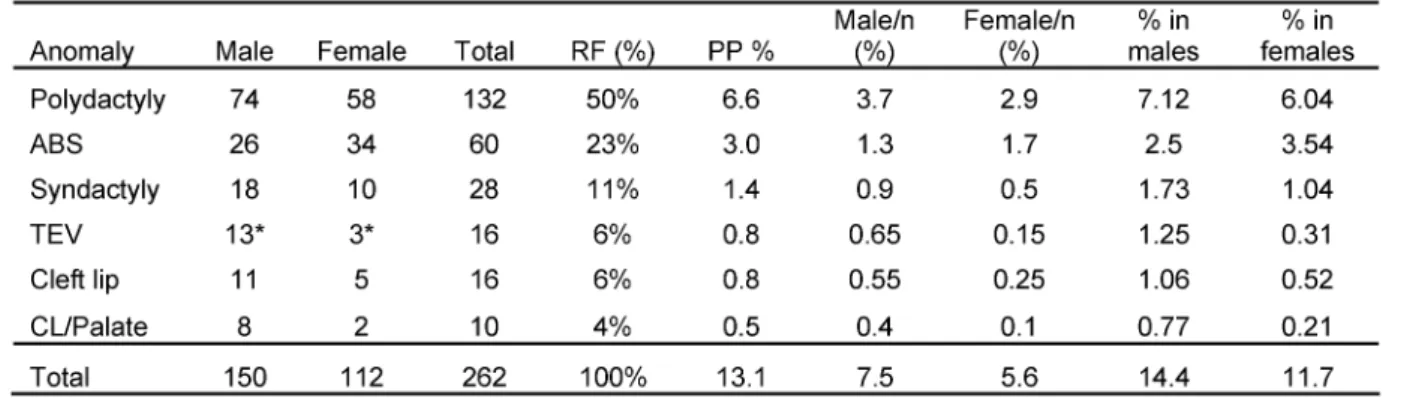
Table 1: Prevalence of some musculoskeletal anomalies in Bayelsa State Nigeria
Undoubtedly, knowledge of prevalence and population distribution of any malady is the first step in scientific planning of preventive and eradication strategies.
Knowledge thus generated would form an invaluable epidemiological data. This study was hence conducted to determine the general population prevalence of amni- otic band syndrome (ABS), cleft lip, cleft palate, polydac- tyly, syndactyly and talipes equinovarus (TEV) in Bayelsa State, Nigeria, since there is no documented study showing that it has been done.
Materials and methods
This study was conducted with two thousand (2000) subjects that were randomly selected within Bayelsa State, Nigeria. Consent was obtained from individuals, though often times a weight gauge was used as a bait to lure would be subjects that may ordinarily not have will- ingly volunteered due to apathy and suspicion. Subjects were physically screened for musculoskeletal anomalies.
Data of individuals who were not born in Bayelsa State or have not domiciled in it for at least one year were ex- cluded. Also excluded were data of individuals with genet- ic syndromes. This study did not discriminate between types or sub-types of any particular birth defect. Sex pre- dilection was assessed by Fisher's exact test (GraphPad InStat V2). Significance level for all comparisons was set atp<0.05. The study design was approved by Research and Ethics Committee of Faculty of Basic Medical Sci- ences, College of Health Sciences, Niger Delta University in accordance with the Helsinki declaration of 1975, as revised in 1983 on human experimentation convention and human right.
Results
A total of 2000 subjects comprising of 960 (48%) females and 1040 (52%) males were included in this study con- ducted in 2007. It was observed that two hundred and sixty two (13%) subjects had one form or the other of musculoskeletal anomaly. Anomalies were seen in a total
of 150 (57%) males and 112 (43%) females respectively.
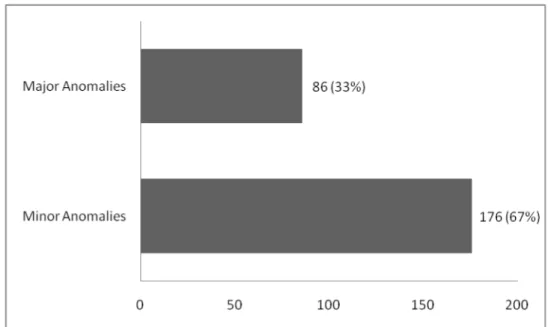
Minor anomalies (polydactyly, syndactyly, and talipes equinovarus) accounted for 67% of the total anomalies, while major anomalies were seen in 33% of subjects.
Polydactyly was the most common and cleft lip/palate the least occurring musculoskeletal anomalies. The gen- eral population prevalence, respective gender prevalence in total population, proportions of male or female with anomalies in total male or female population and the relative prevalence of respective anomalies is as shown (Table 1). We observed that the prevalence of these anomalies were more in males than in females, except for ABS. Cleft lip, cleft lip/palate and talipes equinovarus were predominantly more common in males compared to females in ratios of 2:1, 4:1 and 4:1 (4.3:1) respect- ively. Sex predilection was only statistically significant, p<0.05 (p=0.015) in TEV.
Discussion
The practice of estimating the incidence of a structural anomaly by the number of times it occurred per thousand births (live and still) has dominated studies to the extent that the general population prevalence of anomalies is largely unknown. We criticised this unintended bias on two grounds. First, it is known that structural anomalies are the 5th leading cause of years of potential life lost prior to age 65 [1], and that this age cluster (0 to 65 years) accounts for more than 85% of world’s population [8], [9]. Furthermore, life expectancy in most developing countries is less than 65 years [8], [9]. Hence the effects of structural birth anomalies are not limited to the 1stfew years of life but constitute a recurring decimal for many affected individuals throughout life. Besides, it is now accepted that anomalies like polydactyly, and some types of syndactyly, and talipes or pes calcaneovalgus be ex- cluded from the list of congenital anomalies [10]. There- fore in addition to population estimates employing birth incidence in determining the episode of a particular an- omaly, it is critical to have a general population based picture of any given anomaly because of the reasons already stated. Undeniably, this would generate important
Oyinbo et al.: Prevalence of polydactyly, syndactyly, amniotic band ...
Figure 1: Prevalence of some musculoskeletal anomalies in Bayelsa State Nigeria. N=2000
Figure 2: The relative prevalence of minor and major musculoskeletal anomalies in Bayelsa State Nigeria
epidemiologic and other essential public health data that would add to our understanding of the patterns of health and disease, and help proffer sound logistic planning for preventive and eradication strategies. Though not yet a popular notion, our study is legitimate. It revealed that the overall population prevalence of selected musculo- skeletal anomalies is 13%. The prevalence of individual anomalies is as depicted (Table 1 and Figure 1). We ob- served a higher prevalence amongst males for most anomalies, probably not unconnected with females being more conscious of anomalies or blemishes in their body and a tendency of concealing the same. Despite this nu- merical inequality, gender predilection was not statistically significant in the anomalies studied except in TEV,p<0.05 (P=0.015, significant). Although it have been reported that males are at greater risk for birth defects than fe- males [11]. In this present study TEV was four times more
prevalent in male compared to female. Male preponder- ance was also the case in several other studies [12], [13], [14]. Our study also shows that minor musculoskeletal anomaly accounts for 67% of the 13% population preva- lence (Figure 2). This finding agrees with earlier study that reported higher incidence of minor anomalies in Blacks in contrast to Caucasian with higher incidence of major anomalies [15], [16]. The contemporary reasoning that excluded some structural anomalies from the list of congenital anomalies on the grounds that they have in- significant medical, functional or cosmetic consequences – are medically inconsequential [10], [17], further but- tresses our argument for the need of a general population based prevalence assessment. Our study also recorded a high prevalence of amniotic band syndrome. This may not be unconnected with the alcoholic habits in this en- vironment. Previous work has linked high alcohol con-
sumption to appearance of ABS among others [18]. It has also been postulated that ethnicity, maternal age, order of pregnancy, birth sex, febrile maternal illness or drug use in the first trimester, geographic altitude [19], [20] are important predisposing factors in the episode of ABS. These factors permeate regions with social disorder, juvenile delinquency, conflicts, militant and pirate ac- tivities, as seen in the Niger Delta region of Nigeria [7].
In our study gender predilection of ABS was not significant (p>0.05), agreeing with the study of Badura-Stronka et al. [21], but contradictory with that of Orioli et al. [19].
The roles of gene-environmental interactions in birth anomaly has been stressed [6], [22] but the precise mechanism is yet unknown [2]. A high population preva- lence of structural anomaly can be reduced by decreasing the incidence of congenital anomalies through supple- ments administered during 1st trimester of pregnancy [23], [24]. Currently, antenatal diagnoses of foetal defects are possible through ultrasound scanning, chromosomal and gene analyses. Unfortunately, chromosomal and gene analysis is not routine antenatal practice in developing countries of Africa. In developed countries and Europe the large majority of prospective parents elect for termin- ation of the pregnancy once informed that the foetus is severely affected by a defect [2]. Termination is a last resort rather than an optimal triumph. The only one good solution is primary prevention, which is healthier than the termination of pregnancy. Therefore the prevention of CAs is an extremely important public health issue, since evidently we can secure the health in healthy born people [2]. Quite recently the question as to how to protect against birth defect was answered. It was recommended that daily use of multivitamin including folic acid (dose varies depending on country: in UK it is 4 mg and for US and most of Europe it is between 0.4–0.8 mg), with a healthy diet and lifestyle is essential for women who want to do their best to have a baby without congenital anomaly [2], [25]. As medical science continue to thrive toward zero incidence of birth anomaly, it is pertinent to note that recent advances in identifying previously undetected sub-microscopic chromosomal anomalies have proven to be successful and that the hope of localizing specific genes and the identification of genes within critical chromosomal regions might pave the way for a better understanding of the genetic basis of congenital abnor- malities. Conclusively, we recommend that for a developing country like Nigeria, emphases should be placed on primary prevention. It is glad to note that the use of folic acid and vitamins is routine with antenatal practice in Nigeria. We want to however stress that pregnant women be correctly informed of the importance of these supplements to their developing child. The im- pression generally is that they build up blood and vitality in pregnancy. This is undisputed, but a common erro- neous believes is –if you eat well, you do not really need these drugs. Though, the problem of drug compliance still remains a challenge. Folic acid and vitamins do more – they ensure a healthy congenital anomaly free baby in the absence of any complication. The general population
prevalence of musculoskeletal anomaly in Bayelsa State, Nigeria is comparable in magnitude with known birth in- cidence world wide. Suggestive though premature, of it being a useful estimate index of congenital anomalies incidence in developing countries where record-keeping is largely poor. Not to mention the problems of underes- timation of birth incidence of congenital anomalies due to prenatal loss of foetuses such as blighted ova, miscar- riages and ectopic pregnancies.
Notes
Conflicts of interest
None declared.
References
1. Sadler TW. Langman's Medical Embrology. 10th ed. Baltimore USA: Willliams and Wilkins; 2006. Congenital malformations. p.
122-43.
2. Czeizel AE. The primary prevention of birth defects: Multivitamins or folic acid? Int J Med Sci. 2004;1(1):50-61.
3. Merks JH, Van Karnebeek CD, Caron HN, Hennekam RC.
Phenotypic abnormalities: terminology and classification. Am J Med Genet A. 2003;123(3):211-30. DOI: 10.1002/ajmg.a.20249 4. International Clearinghouse for Birth Defects Monitoring Systems.
Congenital malformations worldwide. Amsterdam: Elsevier, 1991.
p. 22.
5. Bakare T, Sowande OA, Adejuyigbe OO, Chinda Y, Usang UE.
Epidemiology of external birth defects in neonates in South Western Nigeria. Afr J Paediatr Surg. 2009;6:28-3020. DOI:
10.4103/0189-6725.48572
6. Leck I. The contribution of epidemiologic studies to understanding human malformations. In: Stevenson RE, Hall JG, Goodman RM, editors. Human malformations and related anomalies (Oxford monographs on medical genetics no 27) Vol I. New York: Oxford University Press; 1993. p. 65-93.
7. Ikelegbe A. The Economy of Conflict in the Oil Rich Niger Delta Region of Nigeria. Nordic J Afr Studies. 2005;4(2):208-34.
8. Kinsella K, Velkoff VA. An Aging World: 2001. Washington, DC:
U.S. Government Printing Office; 2001. (U.S. Census Bureau Series; P95/01-1). Available from:
http://www.census.gov/prod/2001pubs/p95-01-1.pdf 9. United Nations Department of Economic and Social Affairs,
Population Division (2007). World population ageing 2007. New York: United Nations; 2007.
10. Eurocat. EUROCAT Guide 1.3 and reference documents.
Instructions for the Registration and Surveillance of Congenital Anomalies. Issued: September 2005. Newtownabbey: EUROCAT Central Registry; 2005. Availbale from:
http://www.eurocat.ulster.ac.uk/pdf/EUROCAT-Guide-1.3.pdf 11. Lary JM, Paulozzi LJ. Sex differences in the prevalence of human
birth defects: A population-based study. Teratology. 2001;64:237- 51. DOI: 10.1002/tera.1070
12. Cartlidge I. Observations on the epidemiology of club foot in Polynesian and Caucasian populations. J Med Genet.
1984;21:290-2. DOI: 10.1136/jmg.21.4.290 Oyinbo et al.: Prevalence of polydactyly, syndactyly, amniotic band ...
13. Boo NY, Ong LC. Congenital talipes in Malaysian neonates:
incidence, pattern and associated factors. Singapore Med J.
1990;31:539-42.
14. Miedzybrodzka Z. Congenital talipes equinovarus (clubfoot): a disorder of the foot but not the hand. J Anat. 2003;202:37-42.
DOI: 10.1046/j.1469-7580.2003.00147.x
15. Leck I, Lancashire RJ. Birth prevalence of malformations in members of different ethnic groups and in the offspring of matings between them, in Birmingham, England. J Epidemiology Community Health. 1995;49:171-9. DOI: 10.1136/jech.49.2.171 16. Airede AI, Bello M. Congenital Malformations in the North-east
of Nigeria. Niger J Paediatr. 1994;21:70.
17. Czeizel AE. Birth Defects Are Preventable. Int J Med Sci.
2005;2:91-2.
18. Torfs CP, Velie EM, Oechsli FW, Bateson TF, Curry CJ. A population-based study of gastroschisis: demographic,pregnancy, and lifestyle risk factors. Teratology. 1994;50:44-53. DOI:
10.1002/tera.1420500107
19. Orioli IM, Ribeiro MG, Castilla EE. Clinical and epidemiological studies of amniotic deformity, adhesion, and mutilation (ADAM) sequence in a South American (ECLAMC) population. Am J Med Genet. 2003:118A:135-45. DOI: 10.1002/ajmg.a.10194 20. Werler MM, Louik C, Mitchell AA. Epidemiologic analysis of
maternal factors and amniotic band defects. Birth Defects Res A Clin Mol Teratol. 2003;67:68-72. DOI: 10.1002/bdra.10001 21. Badura-Stronka M, Jamsheer A, Materna-Kiryluk A, Wisniewska
K, Wieckowska B, et al. Epidemiologic characteristics of amniotic band sequence with limb malformations without body wall defect:
data from the Polish Registry of Congenital Malformations. Arch Perinatal Med. 2008;14(1):23-8.
22. Little J, Nicoll A. The epidemiology and service implications of congenital and constitutional anomalies in ethnic minorities in the United Kingdom. Paediat Perinat Epidemiol. 1988;2:161-84.
DOI: 10.1111/j.1365-3016.1988.tb00200.x
23. Czeizel AE. Reducing risks of birth defects with periconceptional micronutrient supplementation. In: Delange FM, West KP Jr, editors. Micronutrient Deficiencies in the First Months of Life - Nestle: Nutrition Workshop Series Pediatric Program. Basel:
Verey/S Karger AG; 2003. p. 309-25. DOI: 10.1159/000074717 24. Botto LD, Olney RS, Erickson JD. Vitamin supplements and the
risk for congenital anomalies other than neural-tube defects. Am J Med Genet 2004;1250:12-21. DOI: 10.1002/ajmg.c.30004 25. Wald NJ. Folic acid and the prevention of neural-tube defects. N
Engl J Med. 2004;350(2):101-3. DOI: 10.1056/NEJMp038186
Corresponding author:
Charles A. Oyinbo
Department of Human Anatomy, Faculty of Basic Medical Sciences, College of Health Sciences, Niger Delta University, Wilberforce Island, Bayelsa State, Nigeria aidemise@yahoo.com
Please cite as
Oyinbo CA, Dare NW, Amain ED. Prevalence of polydactyly, syndactyly, amniotic band syndrome, cleft lip, cleft palate and talipes equinovarus in Bayelsa State, Nigeria. GMS Med Inform Biom Epidemiol.
2009;5(2):Doc14.
This article is freely available from
http://www.egms.de/en/journals/mibe/2009-5/mibe000093.shtml Published:2009-09-09
Copyright
©2009 Oyinbo et al. This is an Open Access article distributed under the terms of the Creative Commons Attribution License
(http://creativecommons.org/licenses/by-nc-nd/3.0/deed.en). You are free: to Share — to copy, distribute and transmit the work, provided the original author and source are credited.