Characterization of Crn7, a novel mammalian Golgi protein
INAUGURAL-DISSERTATION zur
Erlangung des Doktorgrades
der Mathematisch-Naturwissenschaftlichen Fakultät der Universität zü Köln
vorgelegt von
Vasily RYBAKIN
aus
St. Petersurg, Russland
Köln, 2005
Referees/Berichterstatter: Prof. Dr. Angelika A. Noegel Prof. Dr. Karin Schnetz
Date of oral examination:
(Tag der mündlichen Prüfung) 02/12/2005
The present research work was carried out under the supervision of Prof. Dr. Angelika A. Noegel, in the Institute of Biochemistry I, Medical Faculty, University of Cologne (Cologne, Germany) from May 2003 to September 2005.
Diese Arbeit wurde von April 2003 bis September 2005 am Biochemischen Institut I der Medizinischen Fakultät der Universität zu Köln unter der Leitung von Prof. Dr. Angelika
Наташе
00. contents.
O. Opening remarks 5
1. Introduction 6
1.1. Coronin proteins as actin regulators 10 1.1.1. Yeast coronin regulates the actin cytoskeleton by
directly interacting with Arp2/3 complex 11 1.1.2 Drosophila Dpod1: An actin-tubulin linker regulating
the development of the nervous system 13 1.1.3. Mammalian short coronins and the actin cytoskeleton 14 1.2. Coronin and POD-1 proteins at the interface of
cytoskeleton and trafficking 18 1.2.1. Drosophila coro possibly acts as a linker between the
actin cytoskeleton and membrane transport 19 1.2.2. C. elegans POD-1, an actin-binding protein participating
in vesicular trafficking 20 1.3. Mammalian coronins with unexplored functions 21
1.4. Clinical implications 22
2. Aims of the work 25
3. Results 26
3.1. Crn7 sequence analysis 26
3.2. Tissue distribution and developmental dynamics of Crn7 29 3.3. Subcellular localization of Crn7 31
3.3.1. Crn7 is present in the cis-Golgi and in the cytosol 31 3.3.2. Crn7 is localized to the outer side of Golgi membranes 35 3.3.3. Stabile association of Crn7 with the Golgi requires the
integrity of ER-to-Golgi transport 36 3.3.4. Properties of cytosolic and membrane-associated forms of Crn7 39 3.4. Analysis of Crn7 function by RNAi 40 3.4.1. Use of siRNA duplexes to silence Crn7 40 3.4.2. Influence of Crn7 RNAi on the Golgi architecture 42 3.4.3. Block of protein export from the Golgi in Crn7 RNAi cells 44
4. Discussion 51
4.1. Physical structure and inheritance of the Golgi complex 51 4.2. Protein import and progression in the Golgi 54
4.3. Golgi export 55
4.4. Models of Golgi dynamics 57 4.5. Crn7 is a novel WD-repeat protein localized to the Golgi complex 59 4.6. Targeting of Crn7 to the Golgi 60
4.7. Possible function of Crn7 in the Golgi complex 62 4.8. Crn7 is an essential mammalian protein. 63
5. Materials and methods 65
6. Abbreviations 72
7. Literature 73
Abstracts (English/German) 80
Erklärung 82
Curriculum vitae 83
Lebenslauf 84
0. Opening remarks
This work would never have been accomplished without constant help and support from my friends and colleagues. I am deeply grateful to Prof. Angelika A. Noegel for offering me the opportunity to perform my PhD work in her lab. Rainer Duden and Irina Majoul (Cambridge - London), Maria Stumpf, Rolf Müller, Andreas Hasse (Cologne) and Dharamdajal Kalicharan (Groningen) patiently taught me methods and helped with experiments. My quite part-time co- bosses Andreas Hasse and Christoph Clemen sometimes popped up with valuable comments.
Akis Karakesisoglou, Deen Bakthavatsalam, Rainer Duden, Irina Majoul and Stefan Höning read piles of paper I brought along and even tolerated questions. Bettina Lauss, Dörte Püsche and Budi Tunggal (Cologne) are acknowledged for expert administrative support.
I am grateful to all colleagues of mine who openly, and sometimes too frankly (mind the understatement) shared their opinions about my work during scientific meetings and conferences in Barcelona, Annaberg, Washington and Cologne, and for sharing key chemicals. The people in the lab (Soraya, Christoph, Thorsten, Carola, Maria, Yogi, Deen, Wenshu, Sabu, Jessica, Andreas, Kumar, Berthold, Budi, Ria, Martina, Hua, Hafi, Rolf, “Kletten”-André, Christian, Marion, “Commandante” Akis, Maria, Francisco, Gudrun and especially Rosi) made my life here pleasant and definitely not boring.
I am especially thankful to Natasha Gounko (Groningen) for her support, advise and help.
Спасибо, рыжик!
Kölsch is not beer. At all.
1. Introduction
Coronins constitute an evolutionarily conserved family of WD-repeat actin- binding proteins, which can be clearly classified into two distinct groups based on their structural features. All coronins possess a conserved basic N-terminal motif and three to ten WD repeats clustered in one or two core domains.
Dictyostelium and mammalian coronins are important regulators of the actin cytoskeleton, while the fly Dpod1 and the yeast coronin proteins crosslink both actin and microtubules. Apart from that, several coronins have been shown to be involved in vesicular transport. C. elegans POD-1 and Drosophila coro regulate the actin cytoskeleton, but also govern vesicular trafficking as indicated by mutant phenotypes. In both organisms, defects in cytoskeleton and trafficking lead to severe developmental defects ranging from abnormal cell division to aberrant formation of morphogen gradients.
WD-repeat (WD-40-domain-repeat) proteins are defined by the presence of at least four WD repeats located centrally in the protein. These repeats were discovered in 1986 (Fong et al., 1986) and are defined by a partially conserved domain of 40–60 amino acids starting with a glycine-histidine (GH) dipeptide 11–24 residues away from the N-terminus and ending with a tryptophane-aspartic acid (WD) dipeptide at the C terminus. The WD domain has no intrinsic catalytic activity and is thought to serve as a stable platform for simultaneous interactions with other proteins.
WD-repeat proteins have extremely diversified cellular functions. They play central roles in a variety of cellular events including, but not limited to, signal transduction, transcriptional regulation, remodeling the cytoskeleton and regulating vesicular trafficking.
Coronin proteins constitute one of the more than thirty subfamilies (Yu et al., 2000) among the WD-repeat proteins and contain three to five clustered WD repeats forming the characteristic coronin core domain (de Hostos, 1999; Neer and Smith, 1996). Apart from the core domain, common structural features include a short conserved N-terminal motif and a 70 amino acid-region located C-terminally to the WD repeats. Furthermore, each coronin contains a unique, divergent region of about 100 amino acids, which follows the conserved C-terminal extension.
This sequence may confer specific functions to any individual coronin protein while, in contrast, it differs between the species. A second region of variability is present in the fourth β-strand of the third WD repeat (de Hostos, 1999).
The first coronin protein was identified in Dictyostelium discoideum (de Hostos et al., 1991). Its name resulted from the location in the actin-rich crown-shaped cell surface projections.
The Dictyostelium protein was indirectly shown to participate in the regulation of the actin cytoskeleton and vesicular trafficking. Early findings on structures and functions of coronin proteins, especially that of Dictyostelium, have been reviewed (de Hostos, 1999, see also A.
Schulze, Diploma thesis, Univesity of Cologne, 2001). Meanwhile, more than 20 coronins have been identified in vertebrates and invertebrates (de Hostos, 1999; Okumura et al., 1998). In mice and men, the coronin family of proteins comprises at least seven members (Table 1).
Most mammalian representatives typically demonstrate tissue-specific distribution patterns, whereas some are rather ubiquitous. Coronin 3 is the most-widely expressed mammalian member (Iizaka et al., 2000; Okumura et al., 1998; Spoerl et al., 2002). Coronin proteins clearly form two distinct subfamilies, short ‘‘conventional’’ coronins and longer proteins, respectively (Fig. 1). The first subfamily consists of approximately 450–650 amino acid proteins harboring a very carboxy-terminal coiled-coil region of 30–40 amino acids mediating homophilic dimerization and/or oligomerization of coronins (Asano et al., 2001; Spoerl et al., 2002), whereas
the second subfamily contains the closely related mammalian Crn7 and C. elegans and Drosophila POD-1 proteins, which are clearly distinct from the first group in that they possess two core domains rather than one.
coronin synonyms protein accession # main tissue expression references 1
coronin 1A, clabp, clipinA, TACO, p57
461 aa NP_009005 thymus, spleen, bone marrow, lymph nodes,
peripheral leukocytes
(Okumura et al., 1998;
Suzuki et al., 1995)
2
coronin 1B, coroninse,
p66
489 aa NP_065174 gastrointestinal mucosa,
liver, spleen, kidney, lung (Parente et al., 1999)
3 coronin 1C,
CORO1C 474 aa NP_055140 Brain, lung, intestine, kidney
(Hasse et al., 2005; Iizaka et al., 2000; Spoerl et al., 2002)
4 coronin 2A, clipinB ,
IR10 525 NP_438171 colon, prostate, testis, brain, lung, epidermis
(Nakamura et al., 1999;
Okumura et al., 1998;
Zaphiropoulos and Toftgard, 1996)
5 coronin 2B,
clipinC 475 NP_006082 brain (Nakamura et al., 1999)
6 3 splicing ClipinE variants
471 aa 431 aa
NP_624354,6
NP_624355 brain NP_624354−6
7 Crn7 925 aa NP_078811 ubiquitous (Rybakin et al., 2004), this work
Table 1. Summary of the mammalian coronin proteins, their nomenclature, sizes, and major expression sites.
A recently discovered novel Dictyostelium long coronin/POD-1 homologue (A. A.
Noegel and F. Rivero, personal communication) is also classified into the same subfamily. It is important to note that all known coronins belonging to the second group lack coiled-coil domains. However, harboring two core domains, longer coronin proteins may indeed be considered dimers with regard to the core domain functions.
Fig. 1. Comparison of domain structures of representative coronins and POD-1 proteins and their phylogenetic relationships. Phylogenetic analysis was performed using ClustalW algorithm (European Bioinformatics Institute, Hinxton, UK), and the phylogenetic tree was build using the Phylodendron server at the University of Indiana, Bloomington, IN. Right to the phylogram, abbreviations indicating organisms, and protein names are given: F, Drosophila melanogaster;
H, Homo sapiens; W, Caenorhabditis elegans; D, Dictyostelium discoideum; Y, Saccharomyces cerevisiae. On the right, domain structures and amino acid numbers for each protein are given.
All known short coronins are characterized by the presence of an extremely highly basic N-terminal 12-amino acid motif (pI 12.5 for human coronin 1), which can be taken as a novel coronin signature (Table 2A), as it is only present in coronin proteins. In the longer coronins, this
sequence is reduced to a 5-amino acid core motif (Table 2B) that appears in front of each coronin core domain. A recent study suggests that this coronin signature is involved in actin binding (Oku et al., 2003), although this sequence is also present in coronin proteins that have not been associated with the actin cytoskeleton.
A
D coronin 1 mskvvrsskyrhvfaaqpkk 20 H coronin 1 1 msrqvvrsskfrhvfgqpak 20 H coronin 3 1 mrrvvrqskfrhvfgqavkn 20 F coro 1 msfrvvrsskfrhvygqalk 20 Y crn1p 1 msgkfvraskyrhvfgqaak 20 B
W POD-1 (1) 1 mawrfaaskfknttpkvpkk 20 W POD-1 (2) 551 gqitskfrhvdgqqgtksga 570 F Dpod1 (1) 1 mawrfkaskyknaapivpka 20 F Dpod1 (2) 611 stvfgkvskfrhlkgtpghk 630 D POD-1 (1) 1 mfkvskyrhtvgkidkrelw 20 D POD-1 (2) 482 givpkvvrsskyrhisgsa 500
Table 2. Coronin signatures present in short and long coronins. A, short coronins contain a stretch of 12 conserved basic amino acids at the very N-terminus. B, long coronins / POD-1 proteins harbor two copies of a 5-amino acid degenerated core signature at the N-terminus and in the intermediate region. Identical amino acids are indicated in red, similar amino acids in blue. Single-letter organism abbreviations as in Fig. 1.
1.1. Coronin proteins as actin regulators
Actin is one of the most-abundant cellular proteins executing multiple structural and
in α, β- and γ-isoforms in mammals. Monomeric actin (G-actin) is predominantly present in the cytosol. G-actin has the potential to polymerize into filamentous actin (F-actin) in vitro and in vivo given the appropriate conditions. Actin filaments are polarized dynamic structures characterized by the presence of the slow growing (-) and fast growing (+) ends. Actin polymerization is initiated by nucleation of a G-actin trimer. This process, along with filament branching, relies on a variety of regulators, among others, the Arp2/3 complex activated by N- WASP, Rho- GTPases and other factors.
The diversity of F-actin structures and associated cellular functions depends not only on polymerization and depolymerization, but on a variety of actin-binding proteins, which can be grouped in G-actin sequestering or associated proteins (e.g. ADF, cofilin, profilin), F-actin capping and severing proteins (e.g. gelsolin, capping protein, severin), actin filament crosslinking and bundling proteins (i.e. fimbrin, α-actinin, filamin), motor proteins (myosins), and membrane anchoring proteins (i.e. ponticulin, talin, vinculin). Most coronins that have been characterized so far belong to the group of actin filament-crosslinking and bundling proteins.
1.1.1. Yeast coronin regulates the actin cytoskeleton by directly interacting with Arp2/3 complex
Crn1p, the only yeast coronin, was independently isolated by two groups using homology cloning and microtubule affinity chromatography, respectively (Goode et al., 1999;
Heil-Chapdelaine et al., 1998). The 651-amino-acid protein comprises five WD-repeats, a coiled- coil carboxyl terminus and a central region enriched in proline and charged amino acids.
Additionally, it includes a predicted microtubule-binding domain similar to that of the microtubule-binding protein MAP1B. Crn1p was shown to localize to cortical actin patches in a latrunculin A-sensitive manner (Goode et al., 1999; Heil-Chapdelaine et al., 1998), implying that
crn1p localization is dependent on F-actin. Crn1p is an abundant protein indicating a high-affinity binding to F-actin at a 1:1 molar ratio suggesting that the binding appears at the sides of actin filaments (Goode et al., 1999). When added to F-actin in vitro, crn1p bundles the actin filaments.
However, if added to actin monomers, yeast coronin instead induces a three-dimensional network formation as shown by electron microscopy and falling ball viscosimetry. Moreover, crn1p accelerates the filament assembly at the barbed end if added to a G-actin solution (Goode et al., 1999). It also binds to microtubules, although considerably weaker than to actin, and can therefore directly crosslink actin filaments with microtubules.
F-actin binding and assembly were mapped to the WD repeats, microtubule binding to the MAP1B homology domain, and actin bundling to the coiled-coil region (Goode et al., 1999).
As the coiled coil was shown to participate in dimerization and oligomerization in other coronins (Asano et al., 2001; Spoerl et al., 2002), it may be argued that F-actin bundling depends on crn1p present in the di- or oligomeric state. In vivo, crn1p localization to the cortical actin patches requires both, coiled-coil and actin-binding domains (Humphries et al., 2002). Moreover, crn1p directly associates with the Arp2/3 complex in vitro and in vivo. Crn1p inhibited the actin nucleation activity of the Arp2/3 complex although crn1p alone has a slightly stimulating effect in an actin polymerization assay (Goode et al., 1999; Humphries et al., 2002). The mechanism for the crn1p inhibition of the Arp2/3 complex was explained by a direct interaction between crn1p and the Arp2/3 complex, as was the case in yeast two-hybrid experiments in which the C- terminus of crn1p interacted with Arc35, the subunit responsible for the binding of the Arp2/3 complex to the sides of actin filaments.
The interaction of crn1p and Arp2/3 was considered to regulate the filament branching and thus the formation of complex actin networks important for cell movement and intracellular
Chapdelaine et al., 1998). However, the absence of crn1p appears to enhance the phenotypes of cofilin and actin (ATP-binding pocket) mutants (Goode et al., 1999). Overexpression of crn1p leads to abnormalities in the cytoskeleton organization resulting in swollen cells with actin patches depolarized from the bud region, as well as in the accumulation of spiral or loop actin structures in the cytoplasm (Humphries et al., 2002). These defects were due to the overproduction of crn1p coiled-coil regions, as a truncated form of crn1p lacking the coiled coil does not cause such a phenotype.
1.1.2. Drosophila Dpod1: an actin–tubulin linker regulating the development of the nervous system
Drosophila dpod1 encodes a novel double-core domain 1074-amino acid coronin strongly expressed in the developing nervous system (Rothenberg et al., 2003). In addition to the two core domains, the Drosophila protein possesses a predicted microtubule-binding domain similar to that of the microtubule-binding protein MAP1B. In adherent S2 cells derived from mixed Drosophila embryonic tissues, Dpod1 co-localizes with both microfilaments and microtubules and is re-localized to microtubules upon the disruption of the actin cytoskeleton. In vitro, Dpod1 crosslinks microtubules as well as microfilaments. Apart from being able to crosslink actin filaments as well as microtubules, Dpod1 is capable of forming bundles containing both microfilaments and microtubules. Co-sedimentation of microtubules with F- actin-coronin bundles has been previously demonstrated in yeast (Goode et al., 1999).
Although Dpod1 is not essential for cytoskeletal organization in S2 cells as shown by the absence of any pronounced phenotype in dpod1 RNAi cells, overexpression of GFP-tagged protein leads to development of highly dynamic neurite-like cell surface projections (Rothenberg et al., 2003). The formation of these projections was further shown to be actin-, but not tubulin-
dependent. Dpod1 acts as an important regulator of neural development in the fly. dpod1 mutant flies are characterized by an aberrant axonal guidance resulting in failures in the target innervation. In particular, axonal fine routing at choice points and turns, but not neurite outgrowth or extension is affected. Overproduction of Dpod1 protein also results in multiple neural phenotypes (Rothenberg et al., 2003). As Dpod1 regulates both microfilament and microtubule architecture, these phenotypes can be explained by aberrant cytoskeleton-related morphogenetic processes in mutant cells, leading to structural abnormalities like axonal breaks, defects in lateral branching and stalling at important decision-making points and thus to defects in target innervation. A model has been proposed linking Dpod1 to scaffolding signalling molecules at the cytoskeleton (Rothenberg et al., 2003), albeit more experimental evidence is necessary to support such a role.
1.1.3. Mammalian short coronin proteins and the actin cytoskeleton
Most of the mammalian coronins belong to the group of actin filament crosslinking and bundling proteins. Coronin 1 is the best-characterized mammalian coronin and is the closest relative of Dictyostelium discoideum coronin (Suzuki et al., 1995). It is most strongly expressed in human immune tissues and immune cells and, to a lesser extent, in the lung and brain. On the mRNA level, the skeletal muscle species is larger in size. During the process of murine thymic cell development, peak expression levels of coronin 1 are found in early thymocytes and in adult CD4+CD8- and CD4-CD8+ thymocytes. In thymocytes, the protein localizes to the cytoplasm and to F-actin-rich membrane protrusions especially in stimulated T-cells (Nal et al., 2004).
ActA-positive Listeria monocytogenes, among other proteins, recruits coronin 1 to their F-actin tails in infected host cells (David et al., 1998).
Two regions that mediate binding to F-actin were determined. One is the N-terminally located KXRHXX-motif conserved in all coronin proteins (Table 1); a second F-actin binding site in coronin 1 was mapped within the domain containing the WD repeats (Oku et al., 2003). A leucine zipper region of the C-terminus mediates homophilic dimerization of coronin 1 (Oku et al., 2005). Human phagocytic leukocytes contain coronin 1 in cytosolic as well as in cytoskeletal fractions. During the course of phagosome formation the peripheral cytoskeletal and cytosolic coronin 1 staining is lost and an association with F-actin around early phagocytic vacuoles can be observed. Dissociation of coronin 1 from the phagosome is accompanied by phosphorylation on serine residues involving PKC. Without the dissociation, the subsequent formation of phagolysosomes is inhibited (Itoh et al., 2002). Recently, coronin 1 has been demonstrated to link the actin cytoskeleton to the plasma membrane in leukocytes (Gatfield et al., 2005). According to the authors, coronin 1 trimerizes using a linker region between the core domain and the C- terminus, while the amino-terminus mediates interaction with plasma membrane (Gatfield et al., 2005). It is unclear whether such coronin 1-mediated interaction between the cytoskeleton and plasma membrane is of biological significance.
Soluble coronin 1 elutes together with phox components in a complex of higher molecular mass from gel filtration, as it binds C-terminally to p40phox, a cytosolic subunit of the NADPH oxidase complex involved in the generation of the microbicidal superoxide burst in neutrophils (Grogan et al., 1997). Furthermore, PKC activation leads to the redistribution of coronin 1 in a phox-protein-dependent manner from the cell cortex to the perinuclear region. A second soluble pool of coronin 1 in human phagocytic leukocytes forms high-molecular-weight complexes independently of the phox proteins. These complexes are solubilized by PI3-kinase activity and may be involved in forming the F-actin structures in early phagosome formation (Didichenko et al., 2000).
In addition, coronin 1 is detected on phagocytic vacuoles of macrophages. Upon internalization of mycobacteria, coronin 1 is transiently recruited to the site of the bacterial entry (Schuller et al., 2001). Moreover, clumps of 10–20 living mycobacteria in phagosomes inhibit the dissociation of coronin 1 and the continued transport of the phagosomes to lysosomes. Retaining coronin 1 on the early phagosome prevents the mycobacterial clumps from lysosomal degradation (Ferrari et al., 1999). Using a dominant-negative approach, Yan and colleagues recently demonstrated that coronin 1 is required for the accumulation of Arp3 on phagosomes, as well as for receptor capping and actin remodeling at forming and early phagosomes (Yan et al., 2005).
Coronin 2 was first described as a phosphoprotein in HCl-secreting gastric parietal cells
(rabbit coroninse, (Brown and Chew, 1989; Chew et al., 1997) and is generally found in the gastrointestinal mucosa, but also highly expressed in secretory cells of the kidney and lung, and in smaller amounts in spleen, adrenal and other tissues (Parente et al., 1999). In the kidney coronin 2 primarily localizes to cortical F-actin structures (Parente et al., 1999). The homologous mouse coronin 2 differs from coroninse in a short part of the unique C-terminal region and is ubiquitously expressed and most prominent in the kidney, lung, spleen and liver (de Hostos, 1999; Morrissette et al., 1999; Okumura et al., 1998). On the subcellular level, coronin 2 shows a perinuclear punctate pattern and is localized to early phagosomes (Morrissette et al., 1999). PKC- dependent serine phosphorylation of coronin 2 leads to a partial redistribution of coronin 2 from vesicular structures, which are neither Golgi membranes nor mitochondria, to the leading edge of the induced actin-rich filopodia (Parente et al., 1999). Recently, coronin 2 was shown to co- localize and interact with Arp2/3 complex (Cai et al., 2005). This interaction is mediated by PKC-dependent phosphorylation of serine-2 on coronin 2, and is important for the leading edge
Coronin 3 is ubiquitously expressed and is most prominent in the brain, lung, intestine
and kidney (Hasse et al., 2005; Iizaka et al., 2000; Spoerl et al., 2002). An additional band of higher molecular weight (60 kDa) is detected in the brain and heart, while in the skeletal muscle this band is the only one present. A single RNA species has been described. Coronin 3 is localized to F-actin-rich punctate structures in the cytosol, which are most pronounced around the nucleus and at the cell cortex, especially in lamellipodia and membrane ruffles (Spoerl et al., 2002). Both N and C termini of coronin 3 are required for its cytoskeletal localization and for coronin-3-mediated regulation of cell morphology. The C terminus (aa 315–474) confers membrane association, and removal of its coiled-coil part (aa 444–474) abolishes membrane localization. In vitro, F-actin binding and bundling occurs through the C-terminal fragment preceding the coiled coil (aa 315–444). This fragment interacts with the N terminus and can lead to decreased binding of the C-terminal fragment to F-actin. Conversely, the entire C terminus can recruit the purified N-terminal region to actin filaments probably reflecting the folding pattern of coronin 3 bound to actin. The dissociation constants of both coronin 3 C-terminal fragments binding to F-actin were evaluated at about 8 mM. F-actin binding was saturated at a 1:3 molar ratio for both fragments.
Surprisingly, the C terminus (aa 315–474) forms trimers while the non-coiled-coil C terminus (aa 315–444) forms dimers. The oligomerization is non-ionic and does not require other proteins. Also, endogenous coronin 3 is extracted as trimer from cytosol and membrane fraction (Spoerl et al., 2002). The coiled-coil region of the C terminus contributes to a simultaneous binding of the N-terminal domain and F-actin and to a trimerization of coronin 3, properties which seem to be essential for cellular membrane localization.
Generally, cytosolic, but not particle-associated coronin 3 indicated a high degree of phosphorylation (Spoerl et al., 2002), and coronin 3 can be dephosphorylated in vitro
(A. Rosentreter and C. Clemen, personal communication). PKC activation did not influence the subcellular distribution, but resulted in a decreased level of the coronin 3 protein. This reduction may be due to the 30 UTR of the coronin 3 mRNA containing 14 CUUU repeats similar to the (CUUU)11(U)8 repeats of MARCKS mRNA (Spoerl et al., 2002). This CUUU14- repeat element is not found in any other mammalian coronin mRNA. In MARCKS mRNA, the element mediates rapid mRNA degradation upon treatment with growth factors or PKC activators (Wein et al., 2003).
1.2. Coronin and POD-1 proteins at the interface of cytoskeleton and trafficking
Intracellular membrane organelles form a highly dynamic continuum intimately connected by means of two major trafficking routes. The membrane flow from the endoplasmic reticulum through the Golgi complex to the cell surface, lysosomes and other organelles is generally described as a biosynthetic pathway. Another trafficking route, the endocytic pathway, connects the plasma membrane with the endosomal system, lysosomes and the Golgi/ER membranes. Subcellular localization and trafficking of the membrane compartments have been shown to rely on the interaction of these structures with cytoskeletal components. Formation of endocytic vesicles at the plasma membrane depends on the interaction of the membrane with cortical actin and several actin-binding proteins including dynamin and cortactin (Cao et al., 2003; Sauvonnet et al., 2005; Yarar et al., 2005). Dynamics of cargo vesicles are mediated by both actin cytoskeleton and microtubules. The interference with any of these systems leads to multiple defects in vesicular trafficking such as an impairment of the formation of the trans-Golgi network and ER export carriers (Waguri et al., 2003; Watson et al., 2005), or of the formation and cytoplasmic progression of the vesicles (Mundy et al., 2002; Yarar et al., 2005). Several
studies hinted at a possible function of coronin proteins at the interface of the cytoskeleton and intracellular membrane transport.
1.2.1. Drosophila coro possibly acts as a linker between the actin cytoskeleton and membrane transport
Recently, a novel conventional coronin gene has been identified in Drosophila (Bharathi et al., 2004). coro encodes a predicted 528-amino-acid protein harboring a core domain and a C-terminal coiled-coil region. The coro gene is ubiquitously expressed at high levels in all cell types with the exception of the larval CNS. Mutations in the gene are lethal at early to late pupal stages and exhibit a number of appendage phenotypes including shortened, ventralized, thick legs, defective wing margins and malformed eyes with improper ommatidial organization.
On the cellular level, coro mutations result in a disruption of the cytoskeleton in the wing imaginal discs. Actin filaments appear retracted and the cortical actin reduced, leading to abnormal cell morphology (Bharathi et al., 2004).
Importantly, it has been suggested that the coro gene genetically interacts with the syx1A gene encoding a Drosophila SNARE protein participating in secretion and calcium channel functions (Schulze et al., 1995; Wu et al., 1999). The coro mutant phenotype is similar to that of syx1A. Furthermore, overexpression of syx1A on a background of coro mutation by imprecise P- element excision enhances lethality and also causes enhancement of the coro phenotype implying an interaction of the syntaxin 1A and coronin genes (Bharathi et al., 2004). In coro mutants, the GFP-fused morphogen Dpp accumulates in endocytic vesicles docked at the inner side of the plasma membrane. Further, Dpp degradation is slowed down, resulting in uniform Dpp presence along the anteroposterior wing disc axis and in disc overgrowth (Bharathi et al., 2004).
Drosophila coro thus participates in establishment of the Dpp morphogen gradient by regulating the intracellular routing of Dpp.
It may well be that the defect in the cortical actin structures reported for coro mutants (Bharathi et al., 2004) causes abnormal subplasmalemmal vesicle transport resulting in accumulation of Dpp-containing vesicles. Such an accumulation by interfering with actin structures was also demonstrated in cytochalasin-D-treated acinar epithelial cells (Da Costa et al., 2003) and in the case of loading actin filaments with N-ethylmaleimide-treated myosin S1 in lamprey reticulospinal synapses (Shupliakov et al., 2002).
1.2.2. C. elegans POD-1, an actin-binding protein participating in vesicular trafficking A POD-1 protein was first purified in a screen designed to isolate C. elegans actin- binding proteins (first named CABP11) (Aroian et al., 1997). In early worm embryos, the protein co-localizes with actin in the cortical region and, in addition, exhibits a punctate cytoplasmic staining pattern. Loss of pod-1 gene activity results in defects in anteroposterior polarity in early embryos in that the second AB and P1 cell division occurs synchronously and in parallel orientations, instead of the wild-type pattern, which is characterized by the P1 cell dividing after AB and having a division plane perpendicular to that of AB (Rappleye et al., 1999). Furthermore, mutant embryos do not separate polar bodies. Additional defects appear in vesicular trafficking processes. Firstly, the polar granules are found throughout the cytoplasm of mutant embryos, whereas normal embryos transport the polar granules towards the posterior pole. Secondly, mutant cells accumulate abnormally large intracellular membrane structures as shown by staining with an antibody recognizing plasma membrane and vesicles of endocytic origin. Finally, a defect in the inner eggshell layer formation also indicates abnormal exocytic functions. This defect has
been illustrated by an increased dye permeability and osmotic sensitivity of the eggshell (Rappleye et al., 1999).
C. elegans POD-1 co-localizes with cortical actin and cytoplasmic structures and facilitates trafficking processes. The function of POD-1 may be to regulate the attachment of vesicular structures to the actin microfilaments and thus facilitating oriented trafficking. The accumulation of abnormal cytosolic membrane structures in pod-1 mutants can be explained by the absence of such vesicle-microfilament binding. Based on the information available on yeast crn1p (see above), it might be reasonable to speculate that such binding is Arp2/3-dependent.
However, the absence of the coiled-coil domain required for Arp2/3 regulation in the yeast protein makes this less likely (Humphries et al., 2002). The eggshell phenotype can be explained by the lack of proper interaction of exocytic vesicles with the cortical actin cytoskeleton or by abnormal intracellular trafficking of such vesicles resulting in misdirection of the Golgi–plasma membrane flow.
1.3. Mammalian coronins with unexplored functions
To date, seven mammalian genes with several transcriptional products have been identified (Table 1). Except for isoform C of coronin 6 lacking the fourth WD-repeat domain, all short coronins contain five predicted central WD repeats. Functional data or biochemical properties are not available for coronins 4, 5 and 6. Coronin 4 is restricted to colon, prostate and testis, but also described in brain tissue (Nakamura et al., 1999; Okumura et al., 1998;
Zaphiropoulos and Toftgard, 1996). Coronin 5 is also present in neuronal tissue and to a lesser degree in heart and ovary (Nakamura et al., 1999). Coronin 6 (accession number NP_624354-6) was detected in the brain.
1.4. Clinical implications
Recent data suggest that coronin proteins participate in such processes as an antimicrobial defense and neuronal development and function. Coronin 1 has been shown to participate actively in the innate defense reactions on the cellular level (Gatfield et al., 2005; Itoh et al., 2002). The functional connection between the actin cytoskeleton, coronin 1 protein and phagocytosis of pathogenic mycobacteria has already been discussed above. Clinical investigations have revealed that coronin 1 and the Arp2/3 complex 20 kDa subunit are downregulated in the fetal Down syndrome brain cortex (Weitzdoerfer et al., 2004). Both are proteins apparently involved in the dysgenesis of the brain and the associated mental disabilities.
In murine brain all areas express coronin 3 during embryogenesis and the first postnatal stages (Hasse et al., 2005). Postnatally, the expression in the gray matter decreases, except for hippocampal and cerebellar Purkinje neurons, while levels in the white matter increase in the course of myelination (Hasse et al., 2005).
Cultured neuro-2a and PC-12 cells transfected with various GFP-tagged coronin 3 versions favor a role for coronin 3 in neuronal function, morphogenesis and possibly migration.
Truncated proteins efficiently suppress neurite formation and either stimulate or inhibit noradrenaline (norepinephrine) secretion of PC-12 cells (A. Hasse and C. Clemen, personal communication). Coronin 5 accumulates at neurite growth cones and co-localizes with focal adhesions as well as with stress fibers. It co-precipitates with vinculin, a major component of focal contacts and also binds directly to F-actin in vitro (Nakamura et al., 1995). Coronins 3 and 5 may be involved in neuronal migration, neurite extension and synapse formation by means of rearranging F-actin and linking it to the plasma membrane.
Coronin 1 is abundantly expressed in T- and B-lymphocytes and macrophages (Didichenko et al., 2000; Goode et al., 1999; Itoh et al., 2002; Okumura et al., 1998; Schuller et al., 2001). Apart from an expression in thymic cells, coronin 1 was shown to be involved in processes of membrane organization of nascent phagosomes and associated with their F-actin coat. Later, a dissociation of coronin 1 seems to be necessary for further phagosome processing.
Clumps of phagocytosed mycobacteria cause the retention of coronin 1 on the early phagosome and inhibit their delivery to lysosomal degradation (Ferrari et al., 1999). The recruitment of coronin 1 to the phagosome may be regulated endogenously by PKC and PI3-kinase activity, and exogenously by factors like that of living mycobacteria. Soluble coronin 1 is involved in generating focal microbicidal superoxide bursts. These data strongly suggest an important contribution of coronin 1 to innate defense reactions.
Coronin proteins play important roles in development and disease: In Drosophila and C.
elegans, coronin and POD-1 mutants exhibit a number of developmental defects ranging from abnormal determination of cell polarity and formation of morphogenetic gradients to aberrant axonal guidance and target innervation (Bharathi et al., 2004; Rappleye et al., 1999; Rothenberg et al., 2003). Several mammalian coronins are strongly expressed in the CNS and possibly involved in the development of the nervous system. Coronin 3 demonstrates a highly dynamic expression pattern in the embryonic brain implying that differential activity of this proteins may participate in the regulation of brain development, probably together with other coronins expressed in the developing brain (Hasse et al., 2005). Although some coronins have been studied in detail, biochemical and functional properties of their majority is unclear. Future experiments need to be directed at functional properties of coronin proteins. There are still significant gaps in our understanding of the regulation of coronin genes and proteins, too. The positions of coronin proteins in the complex protein interaction networks have not yet been characterized. Functions
of the central core domain particularly require further investigation. Clearly, based on the well- established functions in the regulation of the actin cytoskeleton and membrane trafficking as well as being implicated in many developmental processes and in disease, this protein family deserves further investigation.
2. Aims of the work
Recently, a novel mammalian coronin family member was identified and designated coronin 7 (A. Schulze, diploma thesis, Cologne University, 2001). My primary aims were:
To analyze the cellular localization and dynamics of coronin 7 (Crn7) using
biochemical methods, immunofluorescence and electron microscopy;
To characterize Crn7 interaction partners;
To reveal the function of Crn7 in mammalian cells using gene interference (RNAi).
3. RESULTS
Crn7 is a ubiquitous mammalian coronin family member. The protein is distributed between the cytosol and Golgi, where it is present at the outer side of the membrane. Golgi localization of Crn7 depends on tyrosine phosphorylation and the integrity of ER-to-Golgi transport. The protein intimately associates with the Golgi membrane and does not require coatomer for its localization.
Crn7 is an essential protein, as its knockdown by RNAi leads to a dramatic time- and concentration-dependent decrease in cell viability. Crn7 RNAi cells display scattered Golgi morphology, as demonstrated by electron and light microscopy.
Most importantly, the knockdown leads to the block of protein export from the Golgi complex, while the import into the organelle, both anterograde and retro- grade, remains unaffected. Further, I established that Crn7 interacts with AP-1 adaptor protein complex participating in the Golgi export by linking cargoes to the clathrin coat.
3.1. Crn7 sequence analysis.
3.1.1. Crn7 is a mammalian long coronin and POD-1 homologue.
A complete cDNA for human a novel human WD-repeat protein has been previously cloned by reverse transcription PCR from a HEPG2 cDNA clone HEP08253 (accession number AK025674, obtained from the MRC Centre, Cambridge, UK) using information from the EST database (A. Schulze, Diploma thesis, University of Cologne, 2001). The obtained sequence contains a single open reading frame encoding a 925-amino acid protein (Fig. 2) with a predicted molecular weight of 100.5 kDa and a predicted isoelectric point of 5.6. T he protein is characterized by the presence of six to ten WD repeats depending on the prediction algorithm.
The WD repeats are clustered in two groups. Sequence alignment (see A. Schulze, Diploma thesis, University of Cologne, 2001) allowed us to postulate that the protein belongs to the
coronin family, and to name it coronin 7 (Crn7). As all coronins, Crn7 possesses a characteristic
…SKFRH… motif upstream of both WD repeat clusters.
1 mnrfrvskfr htearpprre swisdiragt apscrnhiks scsliafnsd rpgvlgivpl 61 qgqgedkrrv ahlgchsdlv tdldfspfdd fllatgsadr tvklwrlpgp gqalpsapgv 121 vlgpedlpve vlqfhptsdg ilvsaagttv kvwdaakqqp ltelaahgdl vqsavwsrdg 181 alvgtackdk qlrifdprtk prasqstqah ensrdsrlaw mgtwehlvst gfnqmrerev 241 klwdtrffss alasltldts lgclvplldp dsgllvlagk gerqlycyev vpqqpalspv 301 tqcvlesvlr gaalvprqal avmscevlrv lqlsdtaivp igyhvprkav efhedlfpdt 361 agcvpatdph swwagdnqqv qkvslnpacr phpsftsclv ppaeplpdta qpavmetpvg 421 dadasegfss ppssltspst psslgpslss tsgigtspsl rslqsllgps skfrhaqgtv 481 lhrdshitnl kglnlttpge sdgfcanklr vavpllssgg qvavlelrkp grlpdtalpt 541 lqngaavtdl awdpfdphrl avagedarir lwrvpaegle evlttpetvl tghtekicsl 601 rfhplaanvl asssydltvr iwdlqagadr lklqghqdqi fslawspdgq qlatvckdgr 661 vrvyrprsgp eplqegpgpk ggrgarivwv cdgrcllvsg fdsqserqll lyeaealagg 721 plavlgldva pstllpsydp dtglvlltgk gdtrvflyel lpespfflec nsftspdphk 781 glvllpktec dvrevelmrc lrlrqsslep vafrlprvrk effqddvfpd taviwepvls 841 aeawlqgang qpwllslqpp dmspvsqapr eaparrapss aqyleeksdq qkkeellnam 901 vaklgnredp lpqdsfegvd edewd
Fig. 2. Predicted amino acid sequence of human Crn7 protein. Core domains containing WD repeats are shown in blue, coronin signature motifs (Rybakin and Clemen, 2005) are highlighted in black, a serine, proline and threonine-enriched sequence is underlined. YxxФ motifs are highlighted in yellow.
The predicted WD repeats of Crn7 are grouped in characteristic two coronin core domains, as in C. elegans POD-1 and dPOD-1 from Drosophila. Crn7 protein is 46% and 47%
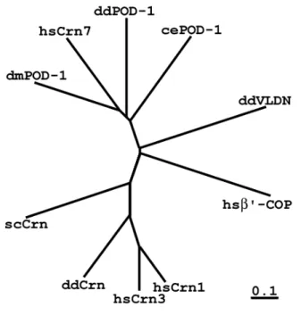
homologous and 30% and 29% identical to Dpod-1 and POD-1, respectively as predicted by NCBI BLAST server using BLOSUM62 matrix. Similarly to the worm and fly homologues, Crn7 lacks the C-terminal coiled-coil region. Phylogenetic analysis using cluster algorithm clearly positions Crn7 and both previously described POD-1 proteins in a group distinct from other coronins (Fig. 3).
A structural peculiarity of Crn7 is the presence of a 47-amino acid long proline, serine and threonine-enriched stretch upstream of the second group of WD repeats designated a PST
motif (Fig. 2). Such sequence is not found in any other coronin proteins, and its function (if any) remains unclear.
Fig. 3. Phylogenetic analysis of the coronin family members, including POD-1 proteins, performed using the cluster algorithm. dmPOD-1 – Drosophila dPOD-1 protein, cePOD1 – C. elegans POD-1, ddPOD-1 – POD-1 protein from Dictyostelium discoideum, hsCrn1, 3, 7 – human coronins 1, 3 and 7, respectively, ddCrn – Dictyostelium coronin, scCrn – yeast (Saccharomyces cerevisiae) coronin, ddVLDN – Dictyostelium villidin, hsβ'-COP – human β'-COP. Multiple WD-repeat proteins villidin and β'-COP are clearly forming outgroups with regard to both coronins and POD-1 proteins. The bar corresponds to 10%
of amino acid substitution within the branch.
Additionally, Crn7 harbours two putative copies of a classical tyrosine-based sorting signal downstream of each core domain (Fig.1). This feature is also unique for Crn7, and will be discussed later.
3.2. Tissue distribution and developmental dynamics of Crn7
Using a monoclonal antibody K37-142-1 raised against the C-terminus of Crn7 (Rybakin et al., 2004), we analyzed the distribution of the protein in murine tissues by western blot. Crn7 was found to be ubiquitously expressed in all studied tissues, except for the heart and skeletal muscle (Fig. 4). These findings are in good agreement with Northern blot data shown previously (A. Schulze, Diploma thesis, University of Cologne, 2001). K37-142-1 mAb is used in all further experiments to visualize Crn7.
Fig. 4. Tissue distribution of the Crn7 mRNA and protein. Top panel, tissue lysates were separated on 10% SDS-polyacrylamide gels, blotted onto a nitrocellulose filter and probed with monoclonal anti-Crn7 antibody. 1 - brain, 2 - heart, 3 - liver, 4 - lung, 5 - kidney, 6 - testis, 7 - muscle, 8 - thymus. Anti-β-actin monoclonal antibody was used to confirm equal loading (not shown).
Using indirect immunofluorescence, we studied the expression pattern of the Crn7 protein in the mouse. The strongest expression is found in the early postembryonic brain, thymus, intestine, skin and in the eye (Fig. 5). In the brain cortex, the Crn7 protein is restricted to the most apical cell layers before postembryonic day 10 and shifted to a population of more basal cells thereafter, whereas in the hypothalamus the protein is detected in the same set of profound big neurons throughout development. In addition, the protein is found in the bodies and dendrites of
Purkinje cells in the cerebellum. In the skin, Crn7 is only present in the apical epidermis layers (Fig. 5). The differentiating cells in the embryonic eye are also found to be Crn7 positive. Here, Crn7 is found in developing lens fibers. In adult mice, the protein is strongly expressed in the outer plexiform layer of the retina, where the rods are located (data not shown). Interestingly, in the intestine, the protein is found not only in terminally differentiated epithelial cells, but also in the crypt epithelium were the stem cells are located (Fig. 5J).
Fig. 5. Immunolocalization of the Crn7 protein in paraformaldehyde-fixed murine early postembryonic tissues. A-D, sections through the brain cortex at the postembryonic days 5, 10, 20 and 30, respectively.
E-G, section through the hippocampus at the postembryonic days 10, 20 and 30, respectively. H, localization of the Crn7 protein in the Purkinje cell bodies (arrows) and dendrites (arrowheads). I-K,
I correspond to the apical side of the corresponding organs. Immunostaining was performed using monoclonal anti-Crn7 antibody (green in I-K). Sections were counter-stained with DAPI (blue in I-K).
3.3. Subcellular localization of Crn7
3.3.1. Crn7 is present in the cis-Golgi and in the cytosol
Using indirect immunofluorescence, we analyzed the cellular distribution of Crn7. The protein was found in vesicle-like structures and in a Golgi-like perinuclear compartment (Fig. 6).
The characteristic Crn7-positive perinuclear structure was prominent in NIH 3T3 fibroblasts, HeLa, Vero and other cell types. Crn7 protein co-localizes with cis-Golgi markers β-COP and Erd2p in the Golgi region as shown by indirect immunofluorescence (Fig. 6). It is noteworthy that (a) in both cases the Crn7 antibody stains a broader region than both Golgi marker antibodies (Fig. 6C, F, arrows), and (b) the cytosolic Crn7-positive vesicles are clearly distinct from the Erd2p- or β-COP-positive ones (not shown). Cis-Golgi localization of Crn7 was further confirmed by immunostaining NIH 3T3 fibroblasts expressing GFP-fused cis-Golgi markers p23 and GM130 with Crn7 antibody (data not shown). Importantly, although we were able to observe a certain co-localization of Crn7 with the trans-Golgi marker TGN38, such co-localization was restricted to the proximal Golgi region and almost absent in the trans-most cisternae (Fig. 6I).
Fig. 6. Immunolocalization of the Crn7 protein in NIH 3T3 cells. Paraformaldehyde-fixed cells were stained with monoclonal anti-Crn7 antibody (B, E, H) and rabbit polyclonal antibodies against either Erd2p (A), β-COP (D), or TGN38 (G). Primary antibodies were detected with goat anti-mouse antibody conjugated with Cy3 (red) and sheep anti-rabbit antibody conjugated with FITC (green). C, F, I, merged false color images. Insets in C, F, I correspond to the areas marked in A-C, D-F and G-I, respectively.
Bar, 10 µm.
Using differential centrifugation, we established that only a minor pool of Crn7 is localized to intracellular membranes. The protein is unequally distributed between the cytosol and membrane fractions, the bulk of it being found in the cytosol (Fig. 7A), where it is present in
membrane-associated Crn7 protein can be extracted from the 10,000g fraction upon treatment with Triton X-100 (Fig. 7B). As a control, we used anti-β-actin antibody (Fig. 7B, lower panel), showing that only the detergent-sensitive membrane components have been extracted, but not cytoskeletal elements.
Fig. 7. Crn7 is present on Triton-soluble membranes. A, differential centrifugation experiment showing the presence of the Crn7 protein in heavy membrane / cytoskeletal fraction (10,000g pellet, lane 1), light membrane fraction (100,000g pellet, lane 2), cytosolic protein complexes (200,000g pellet, lane 3) and cytosol (200,000g supernatant, lane 4). B, solubilization of membrane compartments from the 10,000g pellet with Triton X-100. The 10,000g pellet was resuspended in homogenization buffer (Spoerl et al., 2002) and separated into two aliquots. One aliquot was treated with 0.5% Triton X-100 for 30 min at 4oC, the other served as control. Both aliquots were centrifuged again and pellets and supernatants analyzed by western blot. P - 10,000g pellet, S – supernatant. Left, control pellet; right, Triton X-100- treated pellet. Top panels, western blot with anti-Crn7 antibody, bottom panels, with anti-β-actin antibody. C, Crn7 is extracted from membrane fraction by incubation with sodium carbonate (see 3.3.2).
Upper panel, sodium carbonate was added to PNS of HeLa cells for 30 min on ice to make 100 mM. PNS was then centrifuged at 100,000g to separate membranes from cytosol. Lower panel, PNS was incubated
To localize the protein more precisely within the Golgi complex, we used confocal microscopy to study its co-localization with cis- and the trans-Golgi markers in HeLa cells treated with nocodazole, which is known to partially disrupt Golgi stacks leading to the formation of ministacks where the positions of cis- and the trans-Golgi proteins can be microscopically distinguished (Neubrand et al., 2005). Upon application of 20 µg ml-1 nocodazole for 2 hrs, Crn7 co-localized with a fraction of the cis-Golgi compartments positive for GM130 (Fig. 8A-C), but not with TGN38-positive trans-Golgi-derived compartments (Fig. 8D-F). Thus, Crn7 labels a subcompartment of the cis-Golgi.
Fig. 8. Crn7 is localized to the outer side of the cis-Golgi membranes. A – F, HeLa cells were treated with nocodazole at 20 µg ml-1 for 2 hrs, fixed and stained for Crn7 (A, D), GM130 (B) or TGN38 (E). C, F –
micrograph demonstrating Crn7 immunostaining (red circles) at the outer side of Golgi membranes in HeLa cells. Size bar, 200 nm.
The cytoplasmic Crn7-labelled structures (see Fig. 7) do not correspond to the intermediates of the endocytic pathway, as they do not co-localize with transferrin-positive compartments after 1, 5, 10, 30 or 60 minutes of the internalization of FITC-labelled transferrin in Triton-permeabilized cells (Fig. 9 and not shown) and do not show any Rab5 or LIMP-1 staining (data not shown). We assume that these structures represent large protein complexes constituting the cytosolic pool of the Crn7 protein.
Fig. 9. Crn7 is not present in early and late endosomal compartments positive for transferrin. HeLa cells were allowed to internalize Tf-FITC for 10 (A) or 30 (B-D) min on ice, then fixed and stained for Crn7 (red). C, D, - areas highlighted in B. C - Crn7 antibody staining, D, Tf-FITC signal. Note the exclusion of Crn7 from the late endosome/lysosome accumulation zone (C, D). Golgi Crn7 staining is absent due to permeabilization with Triton X100.
3.3.2. Crn7 is localized to the outer side of Golgi membranes
Further, we exploited the topology of Crn7 - Golgi membrane interactions. We
possess predicted signal sequences, signal cleavage or transmembrane sites (as predicted by PSORTII algorithm, (Nakai and Horton, 1999). To prove this hypothesis, we first used the Crn7 antibody and 5-nm immunogold detection to visualize the protein in rat Purkinje cells. Crn7 could be found only at the outer side of Golgi membranes, but not at the inner side or in the lumen (Fig. 8G). The conclusion that Crn7 is localized to the cytosolic side of the Golgi membrane was additionally confirmed by carbonate extraction (Fig. 7C) and proteinase K protection experiments, where Crn7 could not be protected by membranes from proteinase added to membrane fraction (not shown).
3.3.3. Stabile association of Crn7 with the Golgi requires the integrity of ER-to-Golgi transport Brefeldin A (BFA) causes the GTP-to-GDP exchange on Arf1 GTPase and rapid dissociation of Arf1 (Fig. 10) and COPI coat from the membranes (Peyroche et al., 1999).
Following this, Golgi membranes start fusing with the ER, and resident Golgi proteins are gradually redistributed to the ER (Lippincott-Schwartz et al., 1989). We treated cells with brefeldin A and assayed βCOP and Crn7 dynamics (Fig. 11A-F). Already after one minute of BFA treatment, no typical βCOP Golgi staining could be observed by immunofluorescence (Fig.
11E), well in agreement with published data on Arf1 dynamics (Presley et al., 2002).
Fig. 10. Effect of Brefeldin A (BFA) on the presense of small GTPAse Arf1 on Golgi membranes (from Presley et al., 2002).
Fig. 11. Crn7 is a structural Golgi protein, and depends on ER-to-Golgi transport in its localization. A – F, HeLa cells were treated with 5 µg ml-1 brefeldin A for 1 min (B, E) or 20 min (C, F), fixed and stained for Crn7 (A-C) or βCOP (D-F). Size bar, 20 µm. G, Fixed HeLa cell demonstrating the presence of Crn7 on an ER-Golgi intermediate (arrowheads) formed upon the application of BFA for 15 min. H, I, HeLa cells overexpressing GFP-Syn5 (asterisks) are characterized by reduced presence of Crn7 at Golgi membranes. H, Merged image, red, Crn7, green, GFP-Syn5. I, Crn7. Size bar, 20 µm.
Although Crn7 Golgi staining was gradually reducing upon application of BFA, βCOP dissociation did not result in the same rate of dissociation of Crn7, and some protein was present
on the collapsing Golgi even 20 min after the beginning of treatment (Fig. 11C). Such dynamics of Crn7 dissociation upon the application of BFA suggest that Crn7 is a structural protein intimately connected to the Golgi membrane. Interestingly, Crn7 was present at chimaeric tubular intermediates apparently connecting Golgi remnants with the ER (Fig. 11G). This finding additionally confirms the presence of Crn7 on Golgi membranes, rather than in the Golgi matrix.
Next, we tested whether Crn7 localization on Golgi membranes depends on the integrity of the ER-to-Golgi trafficking system. Recently, a cis-Golgi t-SNAREs complex was shown to participate in the late stages of ER-to-Golgi transport (Zhang and Hong, 2001). The complex consists of four SNARE proteins, syntaxin 5 (Syn5), GS28, Bet1 and Ykt6. Syntaxin 5 was previously shown to specify docking sites for both COPI and COPII vesicles in the Golgi complex (Hui et al., 1997, and references therein). In particular, Syn5 inhibits the import of the vesicular stomatitis virus G-glycoprotein (VSVG) into the Golgi complex if overexpressed, leading to accumulation of cargo in pre-Golgi intermediates (Dascher et al., 1994). We overexpressed GFP-Syn5 in HeLa cells and examined Crn7 localization by immunostaining. The cells expressing GFP-Syn5 exhibit a substantial increase in cytosolic Crn7 staining, and reduced amount of Crn7 on Golgi membranes (Fig. 11H,I). Thus, Crn7 requires the intact influx of certain ER-derived material to be localized to the Golgi.
Because Crn7 lacks any signal peptide and is present in both cytosol and Golgi (see above), we imply that it is recruited to the outer side of Golgi membrane from the cytosol rather than to the Golgi lumen or inner membrane side from the ER. This implication is confirmed by our studies on Crn7 topology using biochemical methods and electron microscopy. Thus, the observed inhibitory effect of Syn5 overexpression on the Golgi localization of Crn7 can hardly be due to the direct block of its import from the ER. It is intriguing to speculate that the Golgi
manner. Interaction of Crn7 with this unidentified partner might be dependent on its tyrosine phosphorylation, as the membrane-associated Crn7 is phosphorylated on tyrosine residues, while the cytosolic protein is not (see above).
3.3.4. Properties of cytosolic and membrane-associated forms of Crn7
We analyzed the Crn7 protein in membrane and cytosolic fractions by means of 2D gel electrophoresis. Our results evidence that the pI values of the cytosolic form of the protein range between 5.2 and 6.0 with a peak corresponding to the predicted value of 5.6, whereas the membrane-associated form has pI values ranging from 4.5 to 6.0 (Fig. 12A), inferring that the membrane bound form is phosphorylated. Furthermore, when we immunoprecipitated Crn7 from the cytosol and membrane fractions (10,000g pellet) using Crn7 antibody coupled to protein G- sepharose beads in the presence of phosphatase inhibitors the precipitated protein specifically reacted with an anti-tyrosine antibody. No labeling was observed with phosphoserine / threonine specific antibodies (data not shown). Moreover, our results indeed confirm that it is the membrane-associated, but not cytosolic form of Crn7, which is phosphorylated on tyrosine residue(s) (Fig. 12B). Importantly, tyrosine phosphorylation does not only correlate with Crn7 presence in the membrane pellet, but also is required for it. We incubated HeLa cells in the presence of tyrosine kinase inhibitor genistein (1 hr, 100 µg/ml, 37oC), prepared PNS and subfractionated it by centrifugation at 100,000g. Fig. 12C demonstrates that inhibition of tyrosine phosphorylation led to a decrease in Crn7 abundance in the membrane fraction.
There are several conserved tyrosines in the protein sequence that might be the targets for phosphorylation. Y738 is conserved among both Crn7, POD-1, Dpod-1 and mammalian coronins 2A and 3, Y712 is conserved in Crn7, and both POD-1 proteins. Several other tyrosine residues are present in Crn7 and one or several coronins or POD-1 proteins.
Fig. 12. Crn7 present in the membrane pellet of HeLa cells is phosphorylated on tyrosine residues. A, two- dimensional gel electrophoresis of the proteins present in the 10,000g supernatant and pellet. After separation, the gel was blotted onto a nitrocellulose membrane and probed with Crn7 antibody. B, analysis of tyrosine phosphorylation of the Crn7 protein. The protein was immunoprecipitated from the 10,000g pellet (P) and supernatant (S) using Crn7 antibody. After separation, the gel was blotted onto the nitrocellulose membrane and probed with anti-phosphotyrosine (left) or anti-Crn7 (right) antibody. C, Tyrosine phosphorylation is required for the Crn7 targeting to membranes. Application of tyrosine kinase inhibitor genistein lowers the amount of Crn7 in the membrane pellet (P).
3.4. Analysis of Crn7 function by RNAi 3.4.1. Use of siRNA duplexes to silence Crn7
To reveal the in vivo function of Crn7 in the Golgi, we used the small interfering RNA (siRNA) methodology allowing a specific and powerful knockdown of the mRNA and corresponding protein (Novina and Sharp, 2004). We used eight siRNA oligonucleotides (see Methods). 48 and 72 h after transfection, cells were harvested and analyzed by Western blotting.
All siRNA constructs were capable of downregulating Crn7 protein at both treatment terms (Fig.
13A). As the construct siRNA(8)2454consistently showed the highest degree of downregulation after 48 and 72 hrs, this construct was used in further experiments. siRNA timescale experiments demonstrated that knocking down Crn7 mRNA and protein leads to a dramatic decrease in cell viability, as compared to mock-transfected cells. This effect was both time- and concentration- dependent (Fig. 13B). Thus, Crn7 is an essential protein in HeLa cells.
Fig. 13. Downregulation of Crn7 by RNAi results in reduced cell viability and profound changes in Golgi architecture. A, The use of eight individual RNA duplexes to downregulate Crn7 (see Methods). Upper panel, 48 h of RNAi application, Western blot using Crn7 antibody. Middle panel, 72 h of RNAi application, Western blot using Crn7 antibody. Lower panel, 72 h of RNAi application, Western blot using a mixture of coronin 3 (Crn3) and actin antibodies. B, Reduced cell viability upon the application of Crn7
siRNA, open circles, 15 nM siRNA. Cells were counted 24, 48, 72 and 96 h after the application of siRNA.
Note logarithmic y-scale.
3.4.2. Influence of Crn7 RNAi on the Golgi architecture
To evaluate the effect of Crn7 knockdown by RNAi on the architecture of the Golgi complex, we treated HeLa cells with siRNA(8)2454 for 24 h and processed them for electron microscopy along with mock-treated cells as described in Methods. Mock-transfected cells displayed the expected Golgi morphology, characterized by the presence of several flat cisternae surrounded by transport intermediates (Fig. 14, left). In contrast, HeLa cells treated with siRNA(8)2454demonstrated different degrees of the Golgi scattering. In most cells, the Golgi was present as a dense accumulation of vesicles still containing one or several cisternae-like structures (Fig. 14, right). In the most extreme cases, no cisternae are observed, and the density of Golgi remnant vesicles decreases. These data suggest that Crn7 is indeed required for the maintenance of the Golgi stability.
Fig. 14, Ultrastructure of the Golgi complex in mock- (left) and Crn7 siRNA-transfected (right) cells.
Bars, 100 nm.
To better understand the effect of Crn7 knockdown on the Golgi architecture, we studied the distribution of cis- and the trans-Golgi markers in fixed mock- and Crn7 siRNA- transfected HeLa cells (Fig. 15). After 24 h of RNAi application, the Golgi ribbon was not present anymore, and smaller Golgi fragments were scattered in the perinuclear area. The trans- Golgi protein TGN38 and the cis-Golgi marker GM130 were still present in predominantly non- overlapping domains localized close to each other, some of them being organized in ministacks (Fig. 15, right), reminiscent of those forming upon nocodazole treatment (Trucco et al., 2004).
Importantly, we could not register any vesicles positive of TGN38 in Crn7 RNAi cells. The absence of TGN38 carriers combined with the accumulation of TGN38 in the Golgi remnants rather than at the cell periphery indirectly indicates an RNAi effect on anterograde, but not retrograde transport of TGN38. At later RNAi terms, the overall structure of the cis-Golgi did not further change, but the trans-Golgi collapsed completely, and TGN38 was only labelling several round-shaped dense compartments per cell (not shown).
Fig. 15. Mock- (left) and Crn7 siRNA-transfected HeLa cells stained with GM130 (red) and TGN38 (green) antibodies. Note TGN38 carrier vesicles (arrows, and a magnification inserted in E, corresponding to the highlighted area). Size bars, 2 µm.
3.4.3. Block of protein export from the Golgi in Crn7 RNAi cells.
As we found the formation of TGN38 carrier intermediates to be blocked upon the Crn7 knockdown, we wanted to know whether such effect of Crn7 downregulation on the Golgi export can be reproduced and quantified using the VSV envelope G glycoprotein. VSVG is known to hijack the anterograde transport system to reach the cell surface after proceeding through the ER and Golgi. A VSVG-tsO45 mutant carries a single point mutation F204S (Gallione and Rose, 1985) rendering it temperature-sensitive with regard to its intracellular trafficking. The mutant protein can be accumulated in the ER at 39.5oC, and released to the Golgi complex upon temperature switch to 32oC (see Methods). This mutant is widely used to study the dynamics of protein trafficking along the biosynthetic pathway (Hirschberg et al., 1998; Presley et al., 1997).
Fig. 16. Block of Golgi export in Crn7 RNAi HeLa cells. Cells were transfected in liquid phase with siRNA and plated for 14 hrs at 37oC, then transfected with VSVG-GFP, kept at 37oC for 2 hrs and at 39.5oC for 8 hrs. For imaging, cells were kept at 32oC to allow VSVG exit from the Golgi. A, Export of