Mechanisms of the Ca
v2.3 Calcium Channel’s Role in Epileptogenesis and Antiepileptic Pharmacotherapy
Inaugural Dissertation zur
Erlangung des Doktorgrades Dr. nat. med.
der Medizinischen Fakultät und
der Mathematisch-Naturwissenschaftlichen Fakultät der Universität zu Köln
vorgelegt von
Maxine Dibué-Adjei aus New York
Köln, 2015
2
Mechanisms of the Ca
v2.3 Calcium Channel’s Role in Epileptogenesis and Antiepileptic Pharmacotherapy
Inaugural Dissertation zur
Erlangung des Doktorgrades Dr. nat. med.
der Medizinischen Fakultät und
der Mathematisch-Naturwissenschaftlichen Fakultät der Universität zu Köln
vorgelegt von
Maxine Dibué-Adjei aus New York
Köln, 2015
3
Berichterstatter/Berichterstatterin: Prof. Dr. Stefan Herzig Prof. Dr. Peter Kloppenburg
Tag der letzten mündlichen Prüfung: 31.10.2014
4
For Mom, Dad and Jason
“If the brain were simple enough for us to understand it, we would be too simple to
understand it.” ― Ken Hill
5
I. Table of Contents
I. TABLE OF CONTENTS 5
II. SUMMARY 7
III. ZUSAMMENFASSUNG 10
1. INTRODUCTION 13
1.1 Epilepsy 13
1.1.1 A Concise History of Epilepsy 13
1.1.2 Epilepsy Today 14
1.1.3 Mesial Temporal Lobe Epilepsy 14
1.1.4 Treatment of Mesial Temporal Lobe Epilepsy 16 1.1.5 The Kainic-Acid Model of Temporal Lobe Epilepsy 17 1.2 The Ca
v2.3 (R-type) Voltage-Gated Calcium Channel 18 1.2.1 Structure of Voltage-gated Calcium Channels 18 1.2.2 Classification of Voltage-gated Calcium Channels 19 1.2.3 The Ca
v2.3 (R-type) Voltage-Gated Calcium Channel and
its Physiological Functions
20 1.3 Ca
v2.3 in Epilepsy and Antiepileptic Pharmacotherapy 22
1.3.1 Ca
v2.3 VGCCs in Epilepsy 22
1.3.2 Experiments with Ca
v2.3KO Mice Experimental Models of Epilepsy
23 1.3.3 Ca
v2.3 as a Target of Antiepileptic Drugs 23
1.4 Cardiac Phenomena during Epilepsy 26
1.4.1 Cardiac Phenomena in Epileptic Patients 26
1.4.2 Effects of AEDs on Cardiac Function 27
1.5 Epilepsy and Sleep 28
1.5.1 Physiological Sleep 28
1.5.2 Regulation of Sleep 29
1.5.3 The Relationship between Sleep and Epilepsy 29
1.6 Experimental Goals 30
2 RESULTS
31 2.1 Ca
v2.3 (R-type) calcium channels are critical for mediating
anticonvulsive and neuroprotective properties of Lamotrigine in vivo
31
2.2 Cardiac Phenomena During Kainic-acid Induced Epilepsy and Lamotrigine Antiepileptic Therapy
33 2.3 Ca
v2.3 E-/R-type voltage-gated calcium channels modulate
sleep in mice
35
3 DISCUSSION 37
3.1 Results from the three presented studies in summary: 37
6
3.2 Ca
v2.3 in Oscillatory Networks 37
3.2.1 R-type Currents in Thalamo-Cortical and Hippocampal Rhythmicity
37 3.2.2 R-type Signaling as a Mediator of Sleep Homeostasis –an
Intersection Point between Sleep and Epilepsy?
39 3.2.3 Ca
v2.3, a “Fine Tuner” of Oscillatory Activity? 40 3.3 Ca
v2.3 as a Key Player in Lamotrigine Pharmacology 41
3.4 Concluding Remarks 42
Reference List 44
Curriculum Vitae 57
Erklärung 61
Publications 63
7
II. SUMMARY
RATIONALE: The Ca
v2.3 (R-type) voltage-gated calcium channel represents the most enigmatic of all voltage-gated calcium channels due to its pharmacoresistance, mixed characteristics of high voltage-activated and low voltage-activated calcium channels and relatively low expression levels. Lamotrigine (LTG) is a modern antiepileptic drug however, its mechanism of action has yet to be fully understood, as it is known to modulate several ion channels and other targets. In heterologous systems, LTG inhibits Ca
v2.3 (R-type) calcium currents, which contribute to kainic-acid (KA)–induced epilepsy in vivo. LTG has been suggested to increase the risk of sudden unexpected death in epilepsy (SUDEP), in which cardiac and respiratory mechanisms are proposed to be involved. In addition to the higher risk of SUDEP during sleep, epileptic patients are at higher risk of seizures during sleep, especially during slow wave sleep (SWS). The bidirectional relationship between sleep and epilepsy has long been acknowledged, however it remains far from understood.
AIM: The goal of the present project was to perform an in depth investigation of the role of R-type signaling in the epileptic brain and heart, by analyzing its contribution to experimental epilepsy, antiepileptic pharmacotherapy and sleep.
METHODS: In the first study we compared the effects of LTG to two other AEDs (Topiramate and Lacosamide) in Ca
v2.3-deficient mice and controls on KA-induced seizures. Behavioral seizure rating and quantitative electrocorticography were performed after KA induced epilepsy, as well as immunohistochemistry and western blot analysis of Ca
v2.3 expression in the brain. In the second study we investigated cardiac parameters during KA-induced epilepsy and LTG treatment in awake and sleeping C57Bl6 mice.
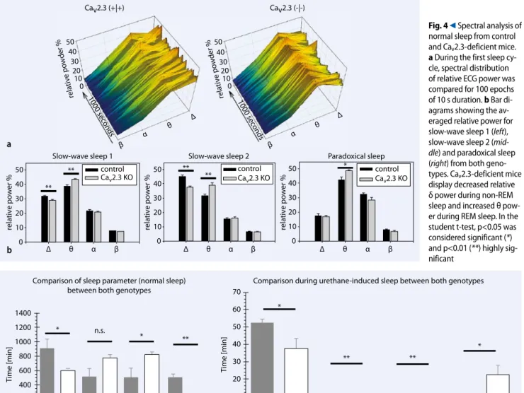
Continuous electrocardiograms and electrocorticograms were collected telemetrically from freely moving mice, and time- and frequency-domain analysis performed on the electrocardiograms. In the third study, we analyzed sleep architecture in Ca
v2.3-deficient and control mice also using radiotelemetric electrocorticography and electromyography during spontaneous and urethane-induced sleep.
RESULTS: LTG treatment displayed no antiepileptic potency in Ca
v2.3-deficient mice,
but contrarily significantly aggravated seizures and increased neurodegeneration in the
8
CA1 region of the hippocampus as well as increasing ultra-high frequency oscillations (ripples) known to be associated with seizure generation. This effect was specific to LTG in Ca
v2.3-deficient mice, as the two other AEDs tested - one with and one without Ca
v2.3 inhibiting capacity- did not aggravate seizures. In our second study we found LTG to alter autonomous nervous control of the heart during SWS after induction of chronic epilepsy promoting sympatho-vagal imbalance. Furthermore, we found LTG to increase the squared-coefficient of variation of the heart rate during SWS, but not during wakefulness. Our third study was able to demonstrate, that ablation of Ca
v2.3 robustly impacts sleep architecture, producing deficits in the amount and depth of SWS.
Interestingly, although Ca
v2.3 mice sleep less and display shorter SWS phases, they do not compensate for this deficit by increasing sleep depth, pointing to disturbances in sleep homeostasis.
DISCUSSION: We provide first in vivo evidence for a crucial role of R-type signaling in LTG pharmacology and shed light on a paradoxical effect of LTG in the absence of Ca
v2.3. LTG appears to promote ictal activity in Cav2.3-deficient mice by increasing high frequency components of seizures, resulting in increased neurotoxicity in the CA1.
This paradoxical mechanism, possibly reflecting rebound hyperexcitation may be key in understanding LTG-induced seizure aggravation observed in patients. Furthermore, we find Ca
v2.3 to be a critical mediator of sleep homeostasis, potentially representing a pivotal link between sleep and epilepsy. Ca
v2.3 has been shown to be crucial for bursting in the reticular thalamus, which underlies delta-rhythm during SWS and generation of spike-and-wave discharges, the hallmark of absence epilepsy. Therefore, seizure resistance and SWS impairment of Ca
v2.3-deficient mice may be symptomatic of impairment of bursting in the thalamus and therefore of the generation and maintenance of highly synchronized slow rhythms. Remarkably, we found LTG to only affect autonomous control of the epileptic heart during SWS, possibly indicating a mechanism by which LTG could increase the risk for SUDEP. LTG-induced increased sympathetic tone during SWS, may also reflect impaired SWS, found in LTG-treated patients and in our Ca
v2.3-deficient mice.
CONCLUSION: Because Ca
v2.3-deficient mice display a subtle phenotype as oppposed
to an obvious one, because of the expression of Ca
v2.3 in rhythmically active tissue and
9
because of Ca
v2.3’s unique electrophysiological properties, it is conceivable that a
general function of R-type currents is the “fine tuning” of oscillatory networks One may
assume that a loss of “fine-tuning” in Ca
v2.3KO mice is only minimally noticeable under
physiological conditions, but becomes evident in certain pathological conditions exerting
a strain on an oscillatory network such as during experimentally induced epilepsy. This
may explain how R-type signaling is crucial for sustaining physiological rhythmic
activity of an entire network despite relatively low expression levels.
10
III. ZUSAMMENFASSUNG
HINTERGRUND: Der Ca
v2.3 (R-Typ) spannungsgesteuerte Kalziumkanal ist der enigmatischste aller Kalziumkanäle aufgrund von seiner Pharmakoresistenz, Charakteristika zwischen hoch- und niedrig- spannungsgesteuerten Kalziumkanälen und relativ niedrigen Expression. Lamotrigin (LTG) ist ein beliebtes modernes Antiepileptikum (AED), jedoch sind seine Wirkmechanismen noch unklar, da LTG viele Ionenkanäle und andere Zielstrukturen moduliert. Im heterologen System, inhibiert LTG R-Typ Ströme, die in vivo zu Kainat (KA)-induzierter Epilepsie beitragen. Es wurde gezeigt, dass LTG das Risiko für plötzlich auftretenden, ungeklärten Tod bei Epilepsie (SUDEP) erhöht, ein Phänomen, bei dem kardiale und respiratorische Mechanismen impliziert sind. Außerdem ist das Risiko für epileptische Anfälle bei Patienten höher im Schlaf, besonders im Langsame-Wellen-Schlaf (SWS). Die bidirektionale Beziehung zwischen Schlaf und Epilepsie ist schon lange bekannt, jedoch dem vollen Verständnis noch fern.
ZIELE: Ziel des gegenwärtigen Projekts, war es eine tiefgründige Analyse der Rolle des R-Typ Kalziumkanals in dem epileptischen Hirn und Herz durchzuführen, indem seine Mitwirkung bei experimenteller Epilepsie, antiepileptischer Pharmakotherapie und Schlaf analysiert wird.
METHODEN: In der ersten Studie wurden die Effekte von LTG und zwei anderen AEDs (Topiramat und Lacosamid) auf Ca
v2.3-defizienten und Kontrollmäusen bei KA–
induzierter Epilepsie untersucht. Neben telemetrischer Erfassung von Elektrokortikogrammen, wurde der Schweregrad der Anfälle quantifiziert.
Immunohistochemie und Westernblot Analyse von Ca
v2.3 Expression im Hirn wurden
auch ausgeführt. In der zweiten Studie wurden kardiale Parameter nach KA-induzierter
Epilepsie und anschließender LTG-Behandlung in wachen und schlafenden C57Bl6
Mäusen untersucht. Langzeit Elektrokardiogramme und Elektrokortikogramme wurden
telemetrisch von sich–frei bewegenden Mäusen erhoben und anschließend Zeit- und
Frequenzdomänen ausgewertet. In der dritten Studie wurde spontanes und Urethan-
11
induziertes Schlafverhalten in Ca
v2.3-defizienten und Kontrollmäusen mittels telemetrische Elektrokortikographie und Elektromyographie untersucht.
ERGEBNISSE: In Ca
v2.3-defizienten Mäusen zeigt LTG keine antiepileptische Potenz, sondern verstärkt epileptische Anfälle und Neurodegeneration in der CA1 Region des Hippokampus und verstärkt ultra-hochfrequente Oszillationen (Ripples), von denen gezeigt wurde, dass sie mit der Entstehung von iktaler Aktivität assoziiert sind. Dieser Effekt war spezifisch für LTG in Ca
v2.3-defizienten Mäusen und entstand nicht bei der Behandlung von Ca
v2.3-defizienten Mäusen oder Kontrollmäusen mit den anderen beiden getesteten AEDs. In der zweiten Studie konnte gezeigt werden, dass bei chronisch epileptischen Mäusen, LTG die autonom-nervöse Kontrolle des Herzes während SWS beeinflusst, indem sympathische Kontrolle der Herzaktivität verstärkt wird. Zudem fanden wir eine Zunahme des Variationskoeffizienten im Quadrat im SWS, jedoch nur bei SWS und nicht bei Wachheit. Unsere dritte Studie konnte zeigen, dass die Ablation von Ca
v2.3 durch verkürzte SWS Phasen und reduzierte SWS Tiefe, robuste Veränderungen des Schlafverhaltens verursacht. Obwohl Ca
v2.3-defiziente Mäuse weniger Schlafen und kürzere SWS Phasen zeigen, kompensieren sie dieses nicht durch Vertiefung des SWS, was auf eine Beeinträchtigung von homöostatischen Mechanismen deutet.
DISKUSSION: Wir zeigen erste in vivo Belege für eine entscheidende Rolle von R-Typ
Kalziumkanälen in dem Wirkmechanismus von LTG und decken einen paradoxen Effekt
von LTG in der Abwesenheit von Ca
v2.3 auf. LTG scheint iktale Aktivität in Ca
v2.3-
defizienten Mäusen zu fördern, indem es ultra-hochfrequente Komponente von Anfällen
verstärkt, was erhöhte Degeneration von CA1 Neurone verursacht. Dieser paradoxe
Mechanismus, der womöglich reaktive Hyperexzitation reflektiert, könnte
ausschlaggebend für das Verständnis von anfallsverstärkenden Effekten von LTG sein,
die bei epileptischen Patienten auftreten. Ca
v2.3 zeigt sich als bedeutender Vermittler der
Schlafhomöostase, und könnte somit ein entscheidender Knotenpunkt zwischen Epilepsie
und Schlaf darstellen. Ca
v2.3 spielt eine entscheidende Rolle beim Bursting im Thalamus,
welches dem Delta-Rhythmus unterliegt aber auch der Generation von Spike-and-Wave
Discharges (SWD). Demzufolge könnte die Resistenz gegenüber Chemokunvulsiva und
gestörtes SWS der Ca
v2.3-defizienten Mäuse symptomatisch für gestörtes Bursting im
12
Thalamus, und somit für die Generation und Aufrechterhaltung von hoch- synchronisierten langsamen Rhythmen sein. Bemerkenswerterweise, verschiebt LTG nur im SWS die autonom-nervöse Kontrolle des Herzes in sympathischer Richtung, was ein potentieller Mechanismus hinter dem erhöhten Risiko für SUDEP im Schlaf sein könnte.
LTG-induzierter erhöhter Sympathikotonus im SWS könnte auch beeinträchtigtes SWS wiederspiegeln, ein Phänomen, das auch in LTG-behandelten Patienten auftritt.
KONKLUSION: Weil Ca
v2.3-defiziente Mäuse einen subtilen statt einen eindeutigen Phänotyp zeigen, weil Ca
v2.3 eine einzigartige Kombination elektrophysiologischer Eigenschaften besitzt und weil Ca
v2.3 in verschieden rhythmisch-aktiven Geweben exprimiert wird, ist es annehmbar, dass eine Hauptfunktion dieses Kanals in der
„Feinjustierung“ von oszillierenden Netzwerken besteht. Es ist möglich, dass ein Verlust von „Feinjustierung“ unter physiologischen Bedingungen kaum auffällt, jedoch unter pathophysiologischen Bedingungen bzw. wenn das Netzwerk stark belastet wird, große Auswirkungen hat wie z.B. bei der Epilepsie. Das würde erklären, wie R-Typ Kanäle, trotz relativ niedriger Expression, ausschlaggebend für die Generation und Aufrechterhaltung von rhythmischer Aktivität in ganzen Netzwerken sein können.
13
1. INTRODUCTION
1.1 Epilepsy
1.1.1 A Concise History of Epilepsy
Epilepsy has been known to man for over 3000 years. The earliest written of account of an epilepsy disorder is an Akkadian text, the Sakikku, written around 1067-1046 BC (Kinnier Wilson and Reynolds, 1990), describing the “falling disease”. Although Hippocrates already identified epilepsy as a disorder of the brain in the 4
thcentury BC, the belief that epilepsy was of supernatural origin, for example due to possession by ghosts, demons, ancestors or even gods persisted into the 18
thcentury in many cultures.
The concept of epilepsy as a disorder of the brain slowly began to gain acceptance in the late 17
thcentury and by the early 19
thcentury it was though that epilepsy was caused by vascular dysfunction. Robert Bentley Todd was the first to recognize electrical dysfunction as the underlying cause of epilepsy refuting the hypothesis of vascular congestion. In the Lumleian Lectures delivered at the Royal College of Physicians in London in 1849, Todd describes animal experiments, in which he found excitation of certain brain regions by “stimulus of galvanism” to cause convulsions (Todd, 2005). The International League Against Epilepsy (ILAE) was founded by a group of physicians in 1909 in Budapest, which greatly influenced the advancement of the understanding of epilepsy. However, it was only until the discovery of the electroencephalogram (EEG) in 1929 by Hans Berger (Berger, 1929), that the electrical basis of epilepsy could be fully confirmed. By this time, phenobarbital, the first antiepileptic drug (AED) (after potassium bromide, which has a very low therapeutic index) had been available for 17 years, offering a first non-toxic method of seizure control, but at a high price, as phenobarbital, a barbiturate is extremely sedative and becomes hypnotic at higher doses.
Interestingly, despite the availability of over 30 FDA approved AEDs today,
phenobarbital remains the most commonly used AED in the world.
14 1.1.2 Epilepsy Today
Epilepsy is defined as the propensity for a person to suffer recurrent unprovoked seizures (WHO, 2005). The prominent British neurologist Gordon Morgan Holmes (1876-1965) defined a seizure as “a sudden, involuntary, time-limited alteration in behavior, motor activity, autonomic function, consciousness, or sensation, accompanied by an abnormal electrical discharge in the brain”.
Today, epilepsy is one of the most prevalent serious neurologic disorders, affecting over 50 million people worldwide (WHO, 2005). The mean number of people with epilepsy per 1000 is higher in low-income countries than in high income countries (9.55 vs 7.99), with 80-90% of people with epilepsy in most developing countries not receiving any treatment at all (WHO, 2005). Incidence rates of epilepsy are also higher in developing countries, which is thought to be due to higher rates of brain trauma, HIV, parasitic infection (particularly neurocysticercosis), perinatal morbidity and consanguinity (WHO, 2005). However, despite many other known circumstances that can cause epilepsy such as stroke, fever, tumors, infection or substance abuse, two thirds of epilepsy cases are idiopathic or cryptogenic (WHO, 2012). According to the ILAEs guidelines for classification, epilepsy, may be classified by etiology (1) and type of seizures (2):
(1): symptomatic: identifiable etiology
cryptogenic: presumed symptomatic but unknown etiology idiopathic: presumed genetic but unknown etiology
(2): partial: focal onset
generalized: initial involvement of both hemispheres unclassified: unknown onset
The ILAE recognizes 32 different epilepsy syndromes (Engel, 2006) with mesial temporal lobe epilepsy (MTLE) representing one of the most common epilepsy syndromes.
1.1.3 Mesial Temporal Lobe Epilepsy
MTLE, the most common type of temporal lobe epilepsy (TLE) is characterized by
seizures originating in the mesial temporal lobe i.e. in the hippocampus and
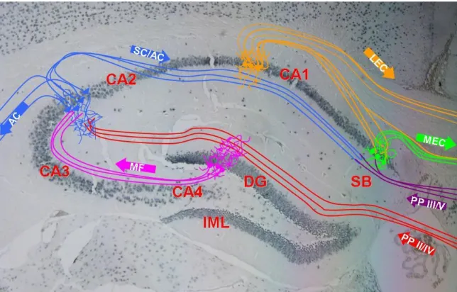
parahippocampal gyrus (Margerison and Corsellis, 1966) (See Figure 1 for connectivity
15
of the mesial temporal lobe). In MTLE aura and automatisms lasting 1-2 minutes, accompanied by autonomic or psychic symptoms precede complex partial seizures, which can evolve into generalized seizures or status epilepticus. If left untreated, neurologic and psychiatric health of MTLE sufferers deteriorates rapidly: seizures occur more frequently and with greater severity causing always greater damage to the brain and manifestation of psychiatric disease such as depression or personality changes. Compared to the general population, morbidity and mortality are increased in people with MTLE, due to accidents occurring from sudden loss of consciousness accompanying seizures.
However, high mortality also results from sudden unexpected death in epilepsy (SUDEP). Patients with therapy refractory MTLE, have a risk of sudden death that is 50 times greater than that in the general population, but are also at risk of dying during status epilepticus (SE). EEG is the primary tool used in diagnosis of MTLE. Interictal spikes and sharp waves (interictal epileptiform discharges (IEDs)), periodic-lateralized epileptiform discharges (PLEDs), lateralized focal or regional polymorphic delta activity (“slowing”) and a build-up of lateralized rhythmic 5-10 Hz sharp activity during seizures are characteristic for MTLE (Javidan, 2012). Generally, MTLE is considered to originate from lesions from febrile convulsions, status epilepticus (SE), mechanical trauma or cerebral infection during childhood, which cause spontaneous seizures after a 5-10 year latency period (Engel, 1993). Hippocampal sclerosis (HS), detectable in a structural MRI using a fluid-attenuated inversion recovery sequence (FLAIR), is considered by many to be the hallmark of MTLE; however one must note that HS does not occur in 30% of MTLE patients, who are therefore diagnosed with paradoxical temporal lobe epilepsy (PTLE). HS describes “scarring” of the hippocampus involving degeneration of neurons and gliosis in the cornu ammonis (Babb and Brown, 1986) as well as pathological sprouting of mossy fibers (MF) in the inner molecular layer of the dentate gyrus (DG) (Sutula et al., 1989). It is presumed that pathological remodeling of circuitry due to aberrant MF sprouts, forming synapses with spines of predominantly excitatory granule cells represents recurrent excitatory circuitry, which may underlie seizure generation (Lothman et al., 1992;Buckmaster et al., 2002). Seizures in MTLE are also referred to as
“limbic” seizures, as the Papez circuit (=subiculum → fornix → mammillary bodies →
mammillothalamic tract → anterior thalamic nucleus → cingulum → entorhinal cortex
16
→ (via perforant path) hippocampus), which is part of the limbic system, may be involved in seizure generation. It has been suggested that pathologic hyperexcitation from the hippocampus is transmitted to other limbic areas via the Papez circuit and then returns to the hippocampus as amplified input resulting in generalized seizures.
Figure 1 Schematic of connectivity of the hippocampus superimposed on a NeuN-stained coronal paraffin section of mouse brain at approximately -1.5 mm Bregma. The hippocampus forms a uni-directional network receiving most input from the entorhinal cortex (EC) and projecting back to the EC. The different layers of the EC project to the dentate gyrus (DG) and CA3 pyramidal layer via the perforant path (PP).
The CA3 also receives input from the DG via the mossy fibers (MF) and projects to the ipsilateral CA1 via the Schaffer Collateral Pathway (SC) and to the contralateral CA1 via the Associational Commissural Pathway (AC). The main output of the hippocampus is from the CA1 to the lateral EC (LEC) and from the subiculum (SB) to the medial EC (MEC).
1.1.4 Treatment of Mesial Temporal Lobe Epilepsy
MTLE is the most common type of epilepsy referred for epilepsy surgery, making it the most medically refractory type of epilepsy (Wass et al., 1996;Engel, 2001).
Approximately half of MTLE patients achieve complete seizure control with antiepileptic
pharmacotherapy, with lamotrigine (LTG) and carbamazepine (CBZ) often being the
AEDs of choice, to which another AED can be added if necessary. A small group of
17
MTLE patients achieve seizure freedom by combination of three or more AEDs, however in many cases, MTLE is susceptible to AED (mono-) therapy in childhood and adolescence and becomes pharmacoresistant with age. For intractable MTLE, anteromedial temporal resection (AMTR) is the treatment of choice, achieving seizure control in 60-90% of patients (Engel, 1996). Despite the high success rate, this surgical procedure comes at a high price, often causing anterograde amnesia and verbal memory deficits. Vagal nerve stimulation is a less invasive surgical procedure that is sometimes recommended before AMTR, however success rates are drastically lower.
1.1.5 The Kainic-Acid Model of Temporal Lobe Epilepsy
2-carboxy-4 (1-methylethenyl)-3-pirrolidiacetic acid or “kainic-acid” (KA) is a non-
degradable cyclic analog of glutamate, which causes hyperexcitation by agonism of
kainate-class ionotropic glutamate receptors (Wang et al., 2005). Rodent models using
systemic or local injections of KA are among the most popular models of epilepsy and
neurodegeneration. MTLE is easily modeled by systemic injection of KA, which induces
automatisms, partial seizures which can evolve into generalized seizures and progressive
sclerosis of the hippocampus leading to recurrent spontaneous seizures (Sharma et al.,
2007). KA, administered by intraventricular injection, was first used to experimentally
model MTLE in rats in 1978 and was found to selectively destroy hippocampal
pyramidal cells (Nadler et al., 1978). Since then numerous KA models have probed
different protocols (several low doses vs one high dose) and administration routes from
intrahippocampal injection to intravenous injection to intraperitoneal injection. Because
systemic administration of KA still causes specific degeneration of hippocampal
pyramidal cells, does not require surgery but is easily executed as an intraperitoneal (i.p)
or subcutaneous (s.c.) injection, it is preferred by many researchers. Rodent KA models
closely mimic phenomena observed in MTLE patients. KA causes necrosis and apoptosis
of pyramidal cells in the CA1 and CA3 subfields of the hippocampus and MF sprouting
in the inner molecular layer (IML) of the DG (Tauck and Nadler, 1985;Okazaki et al.,
1995) as well as changes in expression of AMPA-receptor subunits GluR1 and GluR2
(Sommer et al., 2001). Neurodegeneration is also detectable in associated limbic and
neocortical structures(Ben-Ari et al., 1979;Pollard et al., 1994). Astrogliosis, microgliosis
18
and neurogenesis in the hippocampus have also been confirmed by TUNEL, MHC I and II and BrdU immunohistochemistry, respectively. All in all, histopathologic alterations of the rodent hippocampus after systemic KA injection are very similar to histopathology of surgically resected hippocampal tissue from MTLE patients. Mouse models of human pathology offer several advantages, most notably genetic homogeneity and availability of transgenic animals, but they can be technically challenging due to the small size of mice.
The same evolution of seizures found in MTLE patients i.e. aura → automatisms → partial seizure → generalized seizure →status epilepticus can analogously be observed in mice after systemic KA injection as: immobility (“freezing”)→ automatisms → myoclonic jerking→ tonic-clonic seizure → status epilepticus. When faced with the task of quantifying behavioral severity of seizures, seizure rating scales adapted for mice such as one used by Morrison and colleagues allow for reproducible and accurate assessment of seizure intensity (Morrison et al., 1996).
1.2 The Ca
v2.3 (R-type) Voltage-Gated Calcium Channel
1.2.1. Structure of Voltage-gated Calcium Channels
In vertebrates, calcium is one of the most tightly regulated physiological variables. The intracellular calcium concentration must be kept within precise limits in order to maintain physiological signal transduction, during which calcium ions are crucial as charge carriers and also as second messengers, neurotransmitter release, contraction of myocytes, hormone release and several other processes (Burgen, 1968;Rubin, 1970;Ashley, 1971;Rasmussen et al., 1976;Rasmussen and Barrett, 1984).
Voltage-gated calcium channels (VGCCs) are heterooligomeric complexes of up to four
subunits (α
1, α
2δ, β, and sometimes γ), however they are primarily characterized by their
pore-forming α
1subunit (212-250 kDa), which harbours the voltage-sensing machinery
and the drug/toxin-binding sites and for which ten different encoding genes in the human
genome are known (Catterall, 2011;Catterall, 2000). The α
1subunit consists of four
homologous repeats (I–IV) containing six transmembrane α-helical segments each (S1-
S6)(Lacinova, 2005). The linker between segments S5 and S6 of each domain, which
19
loops back from the extracellular side of the cell membrane (pore loop or P-region), forms the inner pore surface, determining ion conductance and selectivity (Guy and Conti, 1990). With their positively charged residues (arginine or lysine), S4 segments act as the main voltage-sensors, carrying gating charges through the membrane upon opening or closing. It remains a matter of debate how exactly the S4 segments move at gating, and movement as a helical screw or a sweeping paddle have been proposed (Guy and Seetharamulu, 1986;Jiang et al., 2003;Tombola et al., 2006). Negative residues of segments S2 and S3 are thought to create the electric field necessary for movement of S4 (Tombola et al., 2006).
In addition to phosphorylation sites for protein kinases such as PKA, PKC and CAMKII, the α1 subunit also harbours the so-called alpha subunit interaction domain (AID), an 18 amino-acid-long motif in the I-II linker representing the site of interaction with the β subunit (Pragnell et al., 1994). Not only does the β subunit mediate trafficking of the α
1subunit to the plasma membrane, partly by masking an endoplasmic reticulum retention signal in the α
1subunit (Bichet et al., 2000), its association with the α
1subunit specifically modulates biophysical properties of the channel (Sokolov et al., 2000;Dolphin, 2003). Co-expression with some α
2δ subunits such as α
2δ-1 enhances membrane trafficking of α
1subunits and can increase current amplitude and activation and inactivation kinetics as well as induce a hyperpolarizing shift in the voltage dependence of activation (Felix et al., 1997;Gao et al., 2000). Initially, γ subunits were considered to be restricted to skeletal muscle VGCCs, however later studies could prove the existence of a neuronal γ subunit (Letts et al., 1998). In contrast to α
2δ and β subunits, γ subunits do not appear to influence surface expression of the channels, instead they appear to only alter biophysical properties of the channel for example: γ
1and γ
2subunits can exert an inhibitory effect on some calcium currents and can modulate activation and inactivation kinetics (Kang et al., 2001;Rousset et al., 2001).
1.2.2. Classification of Voltage-gated Calcium Channels
In accordance with newer nomenclature used for naming sodium and potassium channels,
VGCCs are named by the chemical symbol of the permeating ion i.e. (“Ca”), followed by
20
the physiological regulator (“v” for voltage) in subscript, followed by the number corresponding to the α1 subunit gene family (1-3), followed by the subunit. Traditionally, VGCCs are grouped into low- and high-voltage activated calcium channels (LVACCs and HVACCs), according to the membrane potential at which the channels activate (Catterall, 2000;Lacinova, 2005;Zamponi et al., 2010). VGCCs can be further grouped into L, N, P/Q, R and T subtypes with L standing for long-lasting current, comprising the dihydropyridine-sensitive channels Ca
v1.1 through Ca
v1.4. The LVACCs Ca
v3.1-Ca
v3.3 are referred to as “T-type” channels, standing for tiny, transient current, whereas the remaining HVACCs i.e. Ca
v2.1-Ca
v2.3 (P/Q-type, N-type and E/R-type) are often referred to as “non-L-type” channels. Non-L-type HVACCs are insensitive to dihydropyridines but can be inhibited by specific spider and marine snail toxins (Ca
v2.1 by -agatoxin IVA, Ca
v2.2 by -conotoxin GVIA and Ca
v2.3 by SNX-482)(Catterall et al., 2005). After blockade of L-type, T-type, Ca
v2.1 and Ca
v2.2 channels, the remaining or resistant current was referred to as R-type current and was later found to be carried by Ca
v2.3 channels (Randall and Tsien, 1995;Zhang et al., 1993). Characterization of this channel proved difficult until the discovery of its fairly specific inhibitor SNX-482 in 1998 (Newcomb et al., 1998), which remains the only specific modulator of Ca
v2.3 to date. However, although not specific, Ca
v2.3 channels are highly sensitive to blockade by Ni
2+and potentially other divalent heavy metal cations (Zamponi et al., 1996;Kang et al., 2006).
1.2.3 The Ca
v2.3 (R-type) Voltage-Gated Calcium Channel and its Physiological Functions
The Ca
v2.3 (R-type) voltage-gated calcium channel represents the most enigmatic of all
voltage-gated calcium channels due to its pharmacoresistance and to its mixed
characteristics of HVA and LVA calcium channels. Its eponymous attribute of
pharmacologic inertness initially made in depth investigation of the channel difficult,
however the identification of the tarantula toxin SNX-482 as a fairly specific inhibitor of
Ca
v2.3 in the nanomolar range has enabled insights into the channels properties.
21
Ca
v2.3 splice variants are expressed in several regions of the central nervous system as well as in the heart and endocrine tissues. In the brain high expression levels are found in the hippocampus, cerebellum, neocortex and reticular thalamus (Soong et al., 1993).
Ca
v2.3 triggers the release of several neurotransmitters such as dopamine in the substantia nigra (Bergquist and Nissbrandt, 2003) and contributes to fast glutamatergic transmission (Gasparini et al., 2001) in the hippocampus, where it is also involved in long term potentiation at the mossy fiber – CA3 synapses. By these mechanisms, Ca
v2.3 is involved in basic processes related to learning and memory formation (Kubota et al., 2001;Isomura et al., 2002;Breustedt et al., 2003;Dietrich et al., 2003). R-type currents are available at resting potential and contribute to after-depolarization, and therfore to the initiation of burst firing in CA1 hippocampal neurons (Metz et al., 2005).
Moreover, R-type currents have been shown to be involved in the secretion of several different hormons. Ca
v2.3KO mice display disturbances in glucose-induced insulin release (Pereverzev et al., 2002;Jing et al., 2005), glucose-mediated glucagon suppression (Pereverzev et al., 2005), and most importantly in glucose-mediated somatostatin-release (Zhang et al., 2007). SNX-482 sensitive R-type currents have been shown to mediate the release of gonadotropin-releasing hormone (Watanabe et al., 2004) and of oxytocin (Wang et al., 1999;Ortiz-Miranda et al., 2005).
Whether Ca
v2.3 is functionally expressed in cardiomyocytes is controversial: although Ca
v2.3 transcripts have been amplified from microscopically identified myocytes (Lu et al., 2004;Weiergräber et al., 2005), Ca
v2.3 protein have yet to be reliably detected in murine cardiomyocytes. Cardiac arrhythmia and impairment of autonomic cardiac control are displayed by Ca
v2.3KO mice, suggesting that in pacemaker cells and in autonomic nerve endings R-type currents may be crucial for cardiac rhythmicity (Galetin et al., 2013).
Because Ca
v2.3KO mice display a subtle phenotype as oppposed to an obvious one, and
because of the expression of Ca
v2.3 in rhythmically active tissue, it is conceivable that a
general function of R-type currents is “fine tuning” of oscillatory networks. The distinct
biophysical properties of Ca
v2.3 bestow upon the channel the unique capacity to elicit a
rapid calcium current at realtively low i.e. close to resting potentials, enabling
modulatory effects on oscillatory activity such as on the after-depolarization, necessary
22
for initiation of burst firing in CA1 hippocampal neurons. One may assume that a loss of
“fine-tuning” in Ca
v2.3KO mice is only minimally noticeable during normal conditions, but becomes evident in certain pathological conditions exerting a strain on an oscillatory network such as during experimentally induced epilepsy.
1.3 Ca
v2.3 in Epilepsy and Antiepileptic Pharmacotherapy
1.3.1 Ca
v2.3 VGCCs in Epilepsy
In some epilepsy syndromes aberrant function or pathologic expression patterns of neuronal VGCCs is a major contributor to hyperexcitability and therefore to epileptogenesis (Turnbull et al., 2005). For example reticular thalamic (RT) neurons of the generalized absence epilepsy rat of Strasbourg (GAERS) display 55% larger LVA calcium currents than controls and also display a 16% increase in mRNA of the LVACC Ca
v3.2 (Talley et al., 2000;tsakiridou et al., 1995), corresponding to data from human studies identifying CACNA1H, the gene coding for Ca
v3.2’s functional subunit, as an epilepsy susceptibility gene (Chen et al., 2003). Findings in mice reveal that pilocarpine induced status epilepticus (SE) causes a selective increase in Ca
v3.2 mRNA and protein in the CA1 region of the hippocampus (Becker et al., 2008). Furthermore, Ca
v3.2KO mice were protected from hippocampal sclerosis found in CA1 and CA3 regions of control mice 10 days after SE.
Interestingly, GAERS also exhibits changes in Ca
v2.3 expression: Ca
v2.3 transcripts have
been found to be significantly reduced in the cerebellum and brain stem (De Borman et
al., 1999;Lakaye et al., 2002), which projects to rostral reticular thalamic nucleus (rRTn),
a key region in the control of thalamic oscillation and bursting and therefore in absence
epilepsy. Interestingly, whole-cell current clamp measurements revealed that
pharmacologic inhibition or genetic ablation of the Ca
v2.3 calcium channel not only
strongly reduces (and in some cases also almost eliminates) rhythmic bursting, but also
reduces the amplitude of the slow AHP following the initial low threshold burst (Zaman
et al., 2011). The same study also demonstrates that for bursting, LVACCs recruit Ca
v2.3
channels to generate depolarizations, providing preliminary evidence for a synergistic
method of action of Ca
v2.3 and Ca
v3.2 in absence epilepsy. In the genetically epilepsy-
23
prone rat (GEPR), a model of acoustically evoked seizures (audiogenic epilepsy), Ca
v2.3 protein and R-type currents have been shown to be increased in the inferior colliculus (IC) compared to controls thereby contributing to hyperexcitability (N'Gouemo and Morad, 2003;N'Gouemo et al., 2010). In the CA1 region of the hippocampus, where R- type currents, have been shown to be increased by stimulation of muscarinic acetylcholine receptors (Meza et al., 1999;Bannister et al., 2004;Tai et al., 2006), increased R-type currents after cholinergic stimulation enhance plateau potentials possibly promoting epileptiform discharges (Williams and Kauer, 1997;Kuzmiski et al., 2005).
1.3.2 Experiments with Ca
v2.3KO Mice Experimental Models of Epilepsy
A 2006 study investigating the effects of two different chemoconvulsants in Ca
v2.3KO mice found that while there was no difference in 4-aminopyridine susceptibility between Ca
v2.3KO and control mice, Ca
v2.3KO mice were less susceptible to seizures induced by pentylenetetrazol (PTZ), a compound that impairs GABA-mediated inhibitory neurotransmission (Weiergräber et al., 2006). In this study, lethality of Ca
v2.3KO mice after 80mg/kg s.c. PTZ was significantly lower, as was the latency, duration and frequency of tonic-clonic-seizures. In a later study, the same authors found that in the KA-model of TLE, Ca
v2.3KO mice displayed strong seizure resistance as well as protection from degeneration of CA3 pyramidal neurons (Weiergräber et al., 2007). 50%
of control mice reportedly died as a result of 30 mg/kg i.p. KA and those surviving exhibited a loss of 90% of CA3 pyramidal neurons 7 days later. Contrastingly not one single Ca
v2.3 mouse died as a result of 30 mg/kg KA with these mice displaying a loss of only 10% of CA3 pyramidal neurons. Interestingly however, although a similar tendency for seizure severity in NMDA induced seizures was found, the authors failed to identify differences in EEG ictal activity between both genotypes, possibly due to the chosen dose of KA, which is quite high.
1.3.3 Ca
v2.3 as a Target of Antiepileptic Drugs
Ion channels are popular targets for many AEDs, however Ca
v2.3 is not considered a
classical target. Lamotrigine (LTG) was approved by the FDA for the treatment of partial
24
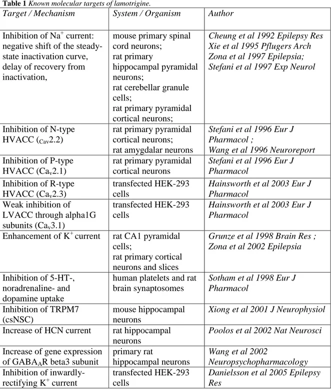
seizures in 1994, showing favorable pharmacokinetics, improved tolerability and lower potential for drug interactions compared to several older antiepileptic drugs (AEDs). In addition to its approval for maintenance treatment of bipolar I disorder, off-label prescription of LTG is becoming increasingly popular, as it shows therapeutic effects on several neurologic and neuropsychiatric diseases of completely diverse etiologies such as borderline syndrome or cocaine dependence. LTG’s wide therapeutic applicability reflects the “unspecificity” of the drug which has shown to modulate several different sodium, calcium and potassium currents (Beck and Yaari, 2012). Anticonvulsive properties of LTG were initially mainly attributed to its capacity to inhibit transient and persistent sodium currents, selectively prolonging slow inactivation thereby suppressing the release of excitatory amino acids (Brodie, 1996;Xie et al., 1995). Over the following years more than 15 other targets of LTG could be identified including several voltage- and ligand- gated cation channels and neurotransmitter receptors and transporters (see Table 1). One of the newly found mechanisms of LTG was potentiation of the outward cation current through hyperpolarization-activated cyclic nucleotide-gated (HCN) channels (Poolos et al., 2002), which are localized on CA1 dendrites, where they are proposed to modulate dendritic integration of excitatory input (Magee, 1998;Magee, 1999).
Furthermore, LTG as well as its derivative sipatrigine have been shown to inhibit R-type currents carried by Ca
v2.3 voltage-gated calcium channels in heterologous systems (Hainsworth et al., 2003). In this study, R-type currents from human α1E subunits with β3 subunits stably transfected in HEK 293 cells were inhibited by 10 μM of LTG, a concentration within the estimated range of therapeutic brain concentrations of 4–40 μM (Leach et al., 1995).
Topiramate (TPM) another broad spectrum anticonvulsant was FDA approved two years
after LTG in 1996 and like LTG enjoys a wide-spectrum of use from treatment of
Lennox-Gastaut Syndrome and bipolar disorder to migraine. In addition to its known
mechanisms involving sodium channel blockade, TPM was later found to be an inhibitor
of R-type currents at concentration within the estimated range of therapeutic brain
concentrations (IC
50= 50.9 μM) (Kuzmiski et al., 2005). In transiently transfected tsA-
201 cells, application of 100 μM TPM causes a negative shift in the voltage dependence
25
of steady-state inactivation of Ca
v2.3. Because the authors also found that R-type inhibition by TPM in CA1 neurons causes a reduction in the Ca
2+influx, necessary for plateau potential activation, they hypothesize that TPM directly inhibits R-type calcium spike generation by increasing steady-state inactivation of Ca
v2.3 calcium channels at resting potentials.
Table 1 Known molecular targets of lamotrigine.