Original article:
WIRELESS CAPSULE ENDOSCOPY EXPLORATION FOR DISEASES OF THE SMALL INTESTINE IN CHINA
Li Yan1, 3, Changqing Bai2, 3, Jian Song1, Qunying Wang1, Guangrong Fang1, Huimin Zhong1*
1Department of Gastroenterology, 401 Hospital of PLA, Qingdao 266071, Shandong prov- ince, P. R. China; 2Department of Respiratory and Gastroenterology, 307 hospital of PLA, Beijing 100044, P. R. China; 3Li Yan and Changqing Bai contributed equally to this work.
*Correspondence should be addressed to Dr. Huimin Zhong, Gastroenterology Department of 401 hospital of PLA, Qingdao, Shandong province, P. R. China; phone, +86 532 83970049 ; Fax, +86 532 83970050. E-mail: bella493@sina.com
ABSTRACT
For small bowel diseases, it is difficult for the ordinary enteroscopy to reach due to its spe- cific curvature and length. Capsule endoscopy (CE) is a unique tool to visualize the mucosa of the small intestine. The aim of this study was to evaluate the detection rate and diagnostic yield of CE in a large group of patients with suspected digestive diseases in China. One hun- dred and two consecutive patients (75 male, mean age 50 years, range 32-87 years) underwent CE in our Gastroenterology Units, for a total of 102 procedures. Referrals were obscure oc- cult/overt gastrointestinal bleeding group (19 patients) and suspected small bowel disease group (83). In our study, the whole detection rate was 92 % (94/102), with a definite diagno- sis yield of 63 % of the patients in the obscure gastrointestinal bleeding and 39 % of the pa- tients in the suspected small bowel diseases. None of the patients developed symptoms of signs of mechanical obstruction, although the capsule was retained in the stomach in 2/102 patients for their somatostatin taken. CE seems to be a very safe, painless and effective proce- dure with a high diagnostic yield. Accurate selection of indications and critical evaluation of the results are essential to explore these diseases.
Keywords: Capsule endoscopy, small bowel disease, diagnostic yield
INTRODUCTION
With the advent of the capsule endo- scopy (CE), the small bowel no longer ap- pears a mysterious territory troubles pa- tients and doctors (Fritscher-Ravens and Swain, 2002). This noninvasive tool plays a very important role in gastrointestinal dis- ease, particularly in small bowel diseases.
There are several methods for screen- ing digestive diseases, including the pri- mary barium X-ray, Computer Tomography (CT) and enteroscopy. However, in small bowel diseases, the mucosal inflammatory and flat lesions can’t be clearly observed by
barium X-ray or CT (Costamagna et al., 2002; Eliakim et al., 2003; Filippone et al., 2007; Hara et al., 2004). Although the im- proved double balloon endoscope(DBE)
was widely applied in the small bowel dis- eases, it is an invasive procedure and expert endoscopists are still needed. In recent years, there is increasing evidence that CE plays a very important role in the diagnostic of small bowel diseases, especially in gas- trointestinal (GI) obscure bleeding, sus- pected small bowel diseases, and other in- dications (Sturniolo et al., 2006; Park et al., 2007; Ziegler et al., 2005; Thomson et al., 2007; Maunoury et al., 2007). Moreover,
with the improved new techniques, includ- ing the “blood indicator” and the “patency capsule,” might make the examination much safer than before (Signorelli et al., 2005; Costamagna et al., 2004).
Although CE is sensitive and specific for detecting small bowel lesions, it is only seven years since the advent of the CE (Id- dan et al., 2000; Appleyard et al., 2000).
Moreover, in China, only a few medical institutions carried out CE detection for small bowel diseases. Therefore, more pro- cedures are still needed to explore the safety, detective and diagnostic value of the CE in Chinese group of patients.
PATIENTS AND METHODS Patients
Between May 2005 to January 2007 a total of 102 patients (75 male), mean age 50 years (range 37-82 years) underwent CE for a total of 102 procedures. Patients were enrolled in the Gastroenterology Units, in 401 hospital of PLA, Qingdao, China.
In all patients, previous upper and lower GI endoscopies were negative. Find- ings of other investigation were not signifi- cant in explaining the clinical picture. Re- ferrals were obscure occult/overt gastroin- testinal bleeding (19 patients) and suspected small bowel disease (83) and none of them have contraindicating EC. Four patients were on somatostatin, three were on antico- agulants, and two were on anti- inflammatory drug (NSAID).
All patients gave their informed con- sent and the study was approved by the Ethic Committees of the 401 Hospital of PLA, China.
Capsule endoscopy procedure
After bowel cleaning with a 250 ml so- lution of mannitol and an overnight fast of 12 h, patients ingested the wireless capsule (PillCam, Given Imaging). Before this, an array of eight sensors was attached to the abdominal wall of each patient, and a re- corder with a battery, attached to a belt, was
worn around each patient’s waist. Patients were allowed to drink 2 h after ingesting the capsule and to eat 4 h later. After ingestion of the capsule with a small amount of wa- ter, patients were free to pursue their own activities. The capsule takes two imagines per second during eight hours, depending on battery power. The frames are transmit- ted directly to a recorder and after the ex- amination they are unloaded onto a com- puter. About 50.000 frames can be re- viewed one by one or as a video sequence, and the results were related to the experi- ence of the investigator.
Patients were asked to look for the cap- sule in the feces. All capsules were re- trieved 12 h to 5 days after ingestion. All patients were contacted 1 month after com- pleting the study for a survey of late com- plications.
RESULTS
Follow-through time of the capsule
After the patients swallowed the cap- sule in the supine position, the median esophagus transit time was 2.46 min (0.5- 21.38 min), the median gastric transit time was 47.4 min(11.73-510 min)including two patients had a gastric distention of 2 hours, and the median small bowel transit time was 275.82 min (124.8-508.8 min). All patients underwent the examination without discomforts and complications. The average recording time was about 4.64 hours (2.08- 8.48 hours).
Endoscopic findings
The overall detection rate of the EC was 92 %. Different abnormities including erosion, ulcer, angiodysplasia, polyp, diver- ticulum and tumor were detected by the CE (Table 1). Moreover, Findings were located in the duodenum (at the third portion) in 17.6 % of patients, in the jejunum in 35.2 % of patients, and in the ileum in 48.1 % of patients. Localization of findings was de- termined by the external localization system supported by the capsule device, the clini-
cian estimations, and surgery in those pa- tients who needed surgical therapy.
Table 1: Total abnormal findings in the study group
Diagnosis N
Positive findings Duodenitis
94 13
Duodenal ulcer 4
Duodenal cancer 1
Angiodysplasia 4 Small-bowel erosions 16
Small bowel ulcer 13
Simple intestinal polyp 15 Intestinal diffuse
lymphangectasia Intestinal interstitialoma
10 2 Lymphoid hyperplasia 11 Small bowel diverticula 2
Crohn’s disease 6
Multiple intestinal polyps 8
Small bowel tumors 2
Negative findings 8
Obscure GI bleeding
Obscure GI bleeding was defined ac- cording to the position of the American Gastroenterological Association. It includes occult obscure bleeding and overt obscure bleeding and the formed was defined by the presence of positive fecal blood test and chronic iron-deficient anemia, without any clinically evident bleeding episode from at least 6 months, whereas the latter indicated a history of recurrent bleeding episodes of melena or lower GI bleeding in the last 6 months (Sturniolo et al., 2006).
In our study, nineteen procedures were performed to investigate the obscure GI bleeding, including 6 patients with overt obscure bleeding and 13 patients with oc-
cult obscure bleeding, among all the pa- tients, there were 15 males, mean age was 51 years (range 34-81), mean Hb value 7.9 ± 3.2 g/dl, 42 % with previous blood transfusion.
The overall detective rate in this group of patients was 100 %, whereas some find- ings (single polyps, small and scattered erosions) were considered not related or adequate to explain the clinical picture.
Thus, the overall diagnostic yield was 63 % (12 medical significant lesions/19 valid explorations) and the detailed types of the lesion detected by CE in this group were as the followings: multiple small bowel ero- sions with bleeding in 8 cases (Fig. 1), se- vere intestine ulcers with bleeding in 2 cases, activated post bulbar ulcer of duode- nal in 1 case (Fig. 2), and jejunal tumor with activated bleeding in 1 case (Fig. 3).
All the abnormities were not known prior to the CE detection.
Figure 1: Multiple erosions with bleeding was observed in the small bowel
Figure 2: Activated post bulbar ulcer of duodenal was detected by wireless capsule
Figure 3: Tumor with activated bleeding was observed in the jejunal
Suspected small bowel disease
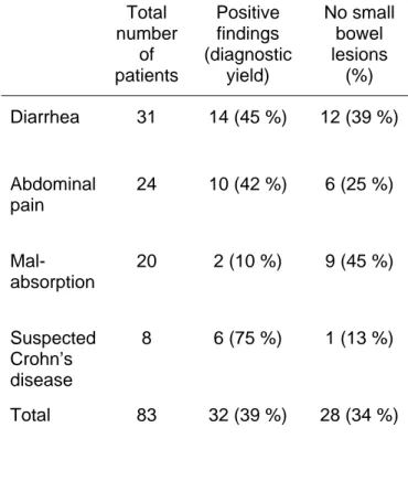
In 83 cases (60 male), mean age 48 years (range 32-87), capsule endoscopy was performed for persistent digestive symp- toms (e.g., diarrhea, abdominal pain) or biochemical abnormities (e.g., increased inflammatory tests, low serum protein) sug- gestive of small bowel disease. In particu- lar, the indication was chronic diarrhea in 31 patients, abdominal pain in 24, malab- sorption in 20, and suspected Crohn’s dis- ease (CD) in 8.
Small bowel lesions were detected in 52 cases (63 %), whereas the exploration was considered insufficient in 3 (3 %) and negative in 28 (34 %) (Table 2), and in the latter group, 11 patients with ileum lym- phoid hyperplasia were included. Capsule explorations were considered significant in 32 cases, final diagnosis being small bowel ulcers in 11 patients, intestinal diffuse lym- phangectasia in 7, CD in 6 (Fig. 4), NSAID enteropathy in 6, and Whipple disease in 3.
Thus the overall diagnostic yield of capsule endoscopy for patients with suspected small bowel disease was 39 %.
Figure 4: Crohn’s disease was observed in the small bowel by wireless capsule
Table 2: Positive and negative findings in the patients with suspected bowel diseases according to the clinical symptoms
Total number
of patients
Positive findings (diagnostic
yield)
No small bowel lesions
(%) Diarrhea 31 14 (45 %) 12 (39 %)
Abdominal pain
24 10 (42 %) 6 (25 %)
Mal- absorption
20 2 (10 %) 9 (45 %)
Suspected Crohn’s disease
8 6 (75 %) 1 (13 %)
Total 83 32 (39 %) 28 (34 %)
DISCUSSION
In this study we evaluated the safety, detective and diagnostic value of CE in a large group of patients in China. Our find- ings demonstrated that a detection rate of 92 % was found, which was higher than the previous report in China (Ge et al., 2004).
This higher detection rate might be associ- ated with the more clinical pictures of the patients when they participated in this study, and the higher sensitivity values of CE in digestive diseases. Moreover, ab- dominal pain had a diagnostic yield of 42 % in our study, which was higher than Bar- dan’s report (Bardan et al., 2003). The higher diagnostic yield was confirmed by the more patients with small bowel ulcers with staxis in our study group.
Compared with the ordinary enterocly- sis, bleeding scanning with labeled red blood cells, push enteroscopy, and double balloon enteroscopy, the CE plays a very important role in the diagnosis of obscure
GI bleeding (Saperas et al., 2007; Leighton et al., 2006; Hadithi et al., 2006). In our study, there were several types of lesions including erosion; ulcer, Crohn’s disease, and tumor were observed in patients with obscure GI bleeding, and the total diagnos- tic yield was 63 % in this group patient, slightly higher than the recent report (Jones et al., 2005). Our results showed that CE should be an ideal tool to explain the reason of obscure GI bleeding. However, for the suspected small bowel disease, the wireless capsule showed a lower diagnostic yield (39 %), although a higher diagnostic yield was found in the abdominal pain group pa- tients (42 %). The results from our study were closely correlated with the patients’
condition in China and the referrals selec- tion.
For small bowel disease, double- balloon push enteroscopy (DBE) has in- creased its diagnostic yield currently, but it is still an invasive procedure and needs ex- pertise endoscopists. In our study, we use CE to detect 8 cases of suspected CD, and found 6 cases of CD at early stage by the following findings: mucosal congestion and edema; mucosal polyps; scattered erosions;
irregular ulcers, and mucosal atrophy. Our results indicated that the CE showed a great superiority in diagnosis of CD, thus con- tributed to early treatment, and prognosis of the CD. Polyp is one common small bowel lesion, and we found 15 cases of single in- testine polyp of 0.2-0.3 cm which needed to follow-up. Moreover, 8 cases of multiple intestine polyps were observed in the upper jejunum and terminal ileum. The location of the polyps detected by the CE was accurate enough to direct polypectomy. Our findings suggest that CE does not only have the ca- pacity to detect the small bowel abnormities but also has the potential to early treatment.
In conclusion, the wireless capsule en- doscopy is a revolutionary technique which allows the visualization of the entire gastro- intestinal (GI) track, especially the small intestine. Our results showed that the CE was safe and effective, with high sensitivity values and diagnostic yield in the small bowel diseases.
Acknowledgements: We greatly thank technician Ping Yao for her excellent tech- nical assistance.
REFERENCES
Appleyard M, Fireman Z, Glukhovsky A, Jacob H, Shreiver R, Kadirkamanathan S, Lavy A, Lewkowicz S, Scapa E, Shofti R, Swain P, Zaretsky A. A randomized trial comparing wireless capsule endoscopy with push enteroscopy for the detection of small- bowel lesions. Gastroenterology 2000;119:
1431-8.
Bardan E, Nadler M, Chowers Y, Fidder H, Bar-Meir S. Capsule endoscopy for the evaluation of patients with chronic abdomi- nal pain. Endoscopy 2003;35:688-9.
Costamagna G, Shah SK, Riccioni ME, Foschia F, Mutignani M, Perri V, Vecchioli A, Brizi MG, Picciocchi A, Marano P. A prospective trial comparing small bowel radiographs and video capsule endoscopy for suspected small bowel disease. Gastro- enterology 2002;123:999-1005.
Costamagna G, Spada C, Spera G, Riccioni ME, Biancone L, Hermano G, Herrerias JM, Lochs H, Neuhaus H, Reddy N, Rut- geerts P, Schreiber S, Pallone F, Warwick S. Evaluation of the given patency system in the GI tract: results of a multi-center study. Gastrointest Endosc 2004;59:145.
Eliakim R, Fischer D, Suissa A, Yassin K, Katz D, Guttman N, Migdal M. Wireless capsule video endoscopy is a superior diag- nostic tool in comparison to barium follow- through and computerized tomography in patients with suspected Crohn's disease. Eur J Gastroenterol Hepatol 2003;15:363-7.
Filippone A, Cianci R, Milano A, Valeriano S, Di Mizio V, Storto ML. Obscure gastro- intestinal bleeding and small bowel pathol- ogy: comparison between wireless capsule endoscopy and multidetector-row CT en- teroclysis. Abdom Imaging 2007 [Epub ahead of print].
Fritscher-Ravens A, Swain CP. The wire- less capsule: new light in the darkness. Dig Dis 2002;20:127-33.
Ge ZZ, Hu YB, Xiao SD. Capsule endo- scopy and push enteroscopy in the diagno- sis of obscure gastrointestinal bleeding.
Chin Med J 2004;117:1045-9.
Hadithi M, Heine GD, Jacobs MA, van Bo- degraven AA, Mulder CJ. A prospective study comparing video capsule endoscopy with double-balloon enteroscopy in patients with obscure gastrointestinal bleeding. Am J Gastroenterol 2006;101:52-7.
Hara AK, Leighton JA, Sharma VK, Flei- scher DE. Small bowel: preliminary com- parison of capsule endoscopy with barium study and CT. Radiology 2004;230:260-5.
Iddan G, Meron G, Glukhovsky A, Swain P. Wireless capsule endoscopy. Nature 2000;405:417.
Jones BH, Fleischer DE, Sharma VK, Heigh RI, Shiff AD, Hernandez JL, Leighton JA. Yield of repeat wireless video capsule endoscopy in patients with obscure gastrointestinal bleeding. Am J Gastroen- terol 2005;100:1058-64.
Leighton JA, Sharma VK, Hentz JG, Musil D, Malikowski MJ, McWane, Fleischer DE.
Capsule endoscopy versus push en- teroscopy for evaluation of obscure gastro- intestinal bleeding with 1-year outcomes.
Dig Dis Sci 2006;51:891-9.
Maunoury V, Savoye G, Bourreille A, Bouhnik Y, Jarry M, Sacher-Huvelin S, Soussan EB, Lerebours E, Galmiche JP, Colombel JF. Value of wireless capsule endoscopy in patients with indeterminate colitis (inflammatory bowel disease type unclassified). Inflamm Bowel Dis 2007;13:
152-5.
Park CH, Kim JO, Choi MG, Kim KJ, Kim YH, Kim YS, Kim TI, Do JH, Ryu JK, Moon JS, Park SH, Shim KN, Lee KM, Lee IS, Chun HJ. Utility of capsule endoscopy for the classification of Crohn's disease: a multicenter study in Korea. Dig Dis Sci 2007;52:1405-9.
Saperas E, Dot J, Videla S, Alvarez- Castells A, Perez-Lafuente M, Armengol JR. Capsule endoscopy versus computed tomographic or standard angiography for the diagnosis of obscure gastrointestinal bleeding. Am J Gastroenterol 2007;102:
731-7.
Signorelli C, Villa F, Rondonotti E, Abbiati C, Beccari G, de Franchis R. Sensitivity and specificity of the suspected blood identifi- cation system in video capsule enteroscopy.
Endoscopy 2005;37:1170-3.
Sturniolo GC, Di Leo V, Vettorato MG, De Boni M, Lamboglia F, De Bona M, Bellu- mat A, Martines D, D'Inca R. Small bowel exploration by wireless capsule endoscopy:
results from 314 procedures. Am J Med 2006;119:341-7.
Thomson M, Fritscher-Ravens A, Mylonaki Murch S, McAlindon M, Furman M. Wire- less capsule endoscopy in children: a study to assess diagnostic yield in small bowel disease in paediatric patients. J Pediatr Gas- troenterol Nutr 2007;44:192-7.
Ziegler KM, Flamm CR, Aronson N. Wire- less capsule endoscopy in patients with ob- scure small-intestinal bleeding. J Am Coll Radiol 2005;2:818-20.