Cellular signalling of adrenomedullin regulating Endothelial barrier function and granulocyte extravasation
Inaugural-Dissertation zur
Erlangung des Doktorgrades
der Mathematisch-Naturwissenschaftlichen Fakultät der Universität zu Köln
vorgelegt von
Qian Mao
aus Jinan (V.R.China)
Berichterstatter: Prof. Dr. Angar Büschges (Gutachter) Prof. Dr. Stefan Herzig
PD. Dr. Ingo Flamme
Tag der mündlichen Prüfung: 19. Oktober. 2012
T
ABLE OFC
ONTENTSTable of Contents I
List of Figures V
List of Tables VII
Abbreviations VIII
1. Abstract 1
2. Zusammenfassung 3
3. Introduction 5
3.1. Adrenomedullin (ADM) 6
3.1.1. Structure and distribution of ADM 6
3.1.2. Receptors and intracellular signalling of ADM 8
3.1.3. Biological actions of ADM in response to inflammation 9
3.2. Regulation of vascular permeability 11
3.2.1. Endothelial cell-cell junctions and vascular integrity 12 3.2.2. Endothelial cytoskeleton and vascular permeability 16 3.2.2.1. Dynamic functions of cytoskeleton and vascular permeability 16 3.2.2.2. Static functions of cytoskeleton in vascular permeability 18 3.2.3. Role of cAMP in regulating vascular permeability 19
3.3. Transendothelial migration of leukocytes 22
3.4. Aims of the study 25
4. Material and Methods 27
4.1. Material 27
4.1.1. Chemical reagents and assay kits 27
4.1.2. Antibodies 30
4.1.3. Buffers 31
4.1.4. Materials 32
4.1.5. Equipments 33
4.2. Methods 35
4.2.1. Molecular biological methods 35
4.2.1.1. Microarray analysis 35
4.2.1.2. Quantitative Real-Time RT-PCR analysis 35
4.2.1.3. RNA interference 37
4.2.2. Biochemical methods 37
4.2.2.1. Measurement of cyclic AMP 37
4.2.2.2. Detection of VASP activation 38
4.2.2.3. Detection of MLC phosphorylation 38
4.2.2.4. Rap1 Pull-down assay 39
4.2.2.5. Co-immunoprecipitation of VE-PTP/VE-cadherin 39
4.2.2.6. Immnuofluorescence staining 41
4.2.3. Cell culture methods 41
4.2.3.1. Cell culture condition 41
4.2.3.2. Aequorin luminescence measurements 43
4.2.3.3. CREB phosphorylation in luciferase-transfected CHO cells 43 4.2.3.4. Cell surface expression of adhesion molecules 44 4.2.3.5. Measurement of transendothelial electrical resistance 44 4.2.3.6. Detection of paracellular macromolecular permeability 45
4.2.3.7. Isolation of human neutrophils 45
4.2.3.8. In vitro-transendothelial migration assay (TEM) 46
4.2.3.9. Adhesion assay 46
4.2.3.10. Myeloperoxidase (MPO) activation assay 46 4.2.3.11. Fluorescence activated cell sorting (FACS) analysis 47
4.2.4. In vivo-experiments 47
4.2.4.1. Animals 47
4.2.4.2. Miles assay to determine vascular permeability 48
4.2.4.3. LPS-induced acute lung injury model 48
4.2.5. Statistical analysis 49
5. Results 50
5.1. Analysis of ADM receptor expression and signalling 50
5.2. Effects of ADM on endothelial barrier integrity 51
5.2.1. Anti-edematous effects of ADM in vitro 52
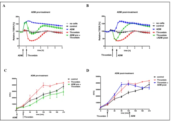
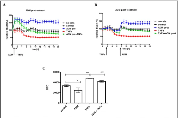
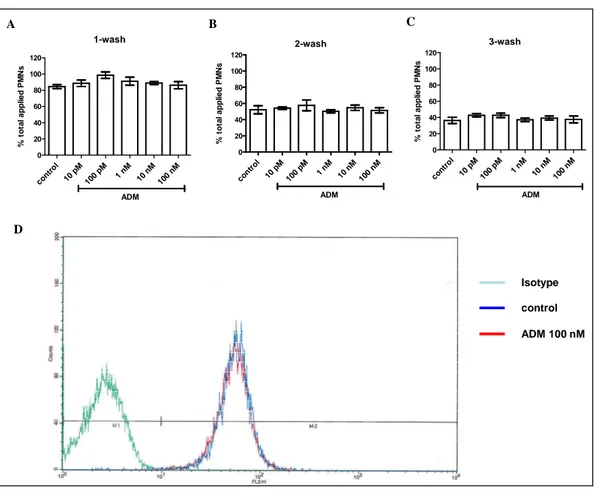
5.2.1.1. Effects of ADM on thrombin-induced hyperpermeability 52 5.2.1.2. Effects of ADM on TNFα-induced hyperpermeability 56 5.2.1.3. Effects of ADM on different stimuli-induced hyperpermeability 57 5.2.2. Effects of ADM on leukocyte transmigration in vitro 60
5.2.3. Anti-edematous effects of ADM in vivo 66
5.2.4. Anti-inflammatory effects of ADM in vivo 68
5.3. Analysis of cAMP-dependent pathway in ADM signalling 70 5.3.1. Comparison of ADM and FSK effects on generation cAMP 71 5.3.2. Comparision of ADM and FSK effects on cAMP accumulation 72 5.3.3. Comparision of ADM and FSK effects on permeability 73 5.3.4. Comparision of ADM and FSK effects on PMN extravasation 75
5.3.5. Comparision of ADM and FSK effects on PKA and Epac/Rap1 activation 76 5.3.6. Comparision of ADM and FSK effects on gene expression 78 5.3.7. Qualitative and quantitative comparision of ADM- and FSK-induced effects 82
5.4. Effects of ADM on gene expression 85
5.4.1. Analysis of time-dependent effects of ADM on CAMs 85 5.4.2. Effects of ADM on TNFα-induced expression of CAMs 86
5.4.3. Effects of ADM on surface expression of CAMs 88
5.5. Role of PKA and Epac/Rap1 in ADM signalling 89
5.5.1. Dissection of PKA and Epac/Rap1 signaling 90
5.5.2. Impact of Epac/Rap1- and PKA-signaling on vascular permeability 92 5.5.3. Role of PKA and Epac/Rap1 in regulation of granulocyte extravasation 94 5.5.3.1. Effects of PKA and Epac/Rap1 in PMN transmigration 94 5.5.3.2. Role of PKA and Epac/Rap1 in PMN-induced hyperpermeability 95 5.5.3.3. Impact of PKA and Epac/Rap1 on endothelial contractility 98 5.6. Direct effects of ADM on endothelial cytoskeleton and cell-cell junctions 102 5.6.1. Role of cortactin in ADM-mediated endothelial barrier modulation 102 5.6.2. Effects of ADM on the background of disrupted actin cytoskeleton 105
5.6.3. Effects of ADM on VE-PTP/VE-Cadherin complex 109
6. Discussion 113
6.1. Effects of ADM in response to inflammation 113
6.1.1. Effects of ADM on endothelial permeability 113
6.1.2. Effects of ADM on granulocyte extravasation 115
6.1.2.1. Direct actions of ADM on human neutrophils 115 6.1.2.2. Effects of ADM on endothelial adhesion receptors 116 6.1.3. Effects of ADM on endothelial cytoskeleton and junctions 118 6.1.3.1. Effects of ADM on endothelial cytoskeleton 118 6.1.3.2. Effects of ADM on endothelial cell-cell junctions 119 6.2. Cellular signalling of ADM: the cAMP-dependent pathway 120 6.2.1. The cAMP signaling in endothelial barrier function 120 6.2.2. Correlation of ADM effects with cAMP signaling 122
6.3. Impacts of PKA and Epac/Rap1 on ADM signalling 123
6.3.1. Impacts of cAMP signaling on anti-edematous effects of ADM 125 6.3.2. Impacts of cAMP signaling on regulating granulocyte extravasation 126 6.4. Role of cortactin in cellular signalling of ADM 128 6.5. Therapeutical potential of ADM for inflammatory diseases 130
7. References 132
8. Appendix 151
9. Acknowledgments 153
10. Erklärung 154
L
IST OFF
IGURESFigure 1 Structure of ADM and post-translational processing of preproADM gene ... 7
Figure 2 Endothelial cell-cell junctions ... 13
Figure 3 Structure of VE-cadherin-catenin complex in endothelial cells ... 16
Figure 3 Contractile machinery and actin cytoskeleton in endothelial cells ... 18
Figure 4 Schematic diagram of leukocyte adhesion and transendothelial migration ... 23
Figure 6 Analysis of ADM receptor expression and signaling in endothelial cells ... 51
Figure 7 Effects of ADM on thrombin-induced hyperpermeability using different in vitro-models. ... 54
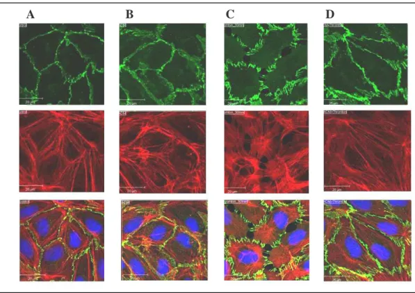
Figure 8 Effect of ADM on thrombin-induced F-actin and VE-cadherin distribution. ... 55
Figure 9 Prophylactic and therapeutic potency of ADM on TNFα stimulation in vitro. ... 57
Figure 10 Effects of ADM on endothelial barrier dysfunction induced by different stimuli ... 60
Figure 11 Effects of ADM on TNFα-induced leukocyte transmigration ... 61
Figure 12 ADM Receptor expression and its effects on MPO release from human neutrophils 64 Figure 13 Effect of ADM on cell adhesion of neutrophils to endothelial cells ... 66
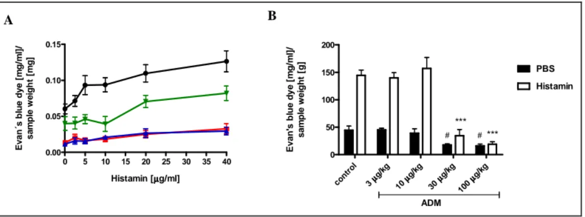
Figure 14 Effects of ADM on histamine-induced vascular hyperpermeability in the skin of rats and mice ... 68
Figure 15 Challenge of LPS induced lung edema and accumulation of leukocytes and proteins in BAL fluid ... 69
Figure 16 Anti-inflammatory effects of ADM in acute lung injury induced by LPS challenge ... 70
Figure 17 Characterization of ADM and FSK in CHO-ADM1-reporter cells ... 72
Figure 18 ADM and FSK increased accumulation of intracellular cAMP ... 73
Figure 19 Comparison of ADM and FSK effects on electrical resistance by using ECIS ... 74
Figure 20 Comparison of ADM and FSK effects on macromolecular permeability... 75
Figure 21 Comparison of ADM and FSK effects on granulocyte extravasation... 76
Figure 22 Effects of ADM and FSK on PKA activation ... 77
Figure 23 Effects of ADM and FSK on activation of Rap1 signaling ... 78
Figure 24 Effects of ADM on gene expression of adhesionce molecules ... 86
Figure 25 Effects of ADM on TNFα-induced gene expression of cell adhesion molecules ... 88
Figure 26 Effects of ADM on cell surface expressions of adhesions molecules after TNFα- stimulation ... 89 Figure 27 Dissection of Benz-cAMP and “007” in different assays. ... 91 Figure 28 Both PKA and Epac/Rap1 signaling are involved in regulating endothelial permeability in vitro and in vivo. ... 93
Figure 29 PKA but not Epac/Rap1 is involved in leukocyte transmigration ... 95 Figure 30 Effects of PKA- and Epac/Rap1 activator in regulating PMN-induced
hyperpermeability ... 97 Figure 31 Regulation of MLC is involved in cAMP-PKA pathway on granulocyte transmigration ... 100 Figure 32 Effects of PKA and Epac/rap1 on thrombin-induced F-actin and VE-cadherin
distribution. ... 101 Figure 33 Effect of ADM on cellular distribution of cortactin by using immunofluorescence microscopy ... 103 Figure 34 Role of cortactin in ADM signaling regulating endothelial permeability and granulocyte transmigration ... 105 Figure 35 Effect of ADM on VE-cadherin and actin fibers in endothelial cells with intact
cytoskeleton. ... 107 Figure 36 Effect of ADM on barrier integrity in endothelial cells with disrupted actin cytoskeleton.
... 109 Figure 37 Effects of ADM on complex of VE-PTP and VE-Cadherin ... 112
L
IST OFT
ABLESTable 1 List of chemical reagents, assay kits and their providers ... 27
Table 2 List of primary antibodies and their providers ... 30
Table 3 List of secondary antibodies and their providers... 30
Table 4 Composition of buffers ... 31
Table 5 List of materials and their providers ... 32
Table 6 List of equipments and their providers ... 33
Table 7 List of primers and fluorescent probes ... 36
Table 8 Microarray analysis in HUVECs after 3 h incubation with ADM and FSK ... 79
Table 9: Microarray analysis in HUVECs after 16 h incubation with ADM and FSK ... 81
Table 10 Effects of ADM and FSK in different models ... 83
Table 11 Quantitative comparison of ADM and FSK in different assays ... 84
Table 12 Quantitative comparison of ∆cAMP induced by ADM and FSK in different assays .... 84
Table 13 Effects of TNFα on gene expression of adhesion molecules and ADM receptors ... 87
A
BBREVIATIONSAC Adenylate cyclase
ADM Adrenomedullin
AJ Adherens junction
ALI Acute lung injury
AMBP-1 Adrenomedullin binding protein-1 ARDS Adult respiratory distress syndrome AUC Area under the curve
BAL Bronchoalveolar lavage
bEnd.5 Brain microvascular endothelial cells
BCA Bicinchoninic acid
BSA Bovine serum albumin
BW Body weight
C5a Complement factor 5a
Ca2+ Calcium
CAFTY Ca2+-free Tyrode solution
cAMP Cyclic adenosine monophosphate cAPK cAMP-dependent protein kinase
CB Cytochalasin B
CBD cAMP-binding domain (CBD)
CCRL Chemokine C-C motified receptor like
CD Cytochalasin D
cDNA Complementary DNA
cGMP Cyclic guanosine monophosphate CGRP Calcitonin gene-related peptide
CHO Chinese hamster ovary
CLP Coecum ligation and puncture CNG cyclic nucleotide-gated ion channels CRE cyclic AMP response element
CREB cyclic AMP response element (CRE)-binding protein CRLR Calcitonin receptor like receptor
CTR Calcitonin receptor
Da Dalton
DAG Diacylglycerol
DMEM Dulbecco’s modified Eagle’s medium DMSO Dimethyl sulfoxide
DNA Deoxyribonucleic acid
DTT Dithiothreitol
EC50 Half-maximal effective concentrations ECIS Electrical cell-substrate impedance sensing
ECL Enhanced chemiluminescence
EDTA Ethylendiamintetra essig acid
ELISA Enzyme-linked immunosorbent assay Epac Exchange protein activated by cAMP ERK Extracellular-signal regulated kinases
ESAM Endothelial cell-selective adhesion molecule et al. Et alii
FACS Fluorescence activated cell sorter
FBS Fetal bovine serum
FCS Fetal calf serum
FITC Fluorescein isothiocyanate
fMLP N-Formyl-L-methionyl-L-leucyl-L-phenylalanine
FSK Forskolin
g Gram
GEF Guanine exchange factor
GFP Green fluorescence protein GPCR G-protein coupled receptor
h Hour(s)
HBSS Hanks balanced salt solution
HEPES N-[2-Hydroxyethyl]-piperazin-N'-[2-Ethansulfonsäure]
HLMEC Human lung microvessel endothelial cell
HOCL Hypochlorous acid
HRPO Horseradish peroxidase
HUVEC Human umbilical vein endothelial cell ICAM Intercellular adhesion molecule
Ig Immunglobulin
IgG Immunoglobulin G; isotype control antibody IL-1α/1β Interleukin 1α/1β
IP Immunoprecipitation
i.v. Intravenously
JAM Junction adhesion molecule
kDa Kilodalton
kg Kilogram
LDS Lithium dodecyl sulfate
LFA Lymphocyte function-associated antigen
LPS Lipopolysaccharid
LSM Laser scanning microscop MAC Membrane-activation complex
MAM Migration assay medium
MAP P38 mitogen-activated protein
MAPK P38 mitogen-activated protein kinase MBS Myosin–binding subunit
MEM Minimal essential medium
MES 2-(N-morpholino)ethanesulfonic acid
Min Minute(s)
MLC Myosin light chain
MLCK Myosin light chain kinase MLCP Myosin light chain phosphatase MMP Matrix metallopeptidase
MOPS 3-(N-morpholino)propanesulfonic acid
MPO Myeloperoxidase
mRNA Messenger ribonucleic acid NEAA Non-essential amino acids
NFκB Nuclear factor κB
NO Nitric oxide
NOS Nitric oxide synthase PAF Platelet-activating factor
PAM Peptidylglycine alpha-amidating monooxygenase PAMP ProADM N-terminal 20 peptide
PAR Protease-activated receptor PBS Phosphate buffered saline PCR Polymerase chain reaction
PDE Phosphodiesterase
PE Phycoerythrin
PECAM Platelet and endothelial cell adhesion molecule-1
PFA Paraformaldehyd
PG Prostaglandine
PI3K Phosphatidylinositol-3 Kinase
PKA Protein kinase A
PKC Protein kinase C
PMNs polymorphonuclear neutrophils
PO Peroxidase
pTyr Phospho-Tyrosine
p-value Probability
PVDF Polyvinylidenefluoride
RAMP Receptor activity modifying proteins
RNA Ribonucleic acid
RND 1 Rho family GTPase 1 ROS Reactive oxygen species
rpm Rounds per minute
RT Room temperature
RT-PCR Reverse transcriptase polymerase chain reaction
SD Standard derivation
SDS Sodium dodecyl sulfate
SDS-PAGE Sodium dodecyl sulfate polyacrylamide gel electrophoresis SEM Standard error of mean
Ser Serine
SiRNA Small interfering ribonucleic acid
S1P Sphingosine-1-phosphate
TBS Tris buffered saline
TCEP Tris(2-carboxyethyl)phosphine
TEER Transendothelial electrical resistance TEM Transendothelial migration
TGFβ transforming growth factor-β Tie-2 Angiopoietin-1 receptor TNFα/β Tumor necrosis factor α/β
TJ Tight junction
TLR Toll like receptor
TMB 3,3′,5,5′-Tetramethylbenzidine Tris Tris(hydroxymethyl)aminomethane
Tyr Tyrosine
VASP Vasodilator stimulated phosphoprotein VCAM Vascular adhesion molecule
VE-cadherin Vascular endothelial cadherin VEGF Vascular endothelial growth factor
VE-PTP Vascular endothelial protein tyrosine phosphatase
WB Western blot analysis
WBC White blood cell
w/v Weight/volume
ZO- Zonular occludings-
% (v/v) Volume percent
% (w/v) Weight percent
1. A
BSTRACTBackground: Disturbed endothelial barrier function is a hallmark of inflammation. Effusion of protein rich plasma fluid leading to edema formation and overshooting transmigration of leukocytes contribute to severe organ dysfunction under conditions of generalized inflammation, e.g. acute lung injury and sepsis. The peptide hormone Adrenomedullin (ADM) and its receptor (CRLR/RAMP2/3) constitute an important signaling system for the protection of endothelial barrier function. Although elevation of cAMP in endothelial cells was identified as signaling pathway of ADM responsible for barrier protection, it was speculated that other mechanisms are involved and precise intracellular signaling routes have not been explored, yet.
Main findings and conclusions: ADM was confirmed to be a strong stabilizer of the endothelial barrier function in vitro, preventing and reversing hyperpermeability independent of the inflammatory stimulus (as determined by electrical impedance and FITC-dextran permeability measurements after stimulation with thrombin, TNFα, histamine, VEGF). Moreover, it was shown for the first time that ADM inhibited TNFα-induced granulocyte transmigration which was solely due to effects of ADM on endothelial cells. These in vitro-findings could be translated to animal models of vascular permeability and inflammation:
ADM dose-dependently reduced vascular permeability in the skin of mice and rats (Miles assay; stimulus histamine). In a murine lipopolysaccharide (LPS)- induced lung injury model, ADM reduced lung edema and leukocyte extravasation.
In endothelial cells, barrier protection could solely be reduced to cAMP signaling via the protein kinase A (PKA) and Epac-Rap1 pathways (dose response comparison with Forskolin). Dissection of the downstream cascade by means of cAMP analogs, specific for PKA and Epac-Rap1 signaling (benz-cAMP and
“007”, respectively), demonstrated that both pathways are with respect to their efficacy equally involved in effects on permeability probably addressing independent effector mechanisms as suggested by enhanced efficacy of combination of both. Consistently, both pathway activators prevented vascular
hyper-permeability in vivo. PKA activation and inhibition of MLC phosphorylation downstream of ADM diminish stimulus induced actin stressfiber formation and contraction of endothelial cells thus counteracting intercellular gap formation and hyperpermeability. Most notably, also effects independent of the contractile apparatus were demonstrated: ADM stabilized barrier function even in the absence of a functional actin cytosceletton after treatment with Cytochalasin D and knockdown of cortactin. ADM induced stabilization of VE-cadherin at the cell borders and increased the amount of detectable VE-cadherin/VE-PTP complex which is important for the regular function of VE-cadherin.
In contrast, granulocyte transmigration was only reduced by PKA activation and ADM effects were lost after knockdown of cortactin. In line with this finding, a PKA inhibitor abolished the effect of ADM. As PKA activation reduces myosin light chain phosphorylation these data collectively link leukocyte extravasation to the contractile apparatus of the endothelial cell. As “007” was fully active with respect to TNFα induced hyperpermeability but had no effect with respect to granulocyte transmigration, endothelial hyperpermeability per se is not a prerequisite for granulocyte transmigration.
2. Z
USAMMENFASSUNGHintergrund: Die Störung der endothelialen Schrankenfunktion ist ein Haupkennzeichen der Entzündung: Die Bildung von Ödemen durch den Austritt Protein-reicher Plasamaflüssigkeit und überschießende Auswanderung weißer Blutzellen ins Gewebe tragen zu schwerer Organfehlfunktion bei generalisierten Entzündungszuständen bei, wie z.B. im akuten Lungenversagen und der Sepsis. Das Peptidhormon Adrenomedullin (ADM) und sein Rezeptor (CRLR/RAMP2/3) stellen ein wichtiges Singalsystem dar, das die endotheliale Schrankenfunktion stabilisiert. Einer Erhöhung des cAMP-Spiegels wurde hierfür als Signalweg von ADM in Endothelzellen identifiziert. Daneben wurde aber auch die Existenz andere Mechanismen vermutet. Bislang sind die intrazellulären Signalwege von ADM in der endothelialen Schrankenfunktion noch nicht präzise untersucht worden.
Hauptbefunde und Schlußfolgerungen: Es konnte bestätigt werden, dass ADM in vitro als starker Stabilisator der endothelialen Schrankenfunktion wirkt, indem es unabhängig vom verwendeten Entzündungsstimulus Hyperpemeabilität verhindert bzw. aufhebt (detektiert als elektrische Impedanz und Permeabilität für FITC-Dextran nach Stimulation mit Thrombin, TNFα, Histamin und VEGF). Darüber hinaus konnte erstmals gezeigt werden, dass ADM auch die durch TNFα-induzierte Transmigration von Granulozyten verhindert – ein Effekt der ausschließlich durch Wirkung von ADM auf das Endothel zustande kommt. Diese In vitro-Ergebnisse konnten auch in Tiermodellen für vaskuläre Permeabilität und Entzündung abgebildet werden.
Mit ADM vorbehandelte Mäuse und Ratten zeigten dosisabhängig eine reduzierte Gefäßpermeabilität in der Haut (Miles-Assay mit Histamin als Stimulus). Darüber hinaus konnte eine Verringerung der Ödembildung und der Rekrutierung neutrophiler Granolozyten in der Lunge in einem murinen Lipopolysaccharid (LPS)-induzierten Lungenschädigungs-Modell gezeigt werden.
In Endothelzellen konnte der die Schrankenfunktion steigernde Effekt von ADM ausschließlich auf den cAMP-Signalweg zurückgeführt werden, und gezeigt
werden, dass sowohl der Protein-Kinase A als auch der Epac/Rap1 Signalweg aktiviert werden (Dosis-Wirkungs-Vergleich mit Forskolin). Mit Hilfe von cAMP- Analoga die spezifische Aktivatoren der Proteinkinase A (PKA) bzw.
Epac/Rap1-Signalwege sind (benz-cAMP bzw. “007”) wurde die funktionelle Bedeutung der Signalwege analysiert. Die maximalen Effekte beider Signalwege in der Regulation der endothelialen Permeabilität sind zwar vergleichbar stark, laufen aber wahrscheinlich über unterschiedliche Effektormechanismen ab, da die Kombination beider Wege zur Verstärkung führt. Übereinstimmend mit dem in vitro-Befund verhinderten beide cAMP- Analoga die Gefäß-Hyperpermeabilität in vivo. Durch Aktivierung des PKA Signalweges und Hemmung der MLC Phosphorylierung reduziert ADM die Stimulus-induzierte Bildung von Aktin Stressfibers und die Kontraktion von Endothelzellen und wirkt so der Bildung interzellulärer Lücken und der daraus folgenden Hyperpermeabilität entgegen. Bemerkenswerter Weise konnten auch Effekte, die unabhängig vom kontraktilen Apparat waren, gezeigt werden: ADM stabilisiert die Schrankenfunktion sogar in der Abwesenheit eines funktionellen Aktin-Cytoskeletts nach Behandlung mit Cytochalasin D und Knockdown von Cortactin. ADM induziert die Stabilisierung von VE-Cadherin an den Zell-Zell Kontakten und die Menge des nachweisbaren VE-cadherin/VE-PTP Komplexes, der wesentlich für die korrekte Funktion von VE-Cadherin ist.
Im Gegensatz dazu wurde die Granulozytentransmigration nur durch die PKA- Aktivierung gehemmt – ein Effekt der nach Knockdown von Cortactin aufgehoben war. Im Einklang mit diesem Befund hebt die Inhibition von PKA die Wirkung von ADM auf die Granulozytentransmigration auf. Da PKA-Aktivierung die Phosphorylierung der MLC hemmt, wird durch diese Befunde insgesamt die Abhängigkeit der Granulozytentransmigration vom kontraktilen Apparat der Endothelzelle deutlich. Da andererseits “007” die gestörte Schrankenfunktion aufhebt aber keinen Effekt auf die Transmigartion ausübt, ist eine gestörte Schrankenfunktion an sich (Hyperpermeabilitä) keine Voraussetzung für die Granulozytentransmigration.
3. I
NTRODUCTIONThe vascular endothelium represents a semipermeable barrier between blood and tissue which is important for tissue homeostasis by regulating the exchange of plasma fluid, electrolytes, proteins and cells. Endothelial barrier dysfunction is a hallmark of acute and severe inflammatory conditions associated with trauma, thrombosis, stroke, ischemia–reperfusion injury, sepsis, and acute lung injury (ALI/ARDS), but also relevant in the course of more chronic conditions such as diabetes and cancer. Increased microvascular permeability leads to plasma effusion and leukocyte extravasation, giving rise to tissue edema formation and eventually fatal organ dysfunction. Although the pathophysiology is well known for many years, a causative therapy to directly improve endothelial barrier function in inflammation is still not available (Temmesfeld-Wollbrück et al., 2007).
Adrenomedullin (ADM) was first discovered as a 52-amino-acid peptide from phaeochromocytoma that increased cAMP levels in a thrombocyte assay and was vasorelaxant when administered to rats (Kitamura et al., 1993). ADM is a multifunctional peptide which is able to act as an autocrine, paracrine, or endocrine mediator in many important and interrelated biological functions, such as in vascular tone regulation, fluid and electrolyte homeostasis and regulation of the reproductive system (Brain & Grant, 2004). More recent studies reveal the importance of ADM in systemic inflammatory response, particularly by improving endothelial barrier function thus establishing an organ protective effect in animal models of sepsis (Allaker et al. 2005; Zudaire et al., 2006).
In the following three chapters, the present knowledge from literature is briefly summarized with respect to the biological function and regulatory effects of ADM in pathological conditions, particularly direct effects on endothelium in response to inflammation. Under this focus the knowledge is further reviewed with respect to the regulation of endothelial permeability as well as transendothelial migration of leukocytes.
3.1. Adrenomedullin (ADM)
3.1.1. Structure and distribution of ADM
Adrenomedullin was first discovered as a peptide from the lysate of an adrenal phaeochromocytoma that increased cyclic adenosine monophosphate (cAMP) levels in a thrombocyte assay and was vasorelaxant when administered to rats (Kitamura et al., 1993). The 52-amino-acid peptide belongs to the calcitonin superfamily of peptides, which also includes calcitonin, calcitonin gene-related peptide (CGRP), amylin, and intermedin (Muff et al., 1995; Wimalawansa, 1997). The amidation at the C-terminus by peptidylglycine alpha-amidating monooxygenase (PAM) and the 6-amino acid ring structure formed by an intramolecular disulphide bond between residues 16 and 21 are essential for the receptor binding of ADM and for its biological activity (Eguchi et al., 1994).
ADM22-52 lacking the intramolecular disulphide ring can only bind to ADM receptor without biological activity and is used as ADM receptor antagonist (Eguchi et al., 1994).
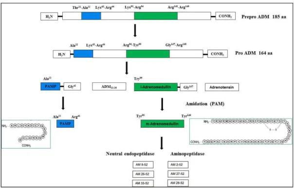
The ADM gene encodes a 185-amino acid preproADM (Figure 1). After cleavage of a 21-residue N-terminal signaling peptide, preproADM is converted into a 164-amino acid proADM peptide, which is a precursor of two biologically active peptides, namely ADM and proADM N-terminal 20 peptide (PAMP) (Kitamura et al., 1994; Kitamura et al., 2002). Under normal healthy conditions, ADM circulates at low picomolar concentrations (2-10 pM) (Kitamura et al., 1994; Suzuki et al., 2004) in the plasma in two forms, a mature 52-amino acid peptide (mADM) and an immature glycine-extended 53-amino acid peptide (iADM), which is subsequently converted to mADM after enzymatic amidation (Kitamura et al., 1998; Asakawa et al., 2001). ). iADM represents 85% of total plasma ADM (Yamaga et al., 2003).
Figure 1 Structure of ADM and post-translational processing of preproADM gene (Modified from Hamid & Baxter, 2005)
After cleavage of a 21-residue N-terminal signaling peptide from the initial 185-amino acid preproADM peptide, the 164-amino acid proADM peptide is converted, which is a precursor of two biologically active peptides, namely ADM and PAMP. Further degradation products derived from the ADM precursor or ADM are also listed.
In the blood circulation, ADM is bound to ADM binding protein 1 (AMBP-1; also called complement factor H) which stabilizes the peptide (Elsasser et al., 1999;
Pio et al., 2001). It is also reported that via its binding to AMBP-1 the receptor- mediated effects of ADM are increased, while its receptor-independent antimicrobial activity is suppressed (Beltoeski & Jamroz, 2004). In addition, the inhibitory effect of factor H in alternative pathway of complement activation is enhanced via its binding to ADM. It is noteworthy that, neither CGRP nor PAMP binds complement factor H from plasma (Beltoeski & Jamroz, 2004).
In humans and rats, circulating ADM is rapidly metabolized with the elimination half-life (T1/2) of about 20 min. However, little is known about its precise metabolism and clearance. ADM is metabolized by neutral endopeptidase and cleared in the pulmonary circulation (Lisy et al., 1998; Dupuis et al., 2005).
Studies using intravenous administration of radioactively labeled ADM derivatives demonstrate a high first pass effect during the passage through the
lung vasculature, where 36% of a bolus is captured in dogs (Dschietzig et al., 2002; Dupuis et al., 2005). Studies in rats show that more than 25% of the lung bound ADM can be displaced by non-labeled ADM, which proportion is therefore likely to be receptor bound. Moreover, ADM is present in human urine with about six-fold higher concentration than in human plasma, suggesting that a renal clearance may be also relevant in addition to pulmonary clearance (Sato et al., 1995). However, circulating ADM is cleared by neutral endopeptidase in kidney, located in the tubular brush border system, and the subsequent urinary excretion of ADM may be derived from renal production rather than from glomerular filtration (Lisy et al. 1998, Nishikimi, 2007).
3.1.2. Receptors and intracellular signalling of ADM
ADM exerts its biological actions through binding to the heterodimeric G-protein coupled receptor complex (GPCR) composed of the calcitonin receptor-like receptor (CRLR) associated with RAMP-2 (ADM-1 receptor) or RAMP-3 (ADM- 2 receptor). RAMPs bind to the receptor molecule in the endoplasmic reticulum and facilitate their translocation to the plasma membrane. Additionally, RAMPs regulate the degree of receptor glycosylation and play an important role in receptor specificity, ligand affinity, and receptor desensitization (McLatchie et al., 1998). Although the three isoforms of RAMPs show a differential tissue distribution, a pharmacological difference of ADM-1 and-2 receptor is not established yet (Chakravarty et al., 2000; Hagner et al., 2002). Besides ADM-1 and -2 receptors, ADM can also mediate its effects via CGRP-1 receptor, especially in the vascular system (Dennis et al., 1990; Chiba et al., 1989).
Indeed, ADM can bind to some regions in the brain where actually CRLR is not expressed, suggesting the existence of specific ADM receptors with a different molecular structure (Hinson et al., 2000).
The cellular signaling mechanisms through which ADM mediates its biological effects vary among species, vascular beds and cell types, but mainly involve cAMP, nitric oxide (NO) and calcium-dependent mechanisms (Lopez &
Martinez., 2002). Moreover, activation of different kinases, such as protein kinase A (PKA), Src, protein kinase C (PKC), p38 mitogen-activated protein
kinase (MAPK), and extracellular-signal regulated kinases (ERK), have been reported to be involved in ADM signaling. The combinations of various signaling may be responsible for numerous biological functions of ADM in different cell types (reviewed by Gibbons et al., 2007).
3.1.3. Biological actions of ADM in response to inflammation
The ADM gene is broadly expressed throughout most organs during embryonic development and adulthood, especially in endothelial cells, vascular smooth muscle cells, cardiac myocytes, and human leucocytes (Garayoa et al., 2002;
Hinson et al., 2000). Moreover, in highly vascularized tissues, such as placenta, lung, heart, and kidney, levels of ADM tend to be relatively higher. Experimental data from literature underscore a role of ADM in a variety of functions, including blood pressure regulation, broncho-dilatation, renal function, hormone secretion, cell growth, differentiation, neurotransmission, and modulation of the immune response. Moreover, ADM plays a crucial role as autocrine factor during proliferation and regeneration of endothelial cells (reviewed by Ishimitsu et al., 2006; Gibbons et al., 2007). However, data from targeted deletion in mice of ADM or its receptor gene, point to another dominant function of this peptide in regulating vascular permeability. While heterozygous littermates show a mild hypertension (Shindo et al., 2000) and exhibit a more pronounced inflammatory response to endotoxin-induced septic shock (Dackor and Caron, 2007), homozygous knockout mice develop a strong and lethal hydrops fetalis (Caron and Smithies, 2001). A similar phenotype is further substantiated by data from knockout mice lacking CRLR or PAM genes (Dackor et al., 2006; Czyzyk et al., 2005). Conversely, mice overexpressing ADM show moderate hypotension and are largely resistant to LPS induced shock (Shindo et al., 2000).
Transcription of the ADM gene is stress sensitive. A variety of stimuli such as hypoxia, inflammatory stimuli (e.g. LPS), as well as pro-inflammatory cytokines (e.g. IL-1α, IL-1β, TNFα, and TNFβ) are shown to induce the expression of ADM (reviewed by Tammesfeld-Wollbrück et al., 2007). Accordingly, elevated plasma ADM levels have been detected in a wide variety of physiological and pathological conditions, including normal pregnancy and pregnancy with
complications, cardiovascular and pulmonary diseases, diabetes, endocrine disorders, hepatic and renal failure, cancer, and sepsis (reviewed by Gibbons et al., 2007). In their analysis, based on published human clinical data, Gibbons and colleagues compared fold change of plasma ADM in a variety of human conditions. One of the highest increases in plasma ADM levels occurred during sepsis, which evidences the importance of ADM under inflammatory conditions.
Moreover, in animal studies, high expression of ADM was observed in the lung in endotoxaemia (Cheung et al., 2004) and in acute lung injury induced by hyperoxia and LPS (Agorreta et al., 2005). In a model of cecal ligation and puncture in rats, the small intestine was demonstrated to be an important source of ADM release during polymicrobial sepsis (Zhou et al., 2001). These observations raised the question as to whether elevated ADM levels reflect a protective defense mechanism rather than being of pathological significance.
Meanwhile, there is strong evidence from the literature that administration of ADM to supra-physiological levels exerts strong anti-edematous effects:
In vitro, using measurement of physiological hydrostatic pressure as indictor of endothelial permeability, ADM was shown to dose-dependently reduce hyperpermeability induced by different stimuli, such as thrombin, hydrogen peroxide, E. coli hemolysin, or S. aureus alpha-toxin, in endothelial cells from different species (human, rat, porcine) and different vasculatures (umbilical vein, lung pulmonary artery) (Hippenstiel et al., 2002; Hocker et al., 2006). The measurable increase of intracellular cAMP levels was supposed to be the key mechanism of action of ADM signaling. Inhibition of MLC phosphorylation via PKA activation was hypothesized to counteract and reduce endothelial contractility and to further stabilize endothelial barrier function (Hippenstiel et al., 2002). Moreover, ADM was shown to tighten blood brain barrier in terms of increased transendothelial electrical resistance (Kis et al., 2001; Kis et al., 2003;
Honda et al., 2006). In ex vivo-models using hydrogen peroxide-exposed rabbit lungs and S. aureus α-toxin-infused rat ileums, ADM showed protective effects in those isolated organs (Hippenstiel et al., 2002; Brell et al., 2005; Hocke et al., 2006).
Further in vivo- studies were in line with the results obtained in vitro and in isolated organs. ADM reduced the ovalbumin-induced airway microvascular leakage in an ovalbumin-sensitized guinea pig model (Ohbayashi et al., 1999).
Intravenous infusion of ADM in rats protected against endotoxemia induced lung injury (Itoh et al. 2007). In a sheep model of endotoxemia induced lung injury, ADM reduced pulmonary hypertension and prevented hypodynamic circulation (Ertmer et al., 2007). ADM was shown to protect lung function significantly in models of oxygen and ventilator induced lung injury (Müller et al., 2010; Tao et al, 2012).
In a rodent model of Gram positive sepsis and in the severe model of coecum ligation and puncture (CLP) induced polymicrobial sepsis, ADM reduced vascular leakage and secondary lung damage, and prolonged survival (Hocke, et al, 2006; Temmesfeld-Wollbrück et al., 2007; Wu et al, 2008). In further models of polymicrobial sepsis (Wang, 2001; Yang et al., 2010; Gonzalez-Rey et al., 2006), endotoxin-related shock (Gonzalez-Rey et al., 2006) and hemorrhage (Cui et al., 2005) ADM showed beneficial effects on survival.
In severe sepsis a loss of plasma ADM coincides with loss of AMBP (the complement factor-H) at the transition from the phase of hyperdynamic to hypodynamic circulation due to increased consumption of components of the complement system (Yang et al., 2002). A decrease of ADM plasma level in sepsis was believed to facilitate generalized edema formation and deterioration of cardiac function (Westphal et al., 2006). In the CLP-induced sepsis model, administration of ADM together with human complement factor-H significantly could halt transition to hypodynamic circulatory failure (Yang et al., 2002), while infusion of ADM stabilized cardiac output in endotoxemic sheep (Ertmer et al., 2007).
3.2. Regulation of vascular permeability
Endothelial permeability is described as a passage through either one of two different routes: one transcellular (cross cells), via caveolae-mediated vesicular transport, and the other paracellular (between cells), through interendothelial
junctions. Although transcytosis plays an important role in the basal permeability of the endothelium, paracellular flux of plasma fluid and proteins through endothelial cell–cell junction is more dominant in the development of vascular inflammation under pathophysiological conditions (Komarova & Malik, 2010; Mehta & Malik, 2006).
3.2.1. Endothelial cell-cell junctions and vascular integrity
Although endothelial cell-cell junctions are differently structured in different organs, the general constituent of endothelial cell–cell junctional structures, with major impact on cell-to-cell adhesion and barrier properties, are tight junctions (TJs) and adherens junctions (AJs). AJs represent the predominant type of junctions within the endothelial barrier, whereas TJs predominate in the epithelium (Vandenbroucke et al., 2008). In the endothelium, the TJs are interwoven with AJs over the lateral intercellular space differing from the structure in epithelium (Figure 2). The molecular structure of AJ as well as TJ is based on the homophilic interaction of transmembrane proteins between two adjacent cells. Specific cytosolic proteins link to the adhesion proteins anchoring them to the cell cytoskeleton and transducing cellular signaling. By way of this protein complex, cell-cell junctions influence the important cellular process, such as proliferation, polarity, protein expression, and permeability (Bazzoni & Dejana, 2004).
Figure 2 Endothelial cell-cell junctions
In the endothelium, the tight junctions and adherence junctions are interwoven with each other over the lateral intercellular space. The tight junction proteins, claudins, occludins and JAMs, are linked to the intracellular actin cytoskeleton via zonular occludins (ZOs). The primary molecular structure of adherence junction is VE-cadherin-catenin complex linking directly or indirectly to actin cytoskeleton.
Tight junctions are primarily found in blood-brain, blood-retinal, or blood-testis barrier microvasculature (Mehta & Malik, 2006; Hawkins & Davis, 2005). Tight junction resembles a zipper-like structure formed by homophilic interactions of transmembrane proteins, including occludin, claudins, and junction adhesion molecule (JAMs). These three TJ proteins represent the backbone for TJs.
They are intracellularly connected to the actin cytoskeleton via zonular occludins (ZO-1; 2) (Bazzoni, 2006). Complex tight junctions as found e.g. in the blood brain barrier are responsible for a high transendothelial electrical resistance and are exclusively impermeable to the passage of solutes (Crone &
Olesen, 1982). Although TJs are well developed in large artery endothelial cells that are exposed to high flow rates, TJs are less complex in capillaries than in arterioles, and even less in venules within the microvasculature (Wallez &
Huber, 2008).
Adherens Junctions have been recognized as a critical component in regulating paracellular permeability of microvascular endothelium, with the exception of blood-brain barrier and blood-retinal barrier endothelium (Mehta & Malik, 2006).
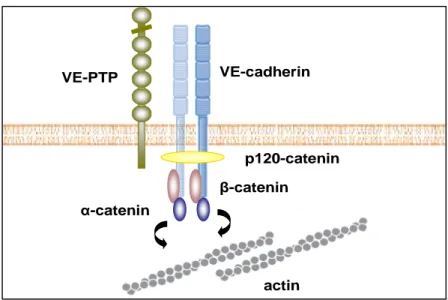
AJs have been identified in nearly all types of vascular beds, especially in the peripheral microvasculature. In the postcapillary venules, where leukocyte adhesion and inflammatory hyperpermeability selectively occur, beside AJs other types of junctions are poorly developed. The primary molecular structure of AJ is based on homophilic interactions of vascular endothelial cadherin (VE- cadherin) (Figure 3). VE-cadherin is a transmembrane protein composed of five extracellular calcium–binding domains, one transmembrane domain and a cytoplasmatic tail domain (Vestweber, 2008). The hemophilic interaction of extracellular domains is calcium dependent. The intracellular domain is anchored to the cell cytoskeleton through binding with catenins (α, β, γ and p120) (Djana, 2004; Vestweber, 2008). This cytosolic connection to the cytoskleton is of importance for maintaining junctional strength and for the regulation of paracellular permeability (Yuan, 2002). It is hypothesized that through these junction-cytoskeleton connections, endothelial cytoskeletal contractile forces can be transmitted to cell–cell junctions and thus dynamically regulate endothelial permeability. Antibodies that inhibit the adhesion function of VE-cadherin cause dissociation of endothelial cell layers in vitro, enhanced accumulation of neutrophils in a peritonitis mouse model and lead to subcutaneous hemorrhage and death of mice (Vestweber et al., 2009).
The stability and composition of the VE-cadherin-catenin complex is dependent on the status of cell contacts and regulated by tyrosine phosphorylation (Vestweber, 2008). In subconfluent cell culture where the endothelial cells are weakly connected to each other, β-catenin and p120 are infirmly linked to VE- cadherin, and the protein components of this complex are highly phosphorylated. In contrast, in confluent cell culture where endothelial cells are strongly connected to each other, γ-catenins are predominant over β-catenins in VE-cadherin-catenin complex, and the phosphorylation of the components is reduced (Lampugnani et al., 1997; Lampugnani et al., 1995). The destabilization of cell contacts is accompanied by a decreased association with
γ-catenin and increased association with β-catenin (Lampugnani et al., 1995).
The underlying mechanism is still not fully understood (Vestweber, 2008).
Furthermore, there is correlation between tyrosine phosphorylation of various components of the VE-cadherin-catenin complex and the decrease of VE- cadherin-mediated adhesion. Many vasoactive stimuli, such as histamine, thrombin, VEGF and TNFα, have in common that they induce phosphorylation of the VE-cadherin-catenin components and thus destabilize the complex (Angelini et al., 2006; Vestweber, 2008). Thrombin is shown to cause disassembly of AJs via PKC-induced modification of VE-cadherin and β-catenin phosphorylation (Konstantoulaki et al., 2003). Vascular endothelial growth factor (VEGF) stimulation induces activation of Src-kinase and thus phosphorylation of VE-cadherin on the residue Serine 665, leading to α-arrestin-2 regulated internalization and loss of endothelial barrier function (Gavard & Gutkind, 2006).
An important counteracting regulator of this phosphorylation is the vascular endothelial protein tyrosine phosphatase (VE-PTP), which is associated with tyrosine kinase receptor (Tie 2) and VE-cadherin. Docking of neutrophil granulocytes or lymphocytes on tumor necrosis factor (TNFα)-stimulated endothelial cells leads to dissociation of VE-PTP from VE-cadherin, (Nottebaum et al., 2008). Down-regulation of VE-PTP expression increases endothelial cell permeability, enhances leukocyte transmigration, and inhibits VE-cadherin- mediated adhesion (Nottebaum et al., 2008).
Figure 3 Structure of VE-cadherin-catenin complex in endothelial cells
The primary molecular structure of adherence junction is based on the homophilic interactions of VE- cadherin. This transmembrane protein VE-cadherin consists from five extracellular domains, one transmembrane domain and a cytoplasmatic tail domain interactingwith catenins (α, β, γ and p120). VE- cadherin is anchored to the cell cytoskeleton, whereas through binding its extracellular domains, VE- cadherin can interact with other binding partners, such as VE-PTP.
.
3.2.2. Endothelial cytoskeleton and vascular permeability
3.2.2.1. Dynamic functions of cytoskeleton and vascular permeability Similar to smooth muscle cells vascular endothelial cells are contractile cells developing forces driven by a mechanochemical interaction between actin and myosin (Figure 4). The increased actomyosin contractility is characterized by the formation of stress fibers, which are bundles of actin filaments associated with nonmuscle myosin II (Wojciak-stothard & Ridley, 2002). The key signal to trigger endothelial contraction is the phosphorylation of the regulatory part of myosin, the myosin light chain (MLC) (Dudek & Garcia, 2001; Tiruppathi, 2002).
Phosphorylation of MLC induces conformational change of myosin, interacting with actin, sliding along actin filaments, subsequently causing contraction (Tiruppathi, 2002; Sandoval et al., 2001). This myosin-actin cross-bridge cycling provides a mechanical basis to develop contractile force in response to physiological and pathological stimulation and is the cause for generating and maintaining a centripetal tension (Yuan, 2002).
VE-cadherin
p120-catenin
α-catenin
β-catenin
actin VE-PTP
In vascular endothelial cells, the phosphorylation status of MLC results from the balance of MLC kinase (MLCK) and MLC phosphatase (MLCP) (Garcia et al., 1995). Firstly, MLCK phosphorylates MLC at Ser-19 and subsequently at Thr- 18, starting actomyosin-interaction thus generating centripetal forces which ultimately may be responsible for dissociation of cell-cell contacts and formation of interendothelial gaps (Moy et al., 1996). MLCK is activated in a Ca2+/calmodulin dependent manner and by tyrosine kinase-mediated phosphorylation at Tyr-464 and Tyr-471 (Goeckeler & Wysolmerski, 1995;
Garcia et al., 1995). Secondly, MLCP dephosphorylates MLC as opposed to MLCK, decreases contractile forces, and subsequently relaxes the endothelial cytoskeleton (Verin et al., 1995). Finally, small GTPases of the Rho family are also involved in regulating the phosphorylation status of MLC (van Nieuw Amerongen & van Hinsbergh, 2001). RhoA, a member of the Rho family of small GTPases, can increase MLC phosphorylation indirectly by activating its downstream effector, Rho C-terminal kinase (ROCK), which subsequently phosphorylates and inhibits MLCP (Noda et al., 1995; Yoshioka et al., 2007).
Besides the formation of actin stress fibers and the induction of contractile forces, activation of Rho and ROCK directly affects the destabilization of cell contacts (Wojciak-Stozhard & Ridley, 2002). In addition to RhoA, additional small GTPases of the Rho superfamiliy, Rac and Cdc42, are also implicated in actomyosin contractility, since their downstream effector, p21-activated kinase (PAK), phosphorylates MLC on Ser-19 (Goeckeler et al., 2000).
MLC phosphorylation is involved in modulating endothelial barrier dysfunction in response to cellular mediators (e.g. activated neutrophils), as well as inflammatory agonists (e.g. thrombin, histamine, cytokines, oxygen radicals).
Inhibition of MLCK by specific inhibitors (ML7 and ML9), prevents phosphorylation of MLC and stabilizes vascular permeability. Using fluorescence microscopic approaches, endothelial cells exposed to inflammatory stimuli present a morphological change characterized by increased staining of MLC phosphorylation accompanied with formation of stress fiber and intercellular gaps (reviewed by Yuan, 2002).
Figure 4 Contractile machinery and actin cytoskeleton in endothelial cells
In vascular endothelial cells, the balance of MLCK and MLCP results in the phosphorylation status of MLC, inducing conformational change of myosin, subsequently interacting with actin, sliding along actin filaments, and causing contractility. Ca2+/calmodulin dependent activation of MLCK, RhoA-ROCK dependent inactivation of MLCP, as well as the small GTPases (Rac, and Cdc42) are implicated in phosphorylation of MLC and acto-myosin based endothelial contractility.
3.2.2.2. Static functions of cytoskeleton in vascular permeability
Besides the role of actin in the active contractile apparatus, disruption and rearrangement of actin cytoskeleton are of equal importance in the development of endothelial gaps (Baldwin & Thurston, 2001; Schnittler et al., 1990). Upon stimulation by inflammatory mediators and neutrophils, as well as during ischemia–reperfusion injury, actin directly undergoes polymerization and redistribution to form stress fibers followed by the formation of intercellular gaps (Korthuis et al., 1991; Schnittler et al., 1990; Shasby et al., 1982). However, those effects can totally be abolished by actin stabilization agents in vitro and ex vivo, such as phalloidin, antamanide, cytochalasin B, or cytochalasin D (Korthuis et al., 1991; Shasby et al., 1982). In a study using confocal microscopic analysis in rat mesenteric venules, the time course of development and recovery of histamine-induced venular leaks is coincident with the rearrangement of endothelial actin fibers (Baldwinand & Thurston, 1995). Local breaks in the peripheral actin rim of endothelial cells are accompanying