Characterization of Phagocytic Pattern Recognition Receptors in
Drosophila melanogaster
Inaugural-Dissertation zur
Erlangung des Doktorgrades
der Mathematisch-Naturwissenschaftlichen Fakultät der Universität zu Köln
vorgelegt von
Yoon-Suk Alexander Chung aus Köln
Köln 2011
Berichterstatter: Prof. Dr. Manolis Pasparakis Prof. Dr. Siegfried Roth Prof. Dr. Christine Kocks Vorsitzender: Prof. Dr. Peter Kloppenburg
Tag der mündlichen Prüfung: 04.07.2011
Our greatest glory is not in never falling, but in rising every time we fall.
Confucius
Table of Contents
Abstract ... 6
Zusammenfassung ... 7
Abbreviations ... 9
1. INTRODUCTION ... 11
1.1. Background ... 11
1.1.1. Innate immune mechanisms in Drosophila ... 11
1.1.2. The NFkB-like Toll and Imd pathways ... 13
1.1.3. Antimicrobial peptides ... 15
1.1.4. Phagocytosis in Drosophila ... 16
1.1.5. Phagocytic receptors in Drosophila ... 17
1.1.6. Eater ... 19
1.1.7. Drosophila as a model for phagocytosis ... 21
1.2. Aims of this Thesis ... 22
2. MATERIAL & METHODS ... 24
3. RESULTS & DISCUSSION ... 36
3.1. Expression, Purification and Characterization of a Recombinant Eater- Fc Protein ... 36
3.1.1. Generation of two Baculovirus expression vectors and small scale purification of Eater-Fc fusion proteins ... 37
3.1.2. Large scale production of biologically active Eater-Fc fusion protein ... 39
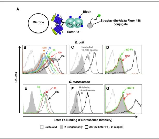
3.1.3. Eater-Fc binding to distinct classes of non-viable microbes ... 41
3.1.4. Characterization of the binding properties Eater-Fc fusion protein ... 42
3.1.5. Discussion and Conclusions ... 46
3.2. Accessibilty of Eater Ligands on Different Classes of Live, Naïve Bacteria ... 47
3.2.1. Eater-Fc binding to live Gram-positive Firmicutes (E. faecalis, S. aureus) ... 47
3.2.2. Absence of Eater-Fc binding to live Gram-negative Proteobacteria (E. coli,
S. marcescens, P. aeruginosa) ... 48
Table of Contents 3.2.3. Membrane bound native Eater receptor on the surface of S2 cells behaves
similarly to Eater-Fc ... 49
3.2.4. Discussion and Conclusions ... 51
3.3. Unmasking of Eater Ligands on Gram-negative Bacteria by Cationic AMPs ... 52
3.3.1. Eater-Fc binds to E. coli killed by exposure to cationic AMP ... 52
3.3.2. Eater-Fc binds to live, AMP-exposed E. coli ... 56
3.3.3. Discussion and Conclusions ... 57
3.4. Eater-Fc binding to bacterial cell wall components ... 60
3.4.1. Differential Eater-N binding to different types of polymeric PGN ... 60
3.4.2. No significant binding of Eater-Fc to LPS and LTA ... 63
3.4.5. Discussion and Conclusions ... 64
3.5. A Limited RNAi Screen to Search for Molecules Involved in Binding and Phagocytosis of the Gram-positive Actinobacterium M. luteus ... 66
3.5.1. Discussion and Conclusions ... 70
3.6. An eater-GAL4 Fly Line to Assess Eater Expression in Different Stages of Fly Development ... 71
3.6.1. Eater expression in different stages of fly development ... 71
3.6.2. Discussion and Conclusions ... 74
References ... 76
Acknowledgements ... 84
Erklärung ... 85
Abstract
Abstract
Drosophila melanogaster has emerged as a powerful model system to study innate immunity. Insects employ multilayered innate immune defenses including antimicrobial peptide responses and phagocytosis. In Drosophila, phagocytosis is carried out by plasmatocytes, a blood cell type similar to mammalian macrophages and neutrophils. The scavenger receptor Eater is expressed by larval and adult plasmatocytes and mediates recognition of a broad range of bacterial pathogens. Eater is required for fly survival after infection with Gram-positive and Gram-negative bacteria. However, the bacterial ligands of Eater, and the mechanisms by which this receptor recognizes these different types of bacteria, remain poorly understood.
To address this problem, I generated a soluble, Fc-tagged receptor variant of Eater comprising the N-terminal 199 amino acids (including four N-terminal EGF- like repeats) and raised antibodies against Eater. Using these tools, I established (i) that Eater is expressed on the surface of macrophage-like Drosophila S2 cells, (ii) that it interacts with broad, yet distinct classes of heat- and ethanol-inactivated microbes and (iii) that it binds peptidoglycan from Gram-negative Proteobacteria (E. coli) and Gram-positive Firmicutes (E. faecalis and S. aureus), but not Gram-positive Actinobacteria (M. luteus). In order to identify genes involved in the phagocytosis of M. luteus, I screened 39 candidate genes by RNA interference-mediated knock down in S2 cells.
A longstanding question was whether Eater recognizes live, naïve bacteria. I found that Eater-Fc bound equally well to naïve or heat-inactivated S. aureus or E. faecalis, suggesting that in vivo Eater directly targets live Gram-positive bacteria, enabling their phagocytic clearance and destruction. By contrast, Eater-Fc was unable to interact with live Gram-negative bacteria (E. coli, S. marcescens and P.
aeruginosa). Eater binding required prior membrane-disrupting treatments. Cecropin
A, a prototypic cationic, membrane-disrupting antimicrobial peptide could promote
Eater-Fc binding to live E. coli, even at sublethal concentrations. These results
suggest a previously unrecognized mechanism by which antimicrobial peptides
cooperate with phagocytic receptors.
Zusammenfassung
Zusammenfassung
Drosophila melanogaster hat sich zu einem nützlichen Modellsystem zur Erforschung angeborener Abwehrmechanismen entwickelt. Insekten besitzen ein vielseitiges Immunsystem welches unter anderem antimikrobielle Peptide und Phagozyten umfasst. In Drosophila wird Phagozytose von sog. Plasmatozyten durchgeführt, einem Blutzelltyp, der den Makrophagen und neutrophilen Granulozyten des Menschen ähnelt. Der ‚Scavenger Rezeptor’ Eater wird von Plasmatozyten in Larven und adulten Insekten ausgeprägt. Er erkennt ein breites Spektrum bakterieller Pathogene und seine Ausprägung ist erforderlich für das Überleben von Infektionen mit Gram-negativen und Gram-positiven Bakterien. Die bakteriellen Liganden und die Mechanismen, mit denen Eater diese verschiedenen Bakterien erkennt, sind unzureichend verstanden.
Um diese Fragen zu erforschen, habe ich eine lösliche Rezeptorvariante hergestellt, Eater-Fc, welche aus den N-terminalen 199 Aminosäuren von Eater (4 EGF-ähnliche Wiederholungen umfassend) und einem C-terminalen Antikörper-Fc-Teil besteht.
Zudem habe ich Antikörper gegen Eater-Fc hergestellt. Mit diesen Reagenzien konnte ich zeigen, dass Eater (i) auf der Oberfläche von Makrophagen-ähnlichen Drosophila S2 Zellen ausgeprägt wird, (ii) mit einem breiten, jedoch differenzierten Spektrum inaktivierter Mikroben interagiert, und (iii) an Peptidoglykan von Gram-negativen Proteobakterien (E. coli) und Gram-positiven Firmicutes (S. aureus, E. faecalis), jedoch nicht von Gram-positiven Actinobakterien (M. luteus), bindet. Um Gene zu finden, die an der Phagozytose von M. luteus beteiligt sind, habe ich S2 Zellen untersucht, in denen 39 Kandidaten-Gene mit Hilfe von RNA-Interferenz ausgeschaltet wurden.
Eine bisher ungeklärte Frage war, ob Eater auch vermag, an lebende, unbehandelte
Bakterien zu binden. Einerseits konnte ich zeigen, dass Eater-Fc lebende Gram-
positive Firmicutes-Bakterien (S.aureus oder E. faecalis) bindet. Es liegt deshalb nahe
zu vermuten, dass Eater diese Gram-positive Bakterien in vivo direkt erkennen und
ihre Phagozytose und Zerstörung einleiten kann. Andererseits war Eater-Fc nicht in
Zusammenfassung
P. aeruginosa) zu reagieren. Um eine Bindung zu ermöglichen, mussten die Bakterien zuvor einer membran-schädigenden Behandlung unterzogen werden. Cecropin A, ein kationisches, membran-permeabilisierendes Peptid bewirkte, dass Eater an lebende E.
coli binden konnte, sogar unter sublethalen Bedingungen. Meine Ergebnisse weisen somit auf einen bisher unbekannten Mechanismus hin, der es Antimikrobielle Peptiden ermöglicht, mit Phagozytose-Rezeptoren zu kooperieren.
Abbreviations
Abbreviations
AMP antimicrobial peptide BHI brain heart infusion
bp base pair
BSA bovine serum albumin CFU colony forming units
Da Dalton
DIC differential interference contrast (Nomarski microscopy) DNA deoxyribonucleic acid
dsRNA doublestranded ribonucleic acid EDTA ethylenediaminetetraacetate EGF-like epidermal growth factor – like EM electron microscopy
FACS fluorescence activated cell sorter FBS fetal bovine serum
Fc Fragment, crystallizable region of antibody constant region FITC fluorescein-5-isothiocyanate
FPLC fast protein liquid chromatography GFP green fluorescent protein
IMD immune deficiency kDa kiloDalton
LB lysogeny broth
LBP LPS binding protein LDL low density lipoprotein LPS lipopolysaccharide LTA lipoteichoic acid
Lys lysine
mDAP meso-diaminopimelic acid
NFκB nuclear factor kappa-light-chain-enhancer of activated B cells OD optical density
PAGE polyacrylamide gel electrophoresis PBS phosphate buffered saline
PBS-T phosphate buffered saline + 1 % Tween 20 PCR polymerase chain reaction
PGN peptidoglycan
PGRP peptidoglycan recognition protein
PI propidium iodide
poly-C polycytidylic acid poly-I polyinosinic acid
PRR pattern recognition receptors S2 Schneider line 2 (S2 cells) SDS sodium dodecyl sulfate Sf-9 Spodoptera frugiperda 9 cells TEV Tobacco etch virus
TFA trifluoroacetic acid
Abbreviations
Tris Tris (hydroxymethyl) aminomethane
TE Tris 10 mM/EDTA 1 mM pH 8
RNA ribonucleic acid RNAi RNA interference
RT room temperature
Introduction
1. INTRODUCTION
1.1. Background
1.1.1 Innate immune mechanisms in Drosophila
Multicellular animals, whether they are invertebrates or vertebrates, are able to defend themselves against pathogens (Kvell et al. 2007). The defense mechanisms against infectious pathogens, namely the innate and the adaptive immune systems, protect animals against attacks from potentially pathogenic bacteria, fungi and viruses. The innate immune system is the first line of defense mounted in response to various microbial invaders (Hoffmann 2003). The innate immune system appeared early during evolution and while this is the only defense mechanism in invertebrates, it is also a major part of the immune system in vertebrates (Janeway 1989). The insect and mammalian innate immune responses against pathogenic microbes show a great amount of evolutionary conservation (Hoffmann 2003; Kimbrell & Beutler 2001;
Aggarwal & Silverman 2008). A great example of this conservation was provided by the discovery of the Toll pathway as an important part of the Drosophila immune response and the following identification of the mammalian Toll-like-receptors (TLRs) (Lemaitre 2004). In addition to an innate immune system, vertebrates however developed a very complex adaptive immune system, which cooperates with the innate immune system in host defense (Medzhitov 2007).
The adaptive immune system has the capacity to specifically recognize and
remember attacks by pathogenic microbes (immunological memory). Adaptive
immunity seemed to dominate pathogen defense, and it seemed that the innate
immune system only played a minor role in the fight against microorganisms that
cause pathology (Kvell et al. 2007). The innate- and the adaptive immune system deal
with the molecular diversity of pathogens in fundamentally different ways. The main
distinction between these are the receptor types used to recognize pathogens
(Medzhitov 2007). Adaptive immune recognition is mediated by antigen receptors on
T and B cells with unlimited specificities generated by somatic rearrangement of
Introduction
uses germline encoded receptors which recognize evolutionarily conserved microbial molecules, the so-called pathogen-associated molecular patterns (PAMPs) (Medzhitov & Janeway 2000; Janeway & Medzhitov 2002). Such germline encoded receptors have been termed pattern recognition receptors (PRRs) (Medzhitov &
Janeway 2000; Janeway & Medzhitov 2002). PRRs initiate signaling cascades leading to the production of immune effectors, such as antimicrobial peptides, cytokines, inflammatory mediators, and the activation of phagocytic and proteolytic cascades.
Figure 1. The fruit fly D. melanogaster possesses multilayered pathogen defense mechanisms.
1. Regulation of the native microbiota in the gut through AMPs and reactive oxygen species. 2. Barrier epithelial responses produce local AMPs and send signals to the rest of the body. 3. Clotting response seals wounds, prevents bleeding and traps bacteria. 4. Phenoloxidase response deposits melanin at the site of an immunereaction releasing potentially antimicrobial reactive oxygen species. 5. Phagocytic response, through which phagocytes kill microbes directly or indirectly (by releasing systemic signals).
6. Systemic AMP response through NfkB pathways (Toll, Imd) involves massive release of AMPs from the fat body into circulation. 7. Virus infected tissues are defended by RNAi. Modified after Figure 1 in Dionne and Schneider, 2008.
A good model organism to study innate immune functions would be an organism, in
which the extra layer of complexity added by the adaptive part of the immune system
does not exist. This is the case for Drosophila melanogaster. In Drosophila, which
rely almost entirely on innate immunity to fight microbial infection, a sophisticated
multilayered pathogen defense system consisting of at least seven subcategories can
be found, including a cellular and a humoral response (Dionne & Schneider 2008).
Introduction The humoral defense response relies on the production of antimicrobial peptides (AMPs), which in response to pathogen attack are secreted from the equivalent of the mammalian liver, the fat body of Drosophila. Members of the peptidoglycan- recognition protein (PGRP) family act as microbe sensors (receptors) and can be found in the hemolymph, on immune cell surfaces and in the immune cells. These receptors recognize bacterial cell wall components like bacterial peptidoglycans (PGNs) that activate immune signaling pathways, such as the nuclear factor kappa (NFk) B-like Toll and Imd signaling pathways (Hoffmann 2003; Hoffmann &
Reichhart 2002).
1.1.2. The NFkB-like Toll and Imd pathways
The Toll pathway is mainly activated by fungi and Gram-positive bacteria, whereas the Imd pathway is activated predominantly by Gram-negative bacteria (Fig. 2). The two pathways are triggered by elicitors released from the microbes, such as Lys-type PGN for Gram-positive bacteria and DAP-type PGN for Gram-negative bacteria, that are recognized by recognition proteins which in turn activate proteolytic cascades leading to the production of antimicrobial peptides and other immune effectors (Lemaitre 2004). Depending on the κB sites present in their promotors, antimicrobial peptide genes are under the control of either the Toll or the Imd signaling cascade or can even be coregulated (Lemaitre & Hoffmann 2007).
Drosophila Toll does not function as a pattern recognition receptor as its vertebrate counterparts, the TLRs, but has to be activated by a proteolytically cleaved form of the cytokine Spätzle which then leads to the activation of NFκB transcription factors Dif and Dorsal and the induction of various target genes encoding humoral factors, including antimicrobial peptides (Lemaitre et al. 1996; Weber et al. 2003; Hu et al. 2004).
The Imd pathway was initially defined by the identification of a mutation named immune deficiency that impaired the expression of several antibacterial peptide genes (Lemaitre 2004; Lemaitre & Hoffmann 2007). imd mutant flies are viable when non- infected however succumb readily to infections by Gram-negative bacteria (Lemaitre
& Hoffmann 2007). The recognition of Gram-negative DAP-type PGN activates a
Introduction
different NFκB transcription factor, Relish, and the induction of genes encoding humoral factors.
Figure 2. Model of Toll and Imd pathway activation. Toll: The Toll pathway is activated by secreted recognition molecules (GNBPs, PGRPs) that sense Gram-positive Lys-type PGN, Glucan and entomopathogenic fungi, which activate the serine-protease SPE which in turn cleaves Spätzle. Mature Spätzle binds to Toll which leads, through a series of phosphorylations, to the release of transcription factors (Dorsal & Dif) into the nucleus and the production of antimicrobial peptides. Imd: Direct binding of peptidoglycan recognition receptors (PGRPs) to monomeric or polymeric PGN and subsequent recruitment of the adaptor Imd leads through proteolytic cascades to the translocation of the transcription factor Rel into the nucleus and the production of AMPs. Modified after Figure 3 in Lemaitre and Hoffmann, 2007.
Certain aspects of both pathways are not yet fully understood, however ultimately
both pathways lead to the rapid and massive release of antimicrobial peptides from
the fat body into the hemolymph as well as transcriptional upregulation of hundreds
of other putative immune effectors whose role remains to be elucidated (Lemaitre
2004; Lemaitre & Hoffmann 2007).
Introduction 1.1.3. Antimicrobial peptides
Even before the mechanisms of the Toll and Imd pathways were elucidated, Boman and his co-workers were able to characterize the inducible antimicrobial peptides (AMPs) Cecropin and Attacin from the giant silk moth Hyalophora cecropia (Steiner et al. 1981; Hultmark et al. 1983). These peptides were rapidly produced by the insect fat body and secreted into the hemolymph after septic injury. Now more than 1200 different AMPs have been either identified or predicted through nucleic acid sequences (Brogden 2005; Bulet & Stöcklin 2005). These include AMPs from many different tissues and cells of a variety of invertebrate, plant and animal species (Ganz 2004; Ganz 2003; Lehrer 2011; Lehrer 2004; Zasloff 2002; Brogden et al. 2003;
Vizioli & Salzet 2002). AMPs are a unique and diverse group of molecules which have been divided into subgroups on the basis of their amino acid composition and structure (Brogden 2005). Here, the focus lies on an evolutionarily conserved subgroup that contains linear and amphipathic α-helical AMPs including cecropins (Fig. 3) and cecropin-like molecules conserved from Diptera
and Lepidoptera even to mammals (pigs) which contain 29-40 amino acid residues (Gazit et al. 1995; Bulet et al. 2004; Bulet et al. 2004). Sequence comparison revealed that cecropins form a homologous group with more than 70 % identity in their amino acid composition (Okada & Natori 1985; Kylsten et al. 1990; Bulet & Stöcklin 2005).
Despite numerous studies, no definitive consensus explanation has emerged for the mechanism of antimicrobial action of cationic AMPs and their modes of action seem to be pleiotropic comprising direct and indirect antimicrobial functions as well as immunomodulatory activities (Hale & Hancock 2007; Hancock & Scott 2000).
However, there is a broad consensus that α-helical AMPs such as cecropins are active with a higher efficacy against Gram-negative than Gram-positive bacteria, are non- toxic for the host and that one site of the antibacterial action is the bacterial plasma
Figure 3. Global fold of Hyalophora cecropin A. Peptide has a long N-terminal, basic, amphipatic α-helix and a shorter, more hydrophobic C-terminal helix, linked by a Gly-Pro hinge region. NH2, N- terminus; CONH2, C-terminus. From Bulet and Stoecklin, 2005.
Introduction
target organism would be electrostatic, as most bacterial surfaces are anionic, in contrast to animal cells. Their amino acid composition, amphipathicity, cationic charge and size allows cecropins or other cationic AMPs to attach to and insert into membrane bilayers to form pores (Brogden 2005). Several groups showed that various cecropins and cecropin analogues initially form selective voltage dependent ion channels, where the positively charged NH
2-terminal helices bind to negatively charged headgroups on the bacterial membrane and the hydrophobic CONH
2-terminal part inserts itself to the membrane core (Christensen et al. 1988; Silvestro et al. 1999;
Shai 1995). After application of a positive potential on the side of the peptides, the positively charged NH
2-terminal helices get pushed into the membrane and the channel is formed by the association of multiple transmembrane NH
2-helices, so that the hydrophilic residues form the aqueous pore and the hydrophobic residues are in contact with the aliphatic phase of the membrane (Christensen et al. 1988; Durell et al. 1992). Despite multiple theories and models, the precise mechanism by which cecropin attacks bacteria is still not known. To date, it has also not been shown whether AMPs by themselves are sufficient to combat bacterial infections. While direct bactericidal activities of cationic AMPs have been demonstrated, mostly under rather non-physiological conditions, a mechanistically poorly defined activity that leads to increased phagocytosis by macrophages was noticed long ago (Finlay and Hancock, 2004). Some experiments in my thesis address this latter aspect of AMP function and suggest a molecular mechanism for this phenomenon: permeabilization of bacterial envelopes may lead to ‘priming’ of AMP exposed bacteria for other innate immune mechanisms, such as phagocytosis.
1.1.4. Phagocytosis in Drosophila
Phagocytosis is an evolutionarily ancient mechanism by which cells internalize
particles (Metchnikoff 1908; Rabinovitch 1995). It requires cell surface receptors that
bind non-self or altered-self molecules displayed on microbes or dying and aberrant
cells (Stuart & Ezekowitz 2005). The engulfment of apoptotic particles by
macrophages in early stages of embryogenesis for instance has been shown to be
essential for development (Tepass et al. 1994; Zhou et al. 1995). Phagocytosis also
plays a major role in innate immunity as one of the first lines of defense against
Introduction invasive microbes and by mobilizing and instructing adaptive immunity. Phagocytes must constantly monitor their environment to quickly recognize, ingest and destroy foreign intruders or altered cells. Once an invader is recognized, phagocytes start an uptake mechanism that is not yet fully understood (Underhill & Ozinsky 2002).
However, multiple studies with a variety of microbes have shown that mammals and Drosophila share certain parts of the uptake machinery such as actin and actin-related proteins as critical participants in phagocytosis (Stuart & Ezekowitz 2008; Pearson et al. 2003; Philips et al. 2005; Agaisse et al. 2005; Stroschein-Stevenson et al. 2006).
In contrast to the nematode C. elegans, Drosophila has circulating and sessile blood cells (called hemocytes in Drosophila), which play an important role in protecting flies against infection by phagocytosing invading microbes. Drosophila melanogaster possesses three major types of blood cells which are derived from the embryonic and larval hematopoietic organs (Meister & Lagueux 2003): 1.
plasmatocytes, 2. crystal cells and 3. lamellocytes (Rizki & Rizki 1984, cited after Stuart & Ezekowitz 2008). 95 % of hemocytes, sessile as well as circulating, are plasmatocytes, the counterpart of the mammalian neutrophils and macrophages. These phagocytic cells are long-lived (Stuart & Ezekowitz 2008; Meister & Lagueux 2003) and devoid of neutrophil-like granules (Rizki & Rizki 1984; Lanot et al. 2001). They play essential roles in tissue remodeling during development (Defaye et al. 2009) and in immunity during infection (Defaye et al. 2009; Nehme et al. 2007; Charroux &
Royet 2009; Avet-Rochex et al. 2007). Phagocytosis by Drosophila hemocytes share many similarities with the process in mammals, but with less anticipated complexity due to Drosophila’s smaller genome, which makes it a good model system to validate known mechanisms of uptake (Cherry & Silverman 2006; Stuart & Ezekowitz 2008).
In all cases, whether in mammals or Drosophila, phagocytes are able to discriminate particles and microbes by an array of receptor molecules on the surface of the cells.
1.1.5. Phagocytic receptors in Drosophila
As described in a series of recent publications (Philips et al. 2005; Kocks et al. 2005;
Rämet et al. 2002; Stroschein-Stevenson et al. 2006), phagocytosis in Drosophila is
initiated by surface receptors on plasmatocytes which either bind directly to microbes,
Introduction
particle (Stuart & Ezekowitz 2008). Genetic screens and other experiments indicate that in Drosophila there are four different classes of molecules involved in phagocytic recognition (reviewed by Stuart & Ezekowitz 2005):
(i) Complement-like opsonins in Drosophila are thioester-containing proteins (TEPs) which have been found to bind microorganisms and enhance phagocytosis (Stroschein-Stevenson et al. 2006). Functional characterizations of TEPs is derived from RNAi screens in S2 cells and in vitro and in vivo analysis in Drosophila and the mosquito Anopheles gambiae (Moita et al. 2005, Bou Aoun et al. 2011).
(ii) Down syndrome adhesion molecule (DSCAM), a member of an immunoglobulin superfamily, is predicted to have more than 38,000 potential splice variants (Schmucker et al. 2000), possibly 18,000 different extracellular domains of DSCAMs, and also exist in soluble forms (Watson et al. 2005). These may prove to be the innate immune system equivalent of immunoglobulins. This hypothesis is based among other evidence on DSCAM crystal structures (Meijers et al. 2007) and needs further investigation.
(iii) Scavenger receptors in Drosophila belong to several classes, which are structurally unrelated and have been shown to bind a wide variety of microbes as well as apoptotic cells: the CD36 homologues croquemort and peste (Franc et al. 1996;
Philips et al. 2005) and scavenger receptors of class C. Scavenger receptors have emerged as important pattern recognition receptors (Janeway 1989) also in many other species.
(iv) EGF-like-repeat containing receptors, a newly emerging family of EGF- like-repeat-containing receptors belonging to the scavenger receptor family, recently termed the Nimrod Superfamily (Kurucz et al. 2007; Somogyi et al. 2008) which has homologues in many invertebrates and vertebrates including humans. An example in mammals are the class F scavenger receptors SCARF1 and 2.
Within the Nimrod family, the phagocytic receptor Eater (Kocks et al. 2005) is
particularly well characterized (see Chapter 1.1.6 below). Apart from Eater, there
have been reports of other Drosophila proteins from this family, such as NimrodC1, a
transmembrane protein with EGF-like repeats similar to Eater, which seems to act as
a phagocytic receptor and a potential adhesion molecule (Kurucz et al. 2007). A more
recently discovered member of this family called SIMU, comprising in it’s
ectodomain 4 EGF-like repeats, is involved in the engulfment of apoptotic neurons by
glial cells in the developing nervous system of Drosophila (Kurant et al. 2008).
Introduction Interestingly there seems to be a connection between SIMU and Draper, another Nimrod family protein containing EGF-like repeats. SIMU is required for the recognition and Draper for the subsequent engulfment of apoptotic neurons (Kurant et al 2008) and programmed axon pruning in the fly central nervous system (Awasaki et al. 2006; Freeman et al. 2003), and has orthologues and homologues in C. elegans (CED-1) (Zhou et al. 2001) and in mammals (MEGF10, MEGF11, Jedi, SREC1 and 2) (Hamon et al. 2006).
1.1.6. Eater
Eater was identified as a putative target for Serpent, a D. melanogaster GATA transcription factor that had been found to be essential for bacterial phagocytosis by an RNAi screen (Rämet et al. 2002). Silencing of Eater expression in S2 cells led to lower bacterial binding and phagocytic activity. The same result was observed in flies lacking the eater gene or after RNAi knock-down of Eater. Such flies show impaired phagocytic activity, with increased bacterial loads and decreased survival rates after bacterial infections (Defaye et al. 2009; Charroux & Royet 2009; Kocks et al. 2005;
Nehme et al. 2011). Induction of AMP expression through NFκB-like pathways Toll and Imd however was not affected (Kocks et al. 2005; Nehme et al. 2011), consistent with results obtained with flies in which phagocytes were ablated altogether. (Defaye et al 2009; Charroux & Royet, 2009). These results indicated that Eater is a major receptor for a broad range of pathogens in D. melanogaster and that it is critical for immune defense.
Although Eater belongs to a superfamily that comprises mammalian class F scavenger
receptors, no clear mammalian orthologue of Eater exists. There are however related
scavenger receptors implicated in the removal of apoptotic cells, p120 in the flesh fly
(Hori et al. 2000) and CED-1 of C. elegans (Zhou et al. 2001) showing overall amino
acid identity of 40 % and 25 %, respectively (Kocks et al. 2005).
Introduction
Figure 4. Schematic depiction of the Eater protein as a type-1 transmembrane protein with an extracellular region consisting of 32 EGF-like repeats, a transmembrane region and a short intracellular tail.
mRNA expression analyses revealed that the eater gene is a rare example of a gene whose expression is restricted exclusively to adult and larval hemocytes, and their pro-hemocyte precursors in the larval ‘lymph gland’ (Kocks et al. 2005). This expression pattern indicates that although it is a phagocytic receptor for bacterial particles, Eater does not seem to be involved in the clearance of apoptotic cells during tissue remodeling in embryogenesis and metamorphosis. Transcriptional silencing of eater in S2 cells did not affect the uptake of apoptotic cells (Cuttell et al. 2008).
Eater consists of 1206 amino acids and forms a large extracellular domain (Fig. 4). It
contains 32 typical, non-calcium binding EGF-like repeats preceded by an N-terminal
extension of 40 amino acids that contains a characteristic cysteine-flanked CCXGY-
motif (Kocks et al. 2005; Kurucz et al. 2007; Somogyi et al. 2008), with an N-
terminal signal sequence, a single membrane spanning domain, and a short C-terminal
membrane anchor followed by an intracellular domain of 28 amino acids containing a
potential tyrosine phosphorylation motif (Fig. 4) (Kocks et al. 2005). It has been
shown that the first four EGF-like repeats which exhibit a high level of amino acid
Introduction diversity, repeat length and N-glycosylation participate in direct microbe binding (Kocks et al. 2005) whereas the remainder of the repeats may play a structural role as a ‘stalk’. Whether this is the case, or whether the ‘stalk’ does participate in binding remains to be determined experimentally. However, analysis of the evolution of repeats in the Nimrod gene family (Somogyi et al. 2008), and the haplotype structure of eater alleles in wild populations of D. melanogaster (Juneja & Lazarro 2010) indicates different modes of evolution of the more ‘unique’ and ‘stalk repeat’ regions, and is in good agreement with this concept.
Direct binding of microbes to Eater was shown after purification of a secreted, truncated ectodomain comprising two complete N-terminal tandem repeats (Eater1- 199His) from stably transfected S2 cell supernatants (Kocks et al. 2005). Eater 1- 199His bound directly and specificly to heat-inactivated Gram-negative S.
marcescens as well as Gram-positive S. aureus and a yeast associated with termites (C. silvatica) (Kocks et al. 2005). Binding experiments aiming at elucidating Eater ligands suggested that Eater is able to recognize multiple polyanionic ligands, a behavior known from scavenger receptors (Greaves & Gordon 2005; Greaves &
Gordon 2009; Plüddemann et al. 2007). Eater’s affinity to certain polyanionic molecules (such as the typical scavenger receptor ligands oxidized low density lipoprotein (LDL) or acetylated LDL), as well as unpublished data suggesting binding of Eater to bacterial LPS and LTA, supported this view (Kocks et al. 2005; J. Cho &
C. Kocks, unpublished). To date, it still remains to be determined whether Eater is also able to recognize other molecules found in the bacterial envelope. Shedding light on this issue would help us understand the mechanism by which Eater is able to bind to bacteria.
1.1.7. Drosophila as a model for phagocytosis
In mammals, the innate and adaptive immune system work in synergy making it
complex to investigate one part of it without the other part interfering. The fruit fly,
however, lacks the adaptive part of the immune system and this makes it inherently
useful to study innate immune responses in the absence of antibody-based, acquired
immunity (Levitin & Whiteway 2008).
Introduction
Drosophila has a short generation time (10-12 days at 25°C) and can be maintained at relatively low cost. Furthermore, there are a number of macrophage-like Drosophila cell lines derived from mixed embryonic tissues including the widely used Schneider line 2 (S2 cells) (Schneider 1972). These hemocyte-derived cells possess properties similar to mammalian macrophages and efficiently phagocytose invading microbes and cell debris in a temperature-dependent manner (Pearson et al 2003;
Rämet et al., 2002; Rämet et al., 2001; Stuart & Ezekowitz 2008). Their morphology after phagocytosis is also very reminiscent of that of professional phagocytes (Meister & Lagueux 2003; Pearson et al. 2003; Rabinowitz et al. 1992). S2 cells have been used as a tool to study Drosophila immune responses, particularly in regard to phagocytosis since they are readily amenable to genetic manipulation such as knock- down of expression of candidate genes by RNAi (Stuart & Ezekowitz 2008).
Therefore, S2 cells have been widely used in high-throughput RNAi screens to identify molecules for their involvement in host pathogen interactions (Rämet et al.
2002; Ramadan et al. 2007; Agaisse et al. 2005; Philips et al. 2005; Boutros et al.
2004; Stroschein-Stevenson et al. 2006; Stuart & Ezekowitz 2008).
With respect to phagocytosis, approximately 600 D. melanogaster proteins were identified to be associated with Drosophila phagosomes, 70 % of which had mammalian orthologues, validating Drosophila as a model system for mammalian phagocytosis (Stuart et al. 2007 reviewed by Stuart & Ezekowitz 2008).
Thus, the powerful genetic tools (Duffy, 2002; Rong et al., 2002) available in Drosophila combined with the ease of using RNAi in cellular systems (Clemens et al., 2000) give researchers many options to study the innate immune system in Drosophila.
1.2. Aims of this Thesis
Characterization of the Drosophila phagocytic pattern recognition receptor Eater
The phagocytic pattern recognition receptor Eater is expressed solely on Drosophila
blood cells (hemocytes) and their precursors (pro-hemocytes) and was shown to play
a critical role in host survival after bacterial infection. Although Eater plays an
Introduction important role during bacterial infection, it remains poorly understood how this receptor recognizes different types of bacteria. Therefore one aim of this thesis was to further elucidate mechanisms by which Eater recognizes various microbes.
Additionally, it was of interest to determine whether Eater recognizes live, naive bacteria since previous binding studies were carried out only with dead bacterial particles. It also remains unclear what the natural ligands of Eater are, and if and how Eater cooperates with other innate immune mechanisms or effectors to exert its protective effect.
Although Eater was found to recognize multiple ligands and broad classes of bacteria, it nevertheless bound specifically to certain microbes (Gram-negative Proteobacteria and Gram-positive Firmicutes) but not to others such as the Actinobacterium M. luteus and yeast C. albicans. This raises the question how Actinobacteria like M. luteus are bound and phagocytosed by Drosophila hemocytes.
It was another aim of this thesis to identify genes involved in the binding and
phagocytosis of M. luteus through a screen of 39 candidate genes.
Materials & Methods
2. MATERIAL & METHODS
2.1. Chemicals and Biologicals
Table 1. Chemicals and Reagents
Product Catalog-No. Manufacturer
Acrylamide Solution BP1410-1 Fisher
Agarose 05066 SIGMA
Ammoniumpersulfat A3678 SIGMA
Anhydrous Sodium Carbonate BioUltra 71345 SIGMA β-Mercaptoethanol (β-M) M7154 SIGMA Blotting Grade Blocker Non-Fat Dry Milk 170-6404 Bio Rad Bovine serum albumin (BSA), Fraction V A3294 SIGMA
Bacto Brain-Heart Infusion Medium 237500 BD Biosciences
CelLytic M C2978 SIGMA
Complete, Mini protease inhibitor cocktail 11836170001 Roche
Dimethylsulfoxide (DMSO) D2650 SIGMA
Ethanol, absolute 200 proof 111ACS200 Pharmco
Ethidium bromide E1510 SIGMA
Fetal Bovine Serum – Heat inactivated
(tested on insect cells) 100 82-147 Invitrogen
Fluorescein-5-isothiocyanate (FITC,
Isomer 1) F-1906 Molecular Probes
Full-Range Rainbow Molecular Weight
Markers 2892534 GE Healthcare
GelCode Blue Stain Reagent 24590 Thermo Scientific
Gentle Ag/Ab Binding and Elution Buffers 21027 PIERCE
Glacial acetic acid, 99.5 % 124040010 ACROS
Glycerol for Molecular Biology G5516 SIGMA
HEPES, 99.5 % H4034 SIGMA
HiTrap Protein A HP 17-0406-01 GE Healthcare
Material & Methods
Human Low Density Lipoprotein (LDL) BT-903 Biomedial Technologies
Isopropanol, 99.9 % BP2632 Fisher
Laemmli-SDS Sample buffer 161-0737 BioRad
LB Broth Lennox L1505 USBiologicals
Lithium Cloride Sigma Ultra L4408 SIGMA
Magnesium chloride 8266 SIGMA
Oxidized Low Density Lipoprotein
(oxLDL) BT-910 Biomedial
Technologies Paraformaldehyde EM Grade 16 % 18814 Polysciences
Phenol 77607 FLUKA
Phenol:Chloroform:Isoamyl Alcohol
25:24:1 77617 FLUKA
Phosphate-buffered saline (PBS) 003000 Invitrogen
Polycytidylic acid (polyC) P4903 SIGMA
Polyinosinic acid (polyI) P4154 SIGMA
Potassium Acetate P1190 SIGMA
Propidium Iodide P3506 Invitrogen
Protein A Carboxylate Beads 17698 Polysciences
Robb’s Drosophila PBS - (Robb 1969)
Schneider’s Drosophila medium 11720-034 Invitrogen
Sodium Acetate S825 SIGMA
Sodium Azide S2002 SIGMA
Sodium chloride S6191 SIGMA
Sodium dodecyl sulfate (SDS), 20 % BP1311 Fisher
Sodium Hydrogen Carbonate S5761 SIGMA
Streptavidin-15 nm colloidal gold
conjugate EM.STP15 Ted Pella Inc.
Sulfo-NHS-LC-Biotin, no weigh 21327 PIERCE
Tetramethylethylenediamine (TEMED) T-9281 SIGMA
Tris-HCl, 1 M, pH 9 T-1190 TEKNOVA
Tris-HCl, 1.5 M, pH 8.8 T-1588 TEKNOVA
Materials & Methods
Tris-HCl, 1 M, pH 6.8 T-1068 TEKNOVA
Trypan Blue (0.4 %) T-8154 SIGMA
Zeba Spin Desalting Columns 89889 PIERCE
Table 2. Enzymes and Enzyme Kits
Product Catalog-No. Manufacturer
All Restriction enzymes New England Bio
labs
Peptide:N-Glycosidase F P07045 New England Bio
labs
‘High Fidelity PCR Master’ Kit 12140314001 Roche Megascript High Yield Transcription Kit AM1334 Ambion
Mutanolysin M9901 SIGMA
Rapid DNA Ligation Kit 1635379 Roche
RedTaq ReadyMix PCR Reaction Mix R2523 SIGMA
THROMBIN CleanCleave Kit RECOM-T SIGMA
Table 3. Bacterial cell wall components
Supplier Cat. No. Compound Origin
Sigma L4524 LPS E. coli
L9143 Pseudomonas aeruginosa
L6136 Serratia marcescens
L3265 LTA Bacillus subtilis
L2515 Staphylococcus aureus
L4015 Streptococcus faecalis
77140 PGN Staphylococcus aureus
69554 Bacillus subtilis
53243 Micrococcus luteus
InvivoGen tlrl-pgnek E. coli
2.2. Microbiology 2.2.1. Bacterial strains
Live E. coli DH10B/TOP10 was purchased from Invitrogen; E. coli DH5alpha GFP,
P. aeruginosa PA14 and S. aureus ALC1435 GFP were gifts of Fred Ausubel, C.
Material & Methods
albicans of Ian Fraser, all at Massachusetts General Hospital, Boston, MA.
S. marcescens Db11-GFP and LPS mutant 20C2 (Nehme et al. 2007), E. faecalis and M. luteus CIPA270 were provided by Dominique Ferrandon, IBMC du CNRS, Strasbourg, France. Surface protein A-negative S. aureus Wood 46 (ATCC10832) was from ATCC. To obtain non-fluorescent S. marcescens Db11, S. marcescens Db11-GFP was cured of the GFP plasmid.
2.2.2. Bacterial cultures and inactivation
Bacteria were grown in LB broth Lennox (US Biological) or brain heart infusion medium (BD) (E. faecalis) and inactivated by heat (60 minutes at 70˚C, or at 95˚C for 30 minutes (PA14)), Carnoy’s fixative (75 % EtOH, 25 % glacial acetic acid for 10 min on ice) or formaldehyde (3 % for 20 min at RT) or used alive. All bacteria were washed in PBS (10 mM sodium phosphate dibasic, 156 mM sodium chloride, 2 mM potassium phosphate monobasic, pH 7.4) before use.
2.3. Molecular Biology
Standard methods of molecular biology were performed according to the respective manufacturer’s guidelines or following protocols described by J. Sambrook et al., unless otherwise stated (Sambrook 2001) .
2.3.1. Quantification of nucleic acids
DNA and RNA concentrations were quantified by measuring the sample absorption at 260 nm and 280 nm with a NanoDrop ND-1000 UV-Vis Spectrophotomoter (Thermo Scientific). An optical density of 1 corresponds to approximately 50 µg/ml of double stranded DNA or to 38 µg/ml of RNA. A 260nm/280nm ratio of > 1.8 was used as an indicator of high nucleic acid purity.
Table 4. Primer sequences, all primer sequences are displayed in 5’à 3’ order. All primers were purchased from MGH DNA Core facility, Boston, MA.
Primer Sequence
pYAC4fwd CGCGGATCCCGCTCAGATCTGCACTGTTAATGT
Materials & Methods
pYAC5fwd ATAGCTCGGTCCGATGTGGATTTGTAGGATAAC
pYAC5rev GCTTACCTTCGA AGGGCCCTCTAGA
pYAC2fwd GTCTAGTCTAGAGTAT ACAACTGATCCCGGTG
pYAC2rev ACCGCGGGTACCGCGGCCGCTGATATC
TCACCTTTGACGA
2.3.2. Extraction of genomic DNA from adult Drosophila
Adapted after a Web protocol from Laura Johnston Lab (www.cumc.columbia.edu/dept/genetics/faculties/Johnston/Potocols/DNA%20Prep.p df); 30 healthy, freshly eclosed flies were collected into 1.5 ml Eppendorf tubes and frozen at –80°C for 5 min. 200 ml Buffer A (RT, 100 mM Tris-HCl, pH 7.5, 100 mM EDTA, 100 mM NaCl, 0.5% SDS) was added and the flies grinded with a tissue grinder until only pieces of cuticle remained. The suspension was incubated at 65°C for 30 min and 800 µl of 1:2.5 [5M]KOAc:[6M]LiCl was added and DNA was precipitated on ice for 10 min. DNA was centrifuged at 14000 rpm for 15 min and the supernatant was transferred to 2 Eppendorf tubes. 7/10 volume of Isopropanol per ml supernatant was added and centrifuged at 14000 rpm for 15 min. The pellet was washed subsequently with 1 ml cold EtOH, 150 ml Phenol (tris-buffered) and aqueous (top) layer was transferred to new eppendorf tube and washed with 150 µl (25:24:1) Phenol : Chloroform : Isoamyl Alcohol. Aqueous (top) layer was transferred to new Eppendorf tube as before and washed with 150 µl (24:1) Chloroform : Isoamyl Alcohol and aqueous (top) layer was transferred to new eppendorf tube. After subsequent addition of 1/10 volume [3M] NaOAc (pH 5.2) and 2x volume 100 % Ethanol, solution was mixed and chilled at -80°C for 15 min then centrifuged at 14000 rpm for 15 min. Ethanol was removed and washed with 1 ml cold 70%
Ethanol. The pellet was dried and resuspended in 100 ml TE (Tris 10 mM EDTA 1 mM).
2.4. Cell culture
Drosophila S2 cells were cultivated at 26.5˚C in Schneider’s Drosophila Medium
supplemented with 10 % FBS heat-inactivated. For production of secreted Eater-Fc
Material & Methods protein, Spodoptera frugiperda 9 (SF-9) cells (Invitrogen) were grown at 27°C in serum-free HyQ-CCM3 medium (Hyclone, Thermo Scientific).
2.5. SDS PAGE and Western Blots
Samples were separated on SDS discontinuous polyacrylamide gels and blotted onto PVDF membranes (Millipore). Membranes were blocked with blocking buffer (2 % w/v dry milk in PBS-T) for one at RT. Subsequently, primary antibodies (Table 5.) diluted in blocking buffer at indicated concentration were applied and incubated for at least 60 minutes at RT. After 3 washes with PBS-T (PBS + 1 % Tween 20) for 10 minutes each, secondary antibody was applied for one hour at RT. After 3 washes with PBS-T, membranes were incubated for one minute in Pierce ECL Western Substrate (PIERCE) and exposed to chemiluminescence films (Kodak). Films were developed in an automatic developer (Kodak).
Table 5. Antibodies and secondary reagents
Product Catalog-No. Manufacturer
Anti-human IgG1-Fc, HRPO conjugate MH1715 Caltag Peroxidase-conjugated AffiniPure Goat
Anti-Rabbit IgG
111-035-144 Jackson
ImmunoResearch
Streptavidin Alexa Fluor 488 S32354 Invitrogen
2.6. Expression and purification of Eater-Fc 2.6.1. Baculovirus Expression Vectors
pYAC4 (encoding Eater
+19-199-Fc; with four additional amino acids at the mature N- terminus)
A 594 bp fragment corresponding to amino acids 19 to 199 of the Eater protein was
amplified using plasmid pMT/V5His-Eater1-199 (Juhyun Cho) as template with
primers pYAC4fwd and pYAC4rev and cloned into pCR2.1-TOPO (TOPO TA
Cloning Kit, Invitrogen Cat. No. K4500-01). The 584 bp BamHI-XhoI fragment
Materials & Methods
Baculovirus expression vector (Ju et al. 2006 , obtained from Bok-Luel Lee) in order to generate an in-frame fusion between the Baculovirus signal sequence gp67 and amino acids 19-199 of Eater, followed by a TEV cleavage site, thrombin cleavage site and the Fc domain of human IgG1. This vector was called pYAC4.
pYAC5 (encoding Eater’19-199-Fc; corresponding to the mature Eater N-terminus) A 648 bp fragment corresponding to amino acids 1 to 199 of the Eater protein was amplified by PCR using plasmid pMT/V5His-Eater1-199 (Juhyun Cho) as template with primers pYAC5fwd and pYAV5rev and cloned into pCR2.1-TOPO (TOPO TA Cloning Kit, Invitrogen Cat. No. K4500-01). The 603 bp BamHI-XhoI fragment corresponding to amino acids 1-199 of Eater was cloned into pFastBactevFc Baculovirus expression vector (Ju et al., 2006, obtained from Bok-Luel Lee) in order to generate an in-frame fusion between the first 199 amino acids of Eater, followed by a TEV cleavage site, thrombin cleavage site and the Fc domain of human IgG1. This vector was called pYAC5. All PCR amplifications were done with High Fidelity PCR Master Kit (Roche). The correct sequence of all of the entire insert was confirmed on both strands.
2.6.2. Expression and purification of Eater-Fc
Recombinant bacmids were generated by transformation into E. coli DH10Bac
(Invitrogen). For production of secreted Eater-Fc protein, Spodoptera frugiperda 9
(SF-9) cells (Invitrogen) were grown at 27°C in serum-free HyQ-CCM3 medium
(Hyclone, Thermo Scientific). High-titer bacmid stock was used to infect 7 liters of
Sf-9 cells (2 x 10
6/ml) and incubated at 27°C for 42 hrs. Cell culture supernatant was
harvested by centrifugation at 5,000 x g for 30 min, filtered (0.22 µm low protein
binding) and loaded onto a 5 ml HiTrap Protein A column on an Äkta FPLC (GE
Healthcare). Bound protein was washed with 5 column volumes of 20 mM Hepes,
100 mM NaCl, pH 7.0, eluted using Gentle Ag/Ab Elution Buffer at pH 6.6 (Pierce,
Thermo Scientific), buffer exchanged into 20 mM Hepes, 150 mM NaCl, pH 7.0
(Zeba Desalt Spin Column; Pierce, Thermo Scientific) and concentrated to 2 mg/ml
with Amicon filter devices (Millipore). To assess purity and size, purified protein was
analysed by Laemmli SDS-PAGE under non-reducing or reducing conditions in the
absence or presence of 710 mM β-mercaptoethanol followed by Coomassie Blue
Material & Methods staining (GelCode Blue; Pierce, Thermo Scientific). For cleavage of Eater-Fc Thrombin CleanCleave KIT (Sigma) was used according to the manufacturer’s instructions.
2.7. Deglycosylation of Eater-Fc
Eater-Fc fusion protein was incubated overnight at 4˚C in PBS under non-denaturing conditions with or without (mock control) Peptide:N-Glycosidase F (New England Biolabs) and subsequently used for SDS-gel analysis (uncleaved or after thrombin- cleavage) or in flow cytometry binding assays (uncleaved).
2.8. Eater-Fc binding to bacteria
Eater-Fc fusion protein, or control human IgG
1or IgG Fc-fragment (Athens Research
& Technology), was biotinylated with EZ-Link Sulfo NHS-LC-Biotin reagent (Pierce, Thermo Scientific) according to the manufacturer’s instructions. For flow cytometry, 2 x 10
6bacteria in Robb’s Drosophila-PBS (Robb 1969) supplemented with 0.5 % BSA and 0.01 % sodium azide were incubated with biotinylated proteins for 30 min at room temperature, sedimented at 9,000 x g for 5 min and washed. For detection of bound biotinylated protein, bacteria were resuspended in the presence of 1 µg/ml streptavidin-Alexa Fluor 488 conjugate (Invitrogen) and incubated for 20 min before analysis on a FACS Calibur (Becton Dickinson). The bacterial population was gated by forward and side scatter and 10,000 events were recorded. For assessment of bacterial viability by propidium iodide (PI) exclusion, 50 µg/ml PI was added on ice immediately before analysis. Fluorescence emissions were detected in the FL-1 channel (Alexa Fluor 488 emission: 519 nm) and, where indicated, in the FL-3 channel (PI emission: 620 nm).
2.9. Generation of anti-Eater-Fc antibodies
Antibodies were generated in two rabbits by Pocono Rabbit Farm & Lab Inc.
Materials & Methods
prescreened towards lack of activity against insect proteins and then injected with Eater-Fc fusion protein. After two bleeds (21 and 28 days after initial injection) the sera were tested for antibodies against Eater by flow cytometry. Rabbits were boosted once more and subjected to a final bleed. Sera from bleed 3 showed a significant improvement in the detection of Eater in flow cytometry as well as in Western Blot analysis and was purified over Protein A Sepharose (Data not shown).
2.10. Cecropin A exposure of bacteria
Chemically synthesized cecropin A peptide from the moth Hyalophora cecropia (KWKLFKKIEKVGQNIRDGIIKAGPAVAVVGQATQIAK) was purchased from Sigma. Cationic control peptide 2K1 with the sequence (GK)
6AS (GK)
6(Fantner et al. 2010) was synthesized by standard solid phase peptide synthesis at the MGH Peptide/Protein Core Facility. Both peptides were dissolved in PBS at 100 µM and stored frozen in aliquots at -80˚C. Bacteria were grown to mid log phase in LB broth Lennox at 37˚C, centrifuged (3,500 x g, 4˚C), resuspended in PBS, counted and adjusted to 10
8/ml. 50 µl of bacterial suspension was added to 50 µl of PBS containing the indicated concentrations of cecropin A and incubated at 25˚C for the indicated times, placed on ice and analysed immediately by flow cytometry in the presence of 50 µg/ml PI. For assessment of Eater-Fc binding to cecropin A-exposed bacteria, subsequent bacterial Eater-Fc binding assays were carried out in PBS at 4˚C, a temperature non-permissive for AMP activity. TFA (used as counterion to maintain charge balance in solid phase peptide synthesis) (Roux et al. 2008) showed no effect on bacterial viability at 9-fold molar excess over peptide (1 counterion per positive charge), and up to 10 mM. For control CFU counts, samples were split, one half was plated on LB Lennox agar, and one half processed for flow cytometry. Colonies were counted the next day.
2.11. Pre-embedding immunogold labeling of E. coli
At 4˚C, 500 µM (25 µg/ml) biotinylated Eater-Fc fusion protein was pre-incubated in
PBS for one hour with 2.7 µg/ml Streptavidin-15 nm colloidal gold conjugate (Ted
Material & Methods Pella Inc.) and then rotated in a total volume of 100 µl for 16 hours with 1 x 10
7E. coli, either heat-inactivated, or cecropin A-killed, or live. Labeled bacteria were washed once with PBS and resuspended in 2 % glutaraldehyde and stored at 4˚C.
Further processing of the samples was carried out by the Microscopy Core of the Program in Membrane Biology at MGH. Fixed bacteria were pelleted, rinsed once with 0.1 M sodium cacodylate buffer (pH 7.4) and re-pelleted. Bacterial pellets were stabilized with 2.0 % agarose before dehydration and embedding in Eponate resin (Ted Pella Inc.). Thin sections were collected onto formvar-coated slot grids, post- stained with 2.0 % aqueous uranyl acetate and examined in a JEOL 1011 transmission electron microscope at 80 kV. Images were collected using an AMT digital imaging system (Advanced Microscopy Techniques).
2.12. S2 cell surface staining
2.5 x 10
5S2 cells were stained with anti-Eater-Fc in PBS supplemented with 0.5 % BSA and 0.01 % sodium azide for 20 min on ice followed by 1 µg/ml goat- anti-rabbit-IgG Alexa Fluor 488 conjugate (Invitrogen). For flow cytometry, S2 cells were gated by forward and side scatter and 5,000 events were recorded. For immunofluorescence microscopy, S2 cells were fixed with formaldehyde (3 % for 20 min at room temperature).
2.13. S2 cell binding and phagocytosis of bacteria and RNA interference, for S2 cell binding to live bacteria and for M.
luteus screen
Flow cytometry based bacterial S2 cell binding assays and RNAi by soaking were
performed and data analysed and presented as described (Kocks et al. 2005; Rämet et
al. 2001). In short: Double-stranded (ds) RNA directed against Eater or pBR322
(control) was synthesized from a PCR product using T7 MegaScript RNA polymerase
(Ambion). 5 x 10
5S2 cells were incubated first with 7.5 µg dsRNA for approx. 60
hours, and then with bacteria in Schneider’s Drosophila Medium (Invitrogen) without
Materials & Methods
to 26.5˚C for 30 min and then returned to ice until flow cytometry analysis. To distinguish bound from phagocytosed particles, samples were quenched with trypan blue prior to analysis (Rämet et al. 2002). To facilitate direct comparison, GFP expressing bacteria were used in all cases; since heating destroyed GFP, heat- inactivated bacteria were labeled with FITC (Isomer I; Invitrogen).
2.14. Peptidoglycan (PGN) cosedimentation
A suspension of polymeric, insoluble PGN from E. coli (InvivoGen), B. subtilis, S. aureus, and M. luteus (all from Sigma) was made in PBS (5 mg/ml), aliquoted and stored at -20˚C. 60 µg of insoluble PGN was mixed with 1 µg thrombin-cleaved Eater-Fc in 50 µl of PBS supplemented with Protease Inhibitor Cocktail (Complete Mini; Roche) and 0.5 % BSA. At 0 and 15 min, 10 µl was removed from the mixture.
After incubation for 15 min at 4˚C, the remaining 30 µl of mixture was centrifuged at 4˚C at 16,000 x g for 15 min (B. subtilis, S. aureus, M. luteus PGN) or 279,000 x g for 1 hour (E. coli PGN). The supernatant was removed and the pellet washed two times with 200 µl supplemented PBS, and resuspended in 30 µl. All samples were mixed with Laemmli sample buffer immediately after preparation and incubated at 95˚C for 5 min. Equal amounts of samples (corresponding to 10 µl of starting sample) were analysed by reducing SDS-PAGE and immunoblotting using anti-Eater-Fc antibodies followed by goat-anti-rabbit IgG conjugated to horseradish peroxidase. As control, 60 µg of insoluble PGN was cleaved with 6 µg (55 units) mutanolysin from S. globisporus (Sigma) at 37˚C for 16 hours before addition of 1 µg thrombin-cleaved Eater-Fc.
2.15. Plasmid vector for GAL4 reporter fly line
Genomic DNA was prepared from the D. melanogaster strain w
1(Bloomington stock no. 145; obtained by JM Reichart) as described above under 2.3.2. Primers were designed and analysed using the sequence analysis software Vector NTI (Invitrogen).
Primers pYAC2fwd and pYAC2rev were used to amplify 2128 bp of the upstream
flanking region of the eater gene (eater promoter). PCR amplifications, using genomic
Material & Methods DNA of D. melanogaster strain w
1as the template, were performed with the ‘High Fidelity PCR Master’ Kit (Roche) according to the manufacturer´s instructions in an Eppendorf master cycler, with a starting step of 2 min at 94°C followed by 10 cycles of (10 sec at 94°C, 70 sec at 60°C, 4:00 min at 72°C) and 20 cycles of (15 sec at 94°C, 30 sec at 60°C, 5:30 min at 72°C) followed by a final elongation step of 7 min at 72°C. The PCR product was ligated into vector pCR2.1-TOPO (TOPO TA Cloning Kit, Invitrogen Cat. No. K4500-01).
The eater promoter (2128 bp) was excised from vector pCR2.1-TOPO with the restriction enzymes XbaI and Acc65I and ligated into vector pJM1398 V (obtained from JM Reichart) using NheI and Acc65I -sites after removal of a 1843bp Acc65I- NheI fragment encoding a different promoter. This resulted in vector pYAC2. pYAC2 was cut with the restriction enzyme NotI and the 5343 bp fragment (containing the eater promoter fused to GAL4) was ligated into vector pCaSpeRSXsNN (obtained from JM Reichhart) pre-cut with NotI. The final vector was pYAC3. All transformation steps were carried out using One Shot® Top10 Competent Cells (Invitrogen, Cat. No. C4040-03).
2.16. Fly strains
Fly cultures and crosses were carried out at 25˚C on a standard medium of yeast, cornmeal, agar and molasses, supplemented with propionic acid and tegasept.
Transgenic eater-GAL4 (pP{eater-GAL4}) flies were generated in the laboratory of our collaborator, Jean-Marc Reichhart from the University of Strasbourg, using plasmid pYAC3 (see Material and Methods section 2.15). eater-GAL4, UAS-GFP recombinants were generated by combining eater-GAL4 (inserted into second chromosome) with UAS-GFP (w[*]; P{w[+mC]=UAS-GFP.S65T}T2; Bloomington stock #1521) on the second chromosome. The recombinant chromosome was then made homozygous.
Results & Discussion
III. Results & Discussion
Passages of the Results & Discussion section were adopted from the following manuscript with no or minor alterations. Those passages are not individually marked.
Chung, Yoon-Suk Alexander and Kocks, Christine. Recognition of Pathogenic microbes by the Drosophila phagocytic pattern recognition receptor Eater. Submitted for publication, under revision.
3.1. Expression, Purification and Characterization of a Recombinant Eater-Fc Protein
Aim: The goal of this part of my thesis was to generate a soluble, recombinant Eater receptor variant comprising the putative ligand binding domain of Eater, and to scale up its production sufficiently to allow follow-up studies on Eater’s microbial binding specificity.
Rationale: Eater mediates phagocytosis of a broad range of microbes, and its binding behavior is reminiscent of the multi-ligand specificity exhibited by scavenger receptors (Kocks et al. 2005). The putative ligand binding domain of Eater was previously produced in S2 cells as a soluble, secreted, histidine-tagged fusion protein (Kocks et al., 2005). This construct comprised the first four EGF-like repeats of Eater (amino acids 19-199). While this recombinant protein showed direct binding activity to microbes, protein yields were very low. I therefore decided to fuse the putative ligand binding domain of Eater to an IgG-Fc tag, and to use a Baculovirus expression system in the hope of generating high amounts of ‘biologically active’ Eater protein.
In order to determine the binding characteristics of Eater-Fc protein, I probed binding
of Eater-Fc to to various heat- or ethanol-inactivated microbes using a previously
established flow cytometry-based, direct microbe binding assay.
Results & Discussion 3.1.1. Generation of two Baculovirus expression vectors and small scale
purification of Eater-Fc fusion proteins
To follow up on previous findings, it was necessary to establish a protein expression system that results in high yields of ‘active’ protein, which would also enable me to generate better antibodies against Eater, since protein yields from the S2 cell expression system were low (Kocks et al., 2005; 50 to 100 µg/liter). I decided to use a Baculovirus expression system to produce large amounts of protein (Atkinson et al.
1992; Verburg et al. 1993). I chose to fuse the putative ligand binding domain of Eater (amino acids 1-199) to the Fc part of human IgG1, in the hope to stabilize the protein and to allow for a simple one step purification by protein A affinity chromatography.
Figure 5. Schematic depiction of Eater-Fc fusion proteins and corresponding Baculovirus expression constructs. (A) Model of Eater-Fc protein. A 611 bp RsrII-XhoI fragment corresponding to amino acids 1-199 of Eater (comprising the signal sequence) was inserted into pFastbactevFc to generate an in-frame fusion of the first 199 amino acids of Eater, with a TEV/thrombin cleavage site and the Fc domain of human IgG1. (B) Model of Eater+-Fc protein. A 584 bp BamHI-XhoI fragment corresponding to amino acids 19-199 of Eater was inserted into pFastbactevFc to generate an in-frame fusion between the Baculovirus gp67 signal sequence and amino acids 19-199 of Eater, followed by a TEV/thrombin cleavage site and the Fc domain of human IgG1. Four additional amino acids generated by the cloning site at the N-terminus are shown in red.