2 nd International Symposium
„Interface Biology of Implants“
May 17-19, 2006
Kurhaus Rostock-Warnemünde
Organizers:
Prof. Dr. Joachim Rychly (University of Rostock) PD Dr. Barbara Nebe (University of Rostock)
Dr. Petra Müller (University of Rostock) Prof. Dr. C. James Kirkpatrick (University of Mainz)
Contact:
Phone: +49 381 5730 or -7771 Fax. +49 381 5739 or -7778
e-mail: joachim.rychly@med.uni-rostock.de
Website: www.uni-rostock.de/ibi
Adamietz, P 6
Adden, N 12
Agniel, R 49
Aldenhoff, YBJ 82
Altankov, G 79
Angenstein, F 115
Anselme, K 13, 47, 48 Ashammakhi, N 71, 75, 98, 125 Aszodi, A 14
Babensee, JE 15
Bader, R 65, 88 Ballestrem, C 16
Baujard-Lamotte, L 49, 101 Beck, U 86
Beckmann, F 76
Behrens, P 91, 124 Bergemann, C 95
Berger, G 70
Berlanda, G 55
Bershadsky, AD 16
Berthold, A 76
Berzina-Cimdina, L 50, 116 Beutner, R 51
Bianco, P 17
Bienengräber, V 80
Bierbaum, S 18, 23, 61, 105 Bigerelle, M 47
Blanchemain, N 29, 56 Bloching, M 115 Blunk, T 52
Bobeth, M 37
Bobzin, K 126
Bongiorno, G 55
Bormann, D 92, 122 Bornhäuser, M 102 Boschin, F 29
Bossert, J 50
Both, S 22
Braceras, I 126
Brandl, F 52
Brennan, K 28
Brochhausen, Ch 53 Bruinink, A 77
Bulnheim, U 54, 81, 95 Burkhardt, C 19
Cabrini, RL 99
Calvacanti-Adam, EA 38 Carbone, R 55
Carramusa, L 16
Carreiras, F 49, 101 Cartmell, SH 24 Castner, DG 12
Chai, F 56
Champion, E 13
Chen, CS 20
Chen, Y 57
Cheval, P 56
Chichkov, B 66, 106 Chierici, E 55
Cho, KS 100
Cho, TH 117
Chung, HJ 100
Cimdins, R 50, 116 Corbellini, F 58
Corsico, M 97
Costa, AM 59
Cravillon, J 91
Curtis, ASG 32
Daculsi, G 21
de Boer, J 22
de Wild, M 111
Dembo, M 27
De Mele, FL 97
Descamps, M 56
Detsch, R 36, 60, 72, 74 Dieter, P 34, 78 Dimpfel, F 124
Dobson, J 24
Donath, T 76
dos Santos, A 48
Douglas, T 23, 61 Dubruel, P 62
Dubs, M 114
El Fray, M 63, 64 El Haj, AJ 24
Erdmann, C 65
Erfani, A 110
Evelson, P 99
Fabian, T 106
Fabry, B 33
Fadeeva, E 66
Farina, M 48
Fedel, M 67
Feldmann, M 63
Feyerabend, F 46, 68, 76 Finke, B 69
Fiorentini, F 55
Fleck, C 97
Franke, K 102
Fulda, G 65
Funk, R 93
Gadegaard, N 32
Gagné, L 25
Gamble, LJ 12
García, AJ 26
Geis-Gerstorfer, J 108, 111 Gerber, T 80
Gerdes, HH 38
Gerling, R 76
Giedrys-Kalemba, S 64 Gildenhaar, R 70
Giljean, S 47
Giorgetti, L 55
Glasmacher, B 109
Goepferich, A 52
Goepfert, C 46, 52 Goikoetxea, L 126
Gomes, ME 125
Gräter, S 58
Grandjean-Laquerriere, A 87
Greil, P 94
Gries, Th 90
Gross, 12, 119 Groth, T 79
Gubskaya, A 30
Guenounou, M 87
Guglielmotti, MB 99
Habibovic, P 22
Hackenbroich, Ch 92, 122 Halttunen, T 71
Hammer, D A 27
Hansmann, D 65, 88 Hardingham, T 28
Hardouin, P 13
Hartmann, J 107
Haulon, S 29
Haverich, A 109
Heidenau, F 72
Heinemann, S 23, 61 Hemmrich, K 90
Hempel, 18, 23,34, 61, 78, 120 Henkel, KO 80
Herklotz, M 73
Hildebrand, HF 29, 56 Hiller, KA 96
Hoene, A 113, 123 Hoffmann, A 12, 119 Hoffmann, B 36, 74 Hoffmann, Ch 63
Hofmann, I 94
Hofmann, N 119
Hollstein, F 93
Hornez, JC 56
Houshmand, A 70
Hughes, S 24
Huolman, R 75
Hwang, SJ 117
Iliev, P 68, 76 Illert, T 105
Israel, I 51
Jakobs, D 118
Jallot, E 87
Jandt, K 50
Jannat, R 27
Kaiser, JP 77
Kalbáãová, M 78, 120 Kaminski, A 89
Kellomäki, M 125
Kenk, H 113, 123 Kholodovych, V 30
Kim, DK 117
Kim, IS 117
Kim, SJ 117
Kirchhof, K 79
Kirchhoff, M 80
Kirkpatrick, C 53, 62, 74, 120, 121 Klee, D 90
Klein,HM 109
Klinkenberg, D 54, 81 Knabe, C 70 Knetsch, MLW 82, 83 Knieb, C 23
Knight, D 30
Koch, J 66
Kohler, T 108
Kohn, J 30
Kollmannsberger, P 33
König, S 84
Koole, LH 82, 83 Köppen, S 85
Kozlov, MM 16
Kratz, K 107
Krause, A 92, 122 Krause, Ch 92
Krueger, I 124
Krylova, V 116
Kuhn, N 121
Kulkarni, A 107
Kunkel, M 121
Lange, R 86
Langel, W 85
Laquerriere, P 87
Laroche, G 25
Laurent-Maquin, D 87
Lefèvre, A 56
Lehmann, E 70
Lenarz, T 106, 112, 119, 124 Lendlein, A 31, 107 Lenz, R 88
Lenz, S 80
Leygue, N 101
Li, WF 20
Liebold, A 95
Limberg, W 76
Linke, R 114
Liu, WF 89
Löbler, M 118
Loebbe, C 103
Lucas, A 122
Lüdtke, B 65
Lugscheider, E 126
Lungwitz, U 52
Lüthen, F 69, 86 Ma, N 89
Magnay, J 24
Mak, AFT 57
Maniglio, D 67
Marque, P 49
Martel, B 29
Mayr, H 60
Meersch, M 90
Menneking, C 91
Menzel, H 12
Meredith, DO 32
Meyer-Lindenberg, A 92
Michael, J 51
Mierke, CT 33
Mietrach, C 34, 78 Migliaresi, C 67
Milani, P 55
Ming, F 56
Mittelmeier, W 88
Mojallal, H 124
Monsees, TK 93
Morcellet, M 29
Motta, A 67
Mueller, PP 124
Mueller, WD 97
Müller, D 104
Müller, FA 59, 94 Müller, L 94
Müller, P 106
Müller, PD 54, 95 Müller, R 96
Münch, K 54
Myrsky, E 71
Nascimento, ML 97
Nebe, B 69, 86, 88 95 Nedelec, JM 87
Nesselmann, C 89
Neumann, HG 54, 65, 81 Neut, C 29, 56 Nienaber, C 118
Nikkola, L 71, 75, 98, 125 Nisch, W 19
Noinville, S 49, 101 Nollo, G 67
Nüsing, R 53
Ohl, A 69
Ohler, B 85
Oldershaw, R 28
Olmedo, DG 99
Olthof, N 83
Ong, LL 89
Oreffo, ROC 35
Öri, F 89
Oswald, J 42
Owen, R 103
Özkucur, N 93
Paasche, G 119
Pallua, N 90
Parco, M 126
Park, J 15
Park, JW 100
Pauli, J 33
Pauthe, E 49, 101 Pelicci, P G 55
Pelsh, J 116
Peschel, A 108
Peters, K 36, 74, 120, 121 Petrie, TA 26
Petzsch, M 118
Pfuch, A 114
Piseri, P 55
Podestà, A 55
Pompe, T 37, 42, 73, 102 Pompe, W 37
Poole, K 103, 104 Prowans, P 64
Puech, PH 104
Rammelt, S 105
Reich, U 106
Reiche, J 107
Reinhart-King, C 27
Reis, RL 125
Renner, L 37
Reuter, G 106
Reyes, CD 26
Richards, RG 32
Richter, E 93
Riehle, MO 32
Rinck-Jahnke, S 38, 58 Rivera, G 25
Röpke, E 115
Rouahi, M 13
Ruhl, S 96
Rupp, F 108, 111 Rustom, A 38
Rychly, J 54, 69, 81,86,95 109 Salber, J 90
Salchert, K 42
Santin, M 39
Sariri, R 110
Sartoris, A 121
Schacht, E 62
Schädler, S 19
Scharnweber, 18, 23, 51, 61,105, 120 Scheideler, L 111
Scheler, S 72
Scheper, V 112
Schlosser, M 113, 123 Schmalz, G 96
Schmitz, KP 118
Schmotzer, H 84
Schnabelrauch, M 18, 114 Schneider, K 78
Schneiders, W 105
Schön, I 115
Schossig, M 68, 76 Schröder, K 69
Schubert, H 76 Schuster, A 68, 76
Schweikl, H 96
Schwenzer, B 51
Seher, A 108
Selent, C 118
Shankar, S 15
Shemesh, T 16
Shestakova, I 116
Siegl, E 118
Smith, L 27
Soares, G A 48
Sommer, F 52
Son, Y-J 100
Song, JK 117
Spatz, JP 38, 58 Stamm, C 89
Starruß, J 37
Staudenmaier, R 94
Steinhoff, G 89, 95 Sternberg, K 118
Stieve, M 124
Stoop, R 19
Stöver, T 106, 112, 119 Suh, JY 100
Suokas, E 71
Sura, H 24
Süß, B 124
Sylvain, G 47
Szentivanyi, A 109
Tasat, D 99
Tessmar, J 52
Textor, M 40
Thimm, B 121
Tsaryk, R 36, 120 Tukiainen, M 125
Turck, C 124
Unger, R 36, 62, 120, 121 van Blitterswijk, C 22
Van Vlierberghe, S 62
Venturini, S 55
Viitanen, P 71, 98 von der Höh, N 122
Walschus, U 113, 123 Wang, M 57
Weisser, J 114
Welsh, WJ 30
Wenzel, MM 94
Werner, C 37, 42, 73, 102 Wetzel, C 93
Wieland, M 111
Wilhelm, L 113, 123 Willumeit, R 68, 76 Windhagen, H 92
Witteck, N 124
Wolburg, H 19
Worch, H 18, 23, 51, 61 Yeo, S-I 100
Yiu, H 24
Ylikauppila, H 125
Yoshida, M 15
Yuan, H 22
Zanardi, A 55
Zeddies, M 97
Zhao, L 126
Ziegler, G 36, 60, 63, 72, 74 Zilberman, Y 16
Zinger, O 84
Zippel, R 113, 123 Zisch, AH 43
Zwipp, H 105
Oral Sessions
BINDING OF BMP-2 TO TITANIUM IMPLANT MATERIALS
N. Adden
1, L. J. Gamble
2, D. G. Castner
2, A. Hoffmann
3, G. Gross
3H. Menzel
11TU Braunschweig, Institute for Technical Chemistry, Hans-Sommer-Str. 10, 38106 Braunschweig, Germany
2University of Washington, NESAC/BIO, Box 351750, Seattle WA 98195, USA
1GBF, Department of Gene Regulation and Differentiation, Mascheroder Weg 1, 38124 Braunschweig, Germany e-mail: h.menzel@tu-bs.de
Introduction
Bone morphogenetic proteins (BMPs) are important factors in bone formation and repair. However, the proteins are highly soluble in vivo and are quickly removed from their site of application [1]. Therefore, self-assembled monolayers (SAMs) of silanes have been employed to couple bone morphogenetic proteins (BMPs) to the titanium surfaces [2]. Recently, phosphonic acid molecules were used for SAM formation on titanium. Advantages of these monolayers in comparison to silanes SAMs are the higher hydrolytic stability [3] and that no surface conditioning (i.e. acid treatment) is required in order to obtain high coverage.
Gawalt et al. developed a simple but effective route to immobilize stable phosphonic acid monolayers onto titanium surfaces [4]. In this study, we investigated the possibility to bind BMP-2 to activated monolayers of hydroxyundecyl phosphonic acid and carboxyundecyl phosphonic acid on titanium. X-ray photoelectron spectroscopy (XPS) and time-of-flight secondary ion mass spectroscopy (ToF-SIMS) were used to characterize the chemistry and structure of the different surface modification steps.
Materials
Ti90/Al6/V4-Pieces (10x10mm) (Goodfellow) were ground with 800, 1200 and 2500 grit SiC-paper, polished with collodial silica (type SBT) and a Multitex SBT polishing cloth, then sonicated with appropriate solvents.
The hydroxyundecyl phosphonic acid and carboxy- dodecyl phosphonic acid monolayers were prepared according to literature [4]. The surfaces were activated using CDI and NHS, respectively. The activity of the surfaces was tested using Trifluoroethylamine (TFEA) hydrochloride. BMP-2 was coupled to the activated surfaces from solution in MES-buffer. Bound BMP-2 was detected by ELISA. XPS was obtained using a Kratos AXIS Ultra DLD instrument. ToF-SIMS spectra were acquired on a Physical Electronics PHI 7200 time- of flight spectrometer.
Results and Discussion
The formation of monolayers of hydroxyundecyl phosphonic acid and carboxyundecyl phosphonic acid was confirmed by XPS and SIMS measurements. In XPS carbon, oxygen and phosphorus were detected close to the theoretical value. Angle dependent composition scans revealed an orientation of the phosphorus towards the surface. Upon activation, the nitrogen content of both surfaces increased, indicating a successful reaction.
Furthermore high resolution carbon scans revealed the
characteristic peakshifts of the amide carbonyl of the NHS and CDI respectively. The binding of TFEA to these activated surfaces was confirmed by the presence of fluorine in the composition scan and the appearance of a high engery (292 eV) carbon peak due to the CF3 group. (see Figure 1) SIMS measurements confirmed the monolayer formation as well. BMP-2 was coupled successfully to the surfaces as evidenced by ELISA.
First in-vivo experiments are under way.
280 285 290 295
Binding Energy [eV]
Figure 1. HighRes C1s spectra of COOH terminated monolayer on titanium (a) after activation with NHS (b) and the binding of trifluoroethylamine (c)
Conclusion
Overall this approach is a promising way to bind proteins like BMP-2 to titanium to improve its bioactivity.
Acknowledgements
This work was supported by the D e u t s c h e Forschungsgemeinschaftas part of the SFB 599 and by the NIH (NIBIB) Grant EB-002027 to the National ESCA and Surface Analysis Center for Biomedical Problems (NESAC/BIO).
References
[1] Hoffmann A, Weich HA, Gross G, Hillmann G (2001) Appl. Microbiol. Biotechnol., 57, 294-308.
[2] Jennissen HP, Zumbrink T, Chatzinilolaidou M, Stepphuhn J (1999) Mat.-Wiss. u. Werkstofftech. 30, 838-845.
[3] Marcinko S, Fadeev AY (2004) Langmuir 20, 2270- 2273.
[4] Gawalt ES, Avaltroni MJ, Koch N, Schwartz J (2001) Langmuir 17, 5736-5738.
(a) (b) (c)
CC, CH CF3 O-C=O N-C=O
QUANTITATIVE KINETIC ANALYSIS OF GENE EXPRESSION DURING HUMAN OSTEOBLASTIC ADHESION ON ORTHOPAEDIC MATERIALS
Anselme Karine
1, Rouahi Myriam
2, Champion Eric
3, Hardouin Pierre
21Institut de Chimie des Surfaces et Interfaces (ICSI), 15 rue jean Starcky, BP2488, 68057 Mulhouse cedex, France
2Laboratoire de Recherche sur les Biomatériaux et les Biotechnologies, Boulogne sur mer, France
3Science des Procédés Céramiques et de Traitement de Surfaces (SPCTS), Limoges, France
e-mail:
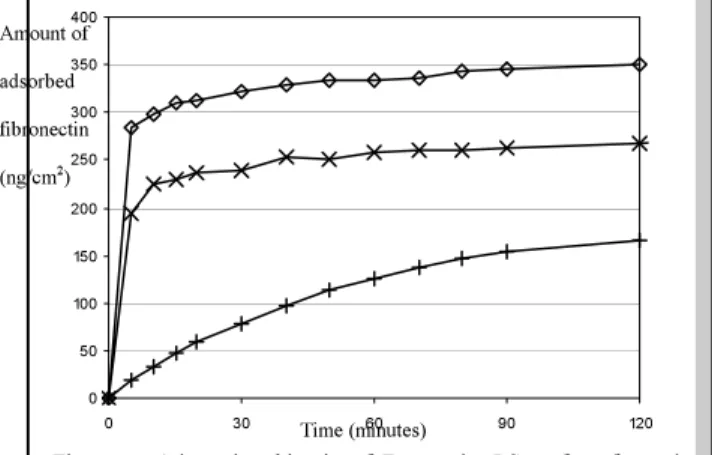
karine.anselme@uha.fr IntroductionLittle information was found in the literature about the expression of genes specific of cellular adhesion molecules on calcium phosphate materials although more were found on titanium-based substrates. Hence, the goal of this work was to study by a kinetic approach from 30 minutes to 4 days the adhesion of human osteosarcoma Saos2 cells on microporous (mHA) and non microporous hydroxyapatite (pHA) in comparison to polished titanium. Our strategy associated the visualization of adhesion proteins inside the cells by immunohistochemistry and the quantitative expression of genes at mRNA level by real-time PCR. The cell morphology was assessed using scanning electron microscopy and the number of cells thanks to biochemical techniques.
Methods
Microporous hydroxyapatite (mHA) discs were provided by BIOCETIS s.a. (France). They were obtained by a humid method using commercial powders of HA (Transtech, USA). The discs were then sintered at high temperature (1250°C). mHA displayed 12.5% of interconnected microporosity. Non microporous hydroxyapatite discs (pHA) were prepared using a laboratory-prepared HA powder which was pressed by uniaxial pressure of 80 Mpa. Discs were sintered at 1220°C and polished with SiC paper. Mirror-polished pure titanium discs, were obtained from the Laboratoire Materiaux (ENSAM, Lille, France). SaOs2 cells were cultured on samples for 30 min, 1h, 4h, 24h or 4 days.
Cells on samples were either treated for immunofluorescence, for adhesion assay or for protein and RNA extraction. For these last measurements, cells on each sample were immersed in 0.25 ml of Extract-All (Eurobio, France) and proceeded for protein and RNA extraction. The expression of genes involved in bone cell adhesion and differentiation was quantified by real-time PCR.
Results
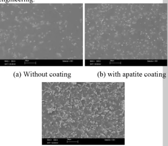
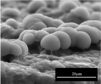
The cellular attachment was the highest on mHA from 30 minutes to 24 hours although the cell growth on mHA was the lowest after 4 days. Generally, the SaOs-2 osteoblastic cells morphology on mHA was radically different than on other surfaces with the particularity of the cytoplasmic edge which appeared un-distinguishable from the surface. The revelation by specific antibodies of proteins of the cytoskeleton (actin) and the focal adhesions (FAK, phosphotyrosine) confirmed that adhesion and spreading were different on the 3 materials.
The actin stress fibers were less numerous and shorter on mHA ceramics. Cells had more focal contacts after 4 hours on mHA compared to other substrates but less after 24 hours. The highest values of total proteins were extracted from mHA at 0.5 and 24 hours and from pHA at 1, 4, and 96 hours. The Dv and E1 integrin, actin, FAK, and ERK gene expression were found to be different with adhesion time and with materials. C-jun expression was comparable on mHA, titanium and plastic but was largely higher than on pHA at 0.5 and 1 hour. On the contrary, c-fos expression was the highest on pHA after 0.5 hours and the lowest after 1 hour. This difference between c-fos and c-jun expression on pHA after 0.5h could be related to the fact that these two genes may differ in their signalling pathways. The expression of the alkaline phosphatase gene after 4 days was lower on mHA compared to other materials demonstrating that the microstructure of the mHA ceramic was not favourable to SaOs-2 cells differentiation (Figure 1).
Figure 1: Normalized gene expression of alkaline phosphatase on plastic, titanium, mHA, pHA at 96h.
Plastic/mHA (*); p=0.02642 and for titanium/mHA (**);
p=0.00886.
Discussion and Conclusions
Finally, it was demonstrated in this study that HA and titanium surfaces influence as well gene expression at early times of adhesion as the synthesis of adhesion proteins but also proliferation and differentiation phases.
The signal transduction pathways involved in adhesion of SaOs-2 cells on HA and titanium were confirmed by the sequential expression ofDv andE1 integrins, FAK, and ERK genes followed by the expression of c-jun and c-fos genes for proliferation and alkaline phosphatase gene for differentiation. These results are a new demonstration that adhesion of cells on materials is linked to their nature, their protein adsorption capacity, their microstructure, and that it influences further cellular proliferation and differentiation phases.
*
**
CELL-MATRIX INTERACTIONS AND SKELETAL DEVELOPMENT A. Aszodi
Department of Molecular Medicine, Max Planck Institute for Biochemistry, Am Klopferspitz 18A, Martinsried, 82152 Germany; e-mail:aszodi@biochem.mpg.de
Introduction
The mammalian skeleton forms via two distinct ways:
mesenchymal cells either directly differentiate into bone producing osteoblast (intramembranous ossification) or first lay down cartilaginous precursors, which are subsequently replaced by bone (endochondral ossification). Skeletal morphogenesis is characterized by the expression of a special set of extracellular matrix (ECM) proteins. Cartilage ECM is enriched in type II collagen and aggrecan, which give tensile strength and resistance against compression to the tissue. In addition, the ECM provides instructive environmental clues for proper bone development by distributing growth factors and interacting with cells. Interactions between chondrocytes and the extracellular matrix are important for cartilage homeostasis regulating various cellular functions such as anchorage, survival, proliferation, matrix biosynthesis and degradation. Chondrocyte adhesion to cartilage ECM molecules is primarily mediated via the integrin family of DE-heterodimeric transmembrane receptors. Chondrocytes mainly express E1 andDv integrins, which mediate adhesion to collagen II and fibronectin. Upon adhesion, ligated integrins are recruited to focal adhesions (FAs) together with adaptor proteins and kinases, such as integrin-linked kinase (Ilk), and initiate downstream signalling events. Cdc42, a member of the family of Rho GTPases, is another intracellular molecule involved in signalling form integrin receptors regulating actin dynamics and cell cycle progression.
Materials
Gene targeting experiments in mice are excellent tools to study the molecular mechanism of skeletogenesis. In the recent years, we have generated a series of conventional and conditional knockout mice lacking specific ECM proteins (e.g. fibronectin, FN), adhesive receptors (E1 integrins andD10E1 integrin) and intracellular signalling molecules (Ilk, Cdc42) in cartilage, and their skeletal phenotypes were analysed.
Results and Discussion
Chondrocyte-specific inactication of the E1 integrin and Ilk genes (Aszodi et al., 2003; Grashoff et al., 2003) results in perinatal chondrodyslasia with severely disorganized growth plates. The phenotypes of these mice were accompanied by various chondrocyte abnormalities such as shape change, adhesion, actin and proliferation defects. In addition, E1 integrin- but not Ilk-deficiency leads to impaired cytokinesis and assembly of collagen II network implicating that Ilk only partially mediates E1 function in chondrocytes. Interestingly, the lack of fibronectin in cartilage has no apparent impact on endochondral bone formation (Aszodi et al., 2003) suggesting that chondrocyte adhesion to FN mediated by D5E1 and DE3 integrins is dispensable for cartilage
function. On the other hand, deletion of the collagen- binding D10E1 integrin leads to a mild growth plate dysfunction (Bengtsson et al., 2005) indicating that this integrin receptor is important but not essential for skeletogenesis and its loss might compensated by other collagen-binding E1 integrins.
To explore the function ofE1 integrins in adult articular cartilage (AC), we have deleted the E1 integrin gene in early limb bud mesenchymal cells. Mutant mice survive and show AC abnormalities of the long bones of the appendicular skeleton. The defects are characterized by a thickening of the AC, chondrocyte clustering, reduced cellularity, altered deposition of collagen X and fibronectin, and the lack of clear tidemarks. At the ultrastructural level, chondrocyte shape change, frequent binucleation, enlarged pericellular matrix compartments, and large collagen bundles near the chondrocyte surface were evident in mutant AC. Compared to wild type AC, surface irregularities and softening of mutant AC were found using atomic force microscopy. The structural abnormalities were accompanied by reduced activation of MAP kinase pathways and reduced MMP13 expression.
Finally, we addressed the role of Cdc42 during skeletogenesis by deleting the Cdc42 gene in limb bud and cranial mesenchymes. In the appendicular skeleton of mutants, the cartilaginous condensations of the long bones were shortened and broadened, the subsequent hypertrophic chondrocyte differentiation, mineralization and the formation of the primary as well the secondary ossification centres were delayed in mutants. These abnormalities were accompanied by delayed recruitment of MMP-9 positive pre-osteoclasts at the vascular invasion zone and a progressive proliferation defect of growth plate chondrocytes. Cdc42-deficient chondrocytes show abnormal spreading and cytoskeletal organizationin vitro. In the calvaria, the intramemranous ossification is greatly retarded resulting in wide sutures and open fontanelles in mutants.
Conclusions
All together, our results demonstrate the critical requirement of integrin-mediated cell-matrix interactions in skeletogenesis and identify several molecular mechanisms mediating E1 integrin functions in chondrocytes.
References
[1] Aszodi A, Hunziker EB, Brakebusch C, Fässler R (2003) Genes Dev, 17, 2465-2479.
[2]Grashoff C, Aszodi A, Sakai T, Hunziker EB, Fässler R. (2003) EMBO Reports, 4, 432-438.
[3] Bengtssson T, Aszodi A, Nicolae C, Hunziker EB, Lundgren-Akerlund E, Fässler R (2005) J Cell Sci, 118, 929-936.
DENDRITIC CELLS AT THE BIOMATERIAL/HOST INTERFACE
J. E. Babensee, J. Park, S. Shankar, M. Yoshida
Wallace H. Coulter Department of Biomedical Engineering, Georgia Institute of Technology and Emory University, Atlanta, GA, 30332
e-mail: julia.babensee@bme.gatech.edu
Introduction
Since biomaterials are used as vehicles for biological components in combination products, it is important to clarify the role of the biomaterial in potentiating any immune response towards the biological component due to a biomaterial adjuvant effect. In tissue engineering applications, immune responses are to be minimized while vaccine strategies seek to enhance the protective immune response. We have shown that poly(lactic-co- glycolic acid) (PLGA) acts as an adjuvant in the immune response against co-delivered antigen1, depending on the form of the PLGA2. As adjuvants act through the maturation of dendritic cells (DCs)3, 4, we have examined the differential effects of various biomaterials on DC maturation to assess their adjuvant effect. Furthermore, we are examining the pattern recognition receptors (PRRs) which may be involved in mediating DC recognition and response to biomaterials, particularly focusing in toll like receptor-4 (TLR4). We are also characterizing the biomaterial-associated molecular patterns which may be recognized by the PRRs. DCs may recognize and respond to biomaterials either indirectly through the adsorbed protein layer, for example through carbohydrate modifications of these proteins or through carbohydrates inherent in the biomaterial chemistry using PRRs to initiate an innate immune response.
Materials
Immature DCs (iDCs) were derived from human peripheral blood monocytes by culturing with GM-CSF and IL-4. The effect of biomaterial contact on maturation was studied by culturing iDCs with biomaterials in the form of microparticles (MPs) or films. The following biomaterials were tested: agarose; 75:25 PLGA; chitosan, alginate, hyaluronic acid. The extent of DC maturation was assessed by evaluating the resultant morphology, expression of co-stimulatory and MHC molecules, release of inflammatory cytokines, and allostimulatory function in a mixed lymphocyte reaction (MLR), as compared to iDCs (negative control) and lipopolysaccharide (LPS)- matured DCs (mDCs) (positive control). The requirement of direct biomaterial contact for DC maturation was examined using a transwell assay. The contribution of MP phagocytosis to DC maturation was evaluated using PLGA or agarose MPs of phagocytosable (2µm) and non- phagocytosable (20µm or 30µm) sizes with constant, but varied exposed surface area. Maturation of C57BL/6 bone marrow (BM)-derived DCs upon biomaterial contact was confirmed. The role of TLR4 in biomaterial-induced maturation of DCs is being evaluated by using mice deficient in TLR4 function as sources of BM-derived DCs or HEK293 cells stably transfected to express TLR4 (InvivoGen, San Diego, CA) for treatment with biomaterials and maturation effects compared to controls.
Enzyme linked lectin assays using
plant lectin probes were used to elucidate carbohydrate modifications, recognized by the mannose receptor, of adsorbed proteins on alkanethiol self-assembled monolayers (SAMs) of defined chemistries.
Results and Discussion
There was a differential effect of the biomaterial on which iDCs were cultured on the extent of DC maturation; chitosan or poly(lactic-co-glycolic) acid (PLGA) but not agarose, alginate or hyaluronic acid films5 supported DC maturation. Culturing iDCs with PLGA MPs or film (but not agarose) resulted in cell morphology similar to mDCs, and expression levels of co-stimulatory and MHC molecules between those exhibited by iDCs and mDCs6. The increase in DC marker expression induced by PLGA MPs required direct contact of DCs with the MPs6. DCs cultured with PLGA or agarose MPs or films secreted several proinflammatory cytokines in a MP-dose dependent manner; higher levels from DCs treated with PLGA than agarose MPs. MLR results showed that iDCs cultured on PLGA films were more efficient at inducing allogeneic T cell proliferation than iDCs cultured on agarose films. DCs cultured with PLGA or agarose MPs of the different sizes did not change their expression level of mDC markers, implying that phagocytosis was not the main contributor to the MP- induced DC maturation but exposed biomaterial surface area was. Different SAM surfaces were associated with differential carbohydrate profiles. NH2 SAM surfaces had highest amounts of carbohydrates and CH3 SAM surfaces the lowest. DCs cultured on NH2 and COOH SAM surfaces were more mature than those cultured on CH3 SAM surfaces. Murine DCs from C57BL/6 mice matured upon contact with PLGA MPs or films.
Conclusions
These studies provide insight into design criteria as well as immunomodulatory strategies for biomaterials for a range of applications.
Acknowledgements
Funding from an NSF CAREER grant (BES-0239142), the Arthritis Foundation Arthritis Investigator Grant, and an NIH RO1 grant 1RO1EB004633-01A1.
References
[1] Matzelle M, Babensee JE (2004) Biomaterials 25, 295-304.
[2] Bennewitz N, Babensee JE (2005) Biomaterials 26, 2991-2999.
[3] Banchereau J, Steinman RM (1998) Nature 392, 245- 252.
[4] Cella M et al. (1997) Nature 388, 782-787.
[5] Babensee JE, Paranjpe A J (2005) J. Biomed. Mater.
Res. 74A, 503-510.
[6] Yoshida M, Babensee JE (2004) J. Biomed. Mater.
Res. 71A, 45-51.
ASSEMBLY AND MECHANOSENSORY FUNCTION OF FOCAL ADHESIONS:
EXPERIMENTS AND MODELS
A.D. Bershadsky
1, C. Ballestrem
1, L. Carramusa
1, Y. Zilberman
1, T. Shemesh
2, M.M. Kozlov
21Department of Molecular Cell Biology, The Weizmann Institute of Science, Rehovot 76100, Israel; 2Department of Physiology and Pharmacology, Sackler Faculty of Medicine, Tel Aviv University, Tel Aviv 69978, Israel
e-mail: alexander.bershadsky@weizmann.ac.il
Focal adhesions as mechanosensors: a physical mechanism
Cells adhere to the extracellular matrix (ECM) via transmembrane integrin family receptors, which together with numerous associated “plaque proteins”
form focal adhesions (FAs) linking the ECM to the actin cytoskeleton. FAs are both adhesion and signal transduction organelles, informing cells about the state of the ECM. We have shown that FAs function as touch receptors responding to the mechanical characteristics of the microenvironment [1].
In our theoretical studies, we demonstrated that the major features of the FA mechanosensitive behavior can be explained by thermodynamic principles, which govern self-assembly of molecules into an aggregate subjected to pulling force. Elastic stresses generated within a protein complex decrease the chemical potential of the aggregated molecules relative to the pool of non-assembled molecules. This means that self- assembly of proteins is favored when pulling forces act on the aggregate and disfavored when these forces are relaxed. Considering various types of linkage between the aggregate and the substrate, we predicted different modes of FA assembly and disassembly and showed that the suggested model accounts for the major types of FA behavior observed experimentally [2]. Thus, this model based on very general assumption allows to explain qualitatively the FA mechanosensitive behavior. A hierarchy of diverse signaling circuits shown to be involved in the focal adhesion mechanosensitivity could be build up as a superstructure onto this basic mechanism.
A mechanism of force-driven focal adhesion assembly can be based on the formin function Signal from small G-protein RhoA is required for the formation of mature FAs and the associated actin filament bundles (stress fibers). Our studies showed that formin homology protein Dia1 is a downstream target of Rho that mediates force-induced FAs formation (reviewed in [1]). More recent experiments with Dia1 knockdown by siRNA (L. Carramusa et al.) confirmed that Dia1 is necessary for the transformation of initial focal complexes into FA and/or for further elongation of FA.
Analysis of formin interaction with actin indicates to a possible direct involvement of formins in the functioning of cellular mechanosensory units.
Specifically, theoretical consideration predicted a novel phenomenon, the force-driven polymerization of actin mediated by proteins of the formin family [3].
Formins localize to the barbed ends of actin filaments, but, in contrast to the regular capping proteins, allow for actin polymerization in the barbed direction (the so-called “leaky” or “processive” capping
mechanism). We proposed that the mechanism of leaky capping is based on the elasticity of the formin dimer [3] or, more precisely, elasticity of the formin dimer/barbed end complex [3, 4]. The phenomenon of force-driven actin polymerization is a direct consequence of the phenomenon of leaky capping of actin filaments by formins. We showed that if a pulling force is applied to the formin capping the filament end, the elastic mechanism drives filament growth.
Specifically, a moderate pulling force of ~3.5 pN (which can be developed by a single myosin molecule) reduces the critical concentration by an order of magnitude [3].
Cross-talk between focal a d h e s i o n s and microtubules. A novel role of Dia1 formin
We have shown that Dia1 activation promotes microtubule interactions with FAs [5]. Thus, Dia1 plays a dual role in the regulation of FAs. First, it is necessary for the FA growth that is induced by force.
Second, Dia1 promotes microtubule targeting to FAs, which locally inhibit myosin II-driven contractility and facilitates FA turnover [5]. Dia1 dramatically suppresses microtubule plus end growth, in an actin dependent manner [6]. These and other results [6]
suggest that Dia1 coordinates the activities of two major cytoskeletal systems, actin and microtubules, in the process of formation and turnover of FAs.
Acknowledgements
Alexander Bershadsky holds the Joseph Moss Chair of Biomedical Research. Grants from the Israel Science Foundation and Minerva (Germany) are acknowledged.
References
[1] Bershadsky A.D., Balaban, N.Q. & Geiger, B.
(2003) Annu Rev Cell Dev Biol 19, 677-695.
[2] Shemesh, T., Geiger, B., Bershadsky A.D., Kozlov, M.M. (2005) Proc. Natl. Acad. Sci. USA 102, 12383- 12388.
[3] Kozlov, M.M., Bershadsky A.D. (2004) J. Cell Biol.
167, 1011-1017.
[4] Shemesh, T., Bershadsky A.D., Kozlov, M.M.
(2005) J. Phys.: Condens. Matter 17, S3913-S3928.
[5] Ballestrem, C., Magid, N., Zonis, J., Shtutman, M.
& Bershadsky A. (2004) In: Cell Motility: From Molecules to Organisms (A. Ridley, M. Peckham, P.
Clark, eds.), Chapter 5, pp. 75-99. John Wiley & Sons Ltd, London.
[6] Bershadsky A.D., Ballestrem, C., Carramusa, L., Zilberman, Y., Gilquin, B., Khochbin, S., Alexandrova, A.Y., Verkhovsky, A.B., Shemesh, T., Kozlov, M.M.
(2005) Eur. J. Cell Biol. 85 [Epub: PMID: 16360240].
Skeletal stem cells Paolo Bianco
La Sapienza University, Rome Skeletal stem cells (SSCs; popularly referred to as
“mesenchymal” stem cells) are found in the clonogenic subset of adherent bone marrow stromal cells. They can be prospectively isolated using a defined set of antibodies recognizing surface epitopes, and their multipotency and self-renewal can be directly demonstrated using appropriate in vivo transplantation assays. As proven by direct comparison of phenotype in vivo and ex vivo, adventitial reticular cells at the abluminal side of sinusoids can now be identified as the in vivo counterpart of clonogenic cells. Cell strains generated from SSCs in vitro form bone, fat, fibroblasts of donor origin when transplanted ectopically in the immunocompromised mouse, or orthotopically as in preclinical models of bone tissue engineering. In addition, they self-renew into adventitial reticular cells in the heterotopic chimeric bone marrow, from which they can be secondarily isolated in culture (self-renewal).
To prove chondrogenic potential, alternate assays are required, either in vitro or in vivo. Adventitial reticular cells in the bone marrow are a local variation of cells called pericytes in other tissues, which can in turn be isolated and probed in vitro and in vivo using approaches conceptually similar to those employed for bone marrow stromal cells. Tissue specific pericytes share the same phenotype, are clonogenic, but exhibit a tissue specific differentiation potential. A general picture emerges outlining the nature of “mesenchymal stem cells”, their origin, mechanisms of their establishment in post-natal tissues, and their function, providing for the first time a rational guideline for their prospective clinical use.
COLLAGEN MATRIX COMPOSITION AND STRUCTURE - INFLUENCE ON BINDING, RELEASE AND ACTIVITY OF TGF-ȕ, BMP-2 AND BMP-4
Bierbaum S
1., Scharnweber D
1., Schnabelrauch M
2., Hempel U
3., Worch H
1.
1Max Bergmann Center for Biomaterials, Technische Universität Dresden, 01059 Dresden, Germany
2Innovent e.V, 07745 Jena, Germany
3Institute for Physiological Chemistry , Technische Universität Dresden, 01307 Dresden, Germany e-mail: Susanne.Bierbaum@tu-dresden.de
Introduction
Components of the extracellular matrix (ECM) can be used as carriers for growth factors. They also provide a microenvironment that can directly influence cells and thus be benefactory to integration and healing. In this context mainly collagen type I has been used, as the protein is the main component of the ECM. Varying the both structure and composition of such collageneous matrices can offer a way of affecting both cell response and growth factor binding.
The collagen matrix can be changed in a number of ways. Among these methods is the inclusion of non- collagenous components with specific functions (i.e.
decorin as collagen and TGF-E binding), the cross- linking of matrices to influence degradation and the release of integrated factors, the stacking of differently composed layers to generate graded structures, the presentation of matrix components/ growth factors at different time points.
Using such different approaches, it may be possible to influence the interaction with growth factors and adherent cells, allowing an adaptation of the artificial ECM coating of implants to specific requirements of the host tissue.
Materials and Methods
Experiments were performed on sand-blasted Ti6Al4V samples with a diameter of 10 or 16 mm. Employed proteins where commercially available collagen I bovine (IBFB) or porcine (Colbar), decorin, and chondroitin sulfate (Sigma) as well as the growth factors TGF-E1, BMP-4 and BMP-2 (R&D). Modified hyaluronic acids were provided by Innovent, Jena. In case of cross-linking EDC was used. Desorption experiments were conducted in cell culture medium with 2 % serum at 37°C. Growth factors were quantified by Elisa.
Cell culture studies were performed with primary osteoblastic cells from rat calvariae. Determined parameters were ALP-activity and collagen synthesis.
For the BMP-Bioassay C2C12 cells were seeded on substrata differing in composition of the collagen matrix adsorbed to titanium alloy and ALP induction was measured.
Results and Discussion
The interaction of growth factors with the collagen matrix depends on several factors. One of them is the matrix composition. Including decorin and chondroitin sulfate into the matrix increases both binding and desorption of TGF-Eand BMP-4, respectively, between 10 % - 20 %, indicating an interaction between these
components as is already described for TGF-E. A more significant influence on the release can be achieved by using a layered matrix architecture, or by integrating the growth factors during the fibrillogenesis into the collagen matrix. Especially for BMP-4 the detectable amount in the latter case increased by a factor of 2 to nearly 100 % of the initially bound amount. For TGF-E the differences were less obvious, here in all cases of layered matrices an increase in the signal by 2 – 5 % could be detected.
Hyaluronic acids modified with anionic groups influenced growth factor binding to a certain degree.
Layering the matrix delayed growth factor desorption though the burst release character was retained. In the combined application of BMP and TGF, this delay was already found to increase the ALP induction in C2C12 cells and primary rat osteoblasts.
The results for BMP-2 compared to BMP-4 were an increased adsorption (detected by Elisa), both on collagen and collagen-CS matrices, but a reduced activity of the adsorbed growth factor as measured in the bioassay. The origin of the collagen type I – (bovine/porcine) did not seem to affect these processes significantly. In the case of BMP-4, over 80% of the desorption took place in the first 24 h, while the detectable amounts of BMP-2 after 300 h desorption were less than 10 % of the bound amount. After 300 h no activity remained detectable for either BMP.
Conclusions
Growth factor binding to and release from a matrix composed of components of the extracellular matrix adsorbed to titanium surfaces can be influenced by inclusion of non-collageneous components specific to the growth factor. The architecture (layering) of the matrix can affect release kinetics as well as the measurable activity, something that may be attributed to a protective effect of the matrix. Using two growth factors, these differences can be used to enhance the effect of the factors. Crosslinking stabilizes the matrix, but not necessarily with positive effects for the adsorbed growth factors as their activity was found to be reduced.
The matrix characteristics thus play an important role in the activity of growth factors, though the behavior of different growth factor preparation can vary significantly in this respect.
Acknowledgements
This work was supported by the Deutsche Forschungs- gemeinschaft (DFG); porcine collagen was provided by Biomet Europe.
HIGH RESOLUTION 3D FIB-SEM MICROSCOPY OF CELL MONOLAYERS ON BIOCOMPATIBLE MATERIALS FOR BIOELECTRONIC DEVICES
C. Burkhardt
1, H. Wolburg
2, R. Stoop
1, S. Schädler
1, W. Nisch
11NMI Natural and Medical Sciences Institute at the University of Tuebingen, Reutlingen, Germany
2Institute of Pathology, University of Tuebingen, Tuebingen, Germany e-mail: claus.burkhardt@nmi.de.de
Introduction
A close contact between living cells or tissue and surfaces of bioelectronic devices like micro electrode arrays (MEA) or neuroprostheses is a crucial parameter for the signal transduction from electrodes to cells and vice versa. Great efforts have been made in the past to investigate ultrastructural details of model systems of this bio-technical interface. Ultramicrotomy of Araldite replicas of thin micro structured metal surfaces was used to investigate cell adhesion by transmission electron microscopy [1]. But this method gives only reliable results for very thin metal layers on materials like Araldite which are compatible to ultramicrotomy. To get access to the interface between hard biomaterials and soft biological systems we have therefore developed a new preparation method.
Sample Preparation 1: Embedding
To obtain high-resolution images from details of the technical substrate, the biological material and the interface in between we combined conventional fixation and embedding techniques, Focused Ion Beam (FIB) technology and low voltage Field Emission Scanning Electron Microscopy (FESEM).
3D FIB-SEM Microscopy
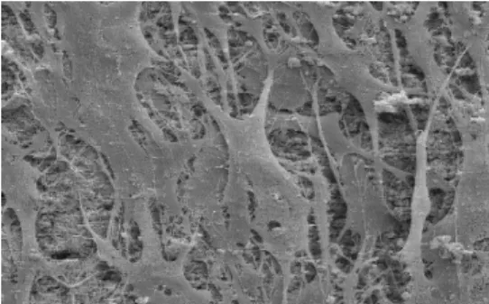
FIB preparation and SEM imaging was performed in a crossbeam 1540 XB microscope (Carl Zeiss, Oberkochen). This FIB-SEM microscope allows an easy selection of the region of interest, fast milling at selected areas and SEM imaging of the FIB prepared micro block face [2]. By serial sectioning at this micro block face (Figure 1), layers with a thickness down to 20 nm are removed by FIB milling while the new block face is imaged in real time with the SEM (live milling).
Figure 1: SEM image of the interface between a single cell and a silicon substrate coated with a thin functional layer of titanium.
Sample Preparation 2: Cryo Techniques
Besides embedding, cryofixation of cell monolayers on thin substrates with plunge freezing in propanol was established. Using a cryo transfer system (Baltec VCT 100) and a cryo stage, the frozen hydrated samples may be further processed by milling and imaging inside the cryo-FIB-SEM microscope ( Figure 2).
Plunge freezing offers several advantages compared to the embedding technique. Cryo-fixation is a fast process, so the chemical composition of the cell monolayer is frozen by this rapid fixation process.
Figure 2: Cryo-SEM of native human vascular endothel cells grown on a lysine coated gold substrate. A part of the lysine layer was removed by in-situ micro fracture with a small needle.
Conclusion and Outlook
3D FIB-SEM microscopy of the interface between cell monolayers and different biomaterials offers deeper insight to the process of cell growth and adhesion on this materials. The influence of functional layers like thin coatings on this materials may now be systematically investigated. Cryo techniques allow a fast preparation of the cell monolayers and opens the potential for a chemical analysis by secondary ion mass spectroscopy which is currently under development in our lab.
Acknowledgements
Thanks to the German BMBF for financial support of part of this project, FKZ 13N8652
References
[1] F. Pfeiffer, et al., Microelect. Eng. (2003) 67-68, 913 [2] C. Burkhardt, W. Nisch, Pract. Metallogr. (2005) 42,
Silicon
Araldit
Gold
Lysin
MECHANOREGULATION AT THE CELLULAR AND MULTICELLULAR SCALES
Wendy F. Liu
1, 2and Christopher S. Chen
21Johns Hopkins University, Baltimore, MD, 21205,
2University of Pennsylvania, Philadelphia, PA 19104 e-mail: chrischen@seas.upenn.edu
C PPrrootteeiinn PPlluurroonniicc CCeellllss
B BB
PDMS
PDMS Stamp
Peel Adsorb Protein B
PDMS PDMS
PDMS PDMS
Stamp
Peel Adsorb Protein
Protein Pluronic Cells
A B
Fig. 1. Microcontact Printing
Fig. 2. Cell spreading regulates proliferation and differentiation
Introduction
Cellular behavior within multicellular organisms is rigorously controlled by many dynamic cues in the surrounding cellular microenvironment. In particular, adhesive interactions with the ECM and with neighboring cells together coordinate growth, migration, and differentiation. Using microfabrication approaches to engineer cellular microenvironments, we are examining how adhesive cues cooperate to control basic processes such as proliferation and differentiation. Our findings suggest that cells integrate biochemical and mechanical cues from their microenvironment to modulate intracellular signaling pathways and cytoskeletal tension, leading to changes in cellular function. These studies may have a significant impact on our understanding of tissue development and the targeting of specific pathways in disease.
Materials and Methods
Microcontact printing (µCP) is a technique that enables the modification of biomaterials surfaces to control cell interactions with the underlying ECM and neighboring cells (Fig. 1). A PDMS stamp is coated with ECM
protein, which is dried and stamped onto a substrate such as glass, polystyrene, or PDMS. A non-adhesive detergent (e.g. Pluronic) is then adsorbed onto the unprinted regions of the surface. Cells adhere to the ECM-stamped regions, but not the detergent-blocked regions. By creating patterns with specific geometries, both cell-ECM and cell-cell adhesion can be controlled.
Results and Discussion
Using micropatterning techniques, we have demonstrated that cell spreading directly regulates cell proliferation and apoptosis [1] (Fig. 2A, B). Recently, we have also demonstrated that spreading affects the commitment of human MSCs to a bone or fat lineage [2]
(Fig. 2C). These effects appear to be dependent cytoskeletal tension and Rho GTPase signaling.
We have also used patterning tools to create bowtie- shaped agarose microwells, which allow the control of both spreading and cell-cell adhesion (Fig. 3). One cell adheres to each half of the bowtie and forms a cell-cell contact with a neighboring cell through the center of the bowtie. Using this system, we have observed that cell- cell contact stimulates proliferation when cell- ECM adhesion remains unchanged. This increase in proliferation has been observed for endothelial and epithelial cells, and depends on cadherins, the major protein within adherens junctions [3].
Using PDMS as a deformable substrate, we have generated a device consisting of an array of microneedles that detects cellular forces. Cells adhere to and pull on the tips of these needles, which are coated with ECM by µCP (Fig. 4). The deflection of the needles is determined by forces exerted by the cell on the underlying substrate. Using
this device, we have demonstrated that both cell spreading and multicellular o r g a n i z a t i o n r e g u l a t e cellular forces [4, 5].
Conclusions
We have demonstrated
using microfabrication tools that adhesive cues within the cellular microenvironment are important regulators of intracellular signalling pathways and cytoskeletal tension, which together modulate cell proliferation and differentiation. Development of new technologies and further biological studies are needed for a complete understanding of how cells integrate the numerous d y n a m i c c u e s i n t h e i r t h r e e - d i m e n s i o n a l microenvironment.
Acknowledgements
This work was supported by the NIH (HL73305 and EB00262). WFL acknowledges the NSF for financial support.
References
[1] Chen, C.S., et al., (1997) Science. 276 (5317): p. 1425-8.
[2] McBeath, R., et al., (2004) Dev Cell. 6(4): p. 483-95.
[3] Nelson, C.M. and C.S. Chen, (2003) J Cell Sci. 116(Pt 17):
p. 3571-81.
[4] Tan, J.L., et al., (2003) Proc Natl Acad Sci U S A. 100(4):
p. 1484-9.
[5] Nelson, C.M., et al., (2005) Proc Natl Acad Sci U S A.
102(33): p. 11594-9.
B A
Fig. 4. Micropost array- detectors Fig. 3. Cell-cell contact
regulates proliferation
HIGH PERFORMANCE OF MICRO MACROPOROUS BIPHASIC CALCIUM PHOSPHATE MATRICES FOR BONE TISSUE RECONSTRUCTION AND BONE TISSUE ENGINEERING
G. Daculsi
INSERM U791, LIOAD, Faculté de Chirurgie Dentaire Nantes France
Introduction
The development of CaP ceramics for bone graft involved a better control of the process of biomaterials resorption and bone substitution particularly to optimize calcium phosphate matrices for tissue engineering and biuoactive factor carrier. Bone graft materials biomaterials are largely represented by calcium hydroxyapatite, HA; tricalcium phosphate, TCP; and macroporous biphasic CaP, MBCP. The concept based on biphasic CaP ceramics is achieved by an optimum balance of the more stable phase of HA and more soluble TCP. The material is soluble and gradually dissolves in the body, seeding new bone formation as it releases Ca and P ions into the biological medium. These bioceramics are largely used for bone reconstruction and will be specially optimized for combination with bone marrow during surgery or for bone tissue engineering using STEM cells. We have optimized matrices in terms of their physico-chemical and crystal properties; to improve cell colonization and to increase kinetic bone ingrowth The fast cell colonization and resorption of the material are associated to the interconnected macropores structure which enhanced the resorption bone substitution process.The micropore content involved biological fluid diffusion and suitable absorption surfaces for circulating growth factors.
Materials and Methods
Inteconnected MicroMacroporous Biphasic CaP (MBCP2000™, CE123, Biomatlante manufacturer) was an improvement of the technology of macropores developed long time ago1 to replace classical naphtalene use. Shortly, CaP deficient apatite CDA, were associated to a mixture of selected particles of naphtalene and sugar. After isostatic compaction, the block was sintered according a specific process of sublimation/calcination. The obtained bioceramics was characterized using X-Rays, FTIR, X-rays micro- tomography, permeability , Hg microporosimetry, BET specific surface area, mechanical test, and SEM.
Cylindrical samples of 6mm in diameter and 8 mm length were implanted in femoral epiphysis of New Zealand rabbits and compared to classical MBCP™ as reference (12 rabbits, 24 implantation sites). After 6 and 12weeks, implants were processed for histology and SEM using image analysis.
Results and Discussion
The density was 0.75 for MBCP2000 and 0.83 for MBCP. The crystal size is 0.5 to 1.5 µm and the specific surface area was 1.6 and 1.7m2/g for MBCP2000 and MBCP respectively. Compression test showed 4MPa and 6MPa for MBCP2000 and MBCP.
Mercury porosimetry gives 73% and 69% of total porosity respectively. The interconnections are evidenced by 3D reconstruction using qualitative and
quantitative microscanner. Permeability was twice higher for MBCP2000, and after incubation with bovine serum, 30% absorption increased with the MBCP2000was observed. The low difference of total porosity between the 2 types cannot explain higher permeability, the performance is due to distribution of pore size particularly mesopores. The HA/TCP ratio was 20/80 for MBCP2000 and 60/40 for MBCP, and FTIR confirms high purity of HA and TCP without carbonate.
After implantation bone ingrowth is observed in the 2 types of bioceramic, and newly-formed bone progressively replaced the bioactive material, followed by haversian bone remodelling. Faster bone ingrowth into the macropores was observed at 6weeks for MBCP2000. After 12weeks no statistical difference was noticed between the 2 implants type. The rate of resorption however is higher for MBCP 2000: 17%
versus 12% at 6weeks, and 19% versus 17% after 12weeks, (no significant difference).
The in vivo experiment indicated higher cell colonization by osteogenic cells in MBCP2000 due to this interconnected and microporous structure associated to higher solubility. However, due to bone ingrowth at the expense of the implant this phenomena is less evident after long term implantation.
MBCP2000 is a more suitable matrice for tissue engineering. The HA/TCP ratio of 20/80 is also more efficient for combination with STEM cell cultivation and expansion then implanted in non bony site comparing to classical MBCP2.
The kinetic of bone ingrowth by the osetogenic cells colonization need to develop inside the macropores.
Without macropores and mesopores the bioactive processes are unable to develop in the deep of the implants. The association of dissolution at the crystal levels, the diffusion of the biological fluid into the micropores, and the resorption by macrophages and osteoclastic cells of the materials at the surface and inside the macropores, involve a progressive bone substitution of the materials by true bone. This is the common process of resorption/bone substitution of the Micro Macroporous Biphasic Calcium phosphate ceramics.
Conclusion
Advanced technologies for macroporous calcium phosphate bioceramics manufacturing involved higher efficacy of such matrices for further relevant surgical technologies as persurgery combination with bone marrow or expanded STEM cell in vitro for bone tissue engineering
References
[1] M. Schmitt, PhD thesis Nantes University 2000 [2] T. Livingston Arinzeh et al, Biomaterials 26 (2005) 3631-3638
0 1 2 3 4 5
Foldinduction
BSP
1300 - 1300 + 1150 - 1150 +
BSP
Foldinduction
0 1 2 3 4 5 6
1300 - 1300 + 1300 - (+1150)1300 +
(+1150)
INSTRUCTIVE PROPERTIES OF BIOMATERIALS IN OSTEOCHONDRAL TISSUE ENGINEERING USING EMBRYONIC STEM CELLS
Sanne Both
1, HuipinYuan
1, Pamela Habibovic
1, Clemens van Blitterswijk
1, Jan de Boer
1Institute for Biomedical Technology, University of Twente, P.O. Box 98, 3720 A.B. Bilthoven, The Netherlands e-mail: j.deboer@tnw.utwente.nl
Introduction
In previous studies with two bi-phasic calcium phosphate ceramics (BCPs) with different microporosity we observed opposite results when implanted intramuscularly in goats. The more microporous ceramic (BCP1150) was able to induce bone whereas the less micro porous (BCP1300) was not (Figure 1).
Mouse embryonic stem cells (mESCs) have the potential to differentiate into all adult cell types and can proliferate indefinitely. Moreover, they can easily be genetically manipulated and are donor independent and thus can serve as a model system for bone tissue engineering. MESCs can be induced into the osteogenic lineage and where therefore assessed in this study as a model to investigate the molecular mechanism underlying the osteoinductive potential of ceramics.
Figure 1: Histological sections of implanted samples in goat muscle. Left, BCP1300, no bone formation visible.
Right, BCP1150, bone formation was present (arrows).
Methods
Embryoid bodies (EBs) were formed from IB10 mESCs, dissociated after 4 days and seeded onto 2x3x3 mm scaffolds of BCP1150 and BCP1300 and cultured in control and differentiation medium.
Results
We seeded mESCs on BCP1150 and BCP1300 scaffolds and assessed gene expression of osteocalcin and BSP, which were chosen as markers of mature osteoblasts. In all cases, differentiation medium enhanced expression of osteocalcin and BSP.
However the expression of osteocalcin and BSP was higher on BCP1150 than on BCP1300 reflecting the in vivo osteogenic behavior of BCP1150 (Figure 2A and data not shown). To investigate whether this was due to differential cell-material interaction or material-medium interaction we started by testing this last option.
Therefore we seeded cells onto BCP1300 in control and differentiation medium and added an empty scaffold of BCP1150 in the same well. The addition of an extra scaffold to the medium induces an increase in expression of both osteocalcin and BSP (Figure 2B), suggesting that
a change in medium composition by BCP1150 enhances gene expression of osteocalcin and BSP.
Figure 2: Differentiation of mouse embryonic stem cells. BSP expression of cells seeded on A. BCP1300, BCP1150 and B. BCP1300 with and without the addition of an empty BCP1150 scaffold (+1150), after 6 days of culture in control (-) and differentiation medium (+).
To investigate whether BCP1150 modifies the medium composition, we incubated medium with BCP1300 and BCP1150 and measured the medium components. We found a small decrease in calcium concentration in the medium after the addition of BCP1300 but a strong decrease after addition of BCP 1150 (Table 1). Medium incubated with both scaffolds, displayed a calcium decrease which was similar to the decrease of BCP1150.
As such, the gene expression correlates to a decrease in calcium concentration (Figure 2A,B). In contrast to the calcium decrease the magnesium concentration was hardly effected.
Table I: Calcium concentration in medium (M) treated with and without BCP1150 and BCP1300 scaffolds
Sample Ca2+(µg/L) Mg2+(µg/L)
M 75000 20000
M + BCP1300 70000 20000
M + BCP1150 55000 22000
M + BCP1300 + 1150 55000 22000 Discussion
In vitro, we found that mESC seeded on BCP1150 displayed enhanced gene expression of osteogenic markers compared to mESCs differentiated on BCP1300, which correlates with the in vivo data from the goat muscle implants. We also found that BCP1150 has an effect on the medium composition, whether this effect is caused by the decrease in the Ca2+concentration is currently under investigation. In conclusion mESCs can be used as a cell source for testing osteogenic
ARTIFICIAL EXTRA-CELLULAR MATRICES BASED ON FIBRILS OF DIFFERENT COLLAGEN TYPES CONTAINING IMMOBILISED PROTEOGLYCANS (PGS) FOR
TITANIUM IMPLANTS
T.Douglas
1, U.Hempel
2, C.Knieb
1, S.Heinemann
1, S.Bierbaum
1, D.Scharnweber
1, H.Worch
11Max Bergmann Center of Biomaterials, Budapester Strasse 27, 01069 Dresden, Germany
2Center of Theoretical Medicine, Institute of Physiological Chemistry, Fiedlerstrasse 42, 01307 Dresden, Germany e-mail: Timothy.Douglas@mailbox.tu-dresden.de
Introduction
Collagen in fibrillar form is used as a coating on titanium implants to mimic the natural bone extra-cellular matrix, as it presents binding sites for integrins and is not desorbed from titanium surfaces when placed in solution.
Proteoglycans (PGs), consisting of a protein core connected to glycosaminoglycan chains, may be bound to these collagen fibrils during fibril formation. These in turn can influence osteoblast behaviour and also bind growth factors (GFs). The PGs decorin and biglycan, present in bone, are able to bind to and modulate the activity of the GFs TGF-ȕ1, which stimulates osteoblast proliferation and chemotaxis, and BMP-4, which enhances osteoblast differentiation [1]. The main aim of this work is to compare fibrils of the collagen types I, II and III with regard to amount of decorin and biglycan bound as well as the resulting changes in fibril formation kinetics and morphology. The reaction of primary osteoblasts and an osteoblastic cell line to titanium surfaces coated with fibrils containing decorin and biglycan was also studied, as was the effect of TGF-ȕ1.
Materials and Methods
Collagen types I (calf skin), II (bovine tracheal cartilage) and III (human placenta) as well as decorin and biglycan (bovine articular cartilage) were obtained from Sigma- Aldrich Chemie GmbH, Germany. Fibrillogenesis took place at 37ºC in a 30 mM phosphate buffer at pH 7.4 at different PG:collagen ratios. Fibril mass was determined by the Lowry protein assay. PG content of fibrils was determined colormetrically by hexosamine and dimethylmethylene blue assays. Fibril morphology was studied by atomic force microscpoy (AFM). The kinetics of fibril formation were studied by means of turbidity measurements. Titanium surfaces were coated with fibrils and in some cases also with TGF-ȕ1 by adsorption, and seeded with an osteoblastic cell line (7F2), or primary rat calvaria (RCO) or human (HO) osteoblasts. Proliferation and collagen synthesis of RCOs and HOs were determined. 7F2 cells were tested for proliferation, calcium accumulation and alkaline phosphatase (ALP) production.
Results and Discussion
The results of both colormetric assays correlated well.
Collagen type II fibrils bound more PGs than fibrils of types I and III. Considerably more biglycan than decorin
was bound by all collagen types (Fig. 1). This could be due to biglycan’s higher molecular weight and longer glycosaminoglycan chains, which may form more stable bonds with collagen by “bridging” more binding sites on collagen. Type II formed fibrils approximately half as thick as type I fibrils. Decorin caused fibrils of types I- III to become thinner, but biglycan had no effect on morphology. Morphological data was supported by kinetic data, which showed deceleration of fibril formation and reduction of turbidity by decorin but not biglycan. Type II may bind more PGs than type I because of its larger relative surface area due to its thinner fibrils. Type II may also have a higher inherent affinity for PGs than types I and III. HO proliferation and RCO collagen synthesis were modulated by the presence of both decorin and biglycan. TGF-ȕ1 modulated the proliferation and collagen synthesis of HOs. PGs did not affect 7F2 behaviour.
Conclusions
The ability of collagen fibrils to bind PGs varies according to PG and collagen type. Biglycan and Decorin influence fibrillogenesis kinetics and fibril morphology differently, and both PGs and TGF-ȕ1 modulate primary osteoblast behaviour on titanium surfaces coated with collagen fibrils. These results may be of importance when designing collagen-based extra- cellular matrices, including those loaded with GFs.
Figure 1: Quantification of PGs immobilised per mg collagen fibrils of collagen types I, II and III.
Acknowledgements
BMBF, Germany for funding and T. Hanke for advice.
References
[1] Mundy et al., Clin Orthop Relat Res1996, 324, 24-8.
Immobilised Decorin, Biglycan
500 100150 200
I II III
Collagen type µgPG/mg collagenfibrils
Decorin Biglycan